Clinical Anatomy
31
Introduction The thorax consists of: • the chest wall • the two pleural cavities surrounding the lungs • the area between these cavities, the mediastinum, in which are found the heart, great vessels, trachea and oesophagus, vagus and phrenic nerves, thymus gland and the thoracic duct. On the external surface of the anterolateral chest wall are the breasts (mammary glands), also considered in this chapter. Below, the thorax is separated from the abdomen by the diaphragm; above, it is continuous with the neck at the thoracic inlet (between sternum and vertebra T1) through which structures pass between thorax and neck (Fig. 10.1). 10.1 Chest wall Thorax 10.1 Chest wall 63 10.2 Pleural cavities, tracheobronchial tree, lungs 68 10.3 Heart and pericardial cavity 75 10.4 Mediastinum 83 10.5 Breast 91 Fig. 10.1 Sagittal section through the thorax: point A (suprasternal border) is at vertebral level T2; point B (sternal angle of Louis) is at vertebral level T4; point C (xiphoid process) is at about vertebral level T9. Overview The chest wall consists of the ribs and sternum laterally and anteriorly, and the vertebral column posteriorly, which provides most of the structural support. Ribs articulate with the vertebral column behind and the sternum in front, these articulations permitting the movements of breathing. Intercostal (between the ribs) spaces are occupied by muscle and a neurovascular bundle that supplies the muscles, the skin over them and the lining of the pleural cavity deep to them. The repeating pattern of vertebrae, ribs and the intercostal neurovascular bundle is an illustration of Learning Objectives You should: • appreciate the orientation of the ribs • know the vertebral levels of the suprasternal notch, angle of Louis, and xiphisternum • understand how ribs articulate with vertebrae and sternum (NN) CA1E Ch 10 18/7/01 4:03 pm Page 63
description
Clinical Anatomy
Transcript of Clinical Anatomy
Introduction
The thorax consists of:
• the chest wall• the two pleural cavities surrounding the lungs• the area between these cavities, the mediastinum, in
which are found the heart, great vessels, trachea andoesophagus, vagus and phrenic nerves, thymusgland and the thoracic duct.
On the external surface of the anterolateral chestwall are the breasts (mammary glands), also consideredin this chapter.
Below, the thorax is separated from the abdomen bythe diaphragm; above, it is continuous with the neck atthe thoracic inlet (between sternum and vertebra T1)through which structures pass between thorax andneck (Fig. 10.1).
10.1 Chest wall
Thorax
10.1 Chest wall 63
10.2 Pleural cavities, tracheobronchial tree, lungs 68
10.3 Heart and pericardial cavity 75
10.4 Mediastinum 83
10.5 Breast 91
Fig. 10.1 Sagittal section through the thorax: point A(suprasternal border) is at vertebral level T2; point B (sternalangle of Louis) is at vertebral level T4; point C (xiphoidprocess) is at about vertebral level T9.
Overview
The chest wall consists of the ribs and sternum
laterally and anteriorly, and the vertebral
column posteriorly, which provides most of the
structural support. Ribs articulate with the
vertebral column behind and the sternum in
front, these articulations permitting the
movements of breathing. Intercostal (between
the ribs) spaces are occupied by muscle and a
neurovascular bundle that supplies the muscles,
the skin over them and the lining of the pleural
cavity deep to them. The repeating pattern of
vertebrae, ribs and the intercostal
neurovascular bundle is an illustration of
Learning Objectives
You should:
• appreciate the orientation of the ribs
•know the vertebral levels of the suprasternal
notch, angle of Louis, and xiphisternum
•understand how ribs articulate with
vertebrae and sternum
(NN) CA1E Ch 10 18/7/01 4:03 pm Page 63
Vertebral levels and surface markings(Fig. 10.1)
Remind yourself of the significance of vertebral levelsand surface markings.
• The suprasternal notch (in which you can palpatethe trachea) is at vertebral level T2.
• The sternal angle (of Louis) is at vertebral level T4.• The xiphisternum (xiphoid process) is at vertebral
level T9 (about).• Note the downwards orientation of the ribs when you
consider an anterior view of the rib cage: the secondrib articulates with vertebra T2 posteriorly, but withthe sternal angle at vertebral level T4 anteriorly.Therefore, a transverse (horizontal) section of thethorax, such as a scan, intersects several ribs.
Bones and joints of the chest wall(Fig. 10.2)
The chest wall is made up of the 12 pairs of ribs, thesternum and the intercostal muscles. Ribs are numbered1–12 from above down and articulate with the vertebralcolumn posteriorly and (mostly) the sternum anteriorly.The ribs are angled with respect to the horizontal plane –the sternal end is lower than the vertebral – and so inhorizontal section, or a transverse CT or MRI scan, severalribs will be seen sectioned obliquely. Ribs are united attheir anterior end to cartilage: the costal cartilages.
Vertebrae
The main features of a typical vertebra are described inChapter 5. A thoracic vertebra is identifiable because ofits facets for articulation with the ribs. Vertebrae T2–T9have two demifacets on each side, other vertebraehaving only one.
Ribs
Each rib has a head, neck, tubercle and shaft. Figure10.3 shows the main features of a typical rib (ribs 2–10).Of the atypical ribs (1, 11, 12), you need only botherwith the first.
First rib
• Scalene tubercle on the upper surface, to which isattached scalenus anterior muscle.
• Groove for subclavian vein, anterior to the tubercle.• Groove for subclavian artery, posterior to the
tubercle.
Sternum
There are three parts, from superior to inferior:manubrium, body and xiphoid process (or xiphisternum).There may be a hole in the centre of the body, since it isformed by a number of individual sternebrae thatshould fuse together, but may fail to do so.
Joints between vertebrae and ribs
Typical ribs have three synovial articulations with thevertebrae.
• The head has upper and lower articular facets setalmost at a right angle to one another, the lowerarticulating with the vertebra corresponding innumber to the rib, and the upper with the vertebraabove. Ribs 1, 10, 11 and 12 have only one facet andarticulate only with the numerically correspondingvertebra.
Thorax
Learning Objectives (cont’d)
•appreciate the structure and contents of an
intercostal space
•know the formation and function of
intercostal nerves and the dermatomes of the
chest wall
Fig. 10.2 Thorax from the front. Note the orientation of theribs: a horizontal section through the thorax (e.g. a CT scan)intersects several ribs.
Vertebrochondral ribs: 8–10
Typical ribs: 2–9
(NN) CA1E Ch 10 18/7/01 4:03 pm Page 64
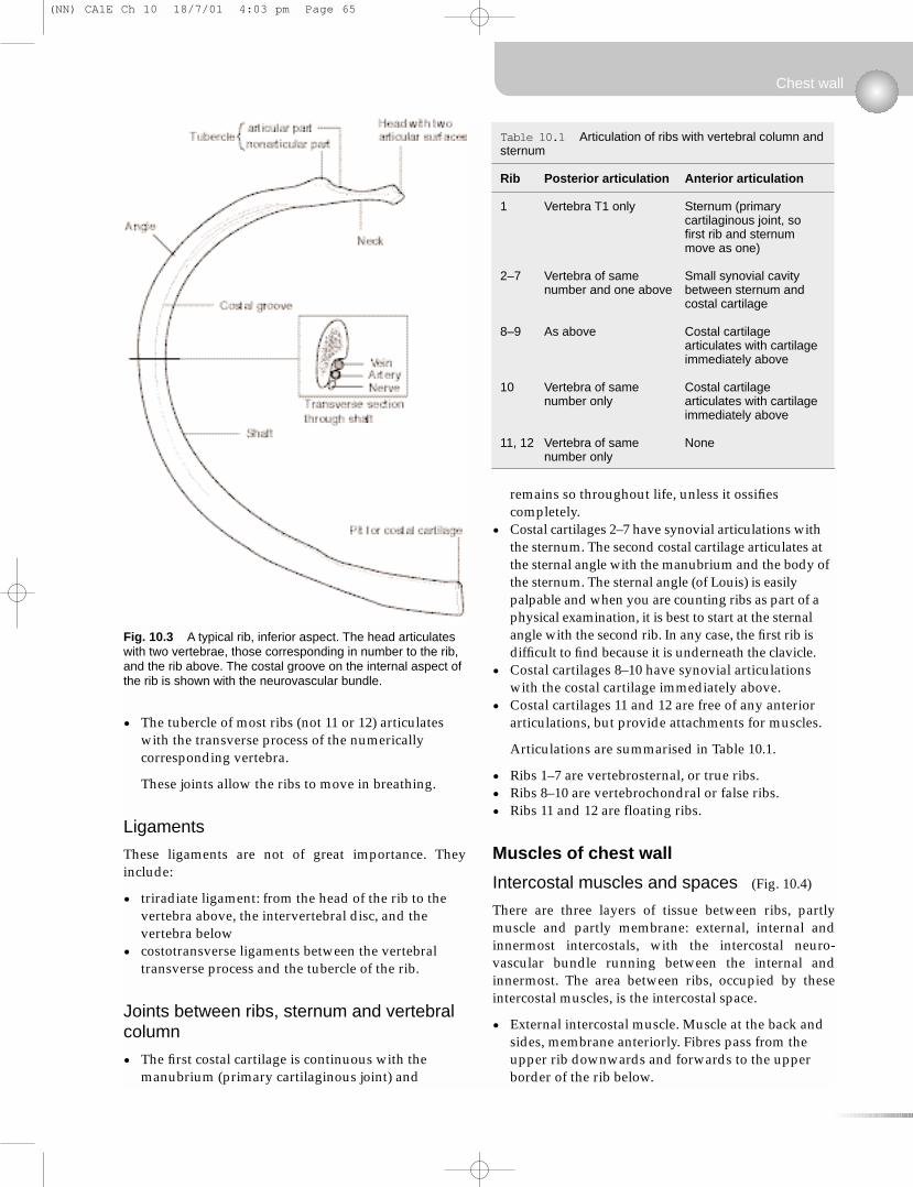
• The tubercle of most ribs (not 11 or 12) articulateswith the transverse process of the numericallycorresponding vertebra.
These joints allow the ribs to move in breathing.
Ligaments
These ligaments are not of great importance. Theyinclude:
• triradiate ligament: from the head of the rib to thevertebra above, the intervertebral disc, and thevertebra below
• costotransverse ligaments between the vertebraltransverse process and the tubercle of the rib.
Joints between ribs, sternum and vertebralcolumn
• The first costal cartilage is continuous with themanubrium (primary cartilaginous joint) and
remains so throughout life, unless it ossifiescompletely.
• Costal cartilages 2–7 have synovial articulations withthe sternum. The second costal cartilage articulates atthe sternal angle with the manubrium and the body ofthe sternum. The sternal angle (of Louis) is easilypalpable and when you are counting ribs as part of aphysical examination, it is best to start at the sternalangle with the second rib. In any case, the first rib isdifficult to find because it is underneath the clavicle.
• Costal cartilages 8–10 have synovial articulationswith the costal cartilage immediately above.
• Costal cartilages 11 and 12 are free of any anteriorarticulations, but provide attachments for muscles.
Articulations are summarised in Table 10.1.
• Ribs 1–7 are vertebrosternal, or true ribs.• Ribs 8–10 are vertebrochondral or false ribs.• Ribs 11 and 12 are floating ribs.
Muscles of chest wall
Intercostal muscles and spaces (Fig. 10.4)
There are three layers of tissue between ribs, partlymuscle and partly membrane: external, internal andinnermost intercostals, with the intercostal neuro-vascular bundle running between the internal andinnermost. The area between ribs, occupied by theseintercostal muscles, is the intercostal space.
• External intercostal muscle. Muscle at the back andsides, membrane anteriorly. Fibres pass from theupper rib downwards and forwards to the upperborder of the rib below.
Chest wall
Fig. 10.3 A typical rib, inferior aspect. The head articulateswith two vertebrae, those corresponding in number to the rib,and the rib above. The costal groove on the internal aspect ofthe rib is shown with the neurovascular bundle.
Table 10.1 Articulation of ribs with vertebral column andsternum
Rib Posterior articulation Anterior articulation
1 Vertebra T1 only Sternum (primary cartilaginous joint, sofirst rib and sternummove as one)
2–7 Vertebra of same Small synovial cavitynumber and one above between sternum and
costal cartilage
8–9 As above Costal cartilage articulates with cartilageimmediately above
10 Vertebra of same Costal cartilage number only articulates with cartilage
immediately above
11, 12 Vertebra of same Nonenumber only
(NN) CA1E Ch 10 18/7/01 4:03 pm Page 65
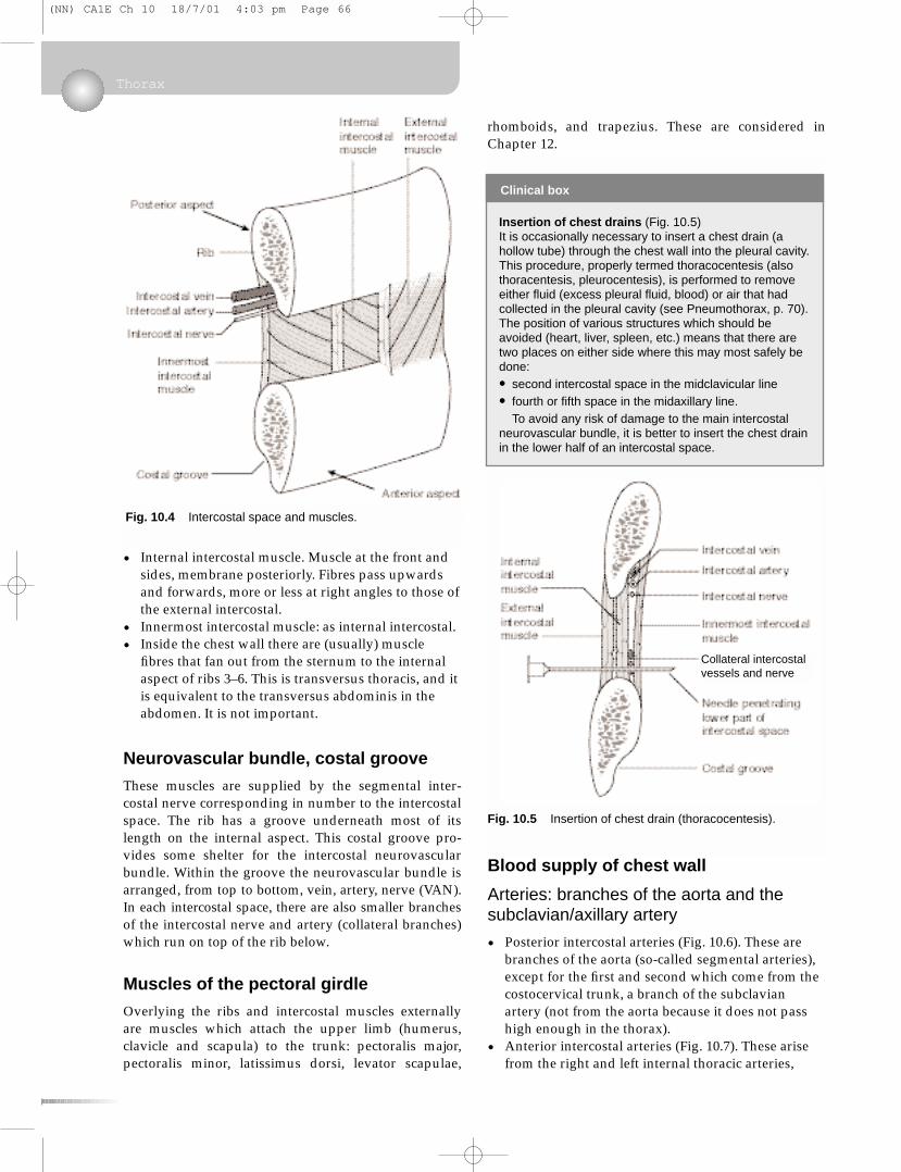
• Internal intercostal muscle. Muscle at the front andsides, membrane posteriorly. Fibres pass upwardsand forwards, more or less at right angles to those ofthe external intercostal.
• Innermost intercostal muscle: as internal intercostal.• Inside the chest wall there are (usually) muscle
fibres that fan out from the sternum to the internalaspect of ribs 3–6. This is transversus thoracis, and itis equivalent to the transversus abdominis in theabdomen. It is not important.
Neurovascular bundle, costal groove
These muscles are supplied by the segmental inter-costal nerve corresponding in number to the intercostalspace. The rib has a groove underneath most of itslength on the internal aspect. This costal groove pro-vides some shelter for the intercostal neurovascularbundle. Within the groove the neurovascular bundle isarranged, from top to bottom, vein, artery, nerve (VAN).In each intercostal space, there are also smaller branchesof the intercostal nerve and artery (collateral branches)which run on top of the rib below.
Muscles of the pectoral girdle
Overlying the ribs and intercostal muscles externallyare muscles which attach the upper limb (humerus,clavicle and scapula) to the trunk: pectoralis major,pectoralis minor, latissimus dorsi, levator scapulae,
rhomboids, and trapezius. These are considered inChapter 12.
Blood supply of chest wall
Arteries: branches of the aorta and thesubclavian/axillary artery
• Posterior intercostal arteries (Fig. 10.6). These arebranches of the aorta (so-called segmental arteries),except for the first and second which come from thecostocervical trunk, a branch of the subclavianartery (not from the aorta because it does not passhigh enough in the thorax).
• Anterior intercostal arteries (Fig. 10.7). These arisefrom the right and left internal thoracic arteries,
Thorax
Clinical box
Insertion of chest drains (Fig. 10.5)It is occasionally necessary to insert a chest drain (ahollow tube) through the chest wall into the pleural cavity.This procedure, properly termed thoracocentesis (alsothoracentesis, pleurocentesis), is performed to removeeither fluid (excess pleural fluid, blood) or air that hadcollected in the pleural cavity (see Pneumothorax, p. 70).The position of various structures which should beavoided (heart, liver, spleen, etc.) means that there aretwo places on either side where this may most safely bedone:
• second intercostal space in the midclavicular line
• fourth or fifth space in the midaxillary line.To avoid any risk of damage to the main intercostal
neurovascular bundle, it is better to insert the chest drainin the lower half of an intercostal space.
Fig. 10.4 Intercostal space and muscles.
Fig. 10.5 Insertion of chest drain (thoracocentesis).
Collateral intercostalvessels and nerve
(NN) CA1E Ch 10 18/7/01 4:03 pm Page 66
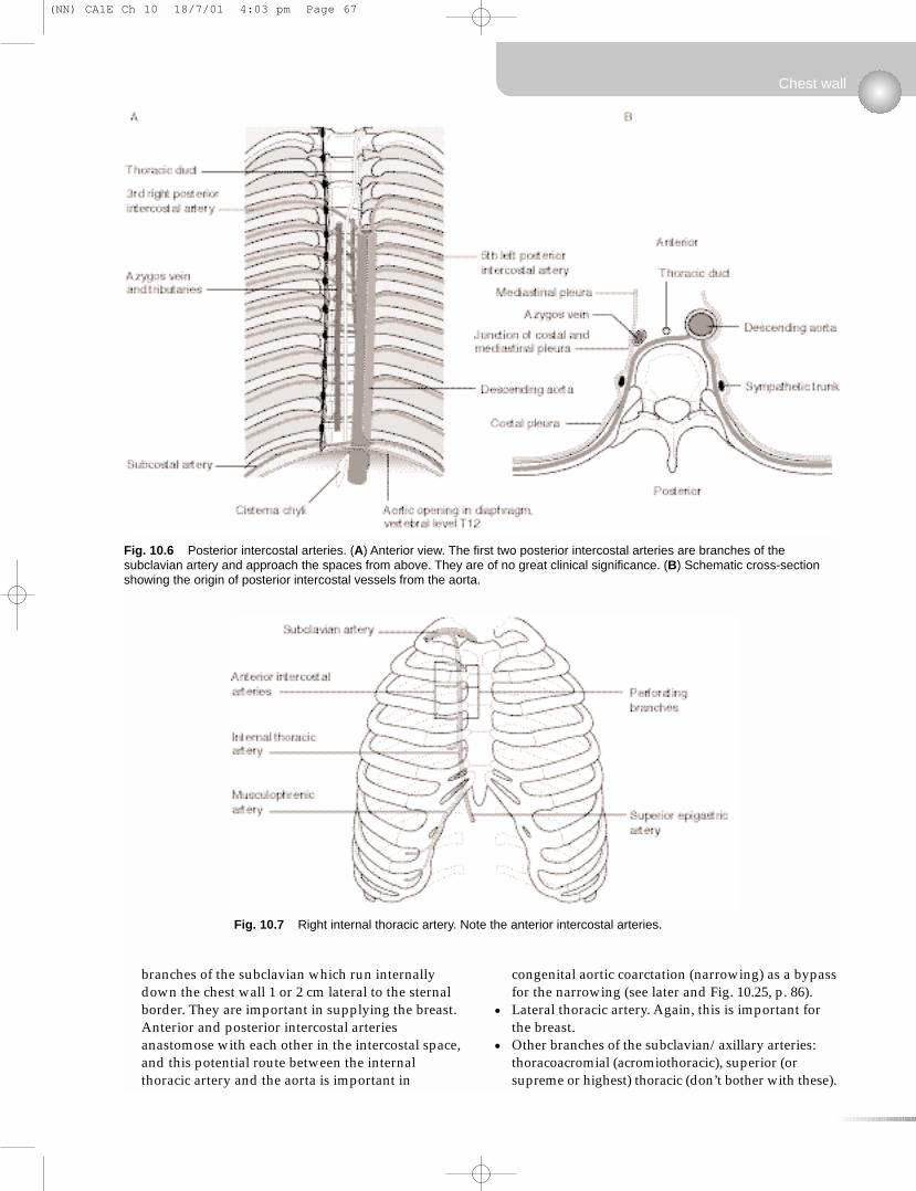
branches of the subclavian which run internallydown the chest wall 1 or 2 cm lateral to the sternalborder. They are important in supplying the breast.Anterior and posterior intercostal arteriesanastomose with each other in the intercostal space,and this potential route between the internalthoracic artery and the aorta is important in
congenital aortic coarctation (narrowing) as a bypassfor the narrowing (see later and Fig. 10.25, p. 86).
• Lateral thoracic artery. Again, this is important forthe breast.
• Other branches of the subclavian/axillary arteries:thoracoacromial (acromiothoracic), superior (orsupreme or highest) thoracic (don’t bother with these).
Chest wall
Fig. 10.6 Posterior intercostal arteries. (A) Anterior view. The first two posterior intercostal arteries are branches of thesubclavian artery and approach the spaces from above. They are of no great clinical significance. (B) Schematic cross-sectionshowing the origin of posterior intercostal vessels from the aorta.
Fig. 10.7 Right internal thoracic artery. Note the anterior intercostal arteries.
(NN) CA1E Ch 10 18/7/01 4:03 pm Page 67
Veins
• Anterior intercostal veins drain to the internalthoracic veins, thence to the subclavian –brachiocephalic – superior vena cava (SVC).
• Posterior intercostal veins drain to the azygossystem and the SVC. As the first one or twoposterior intercostal arteries arise differently fromthe rest, so the posterior intercostal veins of the firstfew spaces drain differently: to the brachiocephalicveins (see later).
Intercostal nerves (Fig. 10.8)
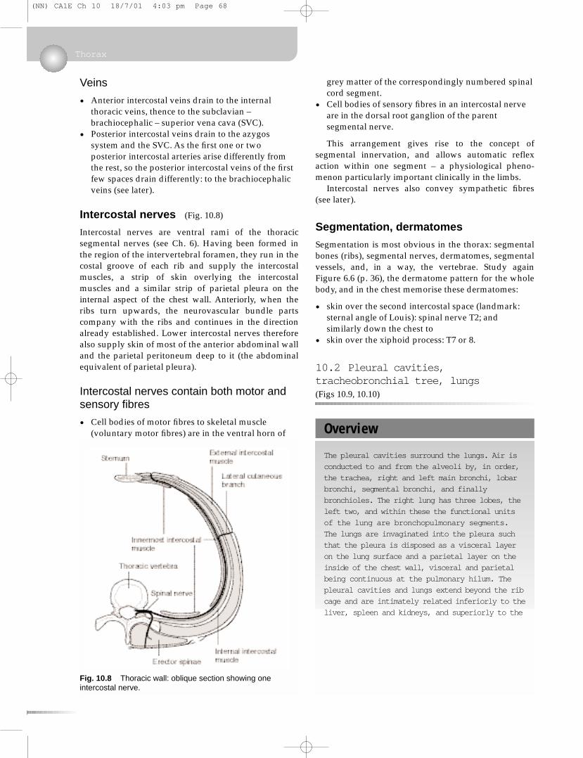
Intercostal nerves are ventral rami of the thoracicsegmental nerves (see Ch. 6). Having been formed inthe region of the intervertebral foramen, they run in thecostal groove of each rib and supply the intercostalmuscles, a strip of skin overlying the intercostalmuscles and a similar strip of parietal pleura on theinternal aspect of the chest wall. Anteriorly, when theribs turn upwards, the neurovascular bundle partscompany with the ribs and continues in the directionalready established. Lower intercostal nerves thereforealso supply skin of most of the anterior abdominal walland the parietal peritoneum deep to it (the abdominalequivalent of parietal pleura).
Intercostal nerves contain both motor andsensory fibres
• Cell bodies of motor fibres to skeletal muscle(voluntary motor fibres) are in the ventral horn of
grey matter of the correspondingly numbered spinalcord segment.
• Cell bodies of sensory fibres in an intercostal nerveare in the dorsal root ganglion of the parentsegmental nerve.
This arrangement gives rise to the concept ofsegmental innervation, and allows automatic reflexaction within one segment – a physiological pheno-menon particularly important clinically in the limbs.
Intercostal nerves also convey sympathetic fibres(see later).
Segmentation, dermatomes
Segmentation is most obvious in the thorax: segmentalbones (ribs), segmental nerves, dermatomes, segmentalvessels, and, in a way, the vertebrae. Study againFigure 6.6 (p. 36), the dermatome pattern for the wholebody, and in the chest memorise these dermatomes:
• skin over the second intercostal space (landmark:sternal angle of Louis): spinal nerve T2; andsimilarly down the chest to
• skin over the xiphoid process: T7 or 8.
10.2 Pleural cavities,tracheobronchial tree, lungs(Figs 10.9, 10.10)
Thorax
Fig. 10.8 Thoracic wall: oblique section showing oneintercostal nerve.
Overview
The pleural cavities surround the lungs. Air is
conducted to and from the alveoli by, in order,
the trachea, right and left main bronchi, lobar
bronchi, segmental bronchi, and finally
bronchioles. The right lung has three lobes, the
left two, and within these the functional units
of the lung are bronchopulmonary segments.
The lungs are invaginated into the pleura such
that the pleura is disposed as a visceral layer
on the lung surface and a parietal layer on the
inside of the chest wall, visceral and parietal
being continuous at the pulmonary hilum. The
pleural cavities and lungs extend beyond the rib
cage and are intimately related inferiorly to the
liver, spleen and kidneys, and superiorly to the
(NN) CA1E Ch 10 18/7/01 4:03 pm Page 68
Pleural cavities, mediastinum
The right and left pleural cavities surround the rightand left lungs. The tissue between them that containsheart and great vessels etc. is the mediastinum.
The pleural cavities are, like the pericardial andperitoneal cavities, serous cavities derived from theembryonic coelom. Serous cavities, which also includesynovial cavities, are lined by mesothelium, a simplesquamous lining which, with associated connectivetissue and basement membrane, makes up the serous membranes (pleura, pericardium, peritoneum,synovium).
Pleural cavities, tracheobronchial tree, lungs
Learning Objectives
You should:
• know the structure of the tracheobronchial
tree and its surface landmarks
•know the extent of the pleural cavities and
the lungs within them in relation to surface
landmarks
•know where major abdominal organs are most
closely related to the lungs
•know the pattern of lobes and
bronchopulmonary segments for each lung
•understand the movements of breathing and
how disease can modify the pattern
•know what you are listening to when you
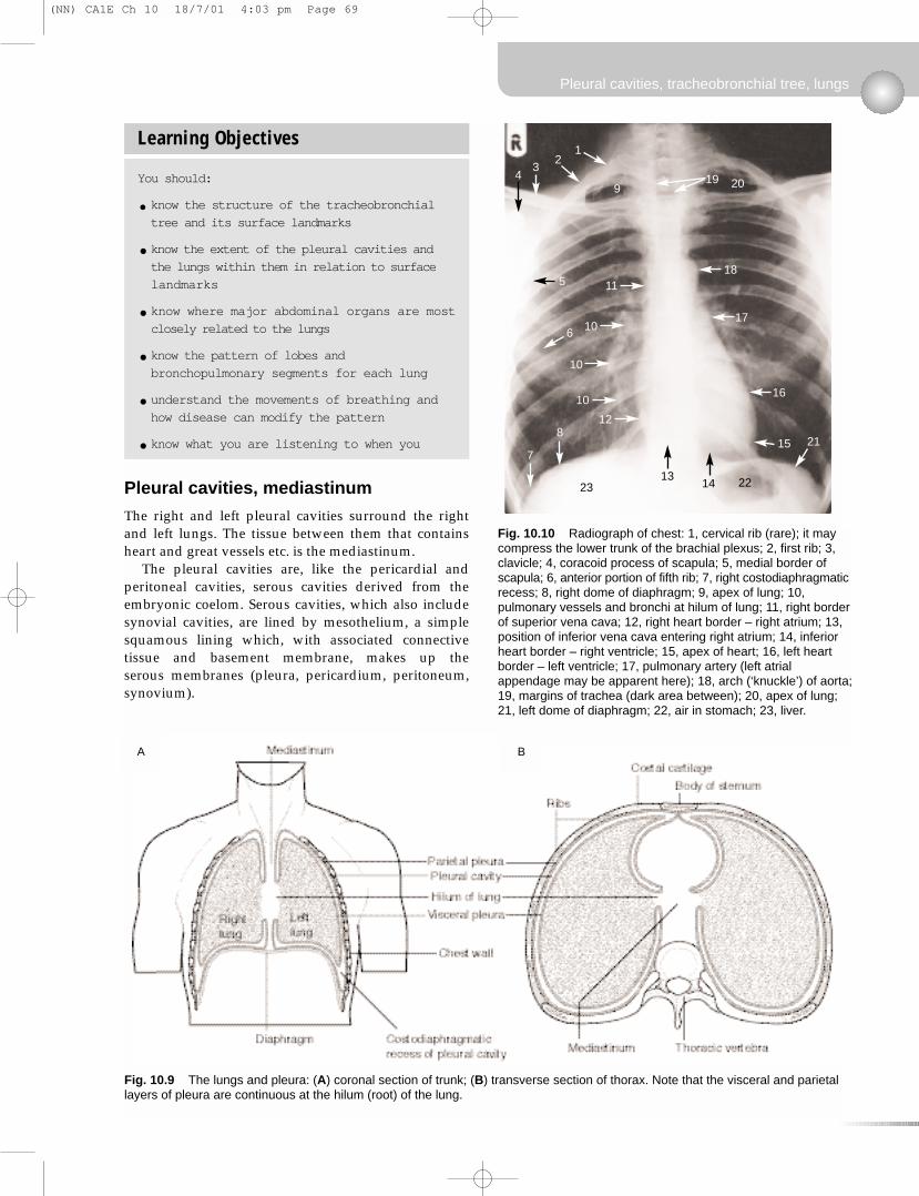
Fig. 10.9 The lungs and pleura: (A) coronal section of trunk; (B) transverse section of thorax. Note that the visceral and parietallayers of pleura are continuous at the hilum (root) of the lung.
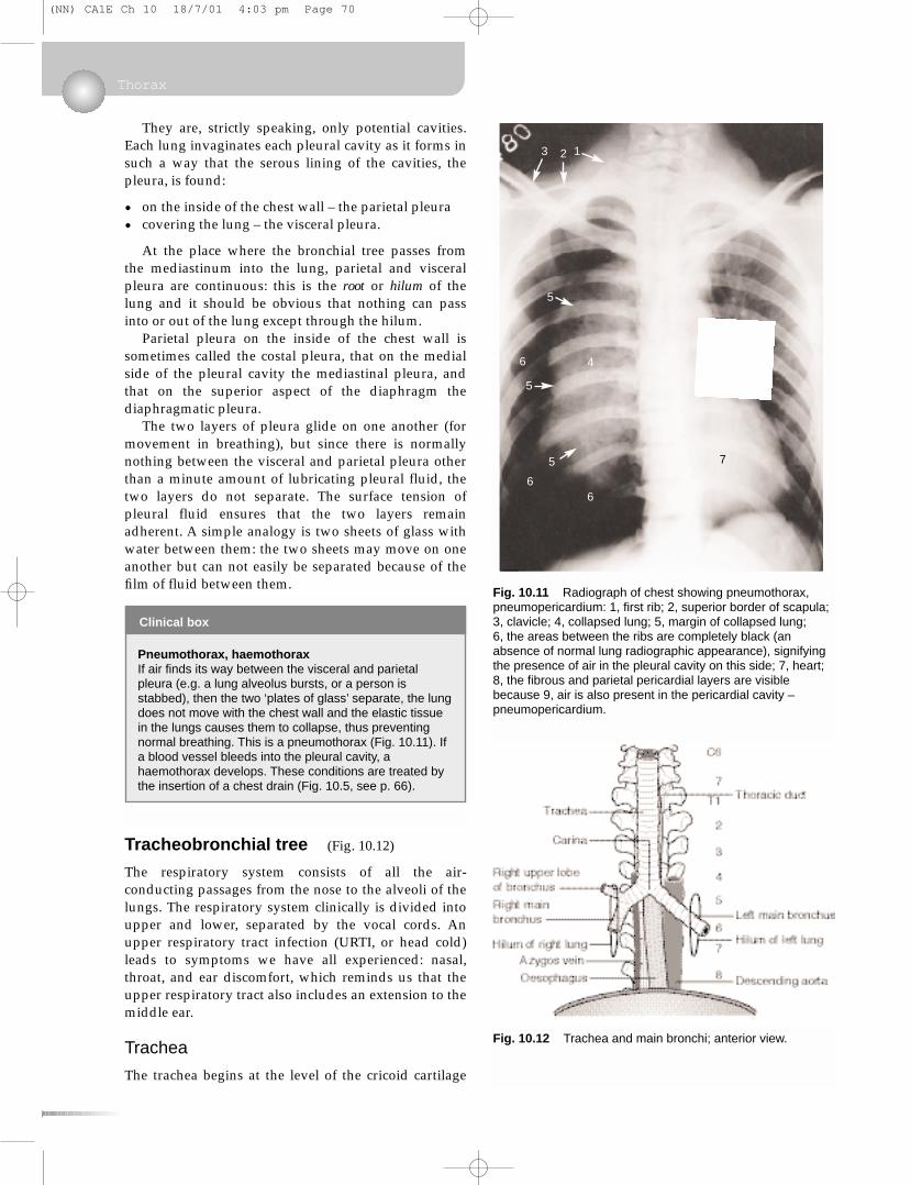
Fig. 10.10 Radiograph of chest: 1, cervical rib (rare); it maycompress the lower trunk of the brachial plexus; 2, first rib; 3,clavicle; 4, coracoid process of scapula; 5, medial border ofscapula; 6, anterior portion of fifth rib; 7, right costodiaphragmaticrecess; 8, right dome of diaphragm; 9, apex of lung; 10,pulmonary vessels and bronchi at hilum of lung; 11, right borderof superior vena cava; 12, right heart border – right atrium; 13,position of inferior vena cava entering right atrium; 14, inferiorheart border – right ventricle; 15, apex of heart; 16, left heartborder – left ventricle; 17, pulmonary artery (left atrialappendage may be apparent here); 18, arch (‘knuckle’) of aorta;19, margins of trachea (dark area between); 20, apex of lung;21, left dome of diaphragm; 22, air in stomach; 23, liver.
A B
4 19
21
9
5 11
10
10
10
12
7
8
6
20
18
17
16
15 21
1314 2223
3
(NN) CA1E Ch 10 18/7/01 4:03 pm Page 69
They are, strictly speaking, only potential cavities.Each lung invaginates each pleural cavity as it forms insuch a way that the serous lining of the cavities, thepleura, is found:
• on the inside of the chest wall – the parietal pleura• covering the lung – the visceral pleura.
At the place where the bronchial tree passes fromthe mediastinum into the lung, parietal and visceralpleura are continuous: this is the root or hilum of thelung and it should be obvious that nothing can passinto or out of the lung except through the hilum.
Parietal pleura on the inside of the chest wall issometimes called the costal pleura, that on the medialside of the pleural cavity the mediastinal pleura, andthat on the superior aspect of the diaphragm thediaphragmatic pleura.
The two layers of pleura glide on one another (formovement in breathing), but since there is normallynothing between the visceral and parietal pleura otherthan a minute amount of lubricating pleural fluid, thetwo layers do not separate. The surface tension ofpleural fluid ensures that the two layers remainadherent. A simple analogy is two sheets of glass withwater between them: the two sheets may move on oneanother but can not easily be separated because of thefilm of fluid between them.
Tracheobronchial tree (Fig. 10.12)
The respiratory system consists of all the air-conducting passages from the nose to the alveoli of thelungs. The respiratory system clinically is divided intoupper and lower, separated by the vocal cords. Anupper respiratory tract infection (URTI, or head cold)leads to symptoms we have all experienced: nasal,throat, and ear discomfort, which reminds us that theupper respiratory tract also includes an extension to themiddle ear.
Trachea
The trachea begins at the level of the cricoid cartilage
Thorax
Clinical box
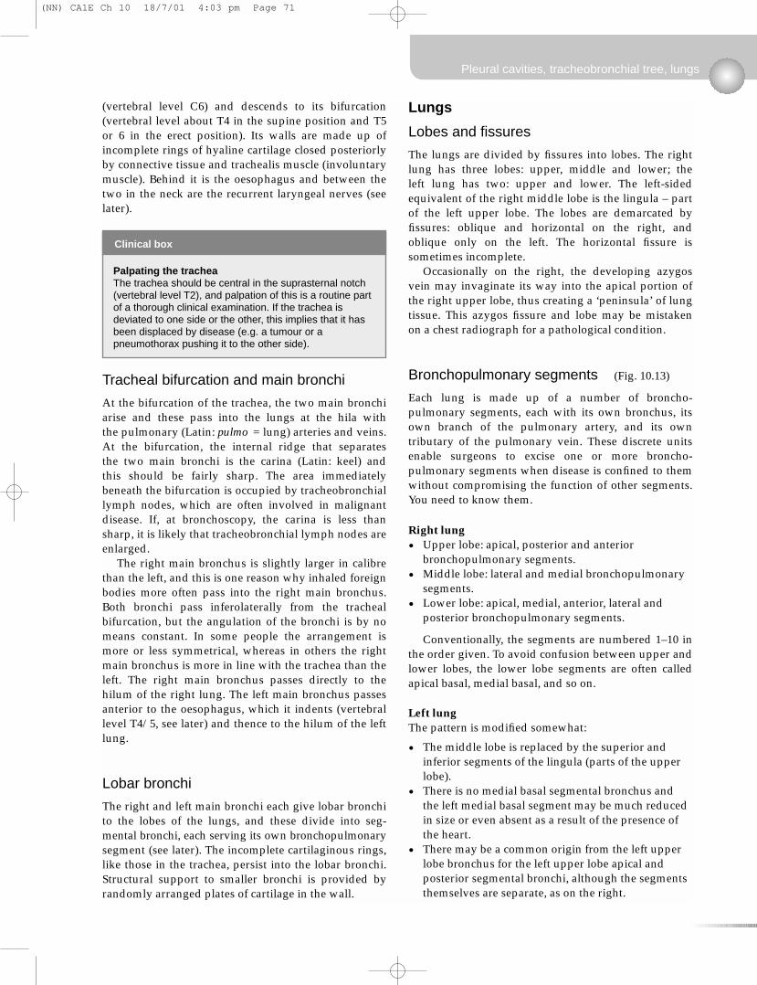
Pneumothorax, haemothoraxIf air finds its way between the visceral and parietalpleura (e.g. a lung alveolus bursts, or a person isstabbed), then the two ‘plates of glass’ separate, the lungdoes not move with the chest wall and the elastic tissuein the lungs causes them to collapse, thus preventingnormal breathing. This is a pneumothorax (Fig. 10.11). Ifa blood vessel bleeds into the pleural cavity, ahaemothorax develops. These conditions are treated bythe insertion of a chest drain (Fig. 10.5, see p. 66).
Fig. 10.11 Radiograph of chest showing pneumothorax,pneumopericardium: 1, first rib; 2, superior border of scapula;3, clavicle; 4, collapsed lung; 5, margin of collapsed lung; 6, the areas between the ribs are completely black (anabsence of normal lung radiographic appearance), signifyingthe presence of air in the pleural cavity on this side; 7, heart;8, the fibrous and parietal pericardial layers are visiblebecause 9, air is also present in the pericardial cavity –pneumopericardium.
Fig. 10.12 Trachea and main bronchi; anterior view.
3 12
5
5
6 4
5
66
7
9
8
(NN) CA1E Ch 10 18/7/01 4:03 pm Page 70
(vertebral level C6) and descends to its bifurcation(vertebral level about T4 in the supine position and T5or 6 in the erect position). Its walls are made up ofincomplete rings of hyaline cartilage closed posteriorlyby connective tissue and trachealis muscle (involuntarymuscle). Behind it is the oesophagus and between thetwo in the neck are the recurrent laryngeal nerves (seelater).
Tracheal bifurcation and main bronchi
At the bifurcation of the trachea, the two main bronchiarise and these pass into the lungs at the hila with the pulmonary (Latin: pulmo = lung) arteries and veins.At the bifurcation, the internal ridge that separates the two main bronchi is the carina (Latin: keel) and this should be fairly sharp. The area immediatelybeneath the bifurcation is occupied by tracheobronchiallymph nodes, which are often involved in malignantdisease. If, at bronchoscopy, the carina is less thansharp, it is likely that tracheobronchial lymph nodes areenlarged.
The right main bronchus is slightly larger in calibrethan the left, and this is one reason why inhaled foreignbodies more often pass into the right main bronchus.Both bronchi pass inferolaterally from the trachealbifurcation, but the angulation of the bronchi is by nomeans constant. In some people the arrangement ismore or less symmetrical, whereas in others the rightmain bronchus is more in line with the trachea than theleft. The right main bronchus passes directly to thehilum of the right lung. The left main bronchus passesanterior to the oesophagus, which it indents (vertebrallevel T4/5, see later) and thence to the hilum of the leftlung.
Lobar bronchi
The right and left main bronchi each give lobar bronchito the lobes of the lungs, and these divide into seg-mental bronchi, each serving its own bronchopulmonarysegment (see later). The incomplete cartilaginous rings,like those in the trachea, persist into the lobar bronchi.Structural support to smaller bronchi is provided byrandomly arranged plates of cartilage in the wall.
Lungs
Lobes and fissures
The lungs are divided by fissures into lobes. The rightlung has three lobes: upper, middle and lower; the left lung has two: upper and lower. The left-sidedequivalent of the right middle lobe is the lingula – partof the left upper lobe. The lobes are demarcated byfissures: oblique and horizontal on the right, andoblique only on the left. The horizontal fissure issometimes incomplete.
Occasionally on the right, the developing azygosvein may invaginate its way into the apical portion ofthe right upper lobe, thus creating a ‘peninsula’ of lungtissue. This azygos fissure and lobe may be mistakenon a chest radiograph for a pathological condition.
Bronchopulmonary segments (Fig. 10.13)
Each lung is made up of a number of broncho-pulmonary segments, each with its own bronchus, itsown branch of the pulmonary artery, and its owntributary of the pulmonary vein. These discrete unitsenable surgeons to excise one or more broncho-pulmonary segments when disease is confined to themwithout compromising the function of other segments.You need to know them.
Right lung• Upper lobe: apical, posterior and anterior
bronchopulmonary segments.• Middle lobe: lateral and medial bronchopulmonary
segments.• Lower lobe: apical, medial, anterior, lateral and
posterior bronchopulmonary segments.
Conventionally, the segments are numbered 1–10 inthe order given. To avoid confusion between upper andlower lobes, the lower lobe segments are often calledapical basal, medial basal, and so on.
Left lungThe pattern is modified somewhat:
• The middle lobe is replaced by the superior andinferior segments of the lingula (parts of the upperlobe).
• There is no medial basal segmental bronchus andthe left medial basal segment may be much reducedin size or even absent as a result of the presence ofthe heart.
• There may be a common origin from the left upperlobe bronchus for the left upper lobe apical andposterior segmental bronchi, although the segmentsthemselves are separate, as on the right.
Pleural cavities, tracheobronchial tree, lungs
Clinical box
Palpating the tracheaThe trachea should be central in the suprasternal notch(vertebral level T2), and palpation of this is a routine partof a thorough clinical examination. If the trachea isdeviated to one side or the other, this implies that it hasbeen displaced by disease (e.g. a tumour or apneumothorax pushing it to the other side).
(NN) CA1E Ch 10 18/7/01 4:03 pm Page 71
Thorax
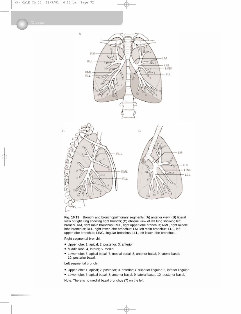
Fig. 10.13 Bronchi and bronchopulmonary segments: (A) anterior view; (B) lateralview of right lung showing right bronchi; (C) oblique view of left lung showing leftbronchi. RM, right main bronchus; RUL, right upper lobe bronchus; RML, right middlelobe bronchus; RLL, right lower lobe bronchus; LM, left main bronchus; LUL, leftupper lobe bronchus; LING, lingular bronchus; LLL, left lower lobe bronchus.
Right segmental bronchi:
• Upper lobe: 1, apical; 2, posterior; 3, anterior
• Middle lobe: 4, lateral; 5, medial
• Lower lobe: 6, apical basal; 7, medial basal; 8, anterior basal; 9, lateral basal; 10, posterior basal.
Left segmental bronchi:
• Upper lobe: 1, apical; 2, posterior; 3, anterior; 4, superior lingular; 5, inferior lingular
• Lower lobe: 6, apical basal; 8, anterior basal; 9, lateral basal, 10, posterior basal.
Note: There is no medial basal bronchus (7) on the left.
(NN) CA1E Ch 10 18/7/01 4:03 pm Page 72
You should bear in mind that there is, as usual, somevariation in this pattern from person to person. It isinteresting, but of no relevance, to know that in pigs,sheep, llamas and tigers one of the right lobar bronchiarises from the trachea itself.
Pulmonary hila
These are the regions where arteries, veins, thebronchial tree, nerves and lymphatics pass betweenmediastinum and lungs. You need not bother with theprecise arrangement of structures at the hilum except toknow that the bronchi are posterior and the pulmonaryarteries superior. The pulmonary ligament is thenarrow inferior extension of the hilum, containingnothing of importance.
Relations of lungs to other organs
A number of important mediastinal structures areintimately related to the lungs and create impressionson the medial aspect of both lungs. On the right lung,those formed by the superior vena cava, the azygosvein and the oesophagus are noteworthy. On the leftside, the impressions of the heart, the aortic arch anddescending aorta are obvious.
Sensory nerve supply of pleura andlungs
Parietal pleura
• Costal: local intercostal nerves.• Diaphragmatic: local intercostal nerves
(peripherally), phrenic nerve (centrally).• Mediastinal: phrenic (mainly).
Parietal pleural sensation is somatic sensation, andpain from irritation of parietal pleura will be sharp andwell localised by the patient.
Lungs and visceral pleura
The lungs and visceral pleura, like all internal organs,are supplied by visceral sensory nerves that travel backto the central nervous system in nerves also conveyingsympathetic and/or parasympathetic fibres. Visceralpain is much more vague and poorly localised by thepatient. Inflammation of the pleura is called pleurisyand the sharp pain commonly associated with it arisesnot from irritation of the visceral pleura but fromirritation of parietal pleura to which the disease hasspread.
Lymphatics of the thorax
Chest wall
Lymph vessels pass:
• posteriorly with the main intercostal neurovascularbundle to reach paravertebral nodes and drain intothe main thoracic duct (except for the upper fewspaces on the right, which drain into the rightlymphatic duct)
• anteriorly with the internal thoracic vessels toparasternal nodes and thence to the thoracic duct.
Both these lymphatic channels communicate withthose of the breast and may be involved in the spreadof breast disease.
Lungs and trachea
There are communicating lymph plexuses immediatelyunder the visceral pleura and around the bronchialtree. From here lymph passes to the hilum where hilarnodes are found and thence to the mediastinal nodesand the thoracic duct.
Tracheobronchial nodes
These are the mediastinal lymph nodes immediatelybeneath the carina. They constitute the largest
Pleural cavities, tracheobronchial tree, lungs
Clinical box
Movements of breathingNormal breathing is the result of movement of thediaphragm. The diaphragm descends, the diaphragmaticparietal pleura descends and, since the two layers ofpleura can not separate, this pulls the visceral pleuradown so that the lung alveoli and airways expand and airis sucked in. In expiration, the diaphragm relaxes and theelastic tissue in the lung recoils, thus expelling air in theairways. In forced inspiration other muscles are used andthese include any muscles that will enlarge the rib cagein any way. Neck muscles (sternocleidomastoid) will pullit up, abdominal muscles (quadratus lumborum) will pull itdown, and by fixing the upper limbs, pectoral girdlemuscles (pectoralis major, latissimus dorsi) will pull it out.These are accessory muscles of respiration and you willsee them in action in any patient suffering a bronchitic orasthmatic attack. The function of the intercostal musclesis not fully understood, despite what anyone may tell you,but obviously they afford a degree of flexibility betweenthe more rigid ribs.
The shape of the costotransverse joints allows thelateral aspect of the lower ribs to swivel up and out, thusincreasing the lateral diameter of the chest. The anteriorends of all ribs can swing up and out, thereby increasingthe anteroposterior diameter of the chest. Disease result-ing in limitation of movement at these joints can be acause of respiratory distress.
(NN) CA1E Ch 10 18/7/01 4:03 pm Page 73
collection of lymph nodes in the body and receivelymph from heart, lungs and mediastinal structuressuch as the oesophagus. Enlargement of these nodeswill cause a widening of the carina when viewed atbronchoscopy (see above).
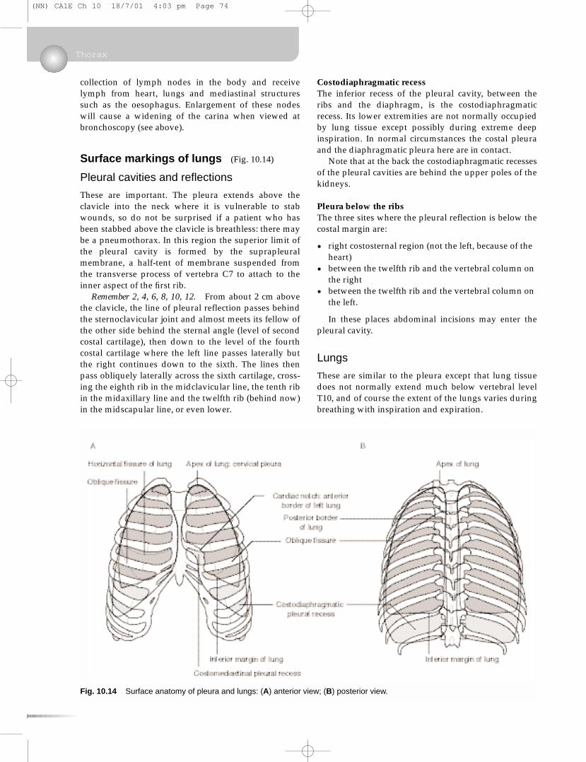
Surface markings of lungs (Fig. 10.14)
Pleural cavities and reflections
These are important. The pleura extends above theclavicle into the neck where it is vulnerable to stabwounds, so do not be surprised if a patient who hasbeen stabbed above the clavicle is breathless: there maybe a pneumothorax. In this region the superior limit ofthe pleural cavity is formed by the suprapleuralmembrane, a half-tent of membrane suspended fromthe transverse process of vertebra C7 to attach to theinner aspect of the first rib.
Remember 2, 4, 6, 8, 10, 12. From about 2 cm abovethe clavicle, the line of pleural reflection passes behindthe sternoclavicular joint and almost meets its fellow ofthe other side behind the sternal angle (level of secondcostal cartilage), then down to the level of the fourthcostal cartilage where the left line passes laterally butthe right continues down to the sixth. The lines thenpass obliquely laterally across the sixth cartilage, cross-ing the eighth rib in the midclavicular line, the tenth ribin the midaxillary line and the twelfth rib (behind now)in the midscapular line, or even lower.
Costodiaphragmatic recessThe inferior recess of the pleural cavity, between theribs and the diaphragm, is the costodiaphragmaticrecess. Its lower extremities are not normally occupiedby lung tissue except possibly during extreme deepinspiration. In normal circumstances the costal pleuraand the diaphragmatic pleura here are in contact.
Note that at the back the costodiaphragmatic recessesof the pleural cavities are behind the upper poles of thekidneys.
Pleura below the ribsThe three sites where the pleural reflection is below thecostal margin are:
• right costosternal region (not the left, because of theheart)
• between the twelfth rib and the vertebral column onthe right
• between the twelfth rib and the vertebral column onthe left.
In these places abdominal incisions may enter thepleural cavity.
Lungs
These are similar to the pleura except that lung tissuedoes not normally extend much below vertebral levelT10, and of course the extent of the lungs varies duringbreathing with inspiration and expiration.
Thorax
Fig. 10.14 Surface anatomy of pleura and lungs: (A) anterior view; (B) posterior view.
(NN) CA1E Ch 10 18/7/01 4:03 pm Page 74
• Oblique fissure (both sides): spine of vertebra T2 orT3 – sixth costal cartilage.
• Horizontal fissure (right lung only): level of fourthcostal cartilage, sternal edge – line of oblique fissure.
Variations in position depending upon posture andbreathingYou should bear in mind that structures move duringbreathing. Vertebral levels are given in Table 10.2.
Finally, it should be obvious that everything will belower when you are standing erect than when you arelying down. For these reasons, surface markings andvertebral levels are only approximate.
10.3 Heart and pericardial cavity
Pericardium
Serous pericardium (Fig. 10.15)
The heart is surrounded by the serous pericardium,
Heart and pericardial cavity
Table 10.2 Effect of breathing on position of lungstructures
Structure In forced expiration In forced inspiration
Bifurcation T4 T5/6 or lowerof trachea
Hilum of T5 T6 or lowerlung
Lower T8 (med.) to T9 (lat.) T9 (med.) to T12 (lat.)border oflung
Clinical box
Listening to (auscultating) the lungsFrom a clinical point of view you should note thefollowing:
• When you place your stethoscope on a patient’s back,you are listening mainly to the lower lobe. There is asmall area of upper lobe, but no middle lobe at all.
• When you place your stethoscope on a patient’santerior chest wall, you are listening mainly to theupper and middle lobes.
• You will listen to the middle lobe best by placing thestethoscope at the side and in the axilla.
• You can not listen to individual bronchopulmonarysegments, or even to individual lobes.
• When a patient is lying in bed on his back, the mostdependent bronchopulmonary segments are the apicaland posterior segments of the lower lobe. Thesesegments are most often affected by lung infections inill, bedridden patients.
• In normal breathing, lung tissue does not occupy thelower extremities of the costodiaphragmatic recesses,but it may in deep inspiration. This means that in thisregion the surface markings of the extent of the lungsis different from the surface markings for the extent ofthe pleural cavities.
Overview
The heart is surrounded by visceral and
parietal layers of serous pericardium
(equivalent to the pleura), and external to these
the fibrous pericardium. The heart and
pericardial cavity are in the anterior
mediastinum, behind the sternum slightly to the
left of the midline. The heart is orientated such
that the right chambers of the heart are more
anterior, and the left chambers more posterior.
The outflow from each chamber is guarded by a
valve: right atrium – tricuspid; right ventricle
– pulmonary; left atrium – mitral; left
ventricle – aortic. The cardiac conducting
system is the means by which contraction is
coordinated. Right and left coronary arteries
from the ascending aorta supply heart muscle
with freshly oxygenated blood. The inherent
rhythmicity of cardiac muscle is modulated by
the autonomic innervation of the heart, amongst
Learning Objectives
You should:
• understand the arrangement of the
pericardium around the heart, and the
importance of cardiac tamponade
•know the surface markings of the heart and
its borders as seen on a PA chest radiograph
•know the route taken by blood as it passes
through the chambers and valves of the
heart, and know how to perform external
cardiac massage
•know the position, surface anatomy and
importance of the cardiac valves
•understand the disposition and importance of
the conducting system
•know the anatomy of the right, left,
circumflex, anterior interventricular and
posterior interventricular coronary arteries
•know where to place your stethoscope in
(NN) CA1E Ch 10 18/7/01 4:03 pm Page 75
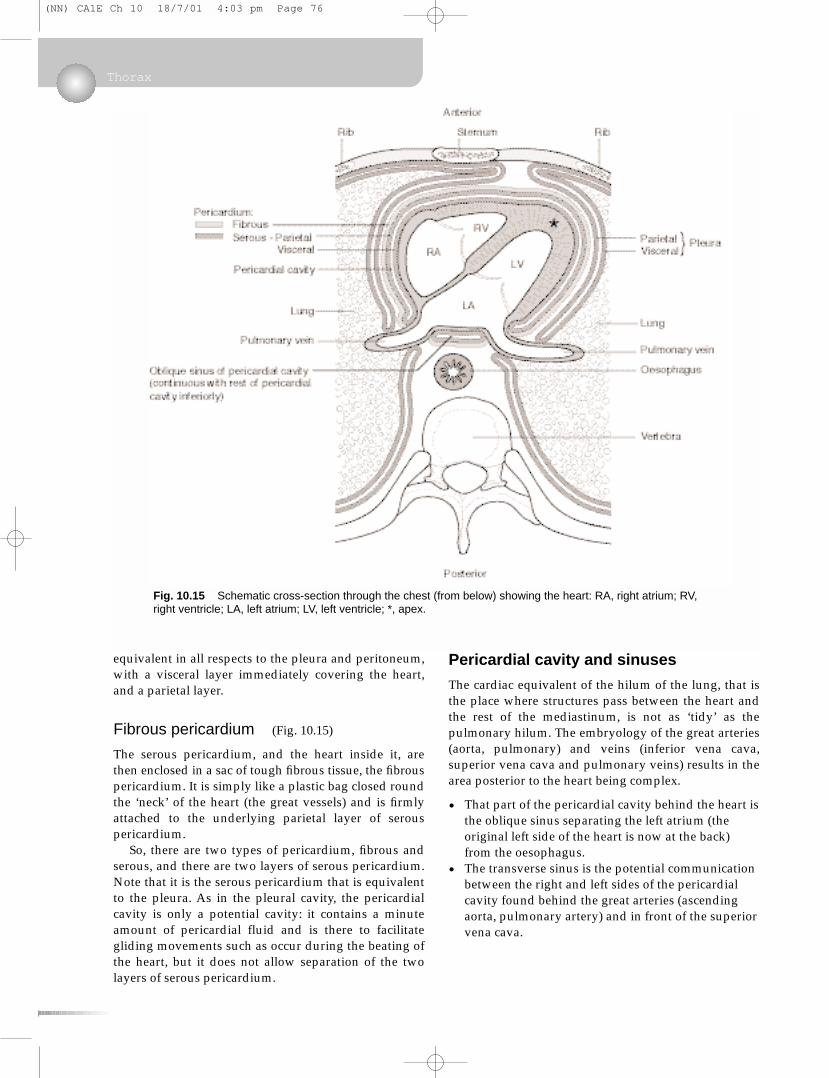
equivalent in all respects to the pleura and peritoneum,with a visceral layer immediately covering the heart,and a parietal layer.
Fibrous pericardium (Fig. 10.15)
The serous pericardium, and the heart inside it, arethen enclosed in a sac of tough fibrous tissue, the fibrouspericardium. It is simply like a plastic bag closed roundthe ‘neck’ of the heart (the great vessels) and is firmlyattached to the underlying parietal layer of serouspericardium.
So, there are two types of pericardium, fibrous andserous, and there are two layers of serous pericardium.Note that it is the serous pericardium that is equivalentto the pleura. As in the pleural cavity, the pericardialcavity is only a potential cavity: it contains a minuteamount of pericardial fluid and is there to facilitategliding movements such as occur during the beating ofthe heart, but it does not allow separation of the twolayers of serous pericardium.
Pericardial cavity and sinuses
The cardiac equivalent of the hilum of the lung, that isthe place where structures pass between the heart andthe rest of the mediastinum, is not as ‘tidy’ as thepulmonary hilum. The embryology of the great arteries(aorta, pulmonary) and veins (inferior vena cava,superior vena cava and pulmonary veins) results in thearea posterior to the heart being complex.
• That part of the pericardial cavity behind the heart isthe oblique sinus separating the left atrium (theoriginal left side of the heart is now at the back)from the oesophagus.
• The transverse sinus is the potential communicationbetween the right and left sides of the pericardialcavity found behind the great arteries (ascendingaorta, pulmonary artery) and in front of the superiorvena cava.
Thorax
Fig. 10.15 Schematic cross-section through the chest (from below) showing the heart: RA, right atrium; RV,right ventricle; LA, left atrium; LV, left ventricle; *, apex.
(NN) CA1E Ch 10 18/7/01 4:03 pm Page 76
Heart
Surfaces and borders
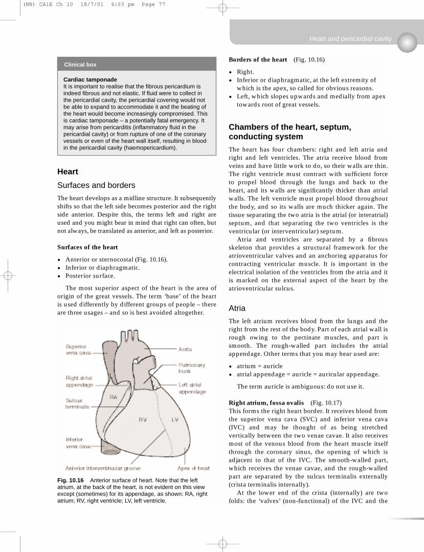
The heart develops as a midline structure. It subsequentlyshifts so that the left side becomes posterior and the rightside anterior. Despite this, the terms left and right areused and you might bear in mind that right can often, butnot always, be translated as anterior, and left as posterior.
Surfaces of the heart
• Anterior or sternocostal (Fig. 10.16).• Inferior or diaphragmatic.• Posterior surface.
The most superior aspect of the heart is the area oforigin of the great vessels. The term ‘base’ of the heartis used differently by different groups of people – thereare three usages – and so is best avoided altogether.
Borders of the heart (Fig. 10.16)
• Right.• Inferior or diaphragmatic, at the left extremity of
which is the apex, so called for obvious reasons.• Left, which slopes upwards and medially from apex
towards root of great vessels.
Chambers of the heart, septum,conducting system
The heart has four chambers: right and left atria andright and left ventricles. The atria receive blood fromveins and have little work to do, so their walls are thin.The right ventricle must contract with sufficient forceto propel blood through the lungs and back to theheart, and its walls are significantly thicker than atrialwalls. The left ventricle must propel blood throughoutthe body, and so its walls are much thicker again. Thetissue separating the two atria is the atrial (or interatrial)septum, and that separating the two ventricles is theventricular (or interventricular) septum.
Atria and ventricles are separated by a fibrousskeleton that provides a structural framework for theatrioventricular valves and an anchoring apparatus forcontracting ventricular muscle. It is important in theelectrical isolation of the ventricles from the atria and itis marked on the external aspect of the heart by theatrioventricular sulcus.
Atria
The left atrium receives blood from the lungs and theright from the rest of the body. Part of each atrial wall isrough owing to the pectinate muscles, and part issmooth. The rough-walled part includes the atrialappendage. Other terms that you may hear used are:
• atrium = auricle• atrial appendage = auricle = auricular appendage.
The term auricle is ambiguous: do not use it.
Right atrium, fossa ovalis (Fig. 10.17)This forms the right heart border. It receives blood fromthe superior vena cava (SVC) and inferior vena cava(IVC) and may be thought of as being stretchedvertically between the two venae cavae. It also receivesmost of the venous blood from the heart muscle itselfthrough the coronary sinus, the opening of which isadjacent to that of the IVC. The smooth-walled part,which receives the venae cavae, and the rough-walledpart are separated by the sulcus terminalis externally(crista terminalis internally).
At the lower end of the crista (internally) are twofolds: the ‘valves’ (non-functional) of the IVC and the
Heart and pericardial cavity
Clinical box
Cardiac tamponadeIt is important to realise that the fibrous pericardium isindeed fibrous and not elastic. If fluid were to collect inthe pericardial cavity, the pericardial covering would notbe able to expand to accommodate it and the beating ofthe heart would become increasingly compromised. Thisis cardiac tamponade – a potentially fatal emergency. Itmay arise from pericarditis (inflammatory fluid in thepericardial cavity) or from rupture of one of the coronaryvessels or even of the heart wall itself, resulting in bloodin the pericardial cavity (haemopericardium).
Fig. 10.16 Anterior surface of heart. Note that the leftatrium, at the back of the heart, is not evident on this viewexcept (sometimes) for its appendage, as shown: RA, rightatrium; RV, right ventricle; LV, left ventricle.
(NN) CA1E Ch 10 18/7/01 4:03 pm Page 77
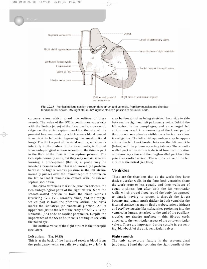
coronary sinus which guard the orifices of thesevessels. The valve of the IVC is continuous superiorlywith the limbus (edge) of the fossa ovalis, a crescenticridge on the atrial septum marking the site of theprenatal foramen ovale by which means blood passedfrom right to left atria, bypassing the non-functionallungs. The thicker part of the atrial septum, which endsinferiorly in the limbus of the fossa ovalis, is formedfrom embryological septum secundum; the thinner partin the floor of the fossa is from septum primum. Thetwo septa normally unite, but they may remain separateforming a probe-patent (that is, a probe may beinserted) foramen ovale. This is not normally a problembecause the higher venous pressure in the left atriumnormally pushes over the thinner septum primum onthe left so that it remains in contact with the thickerseptum secundum.
The crista terminalis marks the junction between thetwo embryological parts of the right atrium. Since thesmooth-walled portion is from the sinus venosus(receiving SVC, IVC, coronary sinus) and the rough-walled part is from the primitive atrium, the cristamarks the sinuatrial (or sinoatrial) junction. At itsupper end, just to the left of the entry of the SVC, is thesinuatrial (SA) node or cardiac pacemaker. Despite theimportance of the SA node, there is nothing to see withthe naked eye.
The outflow valve of the right atrium is the tricuspid(see later).
Left atrium (Fig. 10.15)This is at the back of the heart and receives blood fromthe pulmonary veins (usually two right, two left). It
may be thought of as being stretched from side to sidebetween the right and left pulmonary veins. Behind theleft atrium is the oesophagus, and an enlarged leftatrium may result in a narrowing of the lower part ofthe thoracic oesophagus visible on a barium swallowinvestigation. The left atrial appendage may be appar-ent on the left heart border between the left ventricle(below) and the pulmonary artery (above). The smooth-walled part of the atrium is derived from incorporationof pulmonary veins and the rough-walled part from theprimitive cardiac atrium. The outflow valve of the leftatrium is the mitral (see later).
Ventricles
These are the chambers that do the work: they havethick muscular walls. In the fetus both ventricles sharethe work more or less equally and their walls are ofequal thickness, but after birth the left ventricularwalls, which propel blood round the body (as opposedto simply having to propel it through the lungs)become and remain much thicker. In both ventricles theinternal surface has many fleshy trabeculations (ridges)and papillary muscles like stalagmites projecting into theventricular lumen. Attached to the end of the papillarymuscles are chordae tendineae – thin fibrous cordsattached to the ventricular aspect of the atrioventricularvalves. These are important during systole in prevent-ing ‘blowback’ of the atrioventricular valves.
Right ventricleThe only noteworthy feature is the septomarginal(moderator) band that contains the right bundle of the
Thorax
Fig. 10.17 Vertical oblique section through right atrium and ventricle. Papillary muscles and chordaetendineae not shown. RA, right atrium; RV, right ventricle; *, position of sinuatrial node.
(NN) CA1E Ch 10 18/7/01 4:03 pm Page 78
conducting system. The outflow valve is the pulmon-ary valve, and the part of the ventricle immediatelybeneath it is the conus arteriosus or infundibulum.
Left ventricleThere is nothing noteworthy about the interior of thischamber. The outflow valve is the aortic.
Ventricular septum
Most of this is thick and highly muscular as befits partof the wall of the left ventricle, but near the atrio-ventricular junction, part is membranous (parsmembranacea), in effect a continuation of the atrialseptum. If during development the membranousportion and the muscular portion fail to fuse, a ventri-cular septal defect results. Neural crest cells contributeto the membranous portion of the septum, andventricular septal defects may be associated with otheranomalies of neural crest origin.
Valves
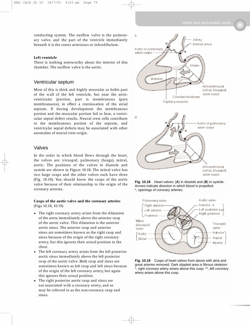
In the order in which blood flows through the heart,the valves are: tricuspid, pulmonary (lungs), mitral,aortic. The positions of the valves in diastole andsystole are shown in Figure 10.18. The mitral valve hastwo large cusps and the other valves each have three(Fig. 10.19). You should know the cusps of the aorticvalve because of their relationship to the origin of thecoronary arteries.
Cusps of the aortic valve and the coronary arteries(Figs 10.18, 10.19)
• The right coronary artery arises from the dilatationof the aorta immediately above the anterior cusp of the aortic valve. This dilatation is the anterioraortic sinus. The anterior cusp and anterior sinus are sometimes known as the right cusp andsinus because of the origin of the right coronaryartery, but this ignores their actual position in thechest.
• The left coronary artery arises from the left posterioraortic sinus immediately above the left posteriorcusp of the aortic valve. Both cusp and sinus aresometimes known as left cusp and left sinus becauseof the origin of the left coronary artery, but againthis ignores their actual position.
• The right posterior aortic cusp and sinus are not associated with a coronary artery, and so may be referred to as the non-coronary cusp andsinus.
Heart and pericardial cavity
Fig. 10.18 Heart valves: (A) in diastole and (B) in systole.Arrows indicate direction in which blood is propelled; *, openings of coronary arteries.
Fig. 10.19 Cusps of heart valves from above with atria andgreat arteries removed. Dark stippled area is fibrous skeleton:*, right coronary artery arises above this cusp; **, left coronaryartery arises above this cusp.
(NN) CA1E Ch 10 18/7/01 4:03 pm Page 79
Conducting system and fibrous skeleton
The pacemaker is the sinuatrial (SA) node in the wall ofthe right atrium at the top of the crista terminalis. Fromthe SA node the impulse passes to the atrioventricular(AV) node in the lower end of the atrial septum besidethe coronary sinus opening. There are probably severaldistinct routes along which impulses pass in the atrialwall, although they are not histologically distinct. Fromthe AV node specialised cardiac muscle fibres, Purkinjefibres, form the bundle of His. This penetrates thefibrous skeleton that separates the atria from the ventri-cles, and it passes into the ventricular septum (ventricularseptal defects may interfere with it, leading to cardiacdysrhythmias). About half way down the septum itdivides into right and left branches. The left bundlepasses to the apex and up the left side of the heart. Theright bundle crosses to the inferior (embryonic right)side of the ventricle in the septomarginal (moderator)band, an identifiable ridge in the wall of the rightventricle.
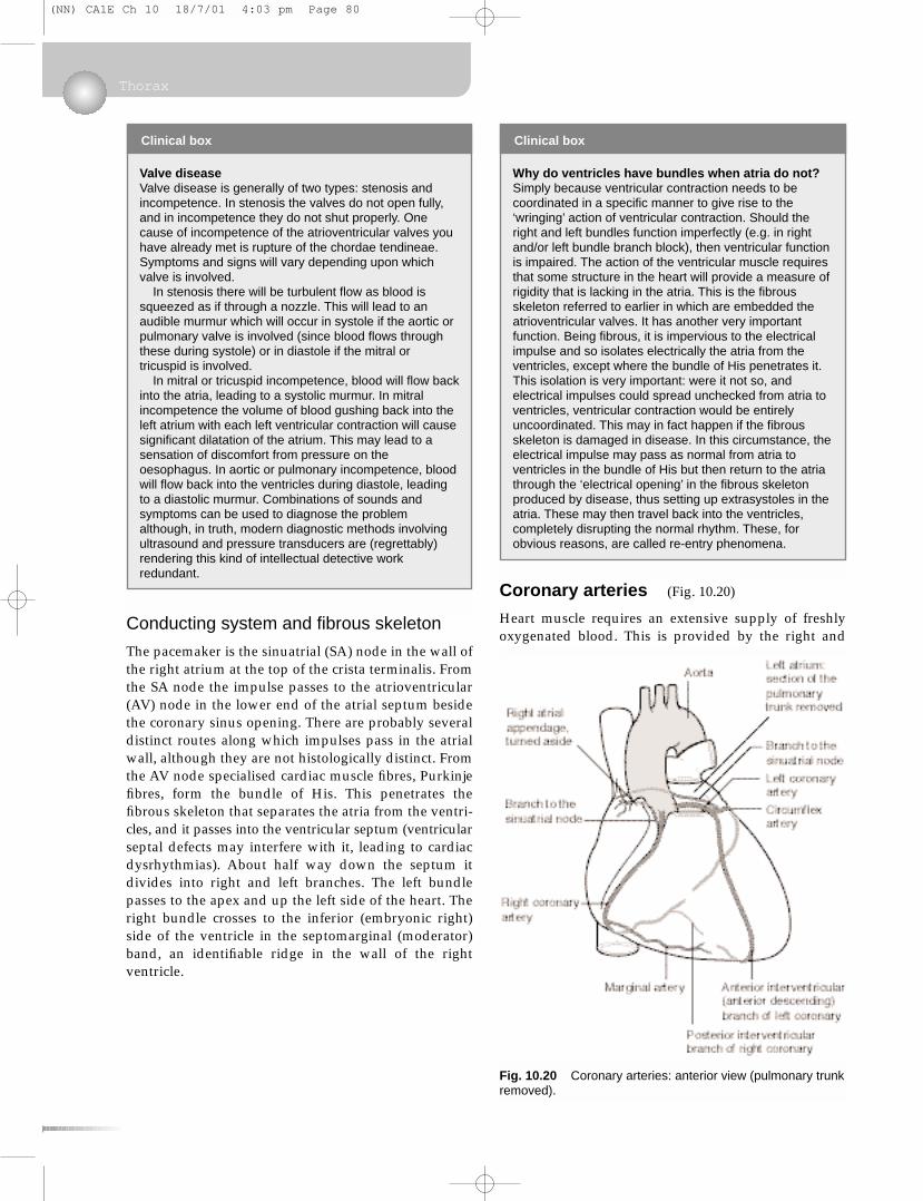
Coronary arteries (Fig. 10.20)
Heart muscle requires an extensive supply of freshlyoxygenated blood. This is provided by the right and
Thorax
Clinical box
Valve diseaseValve disease is generally of two types: stenosis andincompetence. In stenosis the valves do not open fully,and in incompetence they do not shut properly. Onecause of incompetence of the atrioventricular valves youhave already met is rupture of the chordae tendineae.Symptoms and signs will vary depending upon whichvalve is involved.
In stenosis there will be turbulent flow as blood issqueezed as if through a nozzle. This will lead to anaudible murmur which will occur in systole if the aortic orpulmonary valve is involved (since blood flows throughthese during systole) or in diastole if the mitral ortricuspid is involved.
In mitral or tricuspid incompetence, blood will flow backinto the atria, leading to a systolic murmur. In mitralincompetence the volume of blood gushing back into theleft atrium with each left ventricular contraction will causesignificant dilatation of the atrium. This may lead to asensation of discomfort from pressure on theoesophagus. In aortic or pulmonary incompetence, bloodwill flow back into the ventricles during diastole, leadingto a diastolic murmur. Combinations of sounds andsymptoms can be used to diagnose the problemalthough, in truth, modern diagnostic methods involvingultrasound and pressure transducers are (regrettably)rendering this kind of intellectual detective workredundant.
Clinical box
Why do ventricles have bundles when atria do not?Simply because ventricular contraction needs to becoordinated in a specific manner to give rise to the‘wringing’ action of ventricular contraction. Should theright and left bundles function imperfectly (e.g. in rightand/or left bundle branch block), then ventricular functionis impaired. The action of the ventricular muscle requiresthat some structure in the heart will provide a measure ofrigidity that is lacking in the atria. This is the fibrousskeleton referred to earlier in which are embedded theatrioventricular valves. It has another very importantfunction. Being fibrous, it is impervious to the electricalimpulse and so isolates electrically the atria from theventricles, except where the bundle of His penetrates it.This isolation is very important: were it not so, andelectrical impulses could spread unchecked from atria toventricles, ventricular contraction would be entirelyuncoordinated. This may in fact happen if the fibrousskeleton is damaged in disease. In this circumstance, theelectrical impulse may pass as normal from atria toventricles in the bundle of His but then return to the atriathrough the ‘electrical opening’ in the fibrous skeletonproduced by disease, thus setting up extrasystoles in theatria. These may then travel back into the ventricles,completely disrupting the normal rhythm. These, forobvious reasons, are called re-entry phenomena.
Fig. 10.20 Coronary arteries: anterior view (pulmonary trunkremoved).
(NN) CA1E Ch 10 18/7/01 4:03 pm Page 80
left coronary arteries arising from the aorta imme-diately above the aortic valve.
• The right coronary artery arises from the anterioraortic sinus (slight dilatation) above the anteriorcusp of the aortic valve. It passes inferiorly in theanterior atrioventricular groove and gives off themarginal branch as it approaches the diaphragmatic(inferior) surface of the heart. The main trunk passesunder the heart on the diaphragmatic surface andthen turns to the left, running in the posteriorinterventricular groove towards the apex as theposterior interventricular artery. Branches of theright coronary artery supply both atria, most of theright ventricle, the posterior part of the ventricularseptum and the posterior part of the left ventricle,and usually there is a branch to the sinuatrial(pacemaker) node.
• The left coronary artery arises from the left posterioraortic sinus above the left posterior cusp of theaortic valve. It passes forwards behind the origin ofthe pulmonary trunk for about 2 cm and thendivides into the circumflex and anteriorinterventricular arteries. The circumflex passesposteriorly in the atrioventricular groove behind theheart to anastomose with branches of the rightcoronary artery. The anterior interventricular arterypasses down on the anterior aspect of the heart tothe apex. Its alternative name, therefore, much usedby clinicians, is the left anterior descending artery.Branches of the left coronary supply both atria, theanterior part of the left ventricle, the anteriorventricular septum and some adjacent rightventricle.
There are extensive anastomoses between theterritories of the two arteries and variations are com-mon. For example, the posterior interventricular arterymay be a continuation of the circumflex (thus from theleft coronary) rather than of the right coronary artery.
Coronary vessels fill during diastole. A little thoughtwill confirm that when the cusps of the aortic valve are open during systole, blood can not enter thecoronary vessels, and it is propelled quickly past theiropenings by the force of ventricular contraction.During diastole with the cusps of the aortic valve in theclosed position, the elastic recoil of the aortic wallprovides helpful propulsion forcing blood from theaortic sinuses into the coronary vessels. Blood flowthrough coronary vessels is maximal during diastole:obviously, when heart muscle is contracting, coronaryvessels are narrowed and blood flow impeded. Here is another good reason why the openings of thecoronary vessels should be outside, rather than in, the myocardium.
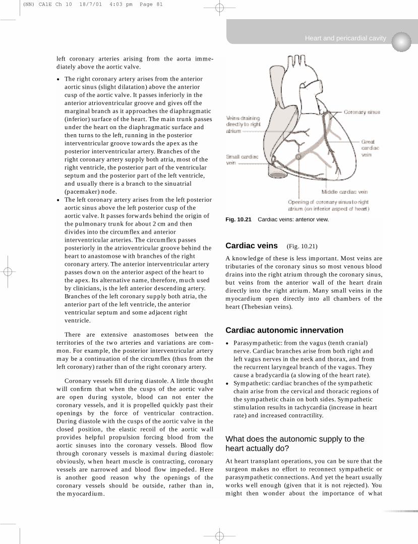
Cardiac veins (Fig. 10.21)
A knowledge of these is less important. Most veins aretributaries of the coronary sinus so most venous blooddrains into the right atrium through the coronary sinus,but veins from the anterior wall of the heart draindirectly into the right atrium. Many small veins in themyocardium open directly into all chambers of theheart (Thebesian veins).
Cardiac autonomic innervation
• Parasympathetic: from the vagus (tenth cranial)nerve. Cardiac branches arise from both right andleft vagus nerves in the neck and thorax, and fromthe recurrent laryngeal branch of the vagus. Theycause a bradycardia (a slowing of the heart rate).
• Sympathetic: cardiac branches of the sympatheticchain arise from the cervical and thoracic regions ofthe sympathetic chain on both sides. Sympatheticstimulation results in tachycardia (increase in heartrate) and increased contractility.
What does the autonomic supply to theheart actually do?
At heart transplant operations, you can be sure that thesurgeon makes no effort to reconnect sympathetic orparasympathetic connections. And yet the heart usuallyworks well enough (given that it is not rejected). Youmight then wonder about the importance of what
Heart and pericardial cavity
Fig. 10.21 Cardiac veins: anterior view.
(NN) CA1E Ch 10 18/7/01 4:03 pm Page 81
physiology textbooks tell you. Heart muscle possessesinherent rhythmicity: its innervation merely modulatesthis.
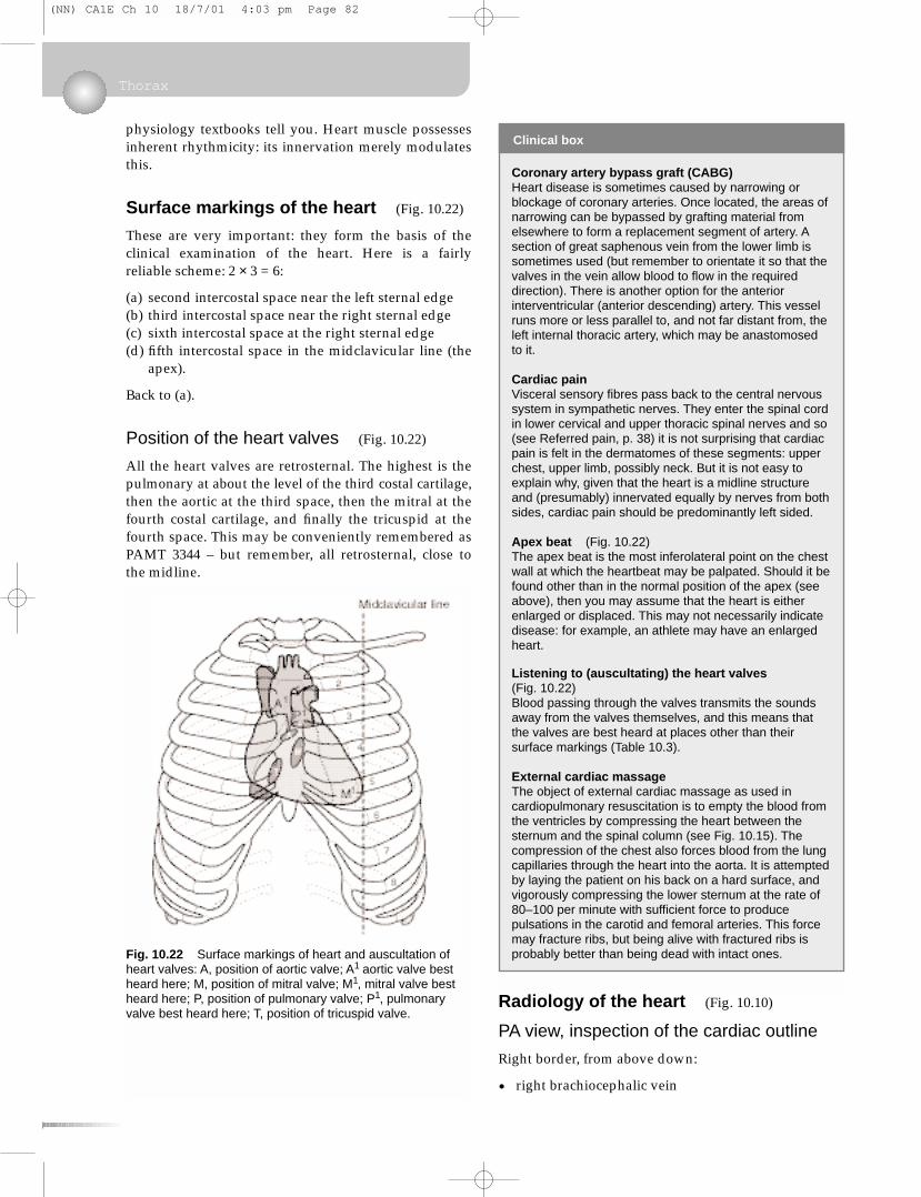
Surface markings of the heart (Fig. 10.22)
These are very important: they form the basis of theclinical examination of the heart. Here is a fairlyreliable scheme: 2 × 3 = 6:
(a) second intercostal space near the left sternal edge(b) third intercostal space near the right sternal edge(c) sixth intercostal space at the right sternal edge(d) fifth intercostal space in the midclavicular line (the
apex).
Back to (a).
Position of the heart valves (Fig. 10.22)
All the heart valves are retrosternal. The highest is thepulmonary at about the level of the third costal cartilage,then the aortic at the third space, then the mitral at thefourth costal cartilage, and finally the tricuspid at thefourth space. This may be conveniently remembered asPAMT 3344 – but remember, all retrosternal, close tothe midline.
Radiology of the heart (Fig. 10.10)
PA view, inspection of the cardiac outline
Right border, from above down:
• right brachiocephalic vein
Thorax
Fig. 10.22 Surface markings of heart and auscultation ofheart valves: A, position of aortic valve; A1 aortic valve bestheard here; M, position of mitral valve; M1, mitral valve bestheard here; P, position of pulmonary valve; P1, pulmonaryvalve best heard here; T, position of tricuspid valve.
Clinical box
Coronary artery bypass graft (CABG)Heart disease is sometimes caused by narrowing orblockage of coronary arteries. Once located, the areas ofnarrowing can be bypassed by grafting material fromelsewhere to form a replacement segment of artery. Asection of great saphenous vein from the lower limb issometimes used (but remember to orientate it so that thevalves in the vein allow blood to flow in the requireddirection). There is another option for the anteriorinterventricular (anterior descending) artery. This vesselruns more or less parallel to, and not far distant from, theleft internal thoracic artery, which may be anastomosedto it.
Cardiac painVisceral sensory fibres pass back to the central nervoussystem in sympathetic nerves. They enter the spinal cordin lower cervical and upper thoracic spinal nerves and so(see Referred pain, p. 38) it is not surprising that cardiacpain is felt in the dermatomes of these segments: upperchest, upper limb, possibly neck. But it is not easy toexplain why, given that the heart is a midline structureand (presumably) innervated equally by nerves from bothsides, cardiac pain should be predominantly left sided.
Apex beat (Fig. 10.22)The apex beat is the most inferolateral point on the chestwall at which the heartbeat may be palpated. Should it befound other than in the normal position of the apex (seeabove), then you may assume that the heart is eitherenlarged or displaced. This may not necessarily indicatedisease: for example, an athlete may have an enlargedheart.
Listening to (auscultating) the heart valves(Fig. 10.22)Blood passing through the valves transmits the soundsaway from the valves themselves, and this means thatthe valves are best heard at places other than theirsurface markings (Table 10.3).
External cardiac massageThe object of external cardiac massage as used incardiopulmonary resuscitation is to empty the blood fromthe ventricles by compressing the heart between thesternum and the spinal column (see Fig. 10.15). Thecompression of the chest also forces blood from the lungcapillaries through the heart into the aorta. It is attemptedby laying the patient on his back on a hard surface, andvigorously compressing the lower sternum at the rate of80–100 per minute with sufficient force to producepulsations in the carotid and femoral arteries. This forcemay fracture ribs, but being alive with fractured ribs isprobably better than being dead with intact ones.
(NN) CA1E Ch 10 18/7/01 4:03 pm Page 82
• SVC• right atrium.
Diaphragmatic surface, right to left:
• right ventricle• very small portion of left ventricle• apex.
Left border, from apex up:
• left ventricle• (left atrial appendage, sometimes)• pulmonary artery• aortic knuckle (junction of aortic arch with
descending aorta).
10.4 Mediastinum
Parts of the mediastinum
The mediastinum is divided horizontally by the planefrom the sternal angle (of Louis) to vertebra T4 into:
• the superior mediastinum, above this plane• the inferior mediastinum below this plane, which is
itself divided into:– anterior mediastinum: anterior to the heart and
pericardium– middle mediastinum: the heart and pericardium– posterior mediastinum: behind the heart and
pericardium, containing the oesophagus, trachea,vagus and phrenic nerves, thoracic duct, andsympathetic chain.
There is nothing in the anterior mediastinum worthyof attention other than the thymus gland extendingdown from the superior mediastinum, and the internalthoracic vessels. Having considered the heart, only thesuperior and posterior compartments remain to beconsidered in any detail.
Mediastinum
Table 10.3 Surface markings and auscultation of the heart valves
Valve Position Best heard
Pulmonary Retrosternal, level of 3rd rib 2nd space just to left of sternal edge
Aortic Retrosternal, level of 3rd space 2nd space just to right of sternal edge
Mitral Retrosternal, level of 4th rib Apex (5th space, midclavicular line)
Tricuspid Retrosternal, level of 4th space Lower sternal edge, side depending upon the condition
Overview
The mediastinum is the area between the two
pleural cavities. It is divided into the superior
mediastinum above vertebral level T4 and,
below this, the anterior mediastinum (in front
of the pericardial cavity), middle mediastinum
(pericardial cavity and contents) and posterior
mediastinum (behind the pericardial cavity).
The organs of significance are the oesophagus,
trachea, great arteries and veins, thoracic
duct, vagus nerve, phrenic nerve and
sympathetic chain. The arch of the aorta gives
rise to the great arteries supplying the head,
neck and upper limbs. The tributaries of the
superior vena cava are important clinical
access routes. The trachea, oesophagus and
vagus and phrenic nerves are closely
interrelated and may be affected in malignant
tumours of the lung. The sympathetic chains are
found in the superior and posterior divisions of
the mediastinum, and give rise to the cardiac
branches and splanchnic nerves supplying
Learning Objectives
You should:
• know the subdivisions and contents of the
mediastinum
•know the disposition of the great arteries and
veins and their principal branches and
tributaries
•know the anatomy of insertion of catheters
etc. into the right brachiocephalic vein
•know the formation, course and functions of
the phrenic nerves
•know the formation, course and functions of
the vagus nerves and branches
•understand how thoracic disease may give
rise to signs and symptoms in the head, neck
and upper limb
•know the basic anatomy of the sympathetic
(NN) CA1E Ch 10 18/7/01 4:03 pm Page 83
Thymus gland
This is found in children anterior to the trachea,extending from the level of the cricoid cartilage(vertebral level C6) down to the retrosternal area.Thymic tumours may compress the tributaries of thesuperior vena cava leading to venous engorgement inthe neck. During childhood the thymus graduallyregresses so that in the adult thymic tissue is more orless confined to the retrosternal connective tissue.
Great arteries
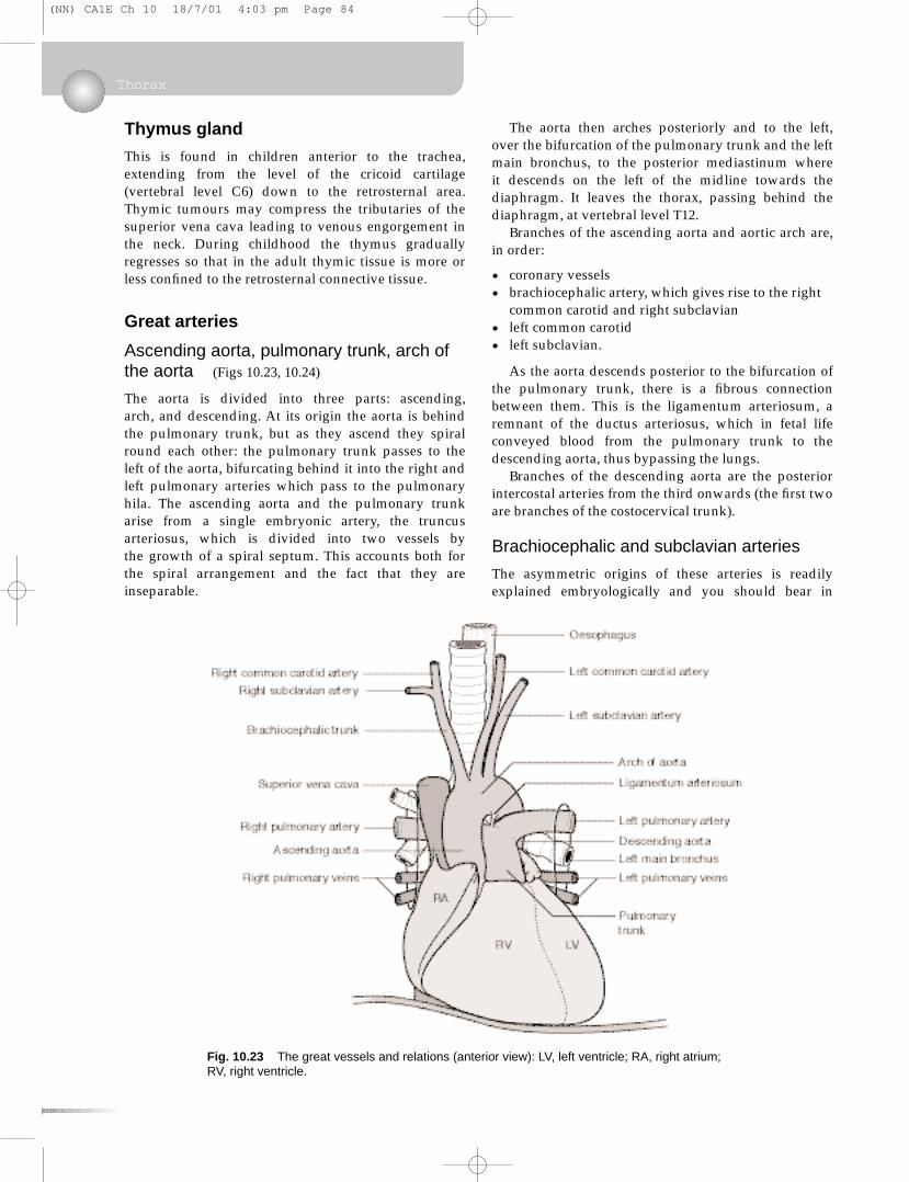
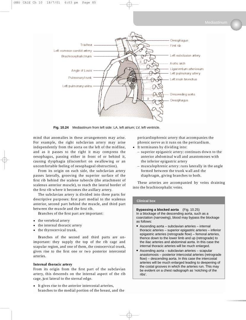
Ascending aorta, pulmonary trunk, arch ofthe aorta (Figs 10.23, 10.24)
The aorta is divided into three parts: ascending, arch, and descending. At its origin the aorta is behindthe pulmonary trunk, but as they ascend they spiralround each other: the pulmonary trunk passes to theleft of the aorta, bifurcating behind it into the right andleft pulmonary arteries which pass to the pulmonaryhila. The ascending aorta and the pulmonary trunkarise from a single embryonic artery, the truncusarteriosus, which is divided into two vessels by the growth of a spiral septum. This accounts both forthe spiral arrangement and the fact that they areinseparable.
The aorta then arches posteriorly and to the left,over the bifurcation of the pulmonary trunk and the leftmain bronchus, to the posterior mediastinum where it descends on the left of the midline towards thediaphragm. It leaves the thorax, passing behind thediaphragm, at vertebral level T12.
Branches of the ascending aorta and aortic arch are,in order:
• coronary vessels• brachiocephalic artery, which gives rise to the right
common carotid and right subclavian• left common carotid• left subclavian.
As the aorta descends posterior to the bifurcation ofthe pulmonary trunk, there is a fibrous connectionbetween them. This is the ligamentum arteriosum, aremnant of the ductus arteriosus, which in fetal lifeconveyed blood from the pulmonary trunk to thedescending aorta, thus bypassing the lungs.
Branches of the descending aorta are the posteriorintercostal arteries from the third onwards (the first twoare branches of the costocervical trunk).
Brachiocephalic and subclavian arteries
The asymmetric origins of these arteries is readilyexplained embryologically and you should bear in
Thorax
Fig. 10.23 The great vessels and relations (anterior view): LV, left ventricle; RA, right atrium; RV, right ventricle.
(NN) CA1E Ch 10 18/7/01 4:03 pm Page 84
mind that anomalies in these arrangements may arise.For example, the right subclavian artery may ariseindependently from the aorta on the left of the midline,and as it passes to the right it may compress theoesophagus, passing either in front of or behind it,causing dysphagia (discomfort on swallowing or anuncomfortable feeling of oesophageal obstruction).
From its origin on each side, the subclavian arterypasses laterally, grooving the superior surface of thefirst rib behind the scalene tubercle (the attachment ofscalenus anterior muscle), to reach the lateral border ofthe first rib where it becomes the axillary artery.
The subclavian artery is divided into three parts fordescriptive purposes: first part medial to the scalenusanterior, second part behind the muscle, and third partbetween the muscle and the first rib.
Branches of the first part are important:
• the vertebral artery• the internal thoracic artery• the thyrocervical trunk.
Branches of the second and third parts are un-important: they supply the top of the rib cage andscapular region, and one of them, the costocervical trunk,gives rise to the first one or two posterior intercostalarteries.
Internal thoracic arteryFrom its origin from the first part of the subclavianartery, this descends on the internal aspect of the ribcage, just lateral to the sternal edge.
• It gives rise to the anterior intercostal arteries,branches to the medial portion of the breast, and the
pericardiophrenic artery that accompanies thephrenic nerve as it runs on the pericardium.
• It terminates by dividing into:– superior epigastric artery: continues down to the
anterior abdominal wall and anastomoses withthe inferior epigastric artery
– musculophrenic artery: runs laterally in the angleformed between the trunk wall and thediaphragm, giving branches to both.
These arteries are accompanied by veins draininginto the brachiocephalic veins.
Mediastinum
Fig. 10.24 Mediastinum from left side: LA, left atrium; LV, left ventricle.
Clinical box
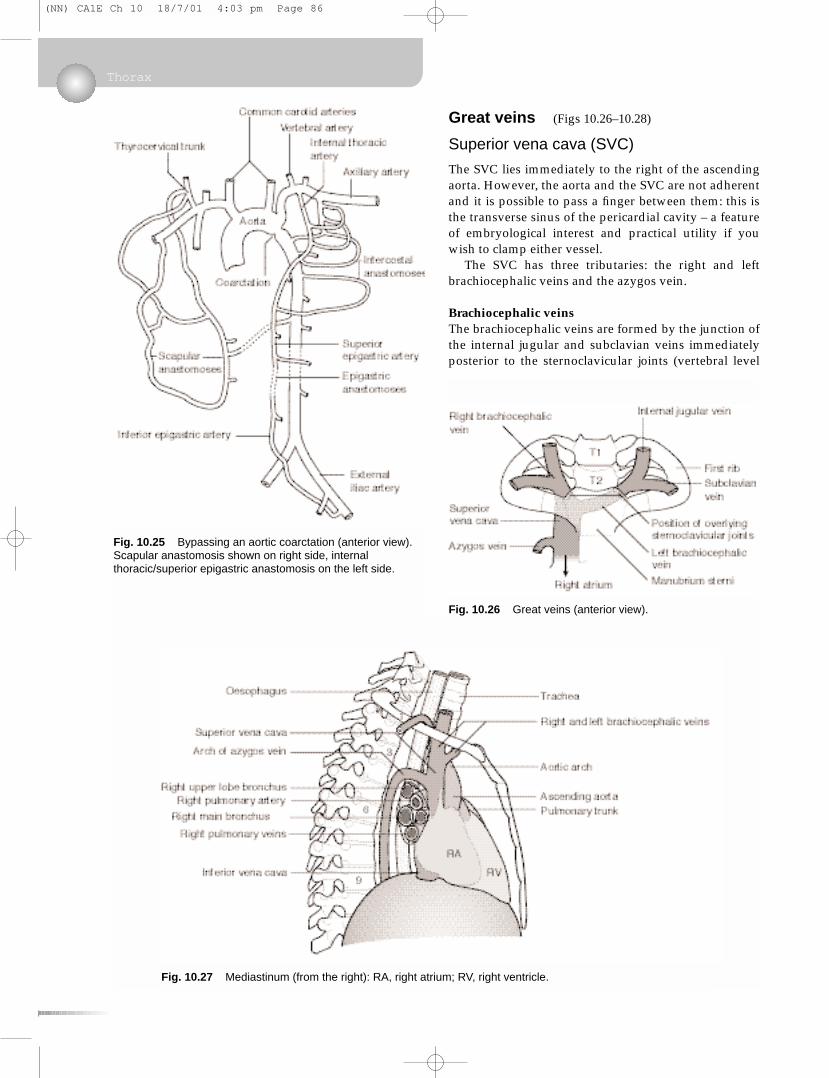
Bypassing a blocked aorta (Fig. 10.25)In a blockage of the descending aorta, such as acoarctation (narrowing), blood may bypass the blockageas follows:
• Ascending aorta – subclavian arteries – internalthoracic arteries – superior epigastric arteries – inferiorepigastric arteries (retrograde flow) – femoral arteries,thence down to the lower limb and up (retrograde) tothe iliac arteries and abdominal aorta. In this case theinternal thoracic arteries will be much enlarged.
• Ascending aorta – subclavian arteries – scapularanastomosis – posterior intercostal arteries (retrogradeflow) – descending aorta. In this case the intercostalarteries will be much enlarged leading to deepening ofthe costal grooves in which the arteries run. This maybe evident on a chest radiograph as ‘notching of theribs’.
(NN) CA1E Ch 10 18/7/01 4:03 pm Page 85
Great veins (Figs 10.26–10.28)
Superior vena cava (SVC)
The SVC lies immediately to the right of the ascendingaorta. However, the aorta and the SVC are not adherentand it is possible to pass a finger between them: this isthe transverse sinus of the pericardial cavity – a featureof embryological interest and practical utility if youwish to clamp either vessel.
The SVC has three tributaries: the right and leftbrachiocephalic veins and the azygos vein.
Brachiocephalic veinsThe brachiocephalic veins are formed by the junction ofthe internal jugular and subclavian veins immediatelyposterior to the sternoclavicular joints (vertebral level
Thorax
Fig. 10.25 Bypassing an aortic coarctation (anterior view).Scapular anastomosis shown on right side, internalthoracic/superior epigastric anastomosis on the left side.
Fig. 10.26 Great veins (anterior view).
Fig. 10.27 Mediastinum (from the right): RA, right atrium; RV, right ventricle.
(NN) CA1E Ch 10 18/7/01 4:03 pm Page 86
about T2): this is an important surface marking. The leftbrachiocephalic vein crosses the midline to join theright, forming the superior vena cava (SVC) just behindthe right sternal edge at the angle of Louis (vertebrallevel T4). The SVC receives the azygos vein and drainsinto the smooth-walled part of the right atrium deep tothe right third costal cartilage.
Azygos venous system and intercostal veinsThe azygos vein receives blood from the posteriorintercostal veins and from the segmental veins of theabdomen (connecting veins from the abdomen ascendbehind the diaphragm). Veins on the right draindirectly into the azygos vein but on the left side thepattern is variable: all you need to know is thatsegmental veins in the thorax and upper abdomendrain into the azygos system of veins, which ultimatelyempty into the SVC.
The termination of the azygos vein arches over thehilum of the right lung, on which there is an azygosimpression, and the azygos vein enters the SVC about 2 cm above the right atrium.
Superior intercostal veins. Since the azygos vein isfound no higher than about vertebral level T4, the firstone or two posterior intercostal veins on each side
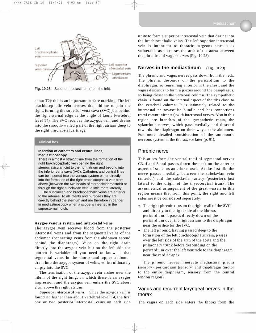
unite to form a superior intercostal vein that drains intothe brachiocephalic veins. The left superior intercostalvein is important to thoracic surgeons since it isvulnerable as it crosses the arch of the aorta betweenthe phrenic and vagus nerves (Fig. 10.28).
Nerves in the mediastinum (Fig. 10.29)
The phrenic and vagus nerves pass down from the neck.The phrenic descends on the pericardium to thediaphragm, so remaining anterior in the chest, and thevagus descends to form a plexus around the oesophagus,so being closer to the vertebral column. The sympatheticchain is found on the internal aspect of the ribs close tothe vertebral column. It is intimately related to theintercostal neurovascular bundle and has connections(rami communicantes) with intercostal nerves. Also in thisregion are branches of the sympathetic chain, thesplanchnic nerves, which pass medially and descendtowards the diaphragm on their way to the abdomen.For more detailed consideration of the autonomicnervous system in the thorax, see later (p. 91).
Phrenic nerve
This arises from the ventral rami of segmental nervesC3, 4 and 5 and passes down the neck on the anterioraspect of scalenus anterior muscle. At the first rib, thenerve passes medially, between the subclavian vein(anterior) and the subclavian artery (posterior), justlateral to the origin of the thyrocervical trunk. Theasymmetrical arrangement of the great vessels in thisregion means that from this point, the right and leftsides must be considered separately.
• The right phrenic runs on the right wall of the SVCand directly to the right side of the fibrouspericardium. It passes directly down on thepericardium over the right atrium to the diaphragmnear the orifice for the IVC.
• The left phrenic, having passed deep to theformation of the left brachiocephalic vein, passesover the left side of the arch of the aorta and thepulmonary trunk before descending on thepericardium over the left ventricle to the diaphragmnear the cardiac apex.
The phrenic nerves innervate mediastinal pleura(sensory), pericardium (sensory) and diaphragm (motorto the entire diaphragm, sensory from the centraltendon region).
Vagus and recurrent laryngeal nerves in thethorax
The vagus on each side enters the thorax from the
Mediastinum
Fig. 10.28 Superior mediastinum (from the left).
Clinical box
Insertion of catheters and central lines,mediastinoscopyThere is almost a straight line from the formation of theright brachiocephalic vein behind the rightsternoclavicular joint to the right atrium and beyond intothe inferior vena cava (IVC). Catheters and central linescan be inserted into the venous system either directlyinto the formation of the right brachiocephalic vein fromabove (between the two heads of sternocleidomastoid) orthrough the right subclavian vein, a little more laterally.
The subclavian and brachiocephalic veins are anteriorto the arteries. To all intents and purposes they aredirectly behind the sternum and are therefore in dangerin mediastinoscopy when a scope is inserted in thesuprasternal notch.
(NN) CA1E Ch 10 18/7/01 4:03 pm Page 87
carotid sheath in the neck, between the commoncarotid artery (medially) and the internal jugular vein(laterally). Again, the asymmetry of the great vesselsrequires that the two sides be considered separately.
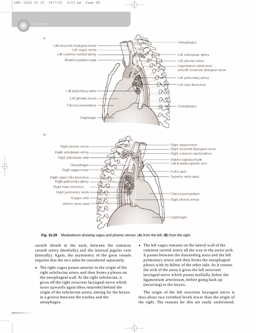
• The right vagus passes anterior to the origin of theright subclavian artery and then forms a plexus onthe oesophageal wall. At the right subclavian, itgives off the right recurrent laryngeal nerve whichturns upwards again (thus recurrent) behind theorigin of the subclavian artery, aiming for the larynxin a groove between the trachea and theoesophagus.
• The left vagus remains on the lateral wall of thecommon carotid artery all the way to the aortic arch.It passes between the descending aorta and the leftpulmonary artery and then forms the oesophagealplexus with its fellow of the other side. As it crossesthe arch of the aorta it gives the left recurrentlaryngeal nerve which passes medially, below theligamentum arteriosum, before going back up(recurring) to the larynx.
The origin of the left recurrent laryngeal nerve isthus about two vertebral levels lower than the origin ofthe right. The reasons for this are easily understood.
Thorax
Fig. 10.29 Mediastinum showing vagus and phrenic nerves: (A) from the left; (B) from the right.
(NN) CA1E Ch 10 18/7/01 4:03 pm Page 88
The recurrent laryngeal nerves innervate muscles of thesixth branchial arch (laryngeal muscles). They areintimately related to the sixth branchial arch arteriesand when both of these arteries are present in theembryo, the arrangement of the recurrent laryngealnerves is symmetrical. However, during subsequentdevelopment, the right sixth arch artery degenerates,thus allowing the right sixth arch nerve to take ashorter course to its destination, so on the right thenerve is held down by the next most cranial branchialarch artery, the fourth (there being no fifth) whichbecomes the subclavian. On the left, the sixth archartery persists as the pulmonary artery and the ductusarteriosus, and since after birth the ductus becomes the ligamentum, this continues to pin down the leftrecurrent laryngeal nerve.
The recurrent laryngeal nerves may be involved inlung tumours, resulting in voice changes. Indeed,hoarseness of the voice thus caused may be a present-ing sign of a lung tumour. This is more common as aresult of a left-sided lesion than a right-sided lesionsince there is more scope for lung disease to affect theleft recurrent laryngeal nerve, the thoracic course of thenerve being longer on the left than the right.
Having formed the oesophageal plexus, the vagusnerves continue through the oesophageal opening inthe diaphragm (vertebral level T10) into the abdomenas the anterior and posterior vagal trunks. The vagusnerve transmits parasympathetic and visceral sensoryfibres to and from the heart (cardiac branches), thelungs, the oesophagus and abdominal organs.
Intercostal nerves (see p. 66; Fig. 10.8)
These lie immediately under the mediastinal pleurabetween the vertebral column and the point at whichthey enter the costal groove with the posterior inter-costal arteries and veins. The intercostal nerves areconnected to the sympathetic chains by means of greyand white rami communicantes.
Intercostal nerves carry:
• motor fibres to intercostal muscles and (in the caseof lower intercostal nerves) muscles of theabdominal wall
• sensory fibres from skin, muscle spindles, parietalpleura and (in the case of lower intercostal nerves)parietal peritoneum
• postganglionic sympathetic fibres to blood vesselsand sweat glands in the territory of the nerve.
Posterior mediastinum (Figs 10.30–10.32)
Trachea and oesophagus
The trachea and oesophagus descend in the midline of
Mediastinum
Clinical box
Structures related to the neck of the first rib,Pancoast’s tumourA tumour of the apex of the lung may present withsymptoms arising from damage by the tumour to nervesrelated to the neck of the first rib. These are:
• fibres of T1 nerve which contribute to the brachialplexus to supply the upper limb; involvement of thesemay mean that the small muscles of the hand areweakened or paralysed, leading to clumsiness and aloss of manual dexterity
• the sympathetic chain; involvement of this mayinterrupt efferent sympathetic impulses to the headand this would result in Horner’s syndrome:– pupilloconstriction because of unopposed
parasympathetic action– facial anhidrosis because of denervation of sweat
glands– ipsilateral ptosis (drooping of the upper eyelid)
because of partial denervation of levator palpebraesuperioris muscle.
Such a tumour was described by Professor H KPancoast and is now commonly known as Pancoast’stumour.
Fig. 10.30 Posterior mediastinum: anterior view.
(NN) CA1E Ch 10 18/7/01 4:03 pm Page 89
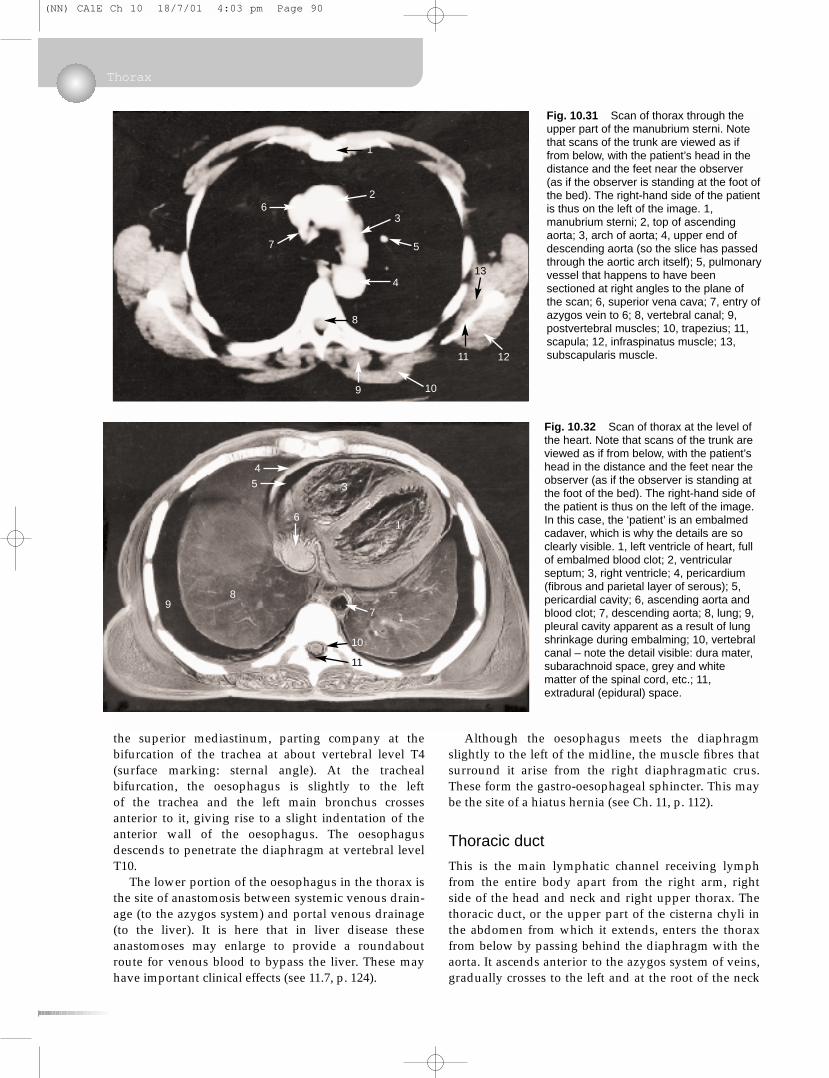
the superior mediastinum, parting company at thebifurcation of the trachea at about vertebral level T4(surface marking: sternal angle). At the trachealbifurcation, the oesophagus is slightly to the left of the trachea and the left main bronchus crossesanterior to it, giving rise to a slight indentation of theanterior wall of the oesophagus. The oesophagusdescends to penetrate the diaphragm at vertebral levelT10.
The lower portion of the oesophagus in the thorax isthe site of anastomosis between systemic venous drain-age (to the azygos system) and portal venous drainage(to the liver). It is here that in liver disease theseanastomoses may enlarge to provide a roundaboutroute for venous blood to bypass the liver. These mayhave important clinical effects (see 11.7, p. 124).
Although the oesophagus meets the diaphragmslightly to the left of the midline, the muscle fibres thatsurround it arise from the right diaphragmatic crus.These form the gastro-oesophageal sphincter. This maybe the site of a hiatus hernia (see Ch. 11, p. 112).
Thoracic duct
This is the main lymphatic channel receiving lymphfrom the entire body apart from the right arm, rightside of the head and neck and right upper thorax. Thethoracic duct, or the upper part of the cisterna chyli inthe abdomen from which it extends, enters the thoraxfrom below by passing behind the diaphragm with theaorta. It ascends anterior to the azygos system of veins,gradually crosses to the left and at the root of the neck
Thorax
Fig. 10.31 Scan of thorax through theupper part of the manubrium sterni. Notethat scans of the trunk are viewed as iffrom below, with the patient’s head in thedistance and the feet near the observer(as if the observer is standing at the foot ofthe bed). The right-hand side of the patientis thus on the left of the image. 1,manubrium sterni; 2, top of ascendingaorta; 3, arch of aorta; 4, upper end ofdescending aorta (so the slice has passedthrough the aortic arch itself); 5, pulmonaryvessel that happens to have beensectioned at right angles to the plane ofthe scan; 6, superior vena cava; 7, entry ofazygos vein to 6; 8, vertebral canal; 9,postvertebral muscles; 10, trapezius; 11,scapula; 12, infraspinatus muscle; 13,subscapularis muscle.
Fig. 10.32 Scan of thorax at the level ofthe heart. Note that scans of the trunk areviewed as if from below, with the patient’shead in the distance and the feet near theobserver (as if the observer is standing atthe foot of the bed). The right-hand side ofthe patient is thus on the left of the image.In this case, the ‘patient’ is an embalmedcadaver, which is why the details are soclearly visible. 1, left ventricle of heart, fullof embalmed blood clot; 2, ventricularseptum; 3, right ventricle; 4, pericardium(fibrous and parietal layer of serous); 5,pericardial cavity; 6, ascending aorta andblood clot; 7, descending aorta; 8, lung; 9,pleural cavity apparent as a result of lungshrinkage during embalming; 10, vertebralcanal – note the detail visible: dura mater,subarachnoid space, grey and whitematter of the spinal cord, etc.; 11,extradural (epidural) space.
4
35
6
98
2
1
10
7
11
6
1
2
7
3
4
8
9 10
11 12
13
5
(NN) CA1E Ch 10 18/7/01 4:03 pm Page 90
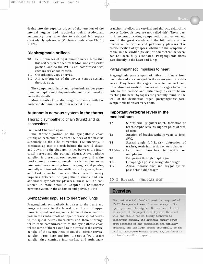
drains into the superior aspect of the junction of theinternal jugular and subclavian veins. Abdominalmalignancy may give rise to enlarged left supra-clavicular lymph nodes (Virchow’s node – see Ch. 11, p. 120).
Diaphragmatic orifices
T8 IVC, branches of right phrenic nerve. Note thatthis orifice is in the central tendon, not a muscularportion, and so the IVC is not compressed witheach muscular contraction.
T10 Oesophagus, vagus nerves.T12 Aorta, tributaries of the azygos venous system,
thoracic duct.
The sympathetic chains and splanchnic nerves pene-trate the diaphragm independently: you do not need toknow the details.
More details of the diaphragm are given with theposterior abdominal wall, from which it arises.
Autonomic nervous system in the thorax
Thoracic sympathetic chain (trunk) and itsconnections
First, read Chapter 6 again.The thoracic portion of the sympathetic chain
(trunk) on each side runs from the neck of the first ribsuperiorly to the side of vertebra T12 inferiorly. Itcontinues up into the neck behind the carotid sheathand down into the abdomen. It lies between the inter-costal nerves and the parietal pleura. A sympatheticganglion is present at each segment, grey and whiterami communicantes connecting each ganglion to itsintercostal nerve. Arising from the ganglia and passingmedially and towards the midline are the greater, lesserand least splanchnic nerves. These nerves conveyimpulses between the sympathetic chains and theabdominal sympathetic plexuses. These will be con-sidered in more detail in Chapter 11 (Autonomicnervous system in the abdomen and pelvis, p. 144).
Sympathetic impulses to heart and lungs
Preganglionic sympathetic impulses to the heart andlungs originate in the lateral grey horns of upperthoracic spinal cord segments. Axons of these neuronspass in the ventral roots of upper thoracic spinal nervesto the spinal nerves themselves and thence throughwhite rami communicantes to the sympathetic chainwhere some of them ascend to the lowest of the cervicalganglia of the sympathetic chain, the inferior cervicalganglion. From here, and from the upper few thoracicganglia, they continue into cardiac and pulmonary
branches: in effect the cervical and thoracic splanchnicnerves (although they are not called this). These pass to intercommunicating sympathetic plexuses on andaround the great vessels and the bifurcation of thetrachea – the cardiac and pulmonary plexuses. Theprecise location of synapses, whether in the sympatheticchain, in the cardiac plexus, or somewhere between,has not been fully elucidated. Postganglionic fibrespass directly to the heart and lungs.
Parasympathetic impulses to heart
Preganglionic parasympathetic fibres originate fromthe brain and are conveyed in the vagus (tenth cranial)nerve. They leave the vagus nerve in the neck andtravel down as cardiac branches of the vagus to contri-bute to the cardiac and pulmonary plexuses beforereaching the heart. Synapses are generally found in thewall of the destination organ: postganglionic para-sympathetic fibres are very short.
Important vertebral levels in themediastinum
T2 Suprasternal (jugular) notch, formation ofbrachiocephalic veins, highest point of archof aorta.
T3 Junction of brachiocephalic veins to formSVC.
T4 Sternal angle (of Louis), bifurcation oftrachea, aortic impression on oesophagus.
T5 (about) Left main bronchus impression onoesophagus.
T8 IVC passes through diaphragm.T10 Oesophagus passes through diaphragm.T12 Aorta, thoracic duct and azygos system
pass behind diaphragm.
10.5 Breast (Figs 10.33–10.35)
Breast
Overview
The postpubertal female breast is composed of
15–20 independent exocrine secretory units
opening around the nipple. It overlies ribs 2–6.
It is part of the superficial layer of the chest
wall and should not be firmly tethered to
underlying muscle. Its arterial supply comes
from branches of the subclavian and axillary
arteries, and its lymph drains principally to the
axilla. Accessory breast tissue may be found in
a line from axilla to groin.
(NN) CA1E Ch 10 18/7/01 4:03 pm Page 91
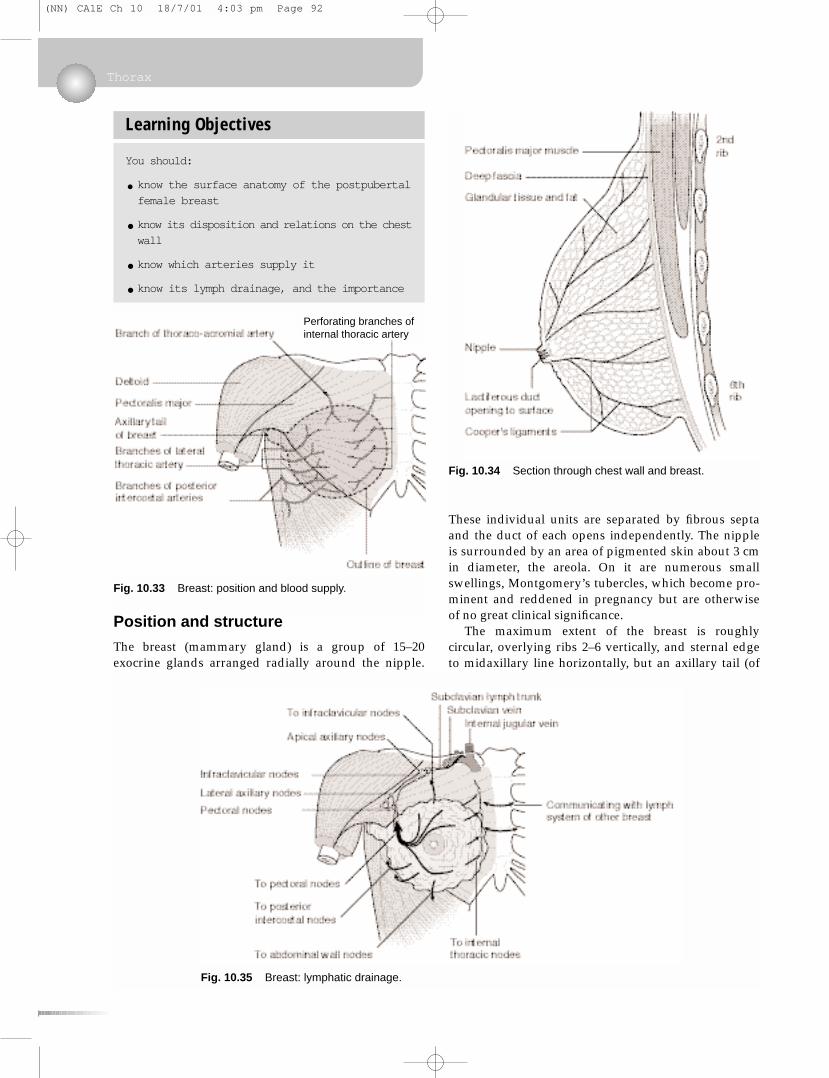
Position and structure
The breast (mammary gland) is a group of 15–20exocrine glands arranged radially around the nipple.
These individual units are separated by fibrous septaand the duct of each opens independently. The nippleis surrounded by an area of pigmented skin about 3 cmin diameter, the areola. On it are numerous smallswellings, Montgomery’s tubercles, which become pro-minent and reddened in pregnancy but are otherwiseof no great clinical significance.
The maximum extent of the breast is roughlycircular, overlying ribs 2–6 vertically, and sternal edgeto midaxillary line horizontally, but an axillary tail (of
Thorax
Learning Objectives
You should:
• know the surface anatomy of the postpubertal
female breast
•know its disposition and relations on the chest
wall
•know which arteries supply it
•know its lymph drainage, and the importance
Fig. 10.33 Breast: position and blood supply.
Fig. 10.34 Section through chest wall and breast.
Fig. 10.35 Breast: lymphatic drainage.
Perforating branches ofinternal thoracic artery
(NN) CA1E Ch 10 18/7/01 4:03 pm Page 92
Spence) extends upwards and laterally into the axilla.The glandular tissue is confined to the superficiallayers of the chest wall, but the axillary tail maypenetrate the deep fascia.
The glandular tissue overlies pectoralis major andserratus anterior muscles but, being confined to thesuperficial fascia, is not attached to these muscles. Anyclinical indication of its tethering to these musclesshould be treated with the utmost seriousness: it maywell be an indication of malignant disease. Asking thepatient to raise the arm and observing the way inwhich the breast moves may reveal tethering. Thebreast tissue is, however, connected to the overlyingskin by fibrous strands, the suspensory ligaments ofCooper. These are more numerous in the upper part of the breast than in the lower. These may becomestretched with advancing years and fat deposition.
Arteries and veins (Fig. 10.33)
Arterial blood supply comes mainly from branches ofthe subclavian artery: the internal thoracic supplies themedial portion, the lateral thoracic the lateral portion.Other arteries that contribute are the thoracoacromialand intercostal arteries 2–5. Venous drainage is bycorresponding veins to the subclavian and brachio-cephalic veins.
Lymph (Fig. 10.35)
The lymph drainage of the breast is of great import-ance. It obeys the general rule that lymphatic drainagemirrors arterial supply.
There are two main lymph plexuses in the breast:
• subareolar, immediately under the skin around thenipple
• submammary (or retromammary), between breastand pectoralis major muscle. It is involvement ofthis that may tether the breast to the muscle.
There are numerous connecting channels runningbetween these two plexuses, each with many tributaries.
From these plexuses, lymph drains as follows:
• From the lateral two-thirds: to axillary lymph nodes,thence in lymph vessels alongside the axillary arteryto the terminations of the thoracic ducts.
• A small amount of lymph passes to posteriorintercostal nodes, and some to nodes of the anteriorabdominal wall.
• From the medial portion: to parasternal nodes alongthe internal thoracic artery, thence up alongside theartery to the terminations of the thoracic ducts. It ispossible for lymph from the medial side of onebreast to drain to the opposite side, which means
that malignant disease of the medial portion of thebreast may spread to the other side.
Blockage of lymph channels in the breast (forexample by a tumour) can lead to engorgement of thebreast tissue by lymph unable to drain away and aparticular appearance of the skin of the breast ‘stretched’over the engorged tissue. This is known as peaud'orange appearance because of its similarity to orangeskin with its multiple pits.
Clinical examination of the breast is not completewithout palpation of the axilla and the breast of theother side. Axillary lymph nodes are often removedduring surgery and this may result in lymph flow fromthe upper limb being disrupted.
Nerve supply
The skin over the breast is supplied by intercostalnerves T2–6, which also convey postganglionic sym-pathetic fibres to the vessels within the breast.
Breast
Clinical box
Male breast and age changes in the female breastThe male breast and the prepubertal female breast aremuch the same: the glands are rudimentary. It is possiblefor carcinoma of the male breast to arise. It is much lesscommon than carcinoma of the female breast, but itssurvival rate is poorer, probably because the possibility ofthis condition is overlooked and so by the time thecondition has been diagnosed, it will have spreadelsewhere.
At puberty in the female, fat deposition takes placetogether with some proliferation of the secretory unitsand at lactation there is substantial proliferation of thesecretory components. After the menopause, secretoryunits atrophy and fat deposition continues. Most breastlumps are innocent tumours of fatty tissue: lipomas.
Cooper’s ligaments and diseaseInvolvement of Cooper’s ligaments in malignant diseasemay lead to their shortening as they become caught up inthe tumour. This can cause dimpling of the overlying skinor even, in extreme cases, inversion of the nipple (butnote that inversion of the nipple is by no means always asign of disease).
Accessory nipplesBreast tissue in other mammals is found along a lineextending from axilla to groin, the milk or mammary line.In humans breast tissue is limited to one organ on eachside. Nevertheless, accessory nipples, and evenaccessory breast tissue, are found in both sexesanywhere along the mammary lines, most commonly inthe thorax and abdomen. Accessory nipples areharmless but may be confused with a mole or suspectedas a melanoma; they were once most definitely life-threatening: the unfortunate possessor was regarded asa witch.
(NN) CA1E Ch 10 18/7/01 4:03 pm Page 93