Circulation and Gas Exchange - Wilmington College
12
3/26/2013 1 Circulation and Gas Exchange Circulatory systems link exchange surfaces with cells throughout the body Diffusion time is proportional to the square of the distance Diffusion is only efficient over small distances In small and/or thin animals, cells can exchange materials directly with the surrounding medium In most animals, cells exchange materials with the environment via a fluid-filled circulatory system Gastrovascular Cavities Some animals lack a circulatory system Some cnidarians, such as jellies, have elaborate gastrovascular cavities digestion and distribution of substances throughout the body The body wall that encloses the gastrovascular cavity is only two cells thick Flatworms have a gastrovascular cavity and a large surface area to volume ratio Figure 42.2 Circular canal Mouth Radial canals 5 cm (a) The moon jelly Aurelia, a cnidarian (b) The planarian Dugesia, a flatworm Gastrovascular cavity Mouth Pharynx 2 mm General Properties of Circulatory Systems A circulatory system has A circulatory fluid A set of interconnecting vessels A muscular pump, the heart The circulatory system connects the fluid that surrounds cells with the organs that exchange gases, absorb nutrients, and dispose of wastes Circulatory systems can be open or closed and vary in the number of circuits in the body A circulatory system minimizes the diffusion distance in animals with many cell layers Open and Closed Circulatory Systems In insects, other arthropods, and most molluscs, blood bathes the organs directly in an open circulatory system general body fluid is called hemolymph In a closed circulatory system, blood is confined to vessels and is distinct from the interstitial fluid Annelids, cephalopods, and vertebrates have closed circulatory systems (a) An open circulatory system Heart Hemolymph in sinuses surrounding organs Pores Tubular heart Dorsal vessel (main heart) Auxiliary hearts Small branch vessels in each organ Ventral vessels Blood Interstitial fluid Heart (b) A closed circulatory system
Transcript of Circulation and Gas Exchange - Wilmington College
3/26/2013
1
Circulation and Gas Exchange Circulatory systems link exchange surfaces with cells throughout the body Diffusion time is proportional to the square of the distance
Diffusion is only efficient over small distances
In small and/or thin animals, cells can exchange materials directly with the surrounding medium
In most animals, cells exchange materials with the environment via a fluid-filled circulatory system
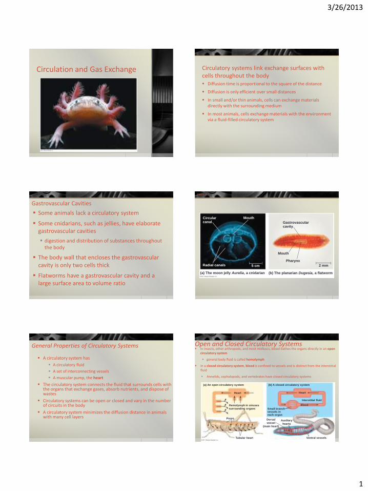
Gastrovascular Cavities
Some animals lack a circulatory system
Some cnidarians, such as jellies, have elaborate gastrovascular cavities
digestion and distribution of substances throughout the body
The body wall that encloses the gastrovascular cavity is only two cells thick
Flatworms have a gastrovascular cavity and a large surface area to volume ratio
Figure 42.2
Circular canal
Mouth
Radial canals 5 cm
(a) The moon jelly Aurelia, a cnidarian (b) The planarian Dugesia, a flatworm
Gastrovascular cavity
Mouth
Pharynx
2 mm
General Properties of Circulatory Systems
A circulatory system has
A circulatory fluid
A set of interconnecting vessels
A muscular pump, the heart
The circulatory system connects the fluid that surrounds cells with the organs that exchange gases, absorb nutrients, and dispose of wastes
Circulatory systems can be open or closed and vary in the number of circuits in the body
A circulatory system minimizes the diffusion distance in animals with many cell layers
Open and Closed Circulatory Systems In insects, other arthropods, and most molluscs, blood bathes the organs directly in an open
circulatory system
general body fluid is called hemolymph
In a closed circulatory system, blood is confined to vessels and is distinct from the interstitial fluid
Annelids, cephalopods, and vertebrates have closed circulatory systems
(a) An open circulatory system
Heart
Hemolymph in sinuses
surrounding organs
Pores
Tubular heart
Dorsal
vessel
(main heart)
Auxiliary
hearts
Small branch vessels in each organ
Ventral vessels
Blood
Interstitial fluid
Heart
(b) A closed circulatory system
3/26/2013
2
Organization of Vertebrate Circulatory Systems
Humans and other vertebrates have a closed circulatory system called the cardiovascular system
The three main types of blood vessels are arteries, veins, and capillaries
Blood flow is one way in these vessels
Arteries branch into arterioles and carry blood away from the heart to capillaries
Networks of capillaries called capillary beds are the sites of chemical exchange between the blood and interstitial fluid
Venules converge into veins and return blood from capillaries to the heart
Single Circulation
Bony fishes, rays, and sharks have single circulation with a two-chambered heart
In single circulation, blood leaving the heart passes through two capillary beds before returning
(a) Single circulation
Artery
Heart:
Atrium (A)
Ventricle (V)
Vein
Gill capillaries
Body capillaries
Key
Oxygen-rich blood
Oxygen-poor blood
Double Circulation
Amphibian, reptiles, and mammals have double circulation
Oxygen-poor and oxygen-rich blood are pumped separately from the right and left sides of the heart
(b) Double circulation
Systemic circuit
Systemic capillaries
Right Left
A A
V V
Lung
capillaries
Pulmonary circuit
Key
Oxygen-rich blood
Oxygen-poor blood
Amphibians
three-chambered heart: two atria and one ventricle
The ventricle pumps blood into a forked artery that splits the ventricle’s output into the pulmocutaneous circuit and the systemic circuit
When underwater, blood flow to the lungs is nearly shut off
Amphibians
Pulmocutaneous circuit
Lung and skin capillaries
Atrium (A)
Atrium (A)
Left Right
Ventricle (V)
Systemic capillaries
Systemic circuit
Key
Oxygen-rich blood
Oxygen-poor blood
Reptiles (Except Birds)
Turtles, snakes, and lizards have a three-chambered heart: two atria and one ventricle
In alligators, caimans, and other crocodilians a septum divides the ventricle
Reptiles have double circulation, with a pulmonary circuit (lungs) and a systemic circuit
Reptiles (Except Birds)
Pulmonary circuit
Systemic circuit
Systemic capillaries
Incomplete septum
Left systemic aorta
Left Right
Right systemic aorta
A
V
Lung capillaries
Atrium (A)
Ventricle (V)
Key
Oxygen-rich blood
Oxygen-poor blood
Mammals and Birds
Mammals and birds have a four-chambered heart with two atria and two ventricles
The left side of the heart pumps and receives only oxygen-rich blood, while the right side receives and pumps only oxygen-poor blood
Mammals and birds are endotherms and require more O2 than ectotherms
3/26/2013
3
Systemic circuit
Lung capillaries
Pulmonary circuit
A
V Left Right
Systemic capillaries
Mammals and Birds
Atrium (A)
Ventricle (V)
Key
Oxygen-rich blood
Oxygen-poor blood
Figure 42.5c
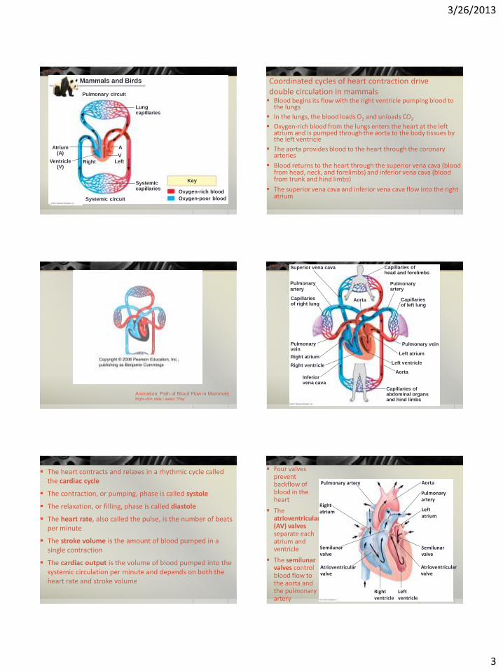
Blood begins its flow with the right ventricle pumping blood to the lungs
In the lungs, the blood loads O2 and unloads CO2
Oxygen-rich blood from the lungs enters the heart at the left atrium and is pumped through the aorta to the body tissues by the left ventricle
The aorta provides blood to the heart through the coronary arteries
Blood returns to the heart through the superior vena cava (blood from head, neck, and forelimbs) and inferior vena cava (blood from trunk and hind limbs)
The superior vena cava and inferior vena cava flow into the right atrium
Coordinated cycles of heart contraction drive double circulation in mammals
Animation: Path of Blood Flow in Mammals Right-click slide / select “Play”
Superior vena cava
Pulmonary
artery
Capillaries of right lung
Pulmonary vein
Aorta
Inferior vena cava
Right ventricle
Capillaries of abdominal organs and hind limbs
Right atrium
Aorta
Left ventricle
Left atrium
Pulmonary vein
Pulmonary artery
Capillaries of left lung
Capillaries of head and forelimbs
Figure 42.6
The heart contracts and relaxes in a rhythmic cycle called the cardiac cycle
The contraction, or pumping, phase is called systole
The relaxation, or filling, phase is called diastole
The heart rate, also called the pulse, is the number of beats per minute
The stroke volume is the amount of blood pumped in a single contraction
The cardiac output is the volume of blood pumped into the systemic circulation per minute and depends on both the heart rate and stroke volume
Four valves prevent backflow of blood in the heart
The atrioventricular (AV) valves separate each atrium and ventricle
The semilunar valves control blood flow to the aorta and the pulmonary artery
Pulmonary artery
Right atrium
Semilunar valve
Atrioventricular valve
Right ventricle
Left ventricle
Atrioventricular valve
Semilunar valve
Left atrium
Pulmonary artery
Aorta
3/26/2013
4
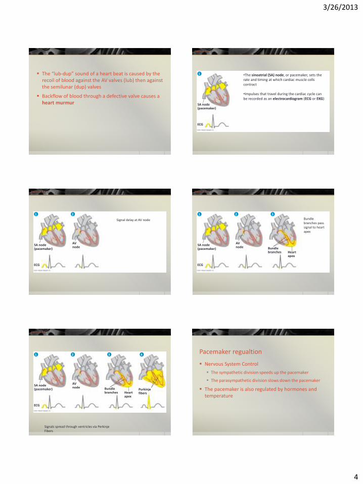
The “lub-dup” sound of a heart beat is caused by the recoil of blood against the AV valves (lub) then against the semilunar (dup) valves
Backflow of blood through a defective valve causes a heart murmur
Figure 42.9-1
SA node (pacemaker)
ECG
1 •The sinoatrial (SA) node, or pacemaker, sets the rate and timing at which cardiac muscle cells contract
•Impulses that travel during the cardiac cycle can be recorded as an electrocardiogram (ECG or EKG)
Figure 42.9-2
SA node (pacemaker)
AV node
ECG
1 2
Signal delay at AV node
Figure 42.9-3
SA node (pacemaker)
AV node Bundle
branches Heart apex
ECG
1 2 3
Bundle branches pass signal to heart apex
Figure 42.9-4
SA node (pacemaker)
AV node Bundle
branches Heart apex
Purkinje fibers
ECG
1 2 3 4
Signals spread through ventricles via Perkinje Fibers
Pacemaker regualtion
Nervous System Control
The sympathetic division speeds up the pacemaker
The parasympathetic division slows down the pacemaker
The pacemaker is also regulated by hormones and temperature

3/26/2013
5
Patterns of blood pressure and flow reflect the structure and arrangement of blood vessels
Artery
Red blood cells
Endothelium
Artery
Smooth muscle
Connective tissue
Capillary
Valve
Vein
Vein
Basal lamina
Endothelium
Smooth muscle
Connective tissue
100 m
LM
Venule
15
m
LM
Arteriole
Red blood cell
Capillary
Figure 42.11
Systolic pressure
Diastolic pressure
0 20 40 60 80
100 120
0 10 20 30 40 50
Pre
ssu
re
(mm
Hg)
V
elo
city
(c
m/s
ec)
A
rea
(cm
2 )
0 1,000 2,000 3,000 4,000 5,000
•Velocity slowest at capillaries •Blood flows from areas of higher pressure to areas of lower pressure
Changes in Blood Pressure During the Cardiac Cycle Systolic pressure is the pressure in the arteries during ventricular
systole; it is the highest pressure in the arteries
Diastolic pressure is the pressure in the arteries during diastole; it is lower than systolic pressure
A pulse is the rhythmic bulging of artery walls with each heartbeat
Blood pressure is determined by cardiac output and peripheral resistance due to constriction of arterioles
Vasoconstriction is the contraction of smooth muscle in arteriole walls; it increases blood pressure
Vasodilation is the relaxation of smooth muscles in the arterioles; it causes blood pressure to fall
Blood pressure reading: 120/70
120
70
Sounds stop
Sounds audible in stethoscope
120
Artery closed
1 2 3
Figure 42.12
Blood pressure is generally measured for an artery in the arm at the same height as the heart
Fainting is caused by inadequate blood flow to the head
Animals with longer necks require a higher systolic pressure to pump blood a greater distance against gravity
Blood is moved through veins by smooth muscle contraction, skeletal muscle contraction, and expansion of the vena cava with inhalation
One-way valves in veins prevent backflow of blood
Direction of blood flow in vein (toward heart) Valve (open)
Skeletal muscle
Valve (closed)
Figure 42.14
Precapillary sphincters
Thoroughfare channel
Arteriole
Capillaries
Venule
(a) Sphincters relaxed
Arteriole Venule
(b) Sphincters contracted
Blood flows through only 510% of the body’s capillaries at a time
Two mechanisms regulate distribution of blood in capillary beds
• Contraction of the smooth muscle layer in the wall of an arteriole constricts the vessel
• Precapillary sphincters control flow of blood between arterioles and venules
3/26/2013
6
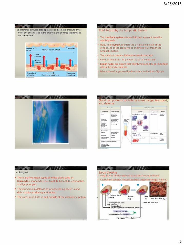
Figure 42.15
INTERSTITIAL FLUID Net fluid movement out
Blood pressure
Osmotic pressure
Arterial end of capillary Direction of blood flow
Venous end of capillary
Body cell
The difference between blood pressure and osmotic pressure drives fluids out of capillaries at the arteriole end and into capillaries at the venule end
Fluid Return by the Lymphatic System
The lymphatic system returns fluid that leaks out from the capillary beds
Fluid, called lymph, reenters the circulation directly at the venous end of the capillary bed and indirectly through the lymphatic system
The lymphatic system drains into veins in the neck
Valves in lymph vessels prevent the backflow of fluid
Lymph nodes are organs that filter lymph and play an important role in the body’s defense
Edema is swelling caused by disruptions in the flow of lymph
Figure 42.16 Figure 42.17
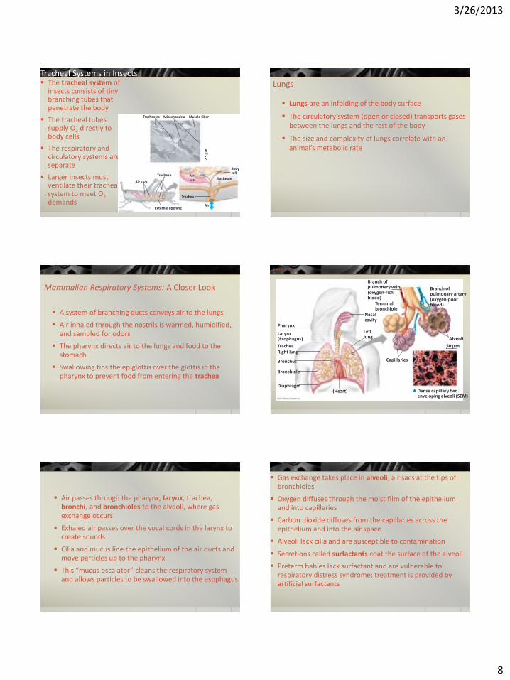
Plasma 55%
Constituent Major functions
Water
Ions (blood electrolytes)
Sodium Potassium Calcium Magnesium Chloride Bicarbonate
Solvent for carrying other substances
Osmotic balance, pH buffering, and regulation of membrane permeability
Plasma proteins
Osmotic balance, pH buffering
Albumin
Fibrinogen
Immunoglobulins (antibodies)
Clotting
Defense
Substances transported by blood
Nutrients Waste products Respiratory gases Hormones
Separated blood elements
Basophils
Neutrophils Monocytes
Lymphocytes
Eosinophils
Platelets
Erythrocytes (red blood cells) 5–6 million
250,000–400,000 Blood clotting
Transport of O2 and some CO2
Defense and immunity
Functions Number per L (mm3) of blood
Cell type
Cellular elements 45%
Leukocytes (white blood cells) 5,000–10,000
Blood components contribute to exchange, transport, and defense
Leukocytes
There are five major types of white blood cells, or leukocytes: monocytes, neutrophils, basophils, eosinophils, and lymphocytes
They function in defense by phagocytizing bacteria and debris or by producing antibodies
They are found both in and outside of the circulatory system
Blood Clotting Coagulation is the formation of a solid clot from liquid blood
A cascade of complex reactions converts inactive fibrinogen to fibrin, forming a clot
A blood clot formed within a blood vessel is called a thrombus and can block blood flow
Collagen fibers
1 2 3
Platelet Platelet plug
Fibrin clot
Fibrin clot formation
Red blood cell 5 m
Clotting factors from: Platelets Damaged cells Plasma (factors include calcium, vitamin K)
Enzymatic cascade
Prothrombin Thrombin
Fibrinogen Fibrin
3/26/2013
7
Stem cells (in bone marrow)
Myeloid stem cells
Lymphoid stem cells
B cells T cells
Lymphocytes
Erythrocytes Neutrophils
Basophils
Eosinophils Platelets Monocytes
Figure 42.19
•The cellular elements of blood wear out and are being replaced constantly •Erythrocytes, leukocytes, and platelets all develop from a common source of stem cells in the red marrow of bones, especially ribs, vertebrae, sternum, and pelvis •Erythropoietin (EPO) stimulates erythrocyte production when O2 delivery is low
Gas exchange occurs across specialized respiratory surfaces
Gas exchange supplies O2 for cellular respiration and disposes of CO2
A gas diffuses from a region of higher partial pressure to a region of lower partial pressure Partial pressure is the pressure exerted by a particular gas in a mixture of gases
Gases diffuse down pressure gradients in the lungs and other organs as a result of differences in partial pressure
Respiratory Media and Surfaces
Animals can use air or water as a source of O2, or respiratory medium
In a given volume, there is less O2 available in water than in air
Obtaining O2 from water requires greater efficiency than air breathing
Animals require large, moist respiratory surfaces for exchange of gases between their cells and the respiratory medium, either air or water
Gas exchange across respiratory surfaces takes place by diffusion
Respiratory surfaces vary by animal and can include the outer surface, skin, gills, tracheae, and lungs
Gills in Aquatic Animals
Gills are outfoldings of the body that create a large surface area for gas exchange
Parapodium (functions as gill)
(a) Marine worm (b) Crayfish
Gills Gills
Tube foot
(c) Sea star
Coelom
Ventilation moves the respiratory medium over the respiratory surface
Aquatic animals move through water or move water over their gills for ventilation
Fish gills use a countercurrent exchange system, where blood flows in the opposite direction to water passing over the gills; blood is always less saturated with O2 than the water it meets
Figure 42.23
Gill arch
O2-poor blood
O2-rich blood
Blood vessels Gill arch
Operculum Water flow
Water flow Blood flow
Countercurrent exchange PO (mm Hg) in water
2 150
PO (mm Hg)
in blood 2
120 90 60 30
140 110 80 50 20 Net diffu- sion of O2
Lamella
Gill filaments
3/26/2013
8
Tracheal Systems in Insects The tracheal system of
insects consists of tiny branching tubes that penetrate the body
The tracheal tubes supply O2 directly to body cells
The respiratory and circulatory systems are separate
Larger insects must ventilate their tracheal system to meet O2 demands
Tracheoles Mitochondria Muscle fiber
2.5
m
Tracheae
Air sacs
External opening
Trachea
Air sac Tracheole
Body cell
Air
Lungs
Lungs are an infolding of the body surface
The circulatory system (open or closed) transports gases between the lungs and the rest of the body
The size and complexity of lungs correlate with an animal’s metabolic rate
Mammalian Respiratory Systems: A Closer Look
A system of branching ducts conveys air to the lungs
Air inhaled through the nostrils is warmed, humidified, and sampled for odors
The pharynx directs air to the lungs and food to the stomach
Swallowing tips the epiglottis over the glottis in the pharynx to prevent food from entering the trachea
Figure 42.25
Pharynx
Larynx (Esophagus)
Trachea Right lung
Bronchus
Bronchiole
Diaphragm (Heart)
Capillaries
Left lung
Dense capillary bed enveloping alveoli (SEM)
50 m
Alveoli
Branch of pulmonary artery (oxygen-poor blood)
Branch of pulmonary vein (oxygen-rich blood)
Terminal bronchiole
Nasal cavity
Air passes through the pharynx, larynx, trachea, bronchi, and bronchioles to the alveoli, where gas exchange occurs
Exhaled air passes over the vocal cords in the larynx to create sounds
Cilia and mucus line the epithelium of the air ducts and move particles up to the pharynx
This “mucus escalator” cleans the respiratory system and allows particles to be swallowed into the esophagus
Gas exchange takes place in alveoli, air sacs at the tips of bronchioles
Oxygen diffuses through the moist film of the epithelium and into capillaries
Carbon dioxide diffuses from the capillaries across the epithelium and into the air space
Alveoli lack cilia and are susceptible to contamination
Secretions called surfactants coat the surface of the alveoli
Preterm babies lack surfactant and are vulnerable to respiratory distress syndrome; treatment is provided by artificial surfactants
3/26/2013
9
Breathing ventilates the lungs
The process that ventilates the lungs is breathing, the alternate inhalation and exhalation of air
An amphibian such as a frog ventilates its lungs by positive pressure breathing, which forces air down the trachea
Anterior air sacs
Posterior air sacs
Lungs
1 mm
Airflow
Air tubes (parabronchi) in lung
Anterior air sacs
Lungs
Second inhalation First inhalation
Posterior air sacs 3
2 4
1
4
3 1
2 Second exhalation First exhalation
Figure 42.27
•Birds have eight or nine air sacs that function as bellows that keep air flowing through the lungs •Air passes through the lungs in one direction only •Every exhalation completely renews the air in the lungs
Mammals ventilate their lungs by negative pressure breathing, which pulls air into the lungs
Lung volume increases as the rib muscles and diaphragm contract
The tidal volume is the volume of air inhaled with each breath
Rib cage expands.
Air inhaled.
Air exhaled.
Rib cage gets smaller.
1 2
Lung
Diaphragm
Control of Breathing in Humans
The medulla oblongata and the pons
The medulla regulates the rate and depth of breathing in response to pH changes in the cerebrospinal fluid
The medulla adjusts breathing rate and depth to match metabolic demands
The pons regulates the tempo
Sensors in the aorta and carotid arteries monitor O2 and CO2 concentrations in the blood
These sensors exert secondary control over breathing
Homeostasis: Blood pH of about 7.4
CO2 level decreases. Stimulus:
Rising level of CO2 in tissues
lowers blood pH. Response: Rib muscles and diaphragm increase rate and depth of ventilation.
Carotid arteries
Aorta Sensor/control center: Cerebrospinal fluid
Medulla oblongata
Figure 42.29
Adaptations for gas exchange include pigments that bind and transport gases
The metabolic demands of many organisms require that the blood transport large quantities of O2 and CO2
3/26/2013
10
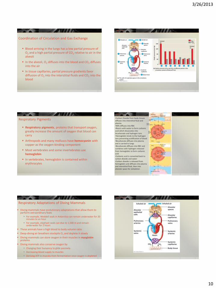
Coordination of Circulation and Gas Exchange
Blood arriving in the lungs has a low partial pressure of O2 and a high partial pressure of CO2 relative to air in the alveoli
In the alveoli, O2 diffuses into the blood and CO2 diffuses into the air
In tissue capillaries, partial pressure gradients favor diffusion of O2 into the interstitial fluids and CO2 into the blood
Exhaled air Inhaled air
Pulmonary arteries
Systemic veins
Systemic arteries
Pulmonary veins
Alveolar capillaries
Alveolar spaces Alveolar
epithelial cells
Inhaled air
160
120
80
40
0 Heart
8 1
2
3
4 6
7
CO2 O2
Systemic capillaries
CO2 O2 Body tissue 5
(a) The path of respiratory gases in the circulatory system
(b) Partial pressure of O2 and CO2 at different points in the circulatory system numbered in (a)
4 3 2 1 5 6 7
Exhaled air
Part
ial
pre
ssu
re (m
m H
g)
PO 2 PCO 2
8
Figure 42.30
Respiratory Pigments
Respiratory pigments, proteins that transport oxygen, greatly increase the amount of oxygen that blood can carry
Arthropods and many molluscs have hemocyanin with copper as the oxygen-binding component
Most vertebrates and some invertebrates use hemoglobin
In vertebrates, hemoglobin is contained within erythrocytes
Figure 42.32 Body tissue
Capillary wall
Interstitial fluid
Plasma within capillary
CO2 transport from tissues CO2 produced
CO2
CO2
CO2 H2O
H2CO3 Hb Red blood cell Carbonic
acid
Hemoglobin (Hb) picks up
CO2 and H+.
H+ HCO3
Bicarbonate
HCO3
HCO3
To lungs
CO2 transport
to lungs
HCO3
H2CO3
H2O
CO2
H+
Hb
Hemoglobin releases
CO2 and H+.
CO2
CO2
CO2
Alveolar space in lung
•Carbon Dioxide from body tissues diffuses into interstitial fluid and plasma. •90% diffuses into RBC •Reacts with water to form carbonic acid which dissociates into bicarbonate and hydrogen ions •Hemoglobin binds to the hydrogen ions preventing acidification of blood •Bicarbonate diffuses into plasma and is carried to lungs •Bicarbonate diffuses into RBC and combines with hydrogen released from hemoglobin to form carbonic acid •Carbonic acid is converted back to carbon dioxide and water •Carbon dioxide is released from hemoglobin and diffuses into plasma and interstitial fluid, then into alveolar space for exhalation
Respiratory Adaptations of Diving Mammals
Diving mammals have evolutionary adaptations that allow them to perform extraordinary feats
For example, Weddell seals in Antarctica can remain underwater for 20 minutes to an hour
For example, elephant seals can dive to 1,500 m and remain underwater for 2 hours
These animals have a high blood to body volume ratio
Deep-diving air breathers stockpile O2 and deplete it slowly
Diving mammals can store oxygen in their muscles in myoglobin proteins
Diving mammals also conserve oxygen by
Changing their buoyancy to glide passively
Decreasing blood supply to muscles
Deriving ATP in muscles from fermentation once oxygen is depleted
Figure 42.UN02
Exhaled air
Alveolar epithelial cells
Pulmonary arteries
Systemic veins
Heart
CO2 O2
Body tissue
Systemic capillaries
Systemic arteries
Pulmonary veins
Alveolar capillaries
Alveolar spaces
Inhaled air
CO2 O2
3/26/2013
11
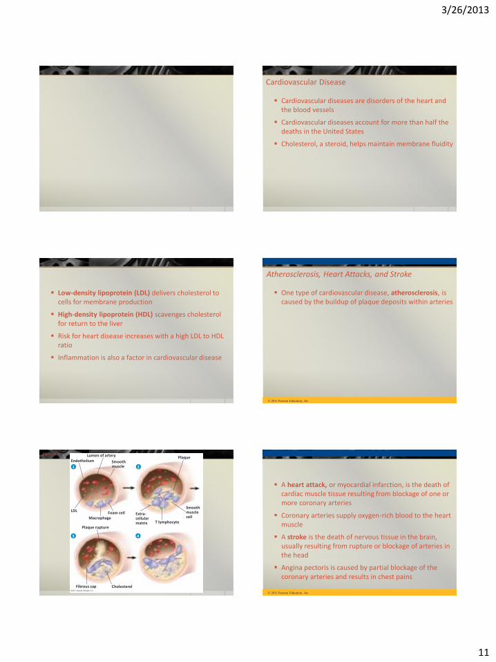
Cardiovascular Disease
Cardiovascular diseases are disorders of the heart and the blood vessels
Cardiovascular diseases account for more than half the deaths in the United States
Cholesterol, a steroid, helps maintain membrane fluidity
Low-density lipoprotein (LDL) delivers cholesterol to cells for membrane production
High-density lipoprotein (HDL) scavenges cholesterol for return to the liver
Risk for heart disease increases with a high LDL to HDL ratio
Inflammation is also a factor in cardiovascular disease
Atherosclerosis, Heart Attacks, and Stroke
One type of cardiovascular disease, atherosclerosis, is caused by the buildup of plaque deposits within arteries
© 2011 Pearson Education, Inc.
Lumen of artery
Smooth muscle
Endothelium Plaque
Smooth muscle cell
T lymphocyte
Extra- cellular matrix
Foam cell Macrophage
Plaque rupture
LDL
Cholesterol Fibrous cap
1 2
4 3
Figure 42.20
A heart attack, or myocardial infarction, is the death of cardiac muscle tissue resulting from blockage of one or more coronary arteries
Coronary arteries supply oxygen-rich blood to the heart muscle
A stroke is the death of nervous tissue in the brain, usually resulting from rupture or blockage of arteries in the head
Angina pectoris is caused by partial blockage of the coronary arteries and results in chest pains
© 2011 Pearson Education, Inc.
3/26/2013
12
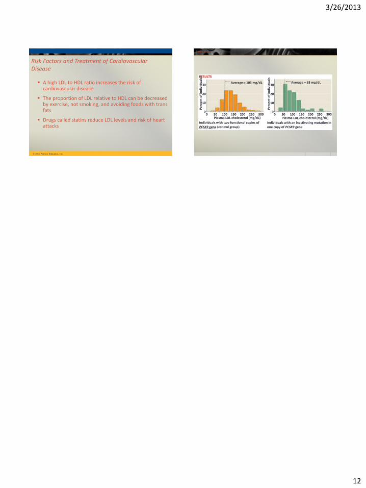
Risk Factors and Treatment of Cardiovascular Disease
A high LDL to HDL ratio increases the risk of cardiovascular disease
The proportion of LDL relative to HDL can be decreased by exercise, not smoking, and avoiding foods with trans fats
Drugs called statins reduce LDL levels and risk of heart attacks
© 2011 Pearson Education, Inc.
Figure 42.21
Individuals with two functional copies of PCSK9 gene (control group)
Plasma LDL cholesterol (mg/dL)
Individuals with an inactivating mutation in one copy of PCSK9 gene
Plasma LDL cholesterol (mg/dL)
Average 63 mg/dL Average 105 mg/dL 30
20
10
0 0 50 100 150 200 250 300 0
0
10
20
30
50 100 150 200 250 300
Per
cen
t o
f in
div
idu
als RESULTS
Per
cen
t o
f in
div
idu
als