CHR No: 11-07249 DOD ADNI
80
CHR No: 11-07249 DOD ADNI 08072012 1 | Page Confidential PROTOCOL: Effects of traumatic brain injury and post traumatic stress disorder on Alzheimer’s disease (AD) in Veterans using ADNI (DoD-ADNI) Sponsored by Department of Defense (DOD) Telemedicine and Advanced Technology Research Center (TATRC) Protocol Principal Investigator, ADNI Principal Investigator, Program Director, Coordinating Center Michael W. Weiner, M.D. University of California, San Francisco Tel. 415-221-4810 x3642 Fax 415-668-2864 [email protected] Psychological Evaluation/PTSD Core Thomas Neylan, M.D. University of California, San Francisco San Francisco Veterans Affairs Medical Center 415-750-6961 [email protected] Traumatic Brain Injury/TBI Core Jordan Grafman, Ph.D., Director Traumatic Brain Injury Research Laboratory Kessler Foundation Research Center 973-243-6995 [email protected] ADNI Clinical Core Paul S. Aisen, M.D. University of California, San Diego Alzheimer’s Disease Cooperative Study 858-622-2028 [email protected] Mayo Clinic Ronald Petersen, M.D., Ph.D. Mayo Clinic, Rochester, Minnesota 507-538-0487 [email protected]
Transcript of CHR No: 11-07249 DOD ADNI

CHR No: 11-07249 DOD ADNI 08072012
1 | P a g e
Confidential
PROTOCOL:
Effects of traumatic brain injury and post traumatic stress disorder on Alzheimer’s disease (AD) in Veterans using ADNI (DoD-ADNI)
Sponsored by
Department of Defense (DOD) Telemedicine and Advanced Technology Research Center (TATRC)
Protocol Principal Investigator, ADNI Principal Investigator, Program Director, Coordinating Center
Michael W. Weiner, M.D. University of California, San Francisco
Tel. 415-221-4810 x3642 Fax 415-668-2864
Psychological Evaluation/PTSD Core Thomas Neylan, M.D.
University of California, San Francisco San Francisco Veterans Affairs Medical Center
415-750-6961 [email protected]
Traumatic Brain Injury/TBI Core Jordan Grafman, Ph.D., Director
Traumatic Brain Injury Research Laboratory Kessler Foundation Research Center
973-243-6995 [email protected]
ADNI Clinical Core Paul S. Aisen, M.D.
University of California, San Diego Alzheimer’s Disease Cooperative Study
858-622-2028 [email protected]
Mayo Clinic
Ronald Petersen, M.D., Ph.D. Mayo Clinic, Rochester, Minnesota
507-538-0487 [email protected]

CHR No: 11-07249 DOD ADNI 08072012
2 | P a g e
ADNI Biomarker Core Leslie M. Shaw, Ph.D.
John Trojanowski, Ph.D. University of Pennsylvania Medical Center
215-662-6575 [email protected] [email protected]
ADNI PET Core William Jagust, M.D.
University of California, Berkeley 510-643-6537
University of Michigan Robert A. Koeppe, PhD University of Michigan
734-763-9247 [email protected]
ADNI MRI Core
Clifford Jack, Ph.D. Mayo Clinic, Rochester
507-284-8548 [email protected]
ADNI Genetics Core Andrew Saykin, PsyD, ABCN
Indiana University School of Medicine 317-278-6947
ADNI Neuropathology Core John Morris, M.D.
Nigel Cairns, Ph.D., F.R.C. Path Washington University School of Medicine
314-286-2881 [email protected] [email protected]

CHR No: 11-07249 DOD ADNI 08072012
3 | P a g e
ADNI Informatics Core Arthur Toga, Ph.D.
LONI, University of California, Los Angeles 310-206-2101
ADNI Biostatistics Core Danielle Harvey, PhD.
University of California, Davis 530-752-8036

CHR No: 11-07249 DOD ADNI 08072012
4 | P a g e
PROTOCOL SYNOPSIS
Title
Effects of traumatic brain injury (TBI) and post traumatic stress disorder
(PTSD) on Alzheimer’s disease (AD) in Veterans using (Alzheimer’s
Disease Neuroimaging Initiative) ADNI
Overall Objective
Primary Objectives
The overall long-term goal of this project is to prevent Alzheimer‟s disease
(AD) in combat Veterans. This project is an important step in that direction
because it will identify risk factors for the development of AD in military
Veterans and will provide information and a network of sites for design,
statistical powering, and performance of clinical treatment and prevention trials
in the future.
The primary objectives to be tested are:
1. Veterans without mild cognitive impairment (MCI)/dementia, with a
history of moderate to severe TBI during military service (but no
PTSD), as well as Veterans without MCI/dementia, with ongoing PTSD
(but no TBI) have increased evidence for AD pathophysiological
markers, when compared with Veteran controls (without TBI, PTSD,
MCI/dementia, and also matched for age, APOE4 status, and
accounting for other comorbidities) as measured by:
a. Greater uptake on Florbetapir F18 amyloid PET scans
b. Lower CSF amyloid beta levels
c. Increased CSF tau/P tau levels
d. Greater brain atrophy in hippocampus, entorhinal cortex, and
parietal/temporal cortices
e. Greater longitudinal rates of brain atrophy in hippocampus,
entorhinal cortex, and parietal/temporal cortices
f. Reduced cognitive function, especially delayed memory
g. Greater rate of change of cognitive function
2. TBI and/or PTSD reduces brain reserve, causing greater cognitive
impairment after accounting for age, educational status, pre-war
cognitive function, as assessed with the Armed Forces Qualifying Exam
score during basic training, to be measured by:
a. Brain amyloid load or hippocampal volume.
b. Greater cognitive impairments at a given level of brain Aβ, or
c. Brain volume in the TBI or PTSD group than controls

CHR No: 11-07249 DOD ADNI 08072012
5 | P a g e
3. TBI, when compared to controls, is associated with changes in brain
regions previously reported to be associated with TBI [37-39].
a. White matter regions frequently noted to have reduced
microstructural integrity due to TBI on DTI include the anterior
corona radiata, uncinate fasciculus, corpus callosum, forceps
minor, forceps major, sagittal stratum, corticospinal tract,
inferior fronto-occipital fasciculus and cingulum bundle [37-
39].
b. Reduced hippocampal volume in PTSD compared to controls,
[32, 33-36].
4. There will be significant correlations between severity of TBI (from
hospital records) and severity of PTSD (CAPS score) on the above-
listed outcomes, i.e. a dose response effect.
5. Exploratory analyses will also be performed to examine other questions,
and compare the patterns (using voxel based methods) of amyloid
deposition (from Florbetapir F18 uptake) and brain atrophy among TBI,
PTSD, and control subjects, and with the patterns from non-Veteran
subjects in ADNI. The results of these studies may provide insight into:
a. The question of whether or not TBI and PTSD alter the pattern
of amyloid distribution or brain atrophy.
b. the relationship between cortical areas with amyloid plaque
(from Florbetapir) and,
c. Underlying white matter integrity as assessed with DTI, to
determine if axonal injury resulting from TBI was associated
with greater amyloid accumulation, or whether regions of brain
with axonal damage have less amyloid accumulation due to
disconnection and reduced brain activity.
Study Design
This is a non-randomized natural history non-treatment study.
1. Using military and Veterans Affairs (VA) Compensation and Pension
records, SFVAMC will identify Vietnam War Veterans with well
documented history of moderate/severe Traumatic Brain Injury (TBI) or
evidence of ongoing Post Traumatic Stress Disorder (PTSD), and
comparable Veteran controls. Subjects meeting criteria for mild
cognitive impairment/dementia will be excluded.
2. SFVAMC will mail out invitation letter, brochure, and response
postcard (a self-addressed stamped envelope will be included for the
CHR No: 11-07249 DOD ADNI 08072012
6 | P a g e
subject to return the response postcard) to large numbers of Veterans
who meet above broad criteria, based on diagnostic codes in military
and VA Compensation and Pension records, and who live within 150
miles of closest ADNI clinic in subject‟s area. Selected sites will have
the General Electric Diffusion Tensor Imaging / Magnetic Resonance
Imaging [GE/DTI MRI] scanner). The mileage distance from the clinics
may be extended if the recruitment numbers are low. The timing of the
mail out effort to the geographic location of a selected ADNI site will
only occur once the specific site has obtained full human subjects
approval from both their agency review boards as well as the
Department of Defense (DOD).
3. SFVAMC will contact subjects by telephone – contact those that have
returned the response post card, as well as those who have not.
a. Obtain verbal consent to administer screening and TBI
questionnaire over the telephone.
b. Administer screening and TBI questionnaires to determine
eligibility criteria to be enrolled in the Psychiatric Assessment
(Clinical Interview) part of the study, using the Structured
Clinical Interview 1 of the Diagnostic and Statistical Manual of
Mental Disorders, Version IV, (Axis 1) - Text Revision (SCID 1
of the DSMIV-TR) and the Clinician Administered PTSD Scale
(CAPS). The SCID/CAPS will be audio recorded for quality
control purposes, essentially to make sure that clinicians are
administering the interview in a similar fashion. Subject will be
asked to sign a separate consent form for permission to audio-
record. Audio-recordings will be stored in a secure and locked
file cabinet, in a secure and locked file room. No names will be
included on the audio recording. They will be stored with study
code only. Audio-recording is OPTIONAL. Subjects who do
not wish to be audio-recorded can still participate in the study.
c. Determine if subject has a study partner. If subject has a reliable
study partner, and is found to be eligible after completing the
screening and clinical interviews, the partner will be asked to
accompany the subject to the clinic visits. Study partners are
helpful in confirming memory problems if they exist. However,
because many subjects may not have a regular person in their
life who can act as a study partner, a study partner is not
required for this study.
d. The screening questions will be an adapted version of the AD8
assessment (70, 71) and will also include some of the questions
of the Clinical Dementia Rating (CDR). For this study, subjects
with mild cognitive impairment (MCI)/dementia may be
screened depending on the answers to the screening questions.
CHR No: 11-07249 DOD ADNI 08072012
7 | P a g e
The Study Coordinator will discuss subject responses to the
Subject Matter Experts to assess whether or not the memory
issues are exclusionary. Questionable items will be discussed
among the clinical core investigators to assess who will be
screened out.
e. At completion of the telephone screener, subjects will be told
whether or not they are eligible to be referred to the clinical
telephone interview, or if some of their responses need to be
discussed with the study doctors. (Some answers to the
screening questions will be discussed with the clinical leaders of
the study to assess whether there are any exclusionary criteria
present, such as MCI/dementia, psychological disorders, alcohol
or substance abuse, etc.). After the discussion with study
doctors, subject will be called back and invited to continue, or
will be thanked and compensated for the time spent in study
thus far.
f. If after the screening interview, subject is eligible for the
SCID/CAPS, SFVAMC staff will mail a written consent form
w/ stamped addressed return envelope to the home of the
subjects along with two self-report questionnaires on MRI
safety and medical history and a consent form for audio-
recording. In a few days, Study staff will call the subject to
make sure that subject received the consent forms, will go over
the consent forms with the subject, answer any questions the
subject may have, and assist with the self report questionnaires
if needed. Subject will be asked to sign the consent forms and
fax or mail them back to SFVAMC in the stamped addressed
envelope along with the completed self-report questionnaires.
The SCID/CAPS cannot be administered without first receiving
the signed consent form for the clinical interview. The
SCID/CAPS cannot be audio-recorded without also receiving
the signed consent form giving permission to audio-record. If
the consent to audio -record the SCID/CAPS is received,
SCID/CAPS will be recorded and the tape will be stored and
filed by study code number only, in a locked and secure file
cabinet in a locked and secure file room. Audio-recording is
optional. Subjects who choose not to be audio-recorded can still
participate in the study.
g. As soon as the signed consent forms and questionnaires are
received back at SFVAMC, by mail or fax, the questionnaires
will be reviewed and if eligible, subject will be referred to the
PTSD core at SFVAMC.
h. Because the project is only enrolling subjects without
MCI/dementia, a study partner (if available) will NEVER act as

CHR No: 11-07249 DOD ADNI 08072012
8 | P a g e
a surrogate for the subject. All subjects will consent for
themselves. A study partner is NOT a requirement for this
study.
4. PTSD core (at the SFVAMC) will call the subject and perform the
Structured Diagnostic Interview I for DSM-IV-TR & Clinician
Administered PTSD Scale (CAPS) by telephone. If a signed consent for
audio recording has been received along with the signed consent for the
clinical interview, this interview will be audio recorded for quality
control purposes, to ensure that the interview is administered in a
consistent manner. The tape will be filed and stored by study code only
in a secure locked file system. Audio recording is optional. Subjects
who choose not to be audio-recorded can still participate in the study.
5. If after clinical telephone interview, the subject is still eligible,
SFVAMC staff will refer subject to local clinic in network of
Alzheimer‟s Disease Neuroimaging Initiative sites (ADNI).
6. SFVAMC staff will mail a packet of self-report questionnaires to
subject, with instructions for subject to complete and bring completed
packet to his or her first in-person clinic visit.
7. SFVAMC will give the subject the contact information of the clinic in
the subject‟s area, and will call the clinic directly to refer the subject‟s
contact information.
8. The local ADNI site will contact subject and make an appointment for
the subject to come to the site. At the ADNI site, trained staff will
conduct the procedures listed below with each subject. Completing all
of the procedures may take 2-3 days, or so, within a few weeks:
a. Explain study and obtain written consent;
b. For those subjects who have a reliable person in their lives who
can act as a study partner (spouse, friend, or relative), the full
CDR will be administered during the clinic visits to confirm that
the subject does not have MCI/dementia, or to further rule out
the presence of MCI/dementia. If a study partner is available,
the study partner will be asked to accompany the subject to all
clinic visits and to communicate changes in the subjects‟ health
status over the period of the study. Study partners will be fully
informed of the study, and will be asked to read and sign the
consent form. There will be a separate signature line on the
subject‟s consent form for study partners (if a study partner is
available).
c. ADNI staff will also discuss autopsy with subject. An ADNI

CHR No: 11-07249 DOD ADNI 08072012
9 | P a g e
clinician will lead a discussion about autopsy with all subjects at
their initial assessment (study partners and families are
welcomed in the discussion). There are 3 objectives of the
discussion:
To convey information about the value of brain autopsy
in confirming the clinical diagnosis and advancing
knowledge regarding mild cognitive impairment and
Alzheimer‟s disease;
To initiate consideration of the individual‟s wishes
concerning an autopsy; and
To answer questions, misconceptions, or concerns about
autopsy. The involvement of the physician in these
discussions emphasizes the importance of autopsy.
There is no pressure on an individual to decide; they are
encouraged to involve family members, clergy,
physicians, or other appropriate persons in their
decision-making. Participants are assured that a
decision not to have an autopsy in no way jeopardizes
their research participation or any other patient
rights. It is important to note that autopsy will not
interfere with funerary arrangements nor will it be a
financial burden to the participant's family. Subjects
will be asked to sign whether or not they are interested
in considering autopsy at the time of his or her death,
and will be told that study personnel will ask
permission to autopsy from his or her next of kin;
therefore, subjects are encouraged to let their next of
kin know their wishes concerning autopsy prior to
death.
d. Collect packet of self-report questionnaires
e. Collect baseline measurements of cognition and function. (This
will confirm that there is no MCI/dementia and/or any other
health issues that may make it unsafe for a subject to continue.)
f. Perform a blood draw by venipuncture
g. Collect cerebrospinal fluid for analyses (lumbar puncture)
h. Conduct MRI scans (structural, diffusion tensor, and resting
state BOLD fMRI). No contrast agent will be used. T2 FSE and
T2*GRE.
i. Conduct an amyloid PET imaging scan with Florbetapir F18.
The dose of Florbetapir F18 is 10 mCi per procedure.
j. Because the Florbetapir F18 can affect a fetus, pregnant women
may not participate in this study. Women of childbearing age
will receive a urine test at the time of the baseline PET scan and
at the 12 month follow-up PET scan to ensure that she is not

CHR No: 11-07249 DOD ADNI 08072012
10 | P a g e
pregnant.
k. Those subjects who have completed the procedures at the ADNI
clinic are eligible for the 12 month (approximate) follow-up
screener. At about 6 months, SFVAMC staff will call each
subject to remind them of the 12 month (approximate) follow-
up screener and to update patient status, with the following
questions:
“We’re calling to remind you that the follow-up telephone interview for the study, Effects of Effects of traumatic brain
injury and post traumatic stress disorder on Alzheimer’s disease
(AD) in Veterans using ADNI, will begin in about 6 months from now, and to check in on how you are doing.
Have you had any change in memory or thinking in the past
6 months? o Yes or no; If yes, specify:
___________________________
Have you had any overall change in activity in the past 6
months?
o Yes or no; If yes, specify: ___________________________
Is there anything that you cannot do now, that you could do
6 months ago?
o Yes or no; If yes, specify: __________________________
Have you had any changes in medications, illness, surgeries
or hospital visits in the last 6 months?
o Yes or no; If yes, specify: ____________________________
Thank you so much for your help. We’ll be calling you in about
6 months to begin the follow-up interview.
l. If at the 6 month reminder/check in call, the subject indicates
that they do not want to continue the study in the next 6 months, we will ask them whether or not they would be willing to move
their one year visit to the time of the 6 month call and make it
an exit visit, and complete as many procedures as they are willing.
9. The follow-up screener, follow-up clinical telephone interview, and
follow-up self administered questionnaires will be identical to the
baseline questions, but will only reference the time period since the last
baseline clinic visit. The follow-up screener will not be screening
anyone out of the study, but will help us determine whether anything
has changed since the last clinic visit that may make a particular
procedure unsafe, and to determine that the subject is still willing and

CHR No: 11-07249 DOD ADNI 08072012
11 | P a g e
able to participate.
10. Mild Cognitive Impairment/dementia at the time of the follow-up
procedures will NOT be exclusionary, unless it would be unsafe for the
subject to participate. With the exception of the lumbar puncture and
the PET scan, all procedures at the clinic sites will be repeated at the
follow-up visit unless there are specific health reasons that may make it
unsafe for the subject to participate (for example, new metal implants).
11. Analysis of the data to test the hypotheses as stated, including the
exploratory analyses, will begin after the first 12 months or so and will
continue until the follow-ups are completed.
12. Deliver final report to the DOD.
13. Should additional funding be obtained, the number of longitudinal
follow-up years may be increased.
Summary of Communication and Data Flow between SFVAMC and Clinic
Sites:
This study will use much of the existing network of communication that
exists for the Alzheimer‟s Disease Neuroimaging Initiative (ADNI)
study, including the ADNI Data and Safety Monitoring Board (DSMB).
The San Francisco site will manage the initial contacts with subjects by
mail and telephone, as well as the collection of telephone screening and
clinical interviews and self-report data. All patient data and telephone
calls will be logged into a currently existing electronic capture system,
modified for the study. All case report forms (CRFS) will be uploaded
to the Alzheimer‟s Disease Cooperative Study (ADCS) database.
The ACDS was formed in 1991 as a cooperative agreement between the
National Institute on Aging (NIA) and the University of California San
Diego. The ADCS is a major initiative for Alzheimer‟s disease (AD)
clinical studies in the Federal government, addressing treatments for
both cognitive and behavioral symptoms.
Upon completion of the mail/telephone assessments, eligible subjects
will be referred to the ADNI sites, and ADCS will coordinate all data
capture. Upon arrival at the ADNI sites, all subjects will have the
complete standardized ADNI assessments. The subjects will be asked
about the presence of all medical conditions and use of medications.

CHR No: 11-07249 DOD ADNI 08072012
12 | P a g e
When the Site Medical Doctors (MDs) and study coordinators have
questions about this, they contact the central ADNI clinical core, and
these issues are adjudicated.
All de-identified data will be available at the ADCS database, displayed
at the Laboratory of Neuroimaging (LONI) website at University of
California, Los Angeles without embargo. LONI is a leader in the
development of advanced computational algorithms and scientific
approaches for the comprehensive and quantitative mapping of brain
structure and function.
The San Francisco Veterans Affairs Medical Center (SFVAMC) site
will maintain the protocol and consent templates, and any changes to
these documents will be distributed to each study site as soon as they are
locally approved.
SFVAMC will be responsible for the mail effort, recruitment, and the
telephone screeners and clinical interviews, as well as the self-report
questionnaires, and maintain the protocol and consent templates, but no
subjects will be seen on the SFVAMC campus.
UCSF will be one of the selected ADNI sites. There will be subjects
seen at the UCSF ADNI clinic and at the China Basin PET scan facility.
The UCSF ADNI site will be submitting their own IRB documents,
using this master template for protocol and consent forms, as will each
of the selected ADNI sites.
Sample Size
Overall Sample of Consented/Screened/Eligible and Enrolled all the way to
the ADNI site with a full battery of assessments will be 195- 300 eligible
subjects.
65-100 Vietnam Veterans w/ TBI, but w/o PTSD, MCI/dementia
65-100 Vietnam Veterans w/ PTSD, but w/o TBI, MCI/dementia
65-100 Vietnam Veteran controls, w/o TBI or PTSD and comparable in
age, gender, and education to above groups
Subjects with mild cognitive impairment (MCI)/dementia will be excluded.
Rationale for this approach is that it„s already established that 50-60 % of
subjects w/MCI have AD pathology in their brain, and it may be difficult to
detect added effects of a history of TBI or ongoing PTSD in MCI/demented
subjects.
The Sample size of 65-100 Vietnam Veterans in each of the study

CHR No: 11-07249 DOD ADNI 08072012
13 | P a g e
cohorts is the estimate for how many subjects will successfully enroll
and complete all study procedures. Our goal is to enroll and complete
one year (approximate) follow-up visits on a total of 210 subjects.
Given expected drop-outs, we plan to enroll and complete all baseline
measurements on 230 subjects. Based on our experience we believe that
we will need to refer approximately 300 subjects to the ADNI sites (that
is about 100/group).
To achieve this number of subjects who pass the screening examination
and the SCID we plan to send out several thousand brochures and will
be making many hundreds, perhaps thousands of telephone contacts. At
the current time it is not possible to precisely identify the exact number
of brochure mailings which will result in satisfactory responses leading
to telephone contacts. Therefore our approach will be to begin with
sending 1000 brochure mailings to veterans in the zip codes of the
ADNI sites and then observe the response rate. If the response rate is
high, then fewer additional mailings will be needed. If the response rate
is low, we will increase the number of mailings until we achieve a
satisfactory number of responses, so that during our follow-up phone
calls we obtain a reasonable number of subjects who pass the screening
telephone interview.
We also do not know the fraction of subjects who will fail to pass the
SCID CAPS telephone interview. However since we are examining the
VA and military records of the subjects prior to mailings, we are
hopeful that we are targeting subjects who have histories of TBI or
PTSD, or neither, and that we are able to exclude subjects with
exclusionary criteria by examination of their records. In summary we
approximate that 300 persons will come in for the in person visit, that
500 will undergo the clinical telephone interview, that 1000 subjects
will undergo the screening interview, and that it will be required to mail
5000 brochures to obtain this number of interested veteran subjects.
Summary of Key
Inclusion/Exclusion
Criteria for TBI
Subjects
TBI Subjects: Identification and inclusion criteria
Subjects must be Veterans of the Vietnam War, 50-90 years of age.
(Subjects 60-80 will be recruited first, but subjects at the lower and
upper ages may be added as study progresses). Subjects <60 or >80 will
be added if recruitment numbers are too low in the 60-80 age range.
Subjects must have a documented history of moderate-severe non-

CHR No: 11-07249 DOD ADNI 08072012
14 | P a g e
penetrating TBI, which occurred during military service in Vietnam
(identified from the Department of Defense or VA records).
Must live within 150 miles of the closest ADNI clinic in subject‟s area.
TBI will be defined as:
o Loss of consciousness,
o Post-traumatic amnesia >24 hours, OR
o Alteration of consciousness or mental state >24 hours
TBI Subjects: Exclusion criteria
Mild Cognitive Impairment/Dementia
Presence of PTSD by SCID-I for DSM-IV-TR criteria, or a CAPS score
of >30 (Both current and/or a history of PTSD will be excluded).
Summary of Key
Inclusion/Exclusion
Criteria for PTSD
Subjects
PTSD Subjects: Identification and inclusion criteria
Subjects must be Veterans of the Vietnam War, 50-90 years of age
(Subjects 60-80 years will be selected first). Subjects <60 or >80 will be
added if recruitment numbers are too low in the 60-80 age range.
Subjects who meet the SCID-I (for DSM-IV-TR)criteria for
current/chronic PTSD (identified by records, and verified by our
telephone assessments)
In addition to meeting DSM-IV-TR criteria for current/chronic PTSD,
subjects must have a minimum current CAPS score of 50 as
determined by telephone assessment.
The PTSD symptoms contributing to the PTSD Diagnosis and Current
CAPS score must be related to a Vietnam War related trauma.
Must live within 150 miles of the closest ADNI clinic in subject‟s area.
PTSD Subjects: Exclusion criteria
Mild Cognitive Impairment/Dementia
Documented or self report history of mild/moderate severe TBI
Any history of head trauma associated with injury onset cognitive
complaints, or
Loss of consciousness for >5minutes
Summary of Key
Inclusion/Exclusion
Criteria for Control
Control Subjects: Identification and inclusion criteria
Subjects must be Veterans of the Vietnam War, 50-90 years of age

CHR No: 11-07249 DOD ADNI 08072012
15 | P a g e
Subjects (Subjects 60-80 years will be selected first). Subjects <60 or >80 will be
added if recruitment numbers are too low in the 60-80 age range.
Comparable in age, gender, and education with TBI and PTSD groups
May be receiving VA disability payments for something other than TBI
or PTSD – or no disability at all.
Must live within 150 miles of the closest ADNI clinic in subject‟s area
Control Subjects: Exclusion criteria
MCI/Dementia
Presence of PTSD by SCID-I for DSM-IV-TR criteria, or a CAPS score
of >30 (Both current and/or a history of PTSD will be excluded).
Documented or self report history of mild/moderate severe TBI
Any history of head trauma associated with injury onset cognitive
complaints, or
Loss of Consciousness for >5 minutes
History of PTSD or current PTSD
Exclusionary criteria applied to TBI/PTSD will be applied to controls
No controls will be enrolled until 25% of the TBI/PTSD enrolled
Exclusion Criteria for
ALL Subjects
All Subjects: Exclusion Criteria for all subjects:
MCI/dementia
History of psychosis or bipolar affective disorder;
History of alcohol or substance abuse/dependence within the past 5
years (by DSM IV – TR criteria);
MRI-related exclusions: aneurysm clips, metal implants that are
determined to be unsafe for MRI; and/or claustrophobia;
Contraindications for lumbar puncture, PET scan, or other procedures in
this study;
Any major medical condition must be stable for at least 4 months prior
to enrollment. These include but are not limited to clinically significant
hepatic, renal, pulmonary, metabolic or endocrine disease, cancer, HIV
infection and AIDS, as well as cardiovascular disease, including:
o cardiac surgery or myocardial infarction within the last 4 weeks;
o unstable angina;
o acute decompensated congestive heart failure or class IV heart
failures;
o current significant cardiac arrhythmia or conduction disturbance
particularly those resulting in ventricular fibrillation, or causing
syncope, or near syncope;

CHR No: 11-07249 DOD ADNI 08072012
16 | P a g e
o Uncontrolled high blood pressure
Seizure disorder or any systemic illness affecting brain function during
the past 5 years will be exclusionary
Clinical evidence of stroke.
Have a history of relevant severe drug allergy or hypersensitivity.
Subjects with current clinically significant unstable medical
comorbidities, as indicated by history or physical exam, that pose a
potential safety risk to the subject.
For Florbetapir Scans:
Subjects who have received an investigational medication within the last
30 days; additionally, the time between the last dose of the previous
experimental medication and enrollment (completion of screening
assessments) must be at least equal to 5 times the terminal half-life of
the previous experimental medication.
Subjects who have received a radiopharmaceutical for imaging or
therapy within the past 24 hours prior to the imaging session for this
study.
Current or recent participation in any procedures involving radioactive
agents such that the total radiation dose exposure to the subject in any
given year would exceed the limits of annual and total dose commitment
set forth in the US Code of Federal Regulations (CFR) Title 21 Section
361.1.
Prohibited medications: Subjects who have ever participated in an
experimental study with an amyloid targeting therapy (e.g.,
immunotherapy, secretase inhibitor, selective amyloid lowering agents)
may not be enrolled without prior sponsor/AVID approval unless it can
be demonstrated that the patient received only placebo in the course of
the trial.
Women of childbearing potential who are not surgically sterile, not
refraining from sexual activity or not using reliable methods of
contraception. Women of childbearing potential must not be pregnant or
breastfeeding at screening. Women must avoid becoming pregnant and
must agree to refrain from sexual activity or to use reliable
contraceptive methods for 24 hours following administration of
Florbetapir F18 injection
o Because the drugs in this study can affect a fetus, pregnant
women may not participate. Women of childbearing age will
receive a urine test for pregnancy on the date of the Florbetapir
F18 scan. Given that the selected age of subjects will be 50-90,
it is unlikely that many pregnancy tests will be needed.
Subjects, who in the opinion of the investigator are otherwise unsuitable

CHR No: 11-07249 DOD ADNI 08072012
17 | P a g e
for a study of this type, are also excluded.
Procedures
After initial mail contact and telephone screening, all eligible subjects
will undergo a clinical psychological interview using the Structured
Diagnostic Interview SCID-I for DSM IV-TR, and the Clinician
Administered PTSD Scale (CAPS) by telephone, conducted by the
PTSD Core at the San Francisco VA Medical Center to determine
eligibility. The SCID/CAPS will be audio-recorded for quality control
purposes. Subject will be asked to sign a separate consent form for
audio recording. All audio recordings will be stored by study code only
in a locked filed cabinet in a locked file room. Audio-recording is
optional. Subjects who choose not to be audio recorded can still
participate in the study.
Those subjects, who are still eligible and live within 150 miles of the
closest ADNI clinic, will be referred to their local site for a full battery
of clinical/cognitive assessments, biomarker and genetic sample
collection (Blood and cerebrospinal fluid), and imaging (MRI and PET
scans).
o Again, because the drugs in this study can affect a fetus,
pregnant women may not participate. Women of childbearing
age will receive a urine test for pregnancy on the date of the
Florbetapir F18 scan. Given that the selected age of subjects
will be 50-90, it is unlikely that many pregnancy tests will be
needed.
All MRI and PET scans will be rapidly assessed for quality so that
subjects may be rescanned if necessary. In the event that the scan fails the quality control procedure and cannot be used, we will repeat the
Florbetapir scan. In ADNI to date, less than .5% of scans require repeat.
Because of this, we include the total dose in the case of the necessity for repeating the scan once. See dosimetry table on page 62.
All clinical data will be collected, monitored, and stored by the
Coordinating Center at University of California, San Diego. The
University of Pennsylvania will collect biomarker samples and, and
NCRAD will collect genetic samples. All raw and processed image data
will be archived at LONI.
SFVAMC will be responsible for the mail effort, recruitment, and the
telephone screeners and clinical interviews. There will be NO subjects
seen on the SFVAMC campus. UCSF will be one of the ADNI sites.
There will be subjects seen at the UCSF ADNI clinic and at the China
Basin PET scan facility. The UCSF ADNI site will submit their own
CHR No: 11-07249 DOD ADNI 08072012
18 | P a g e
IRB documents, using this master template for protocol and consent
forms, as will each of the selected ADNI sites.
Outcome Measures
1. Level of early cognitive functioning as measured by Armed Forces
Qualifying Exam Score.
2. Level of current cognitive function as measured by Cognitive tests.
3. Prevalence of brain AD pathology (measured by amyloid PET, CSF Aβ
and tau, medial temporal lobe atrophy, (after accounting for effects of
age and APOE).
4. Prevalence of AD pathology and other co-morbid diseases/pathology
identified at autopsy.
5. Examine group differences for each imaging and biomarker
measurements:
a. Brain uptake –Florbetapir F18 amyloid PET scans
b. Levels of CSF Amyloid beta, tau/P tau
c. Rate of brain atrophy in hippocampus, entorhinal cortex, and
parietal/temporal cortices (both current and longitudinally)
6. Hippocampal atrophy while accounting for brain amyloid.
Sponsor
Department of Defense

CHR No: 11-07249 DOD ADNI 08072012
19 | P a g e
Schedule of Events
Completed by SFVAMC Recruiters and PTSD Core
Stage of Project Baseline 12 Month (approximate) Follow-up
Who is responsible?
SFVAMC
Recruiters
PTSD
Core
Self
Reports
Local
ADNI
SITE
SFVAMC
Recruiters
PTSD
Core
Self
Report
Local
ADNI
SITE
Invitation letter,
brochure, & response
postcards mailed to
subjects X X
6 month (approximate)
contact call X
Birthday cards/holiday
cards X X X X
Thank you cards X X X X
Subjects contacted by
telephone; Explain Study X X
Verbal Consent obtained
for Screening/TBI
interview /Self Report
Questionnaires (SRQ‟s) X X
Structured interview for
TBI history and screening X X
If Eligible, mail MRI
safety/Medical history
SRQ‟s w/written consents
for Clinical Interview and
Consent for audio
recording (w/stamped/
addressed envelope) X X
Call subject, review
consents. Answer
questions. If interested,
instruct subject to return
consents /SRQ‟s in

CHR No: 11-07249 DOD ADNI 08072012
20 | P a g e
prepared envelope. X X
Once written
consent/SRQ‟s received
and reviewed at
SFVAMC, refer eligible
subjects to clinical core
for SCID/CAPS, X
X
If eligible after
SCID/CAPS; Mail SRQ‟s X
X
Completed Over the Telephone by PTSD CORE
Stage of Project Baseline 12 Month (approximate) Follow-up
Who is responsible?
SFVAMC
Recruiters
PTSD
Core
Self
Reports
Local
ADNI
SITE
SFVAMC
Recruiters
PTSD
Core
Self
Report
Local
ADNI
SITE
Structured Clinical
Interview for
SCID-I for DSM-IV TR X
X
Clinician Administered
PTSD Scale X
X
Life Stressor Checklist -
Revised X X
SELF REPORT QUESTIONNAIRES (SRQ):
Baseline 12 Month (approximate) Follow-Up
Who is responsible?
SFVAMC
Recruiters
PTSD
Core
Self
Reports
Local
ADNI
SITE
SFVAMC
Recruiters
PTSD
Core
Self
Report
Local
ADNI
SITE
MRI Safety X X
Medical History X X
CHR No: 11-07249 DOD ADNI 08072012
21 | P a g e
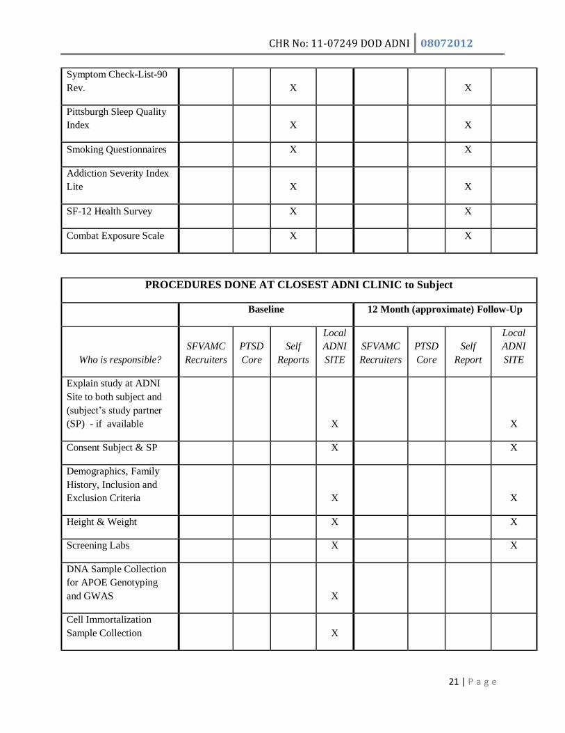
Symptom Check-List-90
Rev. X X
Pittsburgh Sleep Quality
Index X X
Smoking Questionnaires X X
Addiction Severity Index
Lite X X
SF-12 Health Survey X X
Combat Exposure Scale X X
PROCEDURES DONE AT CLOSEST ADNI CLINIC to Subject
Baseline 12 Month (approximate) Follow-Up
Who is responsible?
SFVAMC
Recruiters
PTSD
Core
Self
Reports
Local
ADNI
SITE
SFVAMC
Recruiters
PTSD
Core
Self
Report
Local
ADNI
SITE
Explain study at ADNI
Site to both subject and
(subject‟s study partner
(SP) - if available
X X
Consent Subject & SP X X
Demographics, Family
History, Inclusion and
Exclusion Criteria
X X
Height & Weight X X
Screening Labs X X
DNA Sample Collection
for APOE Genotyping
and GWAS
X
Cell Immortalization
Sample Collection
X
CHR No: 11-07249 DOD ADNI 08072012
22 | P a g e
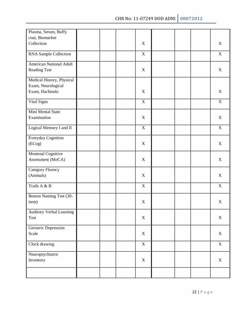
Plasma, Serum, Buffy
coat, Biomarker
Collection
X X
RNA Sample Collection X X
American National Adult
Reading Test
X X
Medical History, Physical
Exam, Neurological
Exam, Hachinski
X
X
Vital Signs X X
Mini Mental State
Examination
X
X
Logical Memory I and II X X
Everyday Cognition
(ECog)
X
X
Montreal Cognitive
Assessment (MoCA)
X
X
Category Fluency
(Animals)
X
X
Trails A & B X X
Boston Naming Test (30-
item)
X
X
Auditory Verbal Learning
Test
X
X
Geriatric Depression
Scale
X
X
Clock drawing X X
Neuropsychiatric
Inventory
X
X

CHR No: 11-07249 DOD ADNI 08072012
23 | P a g e
ADAS-Cog 13 (with
Delayed Word Recall and
Number Cancellation)
X
X
Clinical Dementia Rating
Scale
X
X
Activities of Daily Living
(FAQ)
X
X
AFQT X X
ACT X X
Concomitant Medications X X
Adverse Events X X
Diagnostic Summary X X
Neuropathological
Diagnosis (autopsy)
discussion
X
X
3T MRI with DTI
Imaging
X
X
Florbetapir F 18 Injection
Amyloid Imaging with
PET
X
CSF Collection by
Lumbar Puncture (LP)
X

CHR No: 11-07249 DOD ADNI 08072012
24 | P a g e
Study Glossary
18F-AV-45 Florbetapir F 18 (under IND with Avid Radiopharmaceuticals, Inc.)
AD Alzheimer‟s Disease ADAS-Cog Alzheimer‟s Disease Assessment Scale – Cognitive
ADC Alzheimer‟s Disease Center
ADC‟s Alzheimer‟s Disease Centers (under NIA) ADCS Alzheimer‟s Disease Cooperative Study
ADEAR Alzheimer‟s Disease Education & Referral Center, under the NIA
ADNI Alzheimer‟s Disease Neuroimaging Initiative ADNI-CC Alzheimer‟s Disease Neuroimaging Initiative Coordinating Center
ADRDA Alzheimer's Disease and Related Disorders Association
AE Adverse Event ANART American National Adult Reading Test
ANOVA Analysis of Variance
APOE/APOE4 Apolipoprotein (APOE) epsilon 4 (APOE4) AVLT Auditory Verbal Learning Test
Aβ Beta Amyloid
BNT Boston Naming Test
CDR Clinical Dementia Rating
CN Cognitively Normal
CSF Cerebral Spinal Fluid CT Computerized Tomography
DNA Deoxyribonucleic Acid
DSMB Data Safety Monitoring Board DSM-IV Diagnostic and Statistical Manual of Mental Disorders, Fourth Edition
DTI Diffusion Tensor Imaging
ECG Electrocardiogram ECog Everyday Cognition
eCRF Electronic Case Report Form
FAQ Functional Activities Questionnaire (Activities of Daily Living) FLAIR Fluid Attenuation Inversion Recovery
FDG Fluorodeoxyglucose
fMRI Functional Magnetic Resonance Imaging GDS Geriatric Depression Scale
GWAS Genome Wide Association Studies
HIPAA Health Insurance Portability and Accountability Act ICH International Conference on Harmonization
IRB Institutional Review Board
LONI Laboratory of Neuroimaging

CHR No: 11-07249 DOD ADNI 08072012
25 | P a g e
LP Lumbar Puncture
MCI Mild Cognitive Impairment
MMSE Mini Mental State Examination MoCA Montreal Cognitive Assessment
MPRAGE Magnetization Prepared Rapid Gradient Echo
MR/MRI Magnetic Resonance / Magnetic Resonance Imaging NCRAD National Cell Repository for Alzheimer‟s Disease
NINCDS National Institute of Neurological and Communicative Diseases and Stroke
NPIQ Neuropsychiatric Inventory Questionnaire PET Positron-Emission Tomography
PI Principal Investigator
PTSD Post Traumatic Stress Disorder QA Quality Assurance
QC Quality Control
RARC Resource Allocation Review Committee REB Research Ethics Board
RNA Ribonucleic Acid
SAE Serious Adverse Event SD Standard Deviation
SFVAMC San Francisco Veterans Affairs Medical Center
T Tesla T2* GRE T2 Star-Weighted Gradient-Echo
TBI Traumatic Brain Injury
TFT‟s Thyroid Function Tests VIQ Premorbid Verbal Intelligence
WML White Matter Lesions
WMS-R Wechsler Memory Scale – Revised

CHR No: 11-07249 DOD ADNI 08072012
26 | P a g e
Table of Contents
PROTOCOL SYNOPSIS .......................................................................................................... 4
Schedule of Events .................................................................................................................. 19
Study Glossary ........................................................................................................................ 24
1. Introduction ..................................................................................................................... 30
2. Background: AD, TBI, & PTSD ...................................................................................... 30
3. Imaging and Biomarkers ................................................................................................. 31
4. The Alzheimer’s Disease Neuroimaging Initiative (ADNI) ............................................ 31
5. Hypotheses........................................................................................................................ 32
Primary Hypothesis ........................................................................................................................ 32
Second Hypothesis......................................................................................................................... 32
Third Hypothesis ............................................................................................................................ 32
Fourth Hypothesis ......................................................................................................................... 33
Exploratory Analyses...................................................................................................................... 33
6. Study Design ..................................................................................................................... 33
7. Overall Study Timetable .................................................................................................. 37
Year One: ....................................................................................................................................... 37
Year Two: ...................................................................................................................................... 39
Year Three: .................................................................................................................................... 39
8. Resources in Place to Conduct the Study: ....................................................................... 40
Study Personnel ............................................................................................................................. 41
Site Principal Investigator .............................................................................................................. 41
Site Study Physician ....................................................................................................................... 41
Site Study Coordinator ................................................................................................................... 41
Project interviewer/Psychometrician ............................................................................................. 41
SFVAMC Study Personnel: .............................................................................................................. 41
9. Study Sample.................................................................................................................... 42
10. Recruitment and Retention Overview ............................................................................. 43
Summary of Recruitment and Eligibility Determination: ................................................................. 44
Summary of Where and How Subjects are approached: ................................................................. 44
Compensation for Subjects ............................................................................................................ 44

CHR No: 11-07249 DOD ADNI 08072012
27 | P a g e
Inclusion and Exclusion Criteria: ..................................................................................................... 45
11. Study Procedures at SFVAMC: ...................................................................................... 45
Records Search .............................................................................................................................. 45
Mail Effort ..................................................................................................................................... 45
Initial Telephone Contact & Verbal Consent for Screening ............................................................. 45
Documented Consent for Clinical Interview, Audio-Recording & Self Report Questionnaires .......... 46
Clinical Interview ........................................................................................................................... 46
Additional Self Report Questionnaires to be Mailed ....................................................................... 47
12. Study Procedures at the ADNI clinic sites: ..................................................................... 47
Initial Evaluation /Medical Exam/Cognitive Tests: .......................................................................... 47
Autopsy Discussion ........................................................................................................................ 47
Magnetic resonance Imaging (MRI) Scan ........................................................................................ 48
Magnetic Resonance Imaging (MRI) Qualification: Site Qualification ............................................ 48
Positron Emission Tomography (PET) Scan ..................................................................................... 49
Procedures for assuring scan quality .............................................................................................. 49
Positron Emission Tomography (PET) Scan ..................................................................................... 50
Computed Tomography (CT) Scan .................................................................................................. 50
Lumbar Puncture (LP): ................................................................................................................... 50
Blood Draw & Genetic Testing: DNA, RNA, Immortalized Cell Lines: ............................................... 51
Storage of DNA, RNA & Cell Line Samples ...................................................................................... 51
Plasma / Serum / Buffy Coat for Biomarkers: ................................................................................. 52
Twelve Month (approximate) Follow-up ........................................................................................ 52
13. Time Commitment ........................................................................................................... 53
Baseline Year 1 .............................................................................................................................. 53
Follow-Up Year 2 ........................................................................................................................... 53
Total for BOTH Years ...................................................................................................................... 53
14. List of Assessments that SFVAMC Will Conduct on Telephone ................................... 54
On telephone at time of screening, by SFVAMC ............................................................................. 54
Verbal Consent Screener ........................................................................................................ 54
On telephone at time of Clinical Interview, by PTSD core at SFVAMC........................................... 55
Structured Clinical Interview for SCID-I DSM-IV, Non-Patient edition (SCID-NP), [41]): ............ 55

CHR No: 11-07249 DOD ADNI 08072012
28 | P a g e
Clinician Administered PTSD Scale (CAPS, [40]): ...................................................................... 55
Life Stressor Checklist - Revised (LSC-R): ................................................................................. 55
15. List of Assessments SFVAMC Will Mail to Subjects ..................................................... 55
MRI Safety Checklist (mail before SCID/CAPS) ........................................................................ 55
Medical History Questionnaire (mail before SCID/CAPS) ......................................................... 55
Symptom Check-List-90-Revised (SCL-90-R, [43]) mail after SCID /CAPS): ............................... 55
Pittsburgh Sleep Quality Index [44] (mail after SCID/CAPS): .................................................... 55
Smoking/Lifetime smoking (mail after SCID/CAPS): ................................................................ 55
Addiction Severity Index Lite (ASI-Lite) (mail after SCID/CAPS):............................................... 56
SF-12 Health Survey (SF-12) [50] (mail after SCID/CAPS): ........................................................ 56
Combat Exposure Scale (CES) [51] (mail after SCID/CAPS): ...................................................... 56
16. List of Assessments Conducted by Local ADNI Site: ..................................................... 56
Cognitive, Behavioral, Functional, and Global Assessments .............................................................. 56
Montreal Cognitive Assessment (MoCA) [52]: ........................................................................ 57
Everyday Cognition (ECog)...................................................................................................... 57
Mini-Mental State Exam (MMSE) [53]: ................................................................................... 57
Alzheimer’s Disease Assessment Scale-Cognitive (ADAS-COG 13 [54]: .................................... 57
Logical Memory Test I and II (Delayed Paragraph Recall) [55]: ................................................ 57
Boston Naming Test [56]: ....................................................................................................... 57
Category Fluency Test [57]: .................................................................................................... 57
Clock Drawing Test [58]: ......................................................................................................... 57
American National Adult Reading Test (ANART): [59] ............................................................. 58
The Auditory Verbal Learning Test: ......................................................................................... 58
Trail making A and B: .............................................................................................................. 58
Clinical Dementia Rating (CDR) [63]: ....................................................................................... 58
Activities of Daily Living | Functional Assessment Questionnaire (FAQ) [64]: .......................... 58
Neuropsychiatric Inventory (NPI) [65]: ................................................................................... 58
Geriatric Depression Scale [66]: .............................................................................................. 58
Armed Forces Qualification Test (AFQT)/Army Classification (ACT) ......................................... 58
17. Risks ................................................................................................................................. 59
Initial Screening and Clinical Interview ........................................................................................... 59

CHR No: 11-07249 DOD ADNI 08072012
29 | P a g e
Self-Report Questionnaires ............................................................................................................ 59
Cognitive Testing ........................................................................................................................... 59
Magnetic resonance Imaging (MRI) Scan ........................................................................................ 59
Lumbar Puncture (LP): ................................................................................................................... 60
Research Blood Draws/DNA/RNA/Cell Line .................................................................................... 60
Confidentiality: .............................................................................................................................. 61
Positron Emission Tomography (PET) Scan ..................................................................................... 61
Computed Tomography (CT) Scan .................................................................................................. 63
18. Adverse Events Reporting ............................................................................................... 63
Definition of an Adverse Event ....................................................................................................... 63
Following up on Adverse Events ..................................................................................................... 63
Reporting Serious Adverse Events .................................................................................................. 64
Data and Safety Monitoring Board ................................................................................................. 64
Medical Monitor ............................................................................................................................ 64
19. Benefits: ............................................................................................................................ 65
Benefits to Subjects ....................................................................................................................... 65
Benefits to Society: ........................................................................................................................ 65
20. Neuropathology ................................................................................................................ 66
21. Data Collection and Monitoring ...................................................................................... 66
22. Inclusion of Women and Minorities ................................................................................ 66
23. Data Sharing Policy ......................................................................................................... 66
Sharing of Banked DNA, RNA, Plasma, Serum and CSF.................................................................... 66
24. Data Analysis.................................................................................................................... 67
Primary Analyses: Comparing Groups on Baseline Level (Hypothesis sets 1 & 3): ........................... 68
Primary Analyses: Comparing Groups on Annual Change (Hypothesis sets 1 & 3): .......................... 68
TBI or PTSD associated with reduction in cognitive reserve (Hypothesis set two): .......................... 68
Within-group correlations to assess dose-response: ...................................................................... 69
Exploratory Analyses:..................................................................................................................... 69
Power Analysis:.............................................................................................................................. 69
25. Bibliography & References Cited: .................................................................................. 71

CHR No: 11-07249 DOD ADNI 08072012
30 | P a g e
1. Introduction The overall long-term goal of this project is to prevent Alzheimer‟s disease (AD), which affects almost
50% of the US population over 85 years of age, and is the most common cause of dementia. Clinical
signs and symptoms of AD include cognitive impairments, especially memory and emotional
disturbances. In order to accomplish this goal of prevention, a population at risk must be identified.
Evidence suggests that both traumatic brain injury (TBI) and posttraumatic stress disorder (PTSD)
increase risk for cognitive decline, AD, and dementia.
TBI and PTSD are common problems resulting from military service. Thus far, there have been no
prospective studies using imaging and biomarkers, which directly measure changes in the brain and AD
pathology to study the effects of TBI and PTSD. This proposed study will provide novel data to test these
hypotheses. The results will have major implications for identifying, subjects at increased risk for AD, a
possible need for early detection of AD in military Veterans with histories of TBI and PTSD, and a
possible need to employ prevention and treatment measures to avoid accelerated development of AD in
US military Veterans. This study is a first step toward a larger, more comprehensive study of dementia
risk factors in Veterans. The results will lead to a design and statistical powering of a prevention trial.
Therefore, this project could be the first step toward the prevention of AD in Veterans, and in the general
population.
2. Background: AD, TBI, & PTSD Alzheimer’s disease (AD) is characterized by brain pathology consisting of extracellular plaques
containing amyloid β (Aβ), tangles of phosphorylated tau protein inside neurons, synapse loss (which
begins in the entorhinal cortex and hippocampus in the medial temporal lobe) and neuronal loss, leading
to dementia. It is generally recognized that age and family history are the major risk factors for the
development of AD. Evidence also exists that several factors including occupation, education, and
intellectual/social activity affect cognitive decline and incidence of AD, leading to the concept of
“cognitive reserve” [1]. Although acetylcholine-esterase inhibitors and memantine are approved for
treatment of AD, these produce modest symptomatic improvement, but do not slow the progression of
AD. Currently a large number of treatment trials are underway. Most use immunotherapy (vaccines or
monoclonal antibodies) or secretase inhibitors (to reduce synthesis of Aβ), but none have been successful.
Most treatment trials are conducted using patients with dementia due to AD, although an increasing
number are enrolling subjects with mild cognitive impairment (MCI) who have evidence of AD pathology
(using imaging/biomarkers). A long-term goal of the field will be to prevent the development of
cognitive impairment or dementia by treatment of normal subjects. Previous attempts at prevention trials
with Ginkgo Biloba [2] or non-steroidal anti-inflammatory drugs [3,4] have failed.
Traumatic Brain Injury (TBI) is defined as traumatically induced physiological disruption of brain
function, as manifested by either loss of consciousness, memory impairment, alteration of mental state,
and/or focal neurological deficits. TBI has many effects on the brain including focal injuries such as
cerebral contusions, lacerations and epidural, subdural, intracerebral or intraventricular hemorrhage.
Diffuse injuries include hypoxia/ischemia, vascular damage, and diffuse macro/microstructural axonal
injury.

CHR No: 11-07249 DOD ADNI 08072012
31 | P a g e
Posttraumatic stress disorder (PTSD) is an anxiety disorder that develops in some individuals
following exposure to traumatic stress [26]. Diagnostic symptoms include flashbacks or nightmares,
avoidance of stimuli, increased arousal, anger, and hypervigilance. In addition to experiencing symptoms
of intrusion, avoidance, and hyperarousal following exposure to trauma, individuals with PTSD are at
increased risk of co-morbid psychiatric disorders including depression, alcohol and drug abuse, panic
disorder, and agoraphobia [27-31].
3. Imaging and Biomarkers Although the diagnosis of AD dementia and MCI are made by clinical information and
neuropsychological tests, there has been increasing use of magnetic resonance imaging (MRI) and
positron emission tomography (PET), as well as analysis of proteins in cerebrospinal fluid (CSF) obtained
by lumbar puncture (LP) for diagnosis, early detection, and monitoring of the progression of AD. The
literature in this field is vast, but in brief: AD is associated with 1) low CSF Aβ, and elevated tau; 2) high
uptake of the Aβ [5] imaging agent C-11PIB (Pittsburgh compound B), a ligand which sticks to Aβ
neuritic plaques, and other more recently developed Florbetapir F18 labeled amyloid PET ligands,
including Florbetapir F18 which will be used in this study; and 3) reduced volume of entorhinal cortex,
hippocampus and cortical thinning of the temporal and parietal cortices. Furthermore, many subjects with
MCI show similar patterns, and these biomarkers predict more rapid decline and conversion to AD.
About 20-30% of normal subjects in their early 70s also appear to have imaging/biomarker evidence of
AD, consistent with previous pathological studies. The recognition of the importance of biomarkers in
the AD field, and the need for standardization and validation, led to the Alzheimer‟s disease
Neuroimaging Initiative (ADNI).
4. The Alzheimer’s Disease Neuroimaging Initiative (ADNI)
ADNI is a large multisite project funded by the National Institute on Aging (NIA) of the National
Institutes of Health (NIH), the Alzheimer‟s Association and other nonprofit groups, and private industry
(more than 20 corporate partners) through the Foundation for NIH (FNIH). The overall goal of ADNI is
validation of imaging and biomarkers for AD clinical trials. ADNI was initially funded for $60 million
for 5 years, enrolled more than 800 subjects (200 controls, 400 MCI, and 200 AD subjects) in 57 sites
throughout the US and Canada, and performed standardized longitudinal, clinical and cognitive [6,7],
MRI [8], Fluorodeoxyglucose (FDG)-PET and PIB-PET [9], blood/cerebrospinal fluid biomarker [10],
and genetics measurements [11,12]. All data is centrally data-based and has been available to the
scientific community without embargo at Laboratory of Neuroimaging (LONI) website at University of
California, Los Angeles. More than 200 peer-reviewed publications have sprung from ADNI including
special journal issues [13]. ADNI methods are used in many AD trials, and ADNI results were used to
define the newly proposed research criteria for AD [14]. ADNI sparked similar studies in Australia [15,
16], Japan [17, 18], Europe [19], Taiwan, Korea, and China (known as World Wide ADNI [20]). ADNI
subsequently received $24 million in American Reinvestment and Recovery ACT (ARRA) funds (for
additional data analysis and to enroll 200 subjects with early MCI), and has recently been refunded by
NIA and its partners for an additional $69 million for the next 5 years in order to follow those subjects
originally enrolled and to enroll an additional 550 subjects. The current ADNI uses clinical/cognitive

CHR No: 11-07249 DOD ADNI 08072012
32 | P a g e
tests, lumbar Puncture (LP) for cerebral spinal fluid (CSF), 3T MRI, Florbetapir F18 PET [21-24] and
FDG PET on all subjects.
This project will use many of the ADNI sites, ADNI methods, and the ADNI data collection and analysis
infrastructure for this project concerning TBI and PTSD as risk factors for AD.
5. Hypotheses
Primary Hypothesis The primary hypothesis to be tested (all data analyses will be covaried for age, gender, and
Apolipoprotein E (APOE) 4 genotype) are that Veterans without MCI/dementia with a history of
moderate to severe TBI during military service, as well as Veterans with ongoing PTSD, have
increased evidence for AD pathophysiological markers, when compared with Veteran controls
(without TBI or PTSD and matched for age, APOE4 status and accounting for other comorbidities)
manifested as the following dependent variables:
Greater uptake on Florbetapir F18 amyloid PET scans;
Lower CSF amyloid beta levels;
Increased CSF tau/P tau levels;
Greater brain atrophy in hippocampus, entorhinal cortex, and parietal/temporal cortices;
Greater longitudinal rates of brain atrophy in hippocampus, entorhinal cortex and
parietal/temporal cortices;
Reduced cognitive function, especially delayed memory; and
Greater rate of change of cognitive function.
Second Hypothesis The second major hypothesis to be tested is that TBI and/or PTSD reduce brain reserve, causing
greater cognitive impairment after accounting for age, educational status, and pre-war cognitive
function as assessed with the Armed Forces Qualifying Exam score during basic training, brain
amyloid load or hippocampal volume. Greater cognitive impairments at a given level of brain Aβ or
brain volume in the TBI or PTSD group compared with controls would support the hypothesis of
reduced cognitive reserve.
Third Hypothesis The third hypothesis will be that TBI, when compared to controls, is associated with changes in brain
regions previously reported to be associated with TBI [37-39]. White matter regions frequently noted
to have reduced microstructural integrity due to TBI on DTI include the anterior corona radiata,
uncinate fasciculus, corpus callosum, forceps minor, forceps major, sagittal stratum, corticospinal
tract, inferior fronto-occipital fasciculus and cingulum bundle [37-39]. Previous findings of reduced
hippocampal volume in PTSD compared to controls will also be replicated [32-36].

CHR No: 11-07249 DOD ADNI 08072012
33 | P a g e
Fourth Hypothesis The fourth hypothesis is that there will be significant correlations between severity of TBI (from
hospital records) and severity of PTSD (CAPS score) on the above-listed outcomes, i.e. a dose
response effect.
Exploratory Analyses Exploratory analyses will be performed to examine other questions and to compare the patterns (using
voxel based methods) of amyloid deposition (from Florbetapir F 18 uptake) and brain atrophy among
TBI, PTSD, and control subjects, and with the patterns from non-Veteran subjects in ADNI. These
results of these studies may provide insight into the question of whether or not TBI and PTSD alter
the pattern of amyloid distribution or brain atrophy. The relationship between cortical areas with
amyloid plaque (from Florbetapir) and underlying white matter integrity as assessed with DTI will
also be studied to determine if axonal injury resulting from TBI was associated with greater amyloid
accumulation, or whether regions of brain with axonal damage have less amyloid accumulation due to
disconnection and reduced brain activity.
6. Study Design
This is a non-randomized natural history non-treatment study.
1. Using military and VA Compensation and Pension records identify Vietnam War Veterans with well documented history of moderate/severe TBI or evidence of ongoing PTSD, and comparable
Veteran controls. Subjects meeting criteria for mild cognitive impairment/dementia will be
excluded.
2. SFVAMC will mail out invitation letter, brochure, and response postcard (a self addressed
stamped envelope will be included, so that the subject can return the postcard) to large numbers
of Veterans who meet above broad criteria (based on diagnostic codes in military and VA Compensation and Pension records), and who live within 150 miles of closest ADNI clinic in
subject‟s area. Selected sites will have the General Electric Diffusion Tensor Imaging / Magnetic
Resonance Imaging [GE/DTI MRI] scanner). The mileage distance from the clinics may be extended if the recruitment numbers are low. The timing of the mail out effort to the geographic
location of a selected ADNI site will only occur once the specific site has obtained full human
subjects approval from both their agency review boards as well as the Department of Defense
(DOD).
3. SFVAMC will contact subjects by telephone for those that have returned the response post card,
as well as those who have not.
SFVAMC staff will:
a. Obtain verbal consent to administer screening and TBI questionnaires over the telephone. b. Administer screening and TBI questionnaires to determine eligibility criteria to be enrolled in
the Psychiatric Assessment, using the SCID-I for DSMIV-TR (will be audio recorded for
quality control purposes – subject will be asked to sign a separate consent form for audio-recording). Audio recording is optional. Subjects who choose not to be audio-recorded can
still be in the study.
CHR No: 11-07249 DOD ADNI 08072012
34 | P a g e
c. Determine if subject has a study partner. If subject has a reliable study partner, and is found
to be eligible after completing the screening and clinical interviews, the partner will be asked
to accompany the subject to the clinic visits. Study partners are helpful in confirming memory
problems if they exist. However, because many subjects may not have a regular person in
their life who can act as a study partner, a study partner is not required for this study.
d. The screening questions will be an adapted version of the AD8 assessment (70, 71) and will
also include some of the questions of the Clinical Dementia Rating (CDR). For this study,
subjects with mild cognitive impairment (MCI)/dementia may be screened depending on the
answers to the screening questions. The Study Coordinator will discuss subject responses to
the Subject Matter Experts to assess whether or not the memory issues are exclusionary.
Questionable items will be discussed among the clinical core investigators to assess who will
be screened out.
e. At completion of the telephone screener, subjects will be told whether or not they are eligible to be referred to the clinical telephone interview, or if some of their responses need to be
discussed with the study doctors. (Some answers to the screening questions will be discussed
with the clinical leaders of the study to assess whether there are any exclusionary criteria present, such as possible MCI/dementia, psychological disorders, alcohol or substance abuse,
etc.). After the discussion with study doctors, subject will be called back and invited to
continue, or will be thanked and compensated for the time spent in study thus far.
f. If after the screening interview, subject is eligible for the SCID/CAPS, SFVAMC staff will mail a written consent form w/ stamped addressed return envelope to the home of the subjects
along with two self-report questionnaires on MRI safety and medical history and the consent
for audio-recording. In a few days, study staff will call the subject to make sure that subject received the consent form, will go over the consent form with the subject, answer any
questions the subject may have, and assist with the self report questionnaires if needed.
Subject will be asked to sign the consent forms and fax or mail it back to SFVAMC in the stamped addressed envelope along with the completed self-report questionnaires.
g. As soon as the signed consent forms are received back at SFVAMC, by mail or fax, the
questionnaires will be reviewed and if eligible, subject will be referred to the PTSD core at
SFVAMC. h. Because the project is only enrolling subjects without MCI/dementia, a study partner if
available will never act as a surrogate for the subject. All subjects will consent for
themselves. A study partner is NOT a requirement for this study.
4. PTSD core (at the SFVAMC) will call the subject and perform the Structured Diagnostic
Interview I for DSM-IV-TR & Clinician Administered PTSD Scale (CAPS) by telephone. If a
signed consent for audio recording has been received along with the signed consent for the
clinical interview, this interview will be audio recorded for quality control purposes, to ensure
that the interview is administered in a consistent manner. This audio tape will be filed and stored
by study code and will be locked and secured at all times. Audio-recording is optional. Subjects
who choose not to be audio-recorded can still be in the study.
5. If after the clinical interview, subject is still eligible, SFVAMC will refer subject to a local clinic
in the network of Alzheimer‟s Disease Neuroimaging Initiative sites (ADNI).
6. SFVAMC staff will mail a packet of self-report questionnaires to subject, with instructions for subject to complete and bring completed packet to first in-person clinic visit.

CHR No: 11-07249 DOD ADNI 08072012
35 | P a g e
7. SFVAMC will give the subject the contact information of the clinic in the subject‟s area, and will call the clinic directly to refer the subject‟s contact information
8. The local ADNI site will contact subject and make an appointment for the subject to come to the site. At the ADNI site, trained staff will conduct the procedures listed below with each subject.
Completing all of the procedures may take 2-3 days, or so, within a few weeks:
a. Explain study and obtain written consent; b. For those subjects who have a reliable person in their lives who can act as a study partner
(spouse, friend, or relative), the full CDR will be administered during the clinic visits to
confirm that the subject does not have MCI/dementia, or to further rule out the presence
of MCI/dementia. If a study partner is available, the study partner will be asked to accompany the subject to all clinic visits and to communicate changes in the subjects‟
health status over the period of the study. Study partners will be fully informed of the
study, and will be asked to read and sign the consent form. There will be a separate signature line on the subject‟s consent form for study partners (if available).
c. ADNI staff will also discuss autopsy with subject. An ADNI clinician will lead a
discussion about autopsy with all subjects at their initial assessment (study partners and families are welcomed in the discussion). There are 3 objectives of the discussion:
To convey information about the value of brain autopsy in confirming the clinical
diagnosis and advancing knowledge regarding mild cognitive impairment and
Alzheimer‟s disease;
To initiate consideration of the individual‟s wishes concerning an autopsy; and
To answer questions, misconceptions, or concerns about autopsy. The
involvement of the physician in these discussions emphasizes the importance of autopsy. There is no pressure on an individual to decide; they are encouraged to
involve family members, clergy, physicians, or other appropriate persons in their
decision-making. Participants are assured that a decision not to have an autopsy in no way jeopardizes their research participation or any other patient rights. It is
important to note that autopsy will not interfere with funerary arrangements nor
will it be a financial burden to the participant's family. Subjects will be asked to sign whether or not they are interested in considering autopsy at the time of
death, and will be told that study personnel will ask permission from the next of
kin. Therefore, subjects are encouraged to let their next of kin know their wishes.
d. Collect packet of self-report questionnaires
e. Collect baseline measurements of cognition and function. (This will confirm that there is
no MCI/dementia and/or any other health issues that may make it unsafe for a subject to continue.)
f. Perform a blood draw by venipuncture
g. Collect cerebrospinal fluid for analyses (lumbar puncture)
h. Conduct MRI scans (structural, diffusion tensor, and resting state BOLD fMRI). No contrast agent will be used. T2 FSE and T2* GRE scans as well.
A fast spin-echo T2 (FSE) is included to determine the boundary of the outer
edge of the cerebral spinal fluid (CSF) from the surrounding dura, meninges and
skull. With this sequence it will be possible to calculate the intracranial volume (ICV) for each subject. A T2-weighted SE sequence is standard on all MRI
manufacturers and will be acquired using fat saturation for added contrast
between CSF and non-brain tissue. This T2 image will also be available for use

CHR No: 11-07249 DOD ADNI 08072012
36 | P a g e
in combination with the DWI data to guide distortion correction. This T2 will
have similar contrast to the B0 image acquired with the DWI, yet be free from
EPI distortions.
A T2* gradient echo (T2*GRE) is included to capture evidence of hemosiderin
deposition which could be due to remote cortical contusion, shearing injury, or
prior subarachniod hemorrhage (superficial siderosis). We included resting state
task free fMRI in the protocol because of the increasing interest in functional
network analysis as a possible precursor of future dementia.
T2*GRE: This sequence will be used to capture evidence of traumatic
hemosiderin deposition. We will use a T2*GRE rather than susceptibility
weighted imaging sequence (SWI) because the latter requires an SWI software
license. It is unlikely that every site will have purchased the SWI license. Therefore, in order to acquire data on remote hemorrhagic TBI in a
uniform manner across all sites, a standard sequence (T2*GRE) that is available
on any scanner will be used.
i. Conduct an amyloid PET imaging scan with Florbetapir F18. The dose of Florbetapir F18
is 10 mCi per procedure. j. Because the drugs in this study can affect a fetus, pregnant women may not participate.
Women of childbearing age will receive a urine test for pregnancy on the date of the Florbetapir F18 scan. Given that the selected age of subjects will be 50-90, it is unlikely
that many pregnancy tests will be needed.
k. Those subjects who have completed the procedures at the ADNI clinic are eligible for the 12 month (approximate) follow-up screener. At about 6 months, SFVAMC staff will call
each subject to remind them of the 12 month (approximate) follow-up screener and to
update patient status, with the following questions:
“We’re calling to remind you that the follow-up telephone interview for the study,
Effects of Effects of traumatic brain injury and post traumatic stress disorder on
Alzheimer’s disease (AD) in Veterans using ADNI, will begin in about 6 months from now, and to check in on how you are doing.
Have you had any change in memory or thinking in the past 6 months?
o Yes or no; If yes, specify: ___________________________
Have you had any overall change in activity in the past 6 months?
o Yes or no; If yes, specify: ___________________________
Is there anything that you cannot do now, that you could do 6 months ago?
o Yes or no; If yes, specify: __________________________
Have you had any changes in medications, illness, surgeries or hospital visits in the
last 6 months? o Yes or no; If yes, specify: ____________________________
Thank you so much for your help. We’ll be calling you in about 6 months to begin the
follow-up interview.
l. If at the 6 month reminder/check in call, the subject indicates that they do not want to
continue the study in 6 months, we will ask them whether or not they would be willing to move their one year visit to now and make it an exit visit, and to complete as much as
they are willing.

CHR No: 11-07249 DOD ADNI 08072012
37 | P a g e
9. The follow-up screener, follow-up clinical telephone interview, and follow-up self administered
questionnaires will be identical to the baseline questions, but will only reference the time period
since the last baseline clinic visit. The follow-up screener will not be screening anyone out of the study, but will help us determine whether anything has changed since the last clinic visit that may
make a particular procedure unsafe, and to determine that the subject is still willing and able to
participate.
10. Mild Cognitive Impairment/dementia at the time of the follow-up procedures will NOT be
exclusionary, unless it would be unsafe for the subject to participate. With the exception of the
lumbar puncture and the PET scan, all procedures at the clinic sites will be repeated at the follow-up visit unless there are specific health reasons that may make it unsafe for the subject to
participate (for example, new metal implants).
11. Analysis of the data to test the hypotheses as stated, including the exploratory analyses, will begin
after the first 12 months, or so and will continue until the follow-ups are completed.
12. Deliver final report to the DOD.
13. Should additional funding be obtained, the number of longitudinal follow-up years may
be increased.
7. Overall Study Timetable This study is funded for three years which involves about 6 months of start-up time, about 12 months
of enrollment and about 12 months of follow-up. The first 6 months, or so, will be consumed by
submission of protocol to local IRBs and verification of approval.
Year One:
Hiring of necessary staff to perform this work.
Write research protocols and informed consent documents for all performance sites, including
documents for local IRB, VA, and the Office of Research Protections (ORP), a Headquarters‟
Level Regulatory Oversight Office at the Department of Defense (DOD).”
Successfully establish contacts with individuals in DOD, and VA Compensation and Pension
program who can provide names, medical records, Armed Forces Qualifying Exam scores,
medical records and other information concerning Vietnam era Veterans with documented
traumatic brain injury, PTSD, and other Veteran control subjects.
Obtain approval from all relevant IRBs and VA Privacy Office, so that recruitment and
enrollment can begin.
Prepare recruitment letter, brochure, response post card
During the second half of the year, send mailings to large numbers of Veterans age 50-90
currently receiving compensation for TBI and PTSD occurred during Vietnam War and Veteran

CHR No: 11-07249 DOD ADNI 08072012
38 | P a g e
controls who are neuropsychiatrically healthy. The first group of subjects to be enrolled will be
those between 60-80 years of age. Those subjects between 50-59 years of age, as well as those
over 80 years of age may be recruited later in the project if enrollment numbers are too low in the
60-80 age range.
Between 6-12 months, or so after funding, the recruitment and enrollment of subjects will begin.
All subjects will be identified by DOD and VA records, or will have responded to an
advertisement in a media that caters to Vietnam Veterans, then contacted by mail, followed –up
by telephone contact to explain the study. After the study explanation, verbal consent for the
screening questions and the structured traumatic brain injury questionnaire will be obtained, and
then the screening and TBI questions will be administered. If eligible, a written consent form for
a telephone clinical interview, as well as a consent to audio-record the clinical telephone
interview, and two self-report questionnaires on MRI safety and medical history, will be mailed
to subjects.
Study staff will call a few days after mailing to review the consent forms, answer any questions
the subject has, and assist with the self-report questionnaires as necessary. If subject is interested
in the study, staff will ask subject to sign the consent form(s) and return them along with the
completed self-report questionnaires by fax or mail (in the stamped addressed envelope).
After self-report questionnaires and written/consent form is received and reviewed, subjects will
be called and told of their eligibility status. Eligible subjects will be referred to PTSD core for
comprehensive psychiatric assessment, which involves a telephone administration of the SCID I
for DSM-IV TR and Clinician Administered PTSD scale (CAPS). If the signed consent form to
audio-record the clinical telephone interview is also received, the interview will be audio
recorded. The recording will be stored by study code only in a locked file cabinet in a locked file
room. Audio-recording is optional. Subjects who choose not to be audio recorded can still be in
the study.
Once the SCID/CAPS is received and reviewed for eligibility criteria, SFVAMC staff will call
subject to let him/her know of the eligibility status. Those that are not eligible to continue to the
in-person clinic visits will be thanked and compensated by SFVAMC for the time spent
completing the telephone interviews and self report questionnaires.
All eligible subjects will be thanked and SFVAMC will compensate for the time spent completing
the interviews and self-report questionnaires, and will be subsequently referred to a nearby ADNI
site. SFVAMC staff will give the subject the contact information of the selected clinic, and will
call the clinic give the clinic site the subject contact information.
Upon arrival at the ADNI sites, all subjects will have the complete standardized ADNI
assessments, including medical/cognitive evaluation, lumbar puncture, blood testing, MRI, and
amyloid PET Florbetapir F18 scanning. The subjects will be asked about the presence of all
medical conditions and use of medications before MRI, lumbar puncture, or PET scan.

CHR No: 11-07249 DOD ADNI 08072012
39 | P a g e
This study will use much of the existing network of communication that exists for the
Alzheimer‟s Disease Neuroimaging Initiative (ADNI) study, including the ADNI Data and Safety
Monitoring Board (DSMB). If/when the Site Medical Doctors (MDs) and study coordinators
have questions about any health related issues, they contact the central ADNI clinical core, and
these issues are adjudicated.
All patient data and telephone calls will be logged into a currently existing electronic capture
system, modified for the study. All case report forms (CRFS) will be uploaded to the Alzheimer‟s
Disease Cooperative Study (ADCS) database. The ACDS was formed in 1991 as a cooperative
agreement between the National Institute on Aging (NIA) and the University of California San Diego. The ADCS is a major initiative for Alzheimer‟s disease (AD) clinical studies in the
Federal government, addressing treatments for both cognitive and behavioral symptoms.
All de-identified data will be available at the ADCS database, displayed at the Laboratory of
Neuroimaging (LONI) website at UCLA without embargo. LONI is a leader in the development
of advanced computational algorithms and scientific approaches for the comprehensive and
quantitative mapping of brain structure and function.
The San Francisco Veterans Affairs Medical Center (SFVAMC) site will maintain the protocol
and consent templates, and any changes to these documents will be distributed to each study site
as soon as they are locally approved.
SFVAMC will be responsible for the mail effort, recruitment, and the telephone screeners and
clinical interviews. There will be NO subjects seen on the SFVAMC campus. UCSF will be one
of the ADNI sites. There will be subjects seen at the UCSF ADNI clinic and at the China Basin PET scan facility. The UCSF ADNI will be submitting their own IRB documents, using this
master template for protocol and consent forms, as will the other participating ADNI sites.
Year Two:
Continue and complete enrollment for baseline and begin 1 year (approximate) follow-up
examination.
For follow-up, all subjects will have repeat psychiatric assessment by telephone and return to the
ADNI sites for repeat medical/cognitive evaluation, blood testing, and MRI. Due to budget
restrictions there will be no follow-up Florbetapir F18 scans and no repeat of the lumbar
puncture.
All raw and analyzed data will be uploaded to the ADNI data center.
The biostatistical core will begin data analyses on cross sectional results.
Year Three:
Complete the 1 yr follow-up.
Complete analysis of all data
Data analysis/hypothesis testing by the biostatistical core continues and uses 1 yr longitudinal
data.
The biostatistical core together with the PI and study investigators completes analysis of data
leading to abstracts and publications.

CHR No: 11-07249 DOD ADNI 08072012
40 | P a g e
ADNI publications committee monitors all proposed publication submissions from the grant
investigators and outside investigators, to insure that the funding organizations and ADNI are
properly credited.
Final report submitted to DOD
8. Resources in Place to Conduct the Study: This study will use much of the existing network of communication that exists for the Alzheimer‟s
Disease Neuroimaging Initiative (ADNI) study, including the ADNI Data and Safety Monitoring Board (DSMB).
ADNI is a large multisite project funded by the National Institute on Aging (NIA) of the National
Institutes of Health (NIH), the Alzheimer‟s Association and other nonprofit groups, and private industry (more than 20 corporate partners) through the Foundation for NIH (FNIH). The overall goal of ADNI is
validation of imaging and biomarkers for AD clinical trials. ADNI was initially funded for $60 million
for 5 years, enrolled more than 800 subjects (200 controls, 400 MCI, and 200 AD subjects) in 57 sites throughout the US and Canada, and performed standardized longitudinal, clinical and cognitive [6,7],
MRI [8], FDG-PET and PIB-PET [9], blood/cerebrospinal fluid biomarker [10], and genetics
measurements [11,12].
More than 200 peer-reviewed publications have sprung from ADNI including special journal issues
[13]. ADNI methods are used in many AD trials, and ADNI results were used to define the newly
proposed research criteria for AD [14]. ADNI sparked similar studies in Australia [15, 16], Japan [17, 18], Europe [19], Taiwan, Korea, and China (known as World Wide ADNI [20]). ADNI subsequently
received $24 million in ARRA funds (for additional data analysis and to enroll 200 subjects with early
MCI), and has recently been refunded by NIA and its partners for an additional $69 million for the next 5 years in order to follow those subjects originally enrolled and to enroll an additional 550 subjects. The
current ADNI uses clinical/cognitive tests, LP for CSF, 3T MRI, and Florbetapir PET [21-24] on all
subjects.
This project will use the ADNI sites, ADNI methods, and the ADNI data collection and analysis
infrastructure for this project concerning TBI and PTSD as risk factors for AD. Upon completion of the
mail/telephone assessments, eligible subjects will be referred to the ADNI sites, and ADCS will coordinate all data capture. Upon arrival at the ADNI sites, all subjects will have the complete
standardized ADNI assessments. The subjects will be asked about the presence of all medical conditions
and use of medications. When the Site Medical Doctors (MDs) and study coordinators have questions about this, they contact the central ADNI clinical core, and these issues are adjudicated.
All de-identified data will be available at the ADCS database, displayed at the Laboratory of
Neuroimaging (LONI) website at University of California, Los Angeles without embargo. LONI is a leader in the development of advanced computational algorithms and scientific approaches for the
comprehensive and quantitative mapping of brain structure and function. The ACDS was formed in 1991
as a cooperative agreement between the National Institute on Aging (NIA) and the University of California San Diego. The ADCS is a major initiative for Alzheimer‟s disease (AD) clinical studies in the
Federal government, addressing treatments for both cognitive and behavioral symptoms.
The San Francisco Veterans Affairs Medical Center (SFVAMC) site will maintain the protocol and
consent templates, and any changes to these documents will be distributed to each study site as soon as
they are locally approved.

CHR No: 11-07249 DOD ADNI 08072012
41 | P a g e
Study Personnel:
For each of the ADNI clinic sties, as well as all activities at SFVAMC, there are essentially three staff
functions – a clinician, a study coordinator, and a psychometrist that will be required to conduct this
protocol. At most sites, this will require three persons. At some sites, two persons may suffice.
Site Principal Investigator: This person is responsible for ensuring that the local IRB approves the
protocol and oversees all site activity for the study and ensures enrollment and protocol adherence. This person may also serve as the study physician.
Site Study Physician: This person is responsible for conducting or supervising the clinical evaluation of all participants, including physical and neurological examinations, reviewing adverse events, and
interpreting laboratory results. The study physician will supervise project personnel and ensure that
interviewers, psychometricians maintain a high level of skill and accuracy in conducting assessments.
Site Study Coordinator: This person will be responsible for managing the day-to-day conduct of the
study, ensuring accurate administration of all instruments, maintaining online data and scheduling study procedures, processing laboratory samples, serving as liaison with the clinical monitor, and coordinating
clinic visits. The study coordinator may perform several ratings, including the CDR. The ADAS-Cog
rater must be a different person from the CDR rater. The Site Study Coordinator will act as the liaison with the SFVAMC Study Coordinator.
Project interviewer/Psychometrician: Each site will have at least one clinician to administer
the clinic procedures. All Psychometricians will have experience and be fully trained in their assigned
duties.
SFVAMC Study Personnel: Michael W. Weiner M.D. is the Principal Investigator for this protocol. He will oversee all study activities and procedures at SFVAMC. He is the Director of the Magnetic Resonance Unit at the VA Medical
Center in San Francisco, and Professor of Radiology, Medicine, Psychiatry and Neurology at UC San
Francisco. He has published over 310 peer reviewed journal publications, of which 250 have been clinical MRI/MRS studies.
Dr. Thomas Neylan M.D. is the Director of the Posttraumatic Stress Disorders (PTSD) Program at the
San Francisco Veterans Affairs Medical Center. He has been an active researcher in the study of sleep, neuroendocrinology, neuroimaging, electrophysiology, gene expression, and treatment of Posttraumatic
Stress Disorder for the past 14 years. He has been the Principal Investigator on multiple funded projects
sponsored by the National Institutes of Health, the National Institute of Justice, the Department of Defense, and the Department of Veterans Affairs. He will oversee the Clinical Telephone Interviews.
There is already an identified Study Coordinator at the SFVAMC with 20 years experience managing
various multi-site research projects. Each ADNI site will also have experienced study coordinators
overseeing the above study activities at the sites.
There will be a minimum of two recruiters/screeners and at least one mental health clinician at SFVAMC
who will administer the clinical interviews. This person will have at least a bachelor‟s degree in health
care psychology, social work or a related field, and/or well-documented experience in administering
interviews and neuropsychological tests.

CHR No: 11-07249 DOD ADNI 08072012
42 | P a g e
9. Study Sample From the VA and military records, a sample of Veterans 50-90 years old who served in the Vietnam War
and meet the criteria of each of the study groups, PTSD, TBI, and comparable controls will be obtained.
Those subjects between 60-80 years of age will be selected first. Those between 50-59 years of age, as
well as those over 80 years may be recruited later in the project should recruitment numbers be too low in
the 60-80 year age range. Recruitment letters, brochures, and response post cards (with stamped and
addressed envelopes in which to return the response postcard) will be mailed to thousands of men and
women who meet the study criteria. After mailing the study materials, staff at the SFVAMC will call
sample of subjects, explain the study, obtain verbal consent, screen, and then enroll eligible subjects.
Between 195 and 300Veterans are expected to complete the full battery of assessments at the ADNI Sites.
65 - 100 TBI without PTSD or MCI/dementia
65 - 100 PTSD without TBI or MCI/dementia
65 - 100 Controls without TBI or PTSD or MCI/dementia, comparable in age, gender, and
education to the TBI and PTSD groups.
Subjects with mild cognitive impairment (MCI)/dementia will be excluded. Rationale for this approach is
that it„s already established that 50-60 % of subjects w/MCI have AD pathology in their brain, and it may
be difficult to detect added effects of a history of TBI or ongoing PTSD in MCI/demented subjects.
The Sample size of 65-100 Vietnam Veterans in each of the study cohorts is the estimate for how many
subjects will successfully enroll and complete all study procedures. Our goal is to enroll and complete one
year (as close to one year as possible) follow-up visits on a total of 210 subjects.
Given expected dropouts, we plan to enroll and complete all baseline measurements on 230 subjects.
Based on our experience we believe that we will need to refer approximately 300 subjects to the ADNI
sites (that is about 100/group).
To achieve this number of subjects who pass the screening examination and the SCID we plan to send out
several thousand brochures and will be making many hundreds, perhaps thousands of telephone contacts.
At the current time it is not possible to precisely identify the exact number of brochure mailings which
will result in satisfactory responses leading to telephone contacts. Therefore our approach will be to begin
with sending 1000 brochure mailings to veterans in the zip codes of the ADNI sites and then observe the
response rate. If the response rate is high, then fewer additional mailings will be needed. If the response
rate is low, we will increase the number of mailings until we achieve a satisfactory number of responses,
so that during our follow-up phone calls we obtain a reasonable number of subjects who pass the
screening telephone interview.
We also do not know the fraction of subjects who will fail to pass the SCID CAPS telephone interview.
However since we are examining the VA and military records of the subjects prior to mailings, we are
hopeful that we are targeting subjects who have histories of TBI or PTSD, or neither, and that we are able
to exclude subjects with exclusionary criteria by examination of their records. In summary we
approximate that 300 persons will come in for the in person visit, that 500 will undergo the clinical
telephone interview, that 1000 subjects swill undergo the screening interview, and that it will be required
to mail 5000 brochures to obtain this number of interested veteran subjects.

CHR No: 11-07249 DOD ADNI 08072012
43 | P a g e
10. Recruitment and Retention Overview The Principal Investigator and the Study Coordinator at the SFVAMC have had communication with the
office of the Under Secretary of Health to obtain authorization to obtain /view VA and DOD
Compensation and Pension records. The information that will be collected from these records will not be
re-disclosed except back to the VA, and will not identify any individual patient in any report or otherwise
disclose any patient identities. Only the Study Coordinator and study recruitment staff at the San
Francisco VA Medical Center will view these records for the sole purpose of identifying potential
subjects. The Study Coordinator will be requesting a Waiver of Consent for Authorization for
Recruitment.
Study subjects to be selected for the TBI and PTSD cohorts will have a diagnostic code which indicates
that TBI or PTSD are probable, or will have received a combat action badge (ribbon for Marines), Purple
Heart, Bronze Star or other decoration related to combat, and currently receive disability payments for
these conditions. Control subjects will also be selected from VA and DOD records, but will have no
history or current diagnosis of TBI or PTSD.
All subjects will be initially contacted by mail with telephone follow-up. After initial mailing, subjects
will be contacted by telephone and the study explained and questions answered. After explanation, verbal
consent will be obtained, and subject will be screened for eligibility and interest. Eligible subjects will be
sent a written consent form. Upon receipt of documented consent, subject will be referred to the PTSD
core at SFVAMC for a full clinical diagnostic interview and PTSD evaluation by phone. Subjects who
meet criteria will be referred to an ADNI site near them (within a maximum of 150 miles, (though this
could be increased if recruitment numbers are too low) for all other procedures.
SFVAMC regulations require all research records to be maintained indefinitely. The VA provides long-
term secure storage of research records. All records will be kept, including those subjects who are found
to be ineligible and/or refuse to take part in the study, all forms and data will be kept in secure locked file
cabinets in a locked storage facility provided by the Veterans Affairs.
Several steps will be taken to assure the high follow up rate that is essential to the validity of the study
results. All staff members will be carefully instructed regarding the need for an expectation of full follow
up participation and the process of removing barriers to participation. At entry, each participant, and a
study partner, if available, will be queried regarding plans to change residence or leave the area. Each
participant will receive a thank you note following the clinical evaluation and a personalized greeting card
on his or her birthday or on a major holiday, as well as a six month (approximate) follow-up phone call.
Progress of the study will be placed in newsletters and distributed to the sites for distribution to subject
participants.
Additionally, there is a planned outreach to help inform potential eligible subjects of the existence and
importance of the project and to raise the overall awareness of Alzheimer‟s disease. There will be targeted
messages in flyers, and in specific media that cater to Veterans, especially veterans of the Vietnam War.
Along with the study brochures and press releases, there will presentations at various Veteran Service
Organizations. There will also be an informational DVD explaining the project and the procedures
involved.

CHR No: 11-07249 DOD ADNI 08072012
44 | P a g e
All of our recruitment materials will have a telephone number and study website address so that
respondents can either call or visit the website for more information about the study.
Summary of Recruitment and Eligibility Determination:
Recruitment letters will only be sent to participants who meet basic eligibility criteria of
being a Vietnam veteran with or without PTSD or TBI from the VA Compensation and Pension records and DOD service records.
Interest and eligibility will be further determined by the trained research associates at
SFVAMC who conduct the Initial Screening by Telephone.
Eligibility will be further determined by the trained clinical assessors who conduct the
Clinical Psychological Interview by Telephone at SFVAMC.
Eligibility will be further determined by the clinic staff at the participating ADNI sites who
will further assess that participating in the clinic procedures will be safe for the subject.
Summary of Where and How Subjects are approached:
Vietnam veterans who meet the basic TBI, PTSD or Control criteria according to government
records will be mailed a letter describing the study and asking them to call our screening
center.
Veterans who do not respond to the letter may be contacted directly by phone by our
screening center staff.
We may also place advertisements to which veterans can respond by calling our screening
center. Ads will be placed on Craigslist, in newspapers, or in veterans‟ newspapers,
magazines or websites. Flyers may also be posted at study sites.
Study staff may conduct outreach at veterans‟ events, or within veterans‟ organizations.
Compensation for Subjects:
Subjects MAY be reimbursed for time spent completing the various study procedures in both the
initial and the follow-up study years depending on a subject's eligibility, as well as the successful
enrollment and completion of study procedures. Some travel costs may also be included.
Because each clinic site will have its own human subjects‟ review board, each site may
compensate the subject for successfully enrolling and completing in-person clinic procedures
depending on each site‟s regulatory protocols regarding subject compensation.
SFVAMC will reimburse subjects at a rate of $30 per hour for the time spent completing the
telephone screener, the telephone clinical interview, and the self-report questionnaires in both the
initial and the follow-up study years. Depending on a subject's eligibility, and the successful
completion of these SFVAMC tasks, subjects may earn between $15 and $135 for time spent (30
minutes - up to 4 hours of time, or so) for both year one and year two.
Time estimate for the screening interview is 30-60 minutes, or so, the telephone clinical
interviews are approximately 1-3 hours, and the self report questionnaires is 15-30 minutes, or
so. Therefore the maximum amount a subject may earn for completing tasks for SFVAMC is
$15-$135 for year one and $15-135 for year two - for a total of $30-$270 total.
A check will be mailed about 6 weeks after subject completes the SFVAMC interviews for
which he/she was eligible. In order to receive payment for the SFVAMC tasks, the subject must
provide a social security number.

CHR No: 11-07249 DOD ADNI 08072012
45 | P a g e
Inclusion and Exclusion Criteria: These Items are fully discussed on pages 12-16.
11. Study Procedures at SFVAMC: This is a non-randomized natural history non-treatment study.
Records Search Using military and Veterans Affairs (VA) Compensation and Pension records, SFVAMC will identify
Vietnam War Veterans with well documented history of moderate/severe Traumatic Brain Injury (TBI) or evidence of ongoing Post Traumatic Stress Disorder (PTSD), and comparable Veteran
controls. Subjects meeting criteria for mild cognitive impairment or dementia will be excluded.
Mail Effort Once subjects are identified, SFVAMC will mail out invitation letter, brochure, and response postcard
(a stamped addressed return envelope will be included for the subject to use to return the postcard) to large numbers of Veterans who meet above broad criteria (based on diagnostic codes in military and
VA Compensation and Pension records), and who live within 150 miles of closest ADNI clinic in
subject‟s area. Selected sites will have the General Electric Diffusion Tensor Imaging / Magnetic Resonance Imaging [GE/DTI MRI] scanner). The mileage distance from the clinics may be extended
if the recruitment numbers are low. The timing of the mail out effort to the geographic location of a
selected ADNI site will only occur once the specific site has obtained full human subjects approval from both their agency review boards as well as the Department of Defense (DOD).
Initial Telephone Contact & Verbal Consent for Screening SFVAMC will contact subjects by telephone – contact those that have returned the response post
card, as well as those who have not. Interviewers will explain the study and answer any questions the
subject may have. All interviewers/recruiters are trained in structured interviewing of sensitive
questions, and in following research protocols.
a. Obtain verbal consent to administer screening and TBI questionnaire over the telephone.
b. Administer screening and TBI questionnaires to determine eligibility criteria to be enrolled in
the Psychiatric Assessment (Clinical Interview) part of the study, using the Structured Clinical Interview 1 of the Diagnostic and Statistical Manual of Mental Disorders, Version
IV, (Axis 1) - Text Revision (SCID 1 of the DSMIV-TR) and the Clinician Administered
PTSD Scale (CAPS) which will be audio recorded for quality control purposes. Audio
recording is optional. Subjects who choose not to be audio recorded can still be in the study. c. Determine if subject has a study partner. If subject has a reliable study partner, and is found
to be eligible after completing the screening and clinical interviews, the partner will be asked
to accompany the subject to the clinic visits. Study partners are helpful in confirming memory
problems if they exist. However, because many subjects may not have a regular person in
their life who can act as a study partner, a study partner is not required for this study.
d. The screening questions will be an adapted version of the AD8 assessment (70, 71) and will
also include some of the questions of the Clinical Dementia Rating (CDR). For this study,
subjects with mild cognitive impairment (MCI)/dementia may be screened depending on the
answers to the screening questions. The Study Coordinator will discuss subject responses to
CHR No: 11-07249 DOD ADNI 08072012
46 | P a g e
the Subject Matter Experts to assess whether or not the memory issues are exclusionary.
Questionable items will be discussed among the clinical core investigators to assess who will
be screened out.
e. At completion of the telephone screener, subjects will be told whether or not they are eligible
to be referred to the clinical telephone interview, or if some of their responses need to be
discussed with the study doctors. (Some answers to the screening questions will be discussed with the clinical leaders of the study to assess whether there are any exclusionary criteria
present, such as MCI/dementia psychological disorders, alcohol or substance abuse,
etc.). After the discussion with study doctors, subject will be called back and invited to continue, or will be thanked and compensated for the time spent in study thus far.
Documented Consent for Clinical Interview, Audio-Recording & Self Report
Questionnaires a. If after the screening interview, subject is eligible for the SCID/CAPS, SFVAMC staff will mail a
written consent form w/ stamped addressed return envelope to the home of the subjects along
with two self-report questionnaires on MRI safety and medical history, as well as a consent form
for audio-recording. b. In a few days, Study staff will call the subject to make sure that subject received the consent
forms, will go over the consent forms with the subject, answer any questions the subject may
have, and assist with the self report questionnaires if needed. Subject will be asked to sign the consent forms and mail them back to SFVAMC in the stamped addressed envelope along with the
completed self-report questionnaires.
The SCID/CAPS cannot be administered without first receiving the signed consent form
for the clinical interview.
f. The SCID/CAPS cannot be audio-recorded without also receiving the signed consent form giving permission to audio record. Audio recording is optional. Subjects who choose not to
be audio recorded can still be in the study.
If the consent to audio -record the SCID/CAPS is received, SCID/CAPS will be recorded
and the tape will be stored and filed by study code number only, in a locked and secure
file in a locked and secure room.
Clinical Interview a. As soon as the signed consent form(s) are received back at SFVAMC, by mail or fax, the
questionnaires will be reviewed and if eligible, subject will be referred to the PTSD core at SFVAMC for the Clinical Interview which will be administered by a trained and experienced
clinician.
b. Because the project is only enrolling subjects without MCI/dementia, a study partner if available will never act as a surrogate for the subject. All subjects will consent for themselves. A study
partner is NOT a requirement for this study.
c. PTSD core (at the SFVAMC) will call the subject and perform the Structured Diagnostic
Interview I for DSM-IV-TR & Clinician Administered PTSD Scale (CAPS) by telephone. If a signed consent for audio recording has been received along with the signed consent for the
clinical interview, this interview will be audio recorded for quality control purposes, to ensure
that the interview is administered in a consistent manner. The tape will be filed and stored by study code only in a secure locked file system in a locked file room. Audio-recording is optional.
Subjects who choose not to be audio recorded can still be in the study.

CHR No: 11-07249 DOD ADNI 08072012
47 | P a g e
d. If after clinical telephone interview, the subject is still eligible, SFVAMC staff will refer subject
to local clinic in network of Alzheimer‟s Disease Neuroimaging Initiative sites (ADNI).
Additional Self Report Questionnaires to be Mailed
a. SFVAMC staff will mail a packet of self-report questionnaires to subject, with instructions for
subject to complete and bring completed packet to his or her first in-person clinic visit.
b. For this study, SFVAMC will handle the recruitment and telephone screening and telephone clinical interviews. If after the telephone interviews, subjects are still eligible for an in-person
clinic visit, then subjects will be referred to the nearest ADNI clinic.
12. Study Procedures at the ADNI clinic sites: Trained and experienced clinicians will call the subject, answer any questions the subject may have, and
then set up an appointment to come to the clinic. On the first appointment day the clinician will go over
the consent form with the subject and obtain documented consent for the project as a whole, and also
specifically for the collection and storage of DNA, RNA, and cell immortalization.
Initial Evaluation /Medical Exam/Cognitive Tests:
A trained and qualified clinician will:
Explain study, answer questions, obtain consent of subject and (if available) a study partner.
Review the self administered questionnaires for any exclusionary items
Obtain medical history and record any and all medications subject is taking
Take blood pressure, pulse, temperature, and record height and weight.
Collect a urine sample
Administer cognitive tests which will include tests of daily functioning, behavioral tests, as well
as questions about whether or not subject has any current or past depression.
Both the medical exam and the cognitive/functioning tests will help to ensure that there are no medical
conditions that might interfere with subject‟s participation in the study, or that could be responsible for
any changes in subject‟s condition throughout the study period. If any of these test results show that
subject is not eligible to continue, subject will not be able to complete the study.
Autopsy Discussion
ADNI staff will also discuss autopsy with subject. An ADNI clinician will lead a discussion about
autopsy with all subjects at their initial assessment (study partners and families are welcomed in the
discussion). There are 3 objectives of the discussion:
To convey information about the value of brain autopsy in confirming the clinical diagnosis and
advancing knowledge regarding mild cognitive impairment and Alzheimer‟s disease;
To initiate consideration of the individual‟s wishes concerning an autopsy; and
To answer questions, misconceptions, or concerns about autopsy. The involvement of the
physician in these discussions emphasizes the importance of autopsy.
There is no pressure on an individual to decide; they are encouraged to involve family members,
clergy, physicians, or other appropriate persons in their decision-making.
CHR No: 11-07249 DOD ADNI 08072012
48 | P a g e
o Participants are assured that a decision not to have an autopsy in no way jeopardizes their
research participation or any other patient rights.
o It is important to note that autopsy will not interfere with funerary arrangements nor will
it be a financial burden to the participant's family.
o Subjects will be asked to sign whether or not they are interested in considering autopsy at
the time of death, and will be told that study personnel will ask permission from the next
of kin. Therefore, subjects are encouraged to let their next of kin know their wishes.
Magnetic resonance Imaging (MRI) Scan:
Because MRI will be performed at 3 Tesla, all subjects will be assessed at the ADNI sites for the presence
of disqualifying metal objects in the body. People with pacemakers, aneurysm clips, cochlear implants,
or certain other metal /foreign objects in the body are not permitted to undergo MRI studies. All
participants will be assessed using a standardized checklist of body parts including head, neck, chest,
abdomen and pelvis, arms and legs. A thorough physical exam will be performed at the ADNI sites, and
any scars which might indicate entry wounds (where metal may be located) will be noted. All
participants will also be assessed using a portable magnetic screening wand that detects ferromagnetic
objects. Examination for tattoos will also be performed.
An MRI is an electronic picture of the brain created using a strong magnet instead of x-ray energy.
Subject will lie on his or her back and enter the MRI machine. Subject will hear a series of loud knocking
noises. There are no known biological risks from MRI. For this study no contrast agent will be used.
There is a slight risk of anxiety due to claustrophobia and noise. Any subject who experiences anxiety
when placed into the MR scanner will be removed from the scanner, offered reassurance by the MR tech
doing the scan, and offered the option of continuing or terminating the study. If the subject decides that the anxiety associated with MRI is uncomfortable for them and they wish to terminate the scan, then the
examination will be ended at that time. There will be no attempt to coerce subjects to complete exams that
they are uncomfortable with.
Sedation during the baseline MRI scan is not offered for this protocol. Subjects that are uncomfortable
with MRI scans should not be included in this study. If a subject is uncomfortable with MRI and refuses
to complete the scan without sedation, the study coordinator will be contacted at ADCS ADNI who will consult with the MRI Core. Exceptions may be granted on a case-by-case basis to allow the use of
sedatives for MR scans at the follow-up visit.
Magnetic Resonance Imaging (MRI) Qualification: Site Qualification Each site must be qualified for MRI. If the machine being used has already been certified by the ADNI
MRI Core and has not experienced any software upgrades, re-qualification will not be required. The
procedures for site qualification will be identical for 1.5T and 3T scanners and consists of two parts – phantom and human scanning.
In terms of human scanning, each site will image a volunteer subject with the protocol and send the images to LONI. Each parameter in each of the pulse sequences in the protocol will be checked at Mayo.
In the event that the protocol has not been performed according to protocol, the site will be asked to
perform another human volunteer scan. This will be repeated as many times as necessary until the site has
demonstrated exact execution of the MR protocol in a volunteer subject, at which point they will have
CHR No: 11-07249 DOD ADNI 08072012
49 | P a g e
passed the human scanning portion of MR site qualification. The volunteers do not need to be elderly
controls; in fact scanning for site qualification may be more easily performed with normal younger
volunteers. In the event that repeat attempts are needed, repeat scans need not be on the same volunteer subject. Once a site has demonstrated perfect execution of the protocol, the protocol will be stored
permanently on the scanner at that site that will be used in the study.
Positron Emission Tomography (PET) Scan: The PET scan will measure β-amyloid in the brain. Β-Amyloid is a protein that is found in the brain in
patients with Alzheimer‟s disease. In this study, brain amyloid will be measured using PET with a
radioactive substance, called Florbetapir F18. Blood pressure, pulse, respiration rate, and weight will be
recorded before the scan. A small amount of the Florbetapir F18 (10mCi) per procedure will be injected
in a vein in the arm. After about a 50 minute waiting period, the subject will be positioned in the PET
scanner to take pictures for approximately 20 minutes. During this time, subject must hold head as still as
possible.
Current ADNI activities involve the acquisition of amyloid PET images using the radiotracer [18F] AV-
45, now known generically as Florbetapir F 18 [21-24]. This tracer was selected for ADNI2 for several
reasons, including its long half-life relative to the short 20-min half-life of [11C] that permits delivery to
all current ADNI sites. Furthermore, Florbetapir F 18 distribution in the living brain has been shown to
correlate well with the presence of amyloid plaques, and the pathological criteria for AD after the same
brains were studied pathologically [21-24].
Because the drugs in this study can affect a fetus, pregnant women may not participate. Women of
childbearing age will receive a urine test for pregnancy on the date of the Florbetapir F18 scan. Given
that the selected age of subjects will be 50-90, it is unlikely that many pregnancy tests will be needed.
Procedures for assuring scan quality:
Involve the following and will be performed on all human PET scans.
Download all PET data sets from Laboratory of Neuroimaging (LONI) website at University of
California, Los Angeles. LONI is a leader in the development of advanced computational
algorithms and scientific approaches for the comprehensive and quantitative mapping of brain
structure and function.
Convert raw Image data to store on local computer.
Raw Image Quality Control (QC) Process
o Visual inspection of all images: including both frames (temporal) and planes (spatial).
o Extract and inspect all header information, and check versus required scan protocol.
o Co-register (six degrees of freedom, rigid-body) all frames to first frame of the raw
dynamic image set. Assess subject motion by magnitude of translation and rotation
parameters.
o Recombine co-registered frames to create both registered dynamic and registered average
(averaged over all frames) image sets in native image geometry and orientation.
o Determine image quality metrics (global correlation, global mean square error, global
absolute error) both between frames on raw dynamic image data sets both pre- and post-

CHR No: 11-07249 DOD ADNI 08072012
50 | P a g e
co-registration. This includes comparisons between all frames pairs (e.g.15 comparisons
for a 6-frame study).
o Inspect PET scan information form completed by site for each scan. Note errors and
correct.
o Complete PET Quality Control (QC) form (e.g. pass, fail with reprocessing, fail with
rescan, fail without rescan).
In the event that the scan fails the quality control procedure and cannot be used, we will repeat
the Florbetapir scan. In ADNI to date, less than .5% of scans require repeat.
Positron Emission Tomography (PET) Scan: Site Qualification Each site must be qualified for PET. If the machine being used has already been certified by the ADNI
PET Core and has not experienced any major software upgrades, re-qualification will not be required.
Qualification of the PET scanner applies to 18F-AV-45 Amyloid imaging protocols. Qualification will employ the same methods utilized for site qualification in ADNI. Sites will be provided with a Hoffman
brain phantom (if one is not available to the site) and a technical manual for the data acquisition using
18F.
The phantom must be scanned using the protocol identical to that required for human imaging. This
enables the PET Core to ascertain the characteristics of the scanner (particularly resolution and uniformity) and assure that sites are capable of performing the protocol for acquisition and image
reconstruction. All phantom images will be forwarded to PET Core QC group for review and
qualification.
IRB approval at the site level is required prior to doing the volunteer scan.
Computed Tomography (CT) Scan Subject may also have a computerized x-ray (CT scan) or another type of PET scan (transmission scan) to
correct PET images for photon attenuation. After the scans are completed subject will be asked to drink
fluids and empty the bladder.
Subject should not receive research PET scans if he/she has received radiation therapy, or has been in
another research study involving radiation, or if subject is pregnant. Staff will call to check on subject 1
or 2 business days following procedure.
Lumbar Puncture (LP):
A lumbar puncture is a procedure in which a small amount of the spinal fluid that surrounds the brain and
spinal cord is removed by inserting a needle in the lower back. All samples will be collected in the
morning before breakfast and after an overnight fast or after 8 hours with no food. The ADNI–preferred
method for obtaining CSF is lumbar puncture with a small caliber atraumatic needle (22 gauge Sprotte
needle) and collection by gravity into a labeled polypropylene container. To clear any blood from minor
trauma associated with needle insertion, the first 1-2 mL of CSF are discarded (or more if needed) to
eliminate blood, and then 20 mL of CSF are collected from each patient for use and treatment in the
following manner:

CHR No: 11-07249 DOD ADNI 08072012
51 | P a g e
The first 2 mL will be used for standard tests such as cell counts, glucose, and total protein with
determinations done at local laboratories. The remaining CSF will be collected into labeled polypropylene
collection tubes and transferred to labeled polypropylene shipping tubes as outlined in the Procedures
manual. CSF is frozen upright on dry ice for at least 20 minutes before being packaged along with the
frozen plasma and serum. CSF samples are shipped frozen on dry ice, on the day of collection, if
possible, via Federal Express overnight delivery to the Penn AD Biomarker Fluid Bank Laboratory. The
day after the Lumbar Puncture each study participant or a person designated to speak for them will be
contacted by phone 24 hours after the Lumbar Puncture to confirm the participant‟s well-being and query
about any new adverse events.
Blood Draw & Genetic Testing: DNA, RNA, Immortalized Cell Lines: A small blood sample, approximately 35 milliliters (7 teaspoons) of blood will be drawn from a vein in
the arm. Subject will be asked to NOT eat or drink anything (water is OK) for at least 8 hours before the
blood draw visit.
Researchers will perform a genome wide association study (GWAS) that surveys markers through the
human genome to discover or validate relationships with AD, aging or other health conditions as well as
routine tests.
Deoxyribonucleic acid (DNA) will be extracted from subject‟s blood sample for a variety of type‟s
genetic research and to assess Apolipoprotein (ApoE) gene type. There is evidence that ApoE gene type
may influence the rate of AD disease progression or a subject‟s response to treatment.
Ribonucleic acid (RNA) will be extracted from the blood sample. RNA helps to study the activity of
genes.
Immortalized Cell Lines will be developed from blood cells that will allow researchers to continue to
study genes for many years.
Storage of DNA, RNA & Cell Line Samples: Subjects will be asked to agree to the storage and
sharing of their DNA, RNA, and immortalized cell line data or samples, so that important research can be
done in the future. This research is expected to include complete analysis (sequencing) of their genetic
information and new types of analyses that are just being developed which will utilize the latest molecular
and technology resources to identify genes influencing the risk for late onset AD and the age-at-onset of
disease.
Data will be stored in a secured database and de-identified data will be sent to the National Cell
Repository for Alzheimer‟s disease. DNA and cell lines will be stored at the National Cell Repository for
Alzheimer‟s disease (NCRAD) at Indiana University School of Medicine in Indianapolis, Indiana. In
addition to the blood sample, year of birth, family history of dementia, and diagnosis will be sent to
NCRAD. DNA and associated clinical and demographic data will be made available to qualified and
approved researchers studying the genetics of AD, aging, and related disorders. If the participant agrees,
his/her de-identified samples and data will be made available to investigators studying the genetics of any
human disease on a secure password protected National Institutes of Aging (NIA)/National Institutes of
Health (NIH) or another NIA approved website. Identity of participants will not be shared with NCRAD
or with any investigators. .

CHR No: 11-07249 DOD ADNI 08072012
52 | P a g e
NIA may distribute, through National Institute of Aging Genetics of Alzheimer‟s Disease Data Storage
(NIAGADS) at the University of Pennsylvania, any and all Genetic Analysis Data and Associated
Phenotypic Data to others and to use it for its own purposes. NIAGADS may also make available upon request from NIA, genetic data to be deposited at an NIH database which will continue to ensure the
privacy and confidentiality of the individuals that participated in the original genetic association studies.
The subject will be informed that he/she can withdraw from the study at any time, and that
his/her samples will be destroyed if that is the participant‟s wish; however, subject will also be
informed that investigators won‟t be able to pull back any data that was generated before they
withdrew. Additionally, Subject will be told that he/she may request that demographic and clinical data
and any unused sample be destroyed. However, data and samples that have already been distributed to approved researchers will not be retrieved.
After a subject‟s death, if the next of kin agrees to a brain donation/Autopsy, DNA and RNA can be extracted from the autopsy specimen for a variety of genetic and related studies. The specimen must be 3-
5 grams of frozen cortex. Fixed tissue will not be accepted. Over the years, AD researchers have been
encouraged by National Institute on Aging to utilize the latest molecular and technology resources to identify genes influencing the risk for late onset AD and the age-at-onset of disease. This protocol
revision now includes the use of whole genome technologies to accomplish this goal.
Plasma / Serum / Buffy Coat for Biomarkers:
All samples will be collected in the morning before breakfast and after an overnight fast.
Plasma is collected in a uniform fashion using EDTA as anti-coagulant. Blood is collected into
two 10 mL EDTA plastic tubes, mixed thoroughly, then centrifuged. Plasma is aliquoted into
labeled polypropylene vial, frozen on dry ice and then shipped overnight to Penn AD Biomarker
Fluid Bank Laboratory.
Serum is obtained after allowing the samples collected in two 10 mL plain red top plastic tubes
to clot at room temperature, then centrifuged. Serum is aliquoted into labeled polypropylene vial,
frozen on dry ice and then shipped overnight to Penn AD Biomarker Fluid Bank Laboratory.
Buffy coat is to be extracted from the 2 lavender-top EDTA tubes that are used for the biomarker
lab blood draw at baseline and month 12. Using sterile gloves and a sterile pipette, extract the
buffy coat from one of the lavender-top EDTA tubes and aliquot it into one of the 2mL
cryogenic vials. Repeat this process for the second lavender-top EDTA tube and the remaining,
empty 2mL cryogenic vial. Both cryogenic vials are to be shipped to NCRAD.
Twelve Month (approximate) Follow-up:
There will be an approximate 12 month follow-up for this study. In attempt to retain subjects, the
SFVAMC staff will mail thank you letters, holiday and/or birthday cards, and make a telephone call
around 6 months to remind subjects of the 12 month (approximate) follow-up.

CHR No: 11-07249 DOD ADNI 08072012
53 | P a g e
Those subjects who have completed the procedures at the ADNI clinic are eligible for the 12 month
(approximate) follow-up screener. At about 6 months, SFVAMC staff will call each subject to remind
them of the 12 month (approximate) follow-up screener
The follow-up screener, follow-up clinical telephone interview, and follow-up self administered
questionnaires will be identical to the baseline questions, but will only reference the time period since the last baseline clinic visit. The follow-up screener will not be screening anyone out of the study, but will
help us determine whether anything has changed since the last clinic visit that may make a particular
procedure unsafe, and to determine that the subject is still willing and able to participate. Mild Cognitive Impairment/dementia at the time of the follow-up procedures will NOT be exclusionary, unless it would
be unsafe for the subject to participate. With the exception of the lumbar puncture and the PET scan, all
procedures at the clinic sites will be repeated at the follow-up visit unless there are specific health reasons that may make it unsafe for the subject to participate (for example, new metal implants).
13. Time Commitment Depending on eligibility and whether or not subjects complete tasks, subject could spend:
Baseline Year 1:
Initial Screening Interview: 30-60 minutes, or so
Self-Report Questionnaire: 30 minutes, or so
Clinical Interview: 1-3 hours, or so
Clinic Visits: about 5-8 hours (over 2-3 days or so, spread over a few weeks), or so
Total for Year 1: 7-13 hours (approximate)
Follow-Up Year 2:
Follow-up Screening Interview: 30-45 minutes, or so
Follow-up Self-Report Questionnaire: 30 minutes, or so
Follow-up Clinical Interview: 1-3 hours, or so
Follow-up Clinic Visits: 3-5 hours (over 1-3 days or so)
Total for Year 2: 5-9 hours (approximate)
Total for BOTH Years: 12- to 22 hours (approximate)
OVERVIEW:
Initial Telephone Screening by SFVAMC staff
Mail out recruitment packet → Contact by phone → Explain Study, Obtain Verbal Consent →
Screen for TBI and other exclusionary measures. → If eligible, send written consents and self
report questionnaires → When written consent and questionnaires received and reviewed, refer
eligibles to PTSD core for SCID/CAPS ↓

CHR No: 11-07249 DOD ADNI 08072012
54 | P a g e
Initial Psychological Assessment (over telephone) by PTSD Core & SFVAMC
Contact subject for possible phone interview after receiving written consent →Administer
SCID/CAPS → If eligible, mail out self – report questionnaires and refer to ADNI site ↓
Base line ADNI Site Visit (at closest ADNI clinic) - Within 30 days to 4 months of referral, if
possible.
ADNI Site contacts subject within 30 days -4 months of receiving referral, (if possible)→
schedule appointment, explains study, obtain written consent → administer
clinical/cognitive battery → Blood Draw → Lumbar Puncture → MRI → PET Scan→
Discussion of autopsy↓
Telephone 6 and 12 Month Follow-up by SFVAMC staff + 12 Month Psychological
Assessment (Times are meant to be approximate, not absolute)
SFVAMC and ADNI sites will follow-up with subjects with Thank you cards, holiday,
birthday cards, and an approximate 6 month follow-up call. Around 12 months or so,
SFVAMC will contact subject again re-administer screening questions, if still willing and
able, refer for follow-up SCID/CAPS ↓
One Year Follow-up at SFVAMC and ADNI site:
If after SCID/CAPS subject is still willing and able → refer to in-person clinic visits and
administer follow-up clinical/cognitive battery → Blood Draw → MRI. All procedures will
be repeated with the exception of the PET and the LP.
14. List of Assessments that SFVAMC Will Conduct on Telephone Measures obtained on all subjects by Evaluators on the Telephone at the San Francisco Veteran‟s Affairs
Medical Center (SFVAMC):
On telephone at time of screening, by SFVAMC:
Verbal Consent Screener Structured interview to document history of traumatic brain injury including military associated
injury, as well as all other episodes; Also questions of mental health, substance use, and questions
to assess level of cognitive functioning.
CHR No: 11-07249 DOD ADNI 08072012
55 | P a g e
On telephone at time of Clinical Interview, by PTSD core at SFVAMC:
Structured Clinical Interview for SCID-I DSM-IV, Non-Patient edition (SCID-
NP), [41]):
The SCID-I NP is a structured diagnostic interview protocol for the determination of DSM-IV
diagnoses.
Clinician Administered PTSD Scale (CAPS, [40]): The CAPS provides both a dimensional and categorical measure of PTSD. CAPS will determine
lifetime and current PTSD. The CAPS measures frequency and magnitude of PTSD-related
symptoms.
Life Stressor Checklist - Revised (LSC-R): Structured clinical interview for lifetime exposure to stressful life events [42]: This structured
clinical interview for lifetime exposure to stressful life events will be used to characterize the type
of trauma exposure and age of occurrence(s) of different traumas in all subjects.
15. List of Assessments SFVAMC Will Mail to Subjects
MRI Safety Checklist (mail before SCID/CAPS)
Medical History Questionnaire (mail before SCID/CAPS)
Symptom Check-List-90-Revised (SCL-90-R, [43]) mail after SCID /CAPS): The SCL-90-R is a standard self-report measure of general psychopathology.
Pittsburgh Sleep Quality Index [44] (mail after SCID/CAPS): This self-report measure provides a subjective assessment of sleep quality, sleep latency, sleep
duration, sleep efficiency, sleep disturbances (including nightmares), use of sedative-hypnotics,
and daytime energy. This widely-used index will be used in exploratory analyses to examine the
effect of sleep quality on specific brain regions (e.g. CA3/dentate [45] and cognitive
performance).
Smoking/Lifetime smoking (mail after SCID/CAPS): Will be assessed using two measures: 1) Smoking status [46] is a two-question categorical
measure employed by the Centers for Disease Control and Prevention National Health Interview
Survey, and categorizes individuals into one of three groups: (a) “Never smokers”, adults 18 or
over who have smoked fewer than 100 cigarettes in their lifetime; (b) “Former smokers”, adults
who have smoked at least 100 cigarettes in their lifetime but are not smoking at time of interview;
and (c) “Current smokers”, adults who have smoked at least 100 cigarettes over their lifetime and
who are still smoking at time of interview; 2) Number of pack years [199] is a two-question
continuous measure of smoking that utilizes the number of cigarettes per day multiplied by the
number of years of smoking to calculate pack years of smoking.

CHR No: 11-07249 DOD ADNI 08072012
56 | P a g e
Addiction Severity Index Lite (ASI-Lite) (mail after SCID/CAPS): Alcohol use and non-alcohol substance use; Alcohol and non-alcohol substance use will be
assessed using portions of the Addiction Severity Index Lite (ASI-Lite) [48, 49]. The ASI-Lite
Composite Score for Alcohol Use will provide past month alcohol use severity and the ASI-Lite
number of years of alcohol use will provide a marker of lifetime alcohol use. The ASI-Lite
Composite Score for Drug Use will provide past month non-alcohol substance use severity, and
the ASI-Lite number of years of drug use will provide a marker of lifetime non-alcohol substance
use. The ASI-Lite is a valid and reliable standardized research interview to assess the occurrence
and severity of alcohol and non-alcohol substance abuse. The ASI-Lite includes questions about
the frequency, duration, and severity of problems over the subject‟s lifetime and in the past 30
days.
SF-12 Health Survey (SF-12) [50] (mail after SCID/CAPS): This is a brief inventory measuring functional status in 6 domains and measuring global daily
functioning. Published normative age-adjusted means for each domain and a global functioning
score were derived from US residents.
Combat Exposure Scale (CES) [51] (mail after SCID/CAPS): This is a brief but reliable and valid 7-item Combat Exposure Scale to quantify the subjective
report of wartime traumatic stressors experienced by combatants in the Vietnam War.
16. List of Assessments Conducted by Local ADNI Site: Upon arrival at the ADNI sites, all subjects will complete the standardized ADNI assessments. The
subjects will be screened and tested for conditions which might affect cognition including B12 and TSH.
The subjects will be asked about the presence of all medical conditions and use of medications. Subjects
with medical conditions or taking medications which are judged to affect cognition will be excluded.
Cognitive, Behavioral, Functional, and Global Assessments:
The tests and scales chosen for use in this protocol represent the ADNI battery to take fullest advantage of
the APOE genotype, amyloid and AD trajectories of decline as a reference for interpretation of the data
from this study. ADNI measures were themselves selected because: (1) they represent the domains of
interest in the aging population at risk for AD; (2) they will adequately sample cognitive domains of
interest in subjects who are cognitively normal (CN), have MCI or AD; (3) they can measure change over
two to three years in these patient populations; (4) subjects enrolled will not demonstrate floor or ceiling
effects; (5) they are reasonably efficient and can meet the practical demands of the ADNI as well as this
proposed study. The measures are briefly described below.

CHR No: 11-07249 DOD ADNI 08072012
57 | P a g e
Montreal Cognitive Assessment (MoCA) [52]: The Montreal Cognitive Assessment test
(MoCA) is a brief cognitive assessment designed to detect subjects at the MCI stage of cognitive
dysfunction.
Everyday Cognition (ECog). This instrument is an informant-rated questionnaire developed
to assess functional impairment of a very mild nature as can be seen in MCI. Results of ECog
suggest that it is a useful tool for the measurement of general and domain-specific everyday
functions in the elderly. The performance of the ECog will be followed to determine its ability to
differentiate among the three cognitive groups.
Mini-Mental State Exam (MMSE) [53]: The MMSE is a fully structured screening
instrument frequently used for Alzheimer‟s disease drug studies. The scale evaluates orientation,
memory, attention, concentration, naming, repetition, comprehension, and ability to create a
sentence and to copy two overlapping pentagons.
Alzheimer’s Disease Assessment Scale-Cognitive (ADAS-COG 13 [54]: The ADAS-
COG is a structured scale that evaluates memory (word recall, word recognition), reasoning
(following commands), language (naming, comprehension), orientation, ideational praxis (placing
letter in envelope) and constructional praxis (copying geometric designs). Ratings of spoken
language, language comprehension, word finding difficulty, and ability to remember test
instructions are also obtained. .
Logical Memory Test I and II (Delayed Paragraph Recall) [55]: The Logical
Memory test that will be used is a modification of the episodic memory measure from the
Wechsler Memory Scale-Revised (WMS-R) [55]. In this modified version, free recall of one
short story (Story A) that consists of 25 bits of information will be elicited immediately after it is
read aloud to the subject and again after a thirty-minute delay.
Boston Naming Test [56]: This measure of visual confrontation naming requires the subject
to name objects depicted in outline drawings.
Category Fluency Test [57]: This is a measure of verbal fluency in which the subject is
asked to generate examples from the semantic categories (animals) in successive one-minute
trials. The primary performance measure is the number of correct, unique examples generated.
Perseveration (repetitions of a correct item) and intrusion (non-category items) errors are also
noted.
Clock Drawing Test [58]: In this visuoperceptual constructional task, the subject is given a
blank sheet of 8 ½” x 11” paper and instructed to “Draw a clock, put in all of the numbers, and set
the hands for 10 after 11.” After that task is completed, the “copy” condition ensues in which the
subject attempts to copy a drawing of a clock with the hands set at ten past eleven.

CHR No: 11-07249 DOD ADNI 08072012
58 | P a g e
American National Adult Reading Test (ANART): [59] The ANART is a method for
estimating premorbid verbal intelligence (VIQ) in demented patients based upon their ability to
read words aloud, a skill that is thought to remain relatively preserved until the later stages of
Alzheimer‟s disease [60].
The Auditory Verbal Learning Test: will be used to assess memory function in normal
subjects and has been used extensively in all ADNI protocols [60]. This test has an extensive
normative database and has been used in many Alzheimer‟s Clinics and population-based studies
of aging [61]. Since it is a more difficult memory test than others, it is useful in identifying
individuals at risk for AD.
Trail making A and B: are measures of attention, cognitive flexibility and executive function
[62]. It has been used for decades and is included in ADNI. It was originally developed for
military purposes and has an excellent track record while being efficient to administer.
Clinical Dementia Rating (CDR) [63]: The CDR describes five degrees of impairment in
performance on each of 6 categories of cognitive functioning including memory, orientation,
judgment and problem solving, community affairs, home and hobbies, and personal care.
Activities of Daily Living | Functional Assessment Questionnaire (FAQ) [64]: Based on an interview with a study companion or qualified partner, a subject is rated on their
ability to carry out ten complex activities of daily living.
Neuropsychiatric Inventory (NPI) [65]: The Neuropsychiatric Inventory (NPI) is a well-
validated, reliable, multi-item instrument to assess psychopathology in AD based on an interview
with a study companion or qualified partner.
Geriatric Depression Scale [66]: The Geriatric Depression Scale (Short Form) is a self-
report scale designed to identify symptoms of depression in the elderly. The scale consists of 15
questions that the subject is asked to answer yes or no on the basis of how they felt over the past
week.
Armed Forces Qualification Test (AFQT)/Army Classification (ACT): Preinjury
intelligence is a strong predictor of long term decline in many cohorts including those with
Traumatic Brain Injury among these variables [25]. Preinjury intelligence [67, 68] can be
estimated by the Armed Forces Qualification Test/Army Classification Test which was
administered prior to the completion of basic training. As part of the ADNI cognitive battery
these exact same tests will be re-administered to the subject and the difference in score of these
tests will be used as a major dependent variable in data analysis. The AFQT and ACT will be
used as 1) measures of cognitive reserve; 2) the change in the score from its original use in Basic
Training to the time of the study will be used as an important outcome measure to test
hypotheses.

CHR No: 11-07249 DOD ADNI 08072012
59 | P a g e
17. Risks All of the study procedures are well-established test methods, involving minimal discomfort, though there
could be some risks or discomforts from being in the study. Possible risks for each procedure are
discussed
Initial Screening and Clinical Interview: The screening and interview questions may be
distressing to some participants. It is possible that being asked questions about mental health or traumatic
military experiences may distress subject.
Subject is free to decline to answer any question or to complete any portion of the interview
or, to stop the interview at any time.
The interview will be conducted by an experienced mental health professional trained to ask
sensitive questions, so it is very likely that these feelings will be temporary.
Self-Report Questionnaires: There are no physical risks from answering questionnaires. Some of
the questions may be upsetting.
Subject can take a break from a questionnaire, or call the study staff to discuss.
Cognitive Testing: May be slightly frustrating or produce fatigue and boredom.
Subject is free to take a break and /or ask study staff for assistance
Cognitive Testing is administered by trained clinicians who work with patients with
dementia. They will stop the testing if the subject appears to be excessively disturbed.
Magnetic resonance Imaging (MRI) Scan: There are no known biological risks associated with MR
imaging. An MRI may cause possible anxiety for people due to the loud banging noise made by the
machine and/or the confined space of the testing area. There is also a risk of injury if metal is brought
into the imaging room, which might be pulled into the MRI magnet. People with pacemakers, aneurysm
clips, artificial heart valves, ear implants, or other metal/foreign objects in the body are not permitted to
have an MRI.
The subject is screened for medical history issues at the screening interview and at the clinic
visit, specifically to exclude a subject with any medical conditions that would make it unsafe
for a subject to participate - this includes history such as cardiovascular disease, and immune
system disorders, medications that are not allowed, psychiatric disorders and substance abuse that may make it unsafe for a subject to participate. See exclusions.
Subjects are checked for metal of any kind by use of a magnetic wand, including tattoos.
There is a communication system set up such that subjects inside the scanner talk to the
investigators, and they are removed from the scanner if they become uncomfortable. There is a slight risk of anxiety due to claustrophobia and noise. Any subject who experiences
anxiety when placed into the MR scanner will be removed from the scanner, offered reassurance by the MR tech doing the scan, and offered the option of continuing or
terminating the study. If the subject decides that the anxiety associated with MRI is

CHR No: 11-07249 DOD ADNI 08072012
60 | P a g e
uncomfortable for them and they wish to terminate the scan, then the examination will be
ended at that time. There will be no attempt to coerce subjects to complete exams that they
are uncomfortable with.
Sedation during the baseline MRI scan is not offered for this protocol. Subjects that are
uncomfortable with MRI scans should not be included in this study. If a subject is
uncomfortable with MRI and refuses to complete the scan without sedation, the study
coordinator will be contacted at ADCS ADNI who will consult with the MRI Core.
Exceptions may be granted on a case-by-case basis to allow the use of sedatives for MR scans at the follow-up visit.
Lumbar Puncture (LP): To minimize any and all risks of this procedure, the LP will only be
performed by a neurologist specifically trained in performing LP‟s. In total, up to 20 milliliters (about 1
1/2tablespoons) of spinal fluid may be taken during the study. The human body will make up for the loss
in about 1-2 hours. During this procedure subject may have temporary pain and discomfort in the back or
in some cases a headache. If the headache persists, it may require additional treatment. Although
uncommon, a small leak of spinal fluid may occur. If so, injecting some of the blood into the lumbar
puncture site will patch the spinal fluid leak. If necessary, this often relieves the headache immediately.
Although very rare it is possible that subject may have an allergic reaction to the local anesthetic
(lidocaine 1%) used for the lumbar puncture. This would cause swelling and a rash on the skin where the
anesthetic was injected. Subject will be asked if he/she ever had a reaction to local anesthetic before, such
as when visiting the dentist.
There is a very small and rare chance of infection, damage to some nerves in your back, or bleeding into
the spinal fluid space.
Again, to minimize any of these rare events, only a neurologist trained in the LP
procedure will administer the LP.
The needle used in the LP has been used in evidence based practice. Findings show that it
does help prevent side effects.
Research Blood Draws/DNA/RNA/Cell Line:
There is a small risk of pain with a blood draw when the needle enters the skin. Bruising at the site of the
needle stick may occur, but this is temporary. Some people may experience fainting or dizziness, and
there is also a slight risk of infection at the site of the needle stick.
To minimize these risks, experienced medical personnel will handle all the blood drawing
procedures and sterile conditions will be maintained
A possible risk from participation in this phase of study involves loss of privacy as a result of providing genetic material (DNA) for research. Although genetic information is unique, we share some genetic
information with children, parents, brothers, sisters, other blood relatives and other members of ethnic
group. Consequently, it may be possible that genetic information from these others could be used to help
identify subject. Similarly, it may be possible that genetic information from subject could be used to help identify others. While information traditionally used to identify subject will not be released (i.e. name,

CHR No: 11-07249 DOD ADNI 08072012
61 | P a g e
date of birth, address, and telephone number), people may develop ways in the future that would allow
someone to link your genetic or medical information back.
A new Federal law, called the Genetic Information Nondiscrimination Act (GINA), generally prohibits health insurers or health plan administrators from requesting or requiring genetic information of an
individual or an individual‟s family members, or using such information for decisions regarding coverage,
rates, or preexisting conditions. GINA also prohibits employers from using genetic information for
hiring, firing, or promotion decisions, and for any decisions regarding terms of employment. Furthermore, the researchers have adopted strict privacy and confidentiality procedures for maintaining
your genetic information as described in this consent form. You should be aware, though, that if your
genetic information were accidentally released to the wrong source, federal law does not protect against genetic discrimination by companies that sell life insurance, disability insurance, or long-term care
insurance or by adoption agencies.
Confidentiality: Participation in research inherently involves a loss of privacy. Every effort will be
made to limit the loss of privacy due to participation in this study, as a loss of privacy might adversely
affect the patient's reputation and/or his/her ability to get insurance in the future.
Safeguards are in place to minimize loss of privacy and to maintain confidentiality. All data are de-
identified and coded, meaning data will not include anything that might directly identify a subject. There
is a slight risk that there could be a breach in the security of the database system resulting in the access of
information. However, safeguards are in place to minimize this risk as well. All data from questionnaires,
audio-recording, medical exams, imaging, and specimens will be de-identified.
Data are coded. A subject's protected health information (PHI) is not located on any data collection
documents or on any audio-recording, nor is it kept with data. Hard copies of data are kept in locked file
cabinets, while electronic data are password protected and maintained on a secure network. A data key
linking the subject's personal information to their study code number is kept securely with access limited
to staff designated by the PI.
Positron Emission Tomography (PET) Scan:
Florbetapir F18 is an imaging agent which includes a small amount of radioactivity necessary to create the PET images. Florbetapir F18 is used in very small doses (10 mCi per procedure) that earlier research
studies indicated were well below the levels known to have effects on your body. The total amount of
radiation from the Florbetapir F18 PET scan, and associated “low dose” computer tomography (CT) scan performed as part of the PET scan, is approximately 7.0 mSv, equivalent to 2.5 years of background
radiation in the U.S. ( 3 mSv/yr). Exposure to large amounts of radiation increases the risk of developing
cancer.
To date, Florbetapir F18 has been tested in approximately 2,200 people in completed and ongoing
trials. The most common side effects were headache, injection site reactions, musculoskeletal pain,
nausea and fatigue in the completed registration trials. Back pain, anxiety/claustrophobia, insomnia,
hypertension, and neck pain were also reported. There were other adverse events, but none that occurred
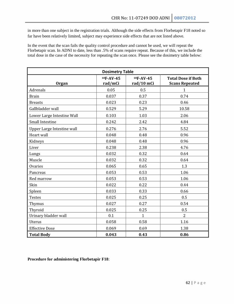
CHR No: 11-07249 DOD ADNI 08072012
62 | P a g e
in more than one subject in the registration trials. Although the side effects from Florbetapir F18 noted so
far have been relatively limited, subject may experience side effects that are not listed above.
In the event that the scan fails the quality control procedure and cannot be used, we will repeat the
Florbetapir scan. In ADNI to date, less than .5% of scans require repeat. Because of this, we include the
total dose in the case of the necessity for repeating the scan once. Please see the dosimetry table below:
Dosimetry Table
Organ
18F-AV-45 rad/mCi
18F-AV-45 rad/10 mCi
Total Dose if Both Scans Repeated
Adrenals 0.05 0.5 1
Brain 0.037 0.37 0.74
Breasts 0.023 0.23 0.46
Gallbladder wall 0.529 5.29 10.58
Lower Large Intestine Wall 0.103 1.03 2.06
Small Intestine 0.242 2.42 4.84
Upper Large Intestine wall 0.276 2.76 5.52
Heart wall 0.048 0.48 0.96
Kidneys 0.048 0.48 0.96
Liver 0.238 2.38 4.76
Lungs 0.032 0.32 0.64
Muscle 0.032 0.32 0.64
Ovaries 0.065 0.65 1.3
Pancreas 0.053 0.53 1.06
Red marrow 0.053 0.53 1.06
Skin 0.022 0.22 0.44
Spleen 0.033 0.33 0.66
Testes 0.025 0.25 0.5
Thymus 0.027 0.27 0.54
Thyroid 0.025 0.25 0.5
Urinary bladder wall 0.1 1 2
Uterus 0.058 0.58 1.16
Effective Dose 0.069 0.69 1.38
Total Body 0.043 0.43 0.86
Procedure for administering Florbetapir F18:

CHR No: 11-07249 DOD ADNI 08072012
63 | P a g e
A needle will be used to inject Florbetapir F18 into your vein. Insertion of the needle may cause pain or a
stinging sensation at the injection site. On rare occasions the insertion of a needle can cause bleeding, a
blood clot, swelling or infection at the site of insertion. There is currently no information on the effects of
Florbetapir F18 on unborn children. However it is known that higher levels of radiation can cause
damage to unborn children. Subject should not participate in this study if pregnant or breastfeeding.
Women of childbearing potential must not be pregnant (negative urine beta-chg. on the day of imaging)
or breast feeding at screening. Women must avoid becoming pregnant and must agree to refrain from
sexual activity or to use reliable contraceptive methods for 24 hours following administration of
Florbetapir F18. Because the age of subjects recruited will be between 50-90 years old, very few subjects
with child bearing potential will be enrolled, nonetheless, should a subject be of childbearing age, a
pregnancy test will first be administered. Pregnancy or breast feeding are exclusionary criteria.
Subject is monitored during the scan for blood pressure, pulse, respiration,
A telephone check will be made one to two business days after the procedure to check on
how subject is feeling, and to see if there are any adverse reactions.
Computed Tomography (CT) Scan. During the Florbetapir F18 PET scan, it is sometimes
necessary to have a CT or another type of scan to help correct the scans for photon attenuation. If so, the
CT or transmission scan also uses a very small dose of radiation.
18. Adverse Events Reporting All subjects will be evaluated for adverse events at each clinical visit. Adverse events (AE) will be
continuously monitored during the imaging session. Subjects who experience any adverse event will not be discharged until the event has resolved or stabilized. A follow-up phone call to the patient (or the
caregiver as applicable) will be conducted approximately 2 (+/- 1) business days after the imaging session
to confirm patient well-being and to collect information about any new adverse events. The injection site
will be observed for excessive inflammation or damage to the surrounding tissue where the dose was injected, and a physician or physician designee will see the patient prior to dosing and prior to discharge.
The most common adverse events in previous human clinical trials include headache, injection site reactions, musculoskeletal pain, nausea and fatigue. Back pain, anxiety/claustrophobia, insomnia,
hypertension, and neck pain were also reported. Because these events could be related in part to the PET
scan apparatus and procedures, careful attention will be taken to make the subject aware of the planned procedures and to maximize subject comfort in the scanner. Previous human clinical trials have revealed
no clinically meaningful changes in vital signs, ECG or laboratory changes.
Definition of an Adverse Event An adverse event is any adverse change from the subject‟s baseline condition including clinical or
laboratory tests, or abnormalities that occur during the course of the study after consent.
Following up on Adverse Events The investigator is obliged to follow subjects with AE‟s until the events have subsided, the conditions are
considered medically stable, or the participants are no longer available for follow up. Subjects who
discontinue due to adverse events will be treated and followed according to established medical practice.

CHR No: 11-07249 DOD ADNI 08072012
64 | P a g e
All pertinent information will be entered into the eCRF. All serious adverse events (SAEs) will be
reported to the independent Data Safety Monitoring Board.
Serious adverse events include any event that is fatal, life threatening, significantly or persistently
disabling or incapacitating, results in hospitalization, prolongs a hospital stay, or is associated with a
congenital abnormality or birth defect. In addition, any experience which the investigator regards as
serious, or which would suggest significant hazard, contraindication, side effect, or precaution associated
with participation in the study should be reported as a serious adverse event.
Reporting Serious Adverse Events Any such experience due to any cause, which occurs during the course of the investigation or within 30
days of the last study visit, must be reported to the Project Director within 24 hours after learning of the
event. This is in turn will trigger a report to be distributed to all participating sites, DSMB and the DOD.
Sites will report based on local IRB requirements.
Eli Lilly must be notified immediately (as soon as possible, and in all cases within 24 hours) of a
florbetapir F 18 drug experience, condition, development, or event, which is considered serious. Eli Lilly
must be notified immediately of any findings with the use of florbetapir F 18 that may suggest significant
hazards, contraindications, adverse drug reactions (ADRs) and precautions pertinent to the safety of the
drug. The investigator will be requested to complete a separate report form.
A Serious adverse event (SAE) spontaneously reported to have occurred within 24-48 hours of florbetapir
F 18 administration will be reported, regardless of the investigator‟s opinion of causation. Thereafter,
SAEs are not required to be reported unless the investigator feels the events were related to either
florbetapir F 18 or the drug delivery system, or a protocol procedure.
If an SAE is determined to be unexpected (not previously reported or described by Avid), and study drug-
related, Eli Lilly, will notify the investigator in writing. The investigator should forward this notification
to the IRB within 24 hours of receipt.
Data and Safety Monitoring Board The ADNI-CC currently has an active Data and Safety Monitoring Board (DSMB) that reviews the safety
of all subjects enrolled in trials on an ongoing basis. The DSMB will review serious adverse event reports
on a quarterly basis.
Medical Monitor The medical monitor may be assigned to assess one or more of the following phases of the research
project: volunteer recruitment, volunteer enrollment, data collection, or data storage and analysis. The
medical monitor will provide an independent evaluation of serious adverse events and unanticipated
problems involving risk to subjects or others to the Institutional Review Board (IRB) /Human Research
Protection Office (HRPO). The medical monitor may be assigned to discuss research progress with the

CHR No: 11-07249 DOD ADNI 08072012
65 | P a g e
principal investigator, interview volunteers, consult on individual cases, or evaluate adverse event
reports. Medical monitor, Adam Fleisher, will promptly report discrepancies or problems to the IRB and
the HRPO. He shall have the authority to stop a research study in progress, remove individual volunteers
from a study, and take whatever steps are necessary to protect the safety and well-being of research
volunteers until the IRB can assess the medical monitor‟s report.
All unanticipated problems involving risk to subjects or others, serious adverse events related to
participation in the study and subject deaths related to participation in the study should be promptly
reported by phone (301-619-2165), by email
([email protected]<mailto:[email protected]>), or by facsimile (301-619-7803) to the
USAMRMC, Office of Research Protections, Human Research Protection Office. A complete written
report will follow the initial notification. In addition to the methods above, the complete report will be
sent to the U.S. Army Medical Research and Materiel Command, ATTN: MCMR-ZB-PH, 504 Scott
Street, Fort Detrick, Maryland 21702-5012.
Our Medical Monitor for this study, Adam Fleisher, M.D., M.A.S., is the Associate director of brain
imaging at the Banner Alzheimer‟s Institute and Associate Professor in the Department of Neurosciences
at the University of California, San Diego.
19. Benefits:
Benefits to Subjects:
This is not a treatment study. There is no direct benefit to subjects who participate in this study.
However, with subject permission, should there be any significant clinical findings from the medical exam, blood test, MRI, PET scan or lumbar puncture, the results will be given to the subjects' physician,
or the subject, should no primary care physician be available.
Benefits to Society: AD is the most common cause of dementia. TBI and PTSD are common problems resulting from military
service, and both may be associated with increased risk of cognitive decline and dementia due to AD or
other causes. The results will have major implications for identifying subjects at increased risk for AD, as
well as possibly establishing a need for early detection of AD in military Veterans with histories of TBI and PTSD, and a possible need to employ prevention and treatment measures to avoid accelerated
development of AD in US military Veterans. This study is a first step towards a larger, more
comprehensive study of dementia risk factors in Veterans. The result will lead to a design and statistical powering of a prevention trial. Therefore, this project could be first step towards the prevention of AD in
Veterans, and in the general population.
While the long-term health risks from participating in this study are small, the benefit to society lies in
having better tests for diagnosis of Alzheimer's disease as well as better tests for assessing drug efficacy.

CHR No: 11-07249 DOD ADNI 08072012
66 | P a g e
20. Neuropathology The ADNI Neuropathology Core will continue to be responsible for coordinating brain autopsies at the
participating sites, shipment of postmortem brain tissues to the Neuropathology Core, and performing a
standardized neuropathological assessment. The Neuropathology Core will maintain a tissue bank to
facilitate clinicopathological research
21. Data Collection and Monitoring The ADNI Clinical Core will be responsible for providing the operational infrastructure for the ADNI
sites. This infrastructure will be provided by the ADCS Administrative, Clinical Operations, Medical and
Data Cores at UCSD.
Clinical data collection and monitoring are standardized with well-established and successful ADCS
operating procedures. Data will be entered directly by sites using web-based data entry screens. Imaging
data collection and monitoring are handled by LONI. Imaging data are handled using a combination of
data entry via web-based forms and automated file transition modules embedded within the web-based
Image Data Archive application. The file translation modules extract metadata directly from the imaging
data, ensuring accuracy and reducing the amount of data entry required.
22. Inclusion of Women and Minorities Both women and members of minority groups will be recruited during this protocol. Based on the
participating sites data regarding enrollment of minorities, it is expected that 12% of subjects enrolled will
be minorities. This is close to the aged minority population in the U. S. which is 14%. The enrollment of
women will be included, though it is expected that the number of women enrolled will be quite small
given the relatively small number of women who participated in the Vietnam War.
23. Data Sharing Policy In order to provide the de-identified clinical data from this project to investigators, the Pharmaceutical
Industry, and the public, the entire clinical database (free of any identifying information such as name,
address, or phone number) will be placed on a public web site, which will be appropriately linked to the
imaging database at LONI. The database will be frequently updated and all cleaned clinical data acquired
by the DOD-ADNI study will be provided in real time. Dr Robert Green, from Harvard University, who
chairs the ADNI Data and publications committee, will continue to monitor all requests for permissions to
access the data and will treat all publications coming forth from this DOD ADNI grant similar to the
ADNI publications. All publications must be submitted to the committee for review, prior to submission
to journals. The committee tracks these and insures that the DOD ADNI project is properly given
acknowledgement in the author line of the paper. There will be no cost to his for this project.
Sharing of Banked DNA, RNA, Plasma, Serum and CSF Specific procedures for requesting and accessing DNA, RNA, or plasma, serum or CSF have been created
by the Resource Allocation Review Committee (RARC) of the ADNI in accordance with

CHR No: 11-07249 DOD ADNI 08072012
67 | P a g e
recommendations proposed in the National Bioethics Advisory Commission (NBAC) Human Biological
Materials Report.
All analysis data will be uploaded to the clinical database at ADCS and will be available on the
UCLA/LONI/ADNI website without embargo
24. Data Analysis
Primary analyses involve comparing groups on baseline levels of CSF, Neuroimaging (MRI and
amyloid PET), and cognitive measures associated with AD pathology and annual change in MRI and
cognitive measures to assess whether PTSD or TBI is associated with increased evidence for AD
compared to Veteran controls. Further analyses will focus on within-group correlations to assess a dose-
response association between outcomes and severity of PTSD or TBI and whether TBI or PTSD reduces
cognitive reserve. Additional exploratory analyses will also be conducted. The specific analytic methods
for hypothesis testing are listed below followed by an assessment of power.
The primary hypotheses to be tested (all data analyses will be covaried for age, gender, and APOe4
genotype) are that Veterans without MCI (or dementia, with a history of moderate to severe TBI during
military service, as well as Veterans with ongoing PTSD, have increased evidence for AD, when
compared with Veteran controls (without TBI or PTSD) manifested as: 1) greater uptake on Florbetapir
amyloid PET scans; 2) lower CSF amyloid beta levels; 3) increased CSF tau/P tau levels; 4) brain atrophy
in hippocampus, entorhinal cortex, and parietal/temporal cortices; and 5) greater rates of brain atrophy in
hippocampus, entorhinal cortex and parietal/temporal cortices; 6) reduced cognitive function, especially
delayed recall.
The second major hypotheses to be tested is that TBI and/or PTSD reduce brain reserve causing greater
cognitive impairment after accounting for age, brain amyloid load or hippocampal volume. Greater
cognitive impairments at a given level of brain Aβ or brain volume in the TBI or PTSD group compared
with controls would support the hypothesis of reduced cognitive reserve.
The third hypothesis will be that TBI, when compared to controls, is associated with changes detected
with DTI in brain regions previously reported to be associated with TBI [37-39].
Finally, the hypothesis that there will be significant correlations between severity of TBI (determined
from medical records) and severity of PTSD (CAPS score) on the above-listed outcomes in the TBI and
PTSD groups respectively will be tested.
Exploratory analyses will be performed to examine other questions; although, after correction for
multiple comparisons, the statistical significance of these will be low, requiring future replication.
Nevertheless, the patterns of amyloid deposition (from Florbetapir uptake) and brain atrophy between
TBI, PTSD, and control subjects will be compared, with similar patterns from non-Veteran subjects in
ADNI. These results of these studies may provide insight into the question of whether or not TBI and
PTSD alter the pattern of amyloid distribution or brain atrophy. Such analyses may also give insight into

CHR No: 11-07249 DOD ADNI 08072012
68 | P a g e
the question of whether TBI or PTSD is associated with reduced cognitive reserve. The relationship
between cortical areas with amyloid plaque (from amyloid PET) and underlying white matter integrity as
assessed with DTI will also be studied to determine if axonal injury resulting from TBI was associated
with greater amyloid accumulation, or whether regions of brain with axonal damage have less amyloid
accumulation due to disconnection and reduced brain activity.
Primary Analyses: Comparing Groups on Baseline Level (Hypothesis sets 1 & 3): Primary outcomes for these analyses include uptake on Florbetapir scans, CSF amyloid beta, CSF tau and
Ptau, volumes of the hippocampus, entorhinal cortex, and parietal/temporal cortices, DTI summary
measures (in the third set of hypotheses) and measures of cognitive function, including delayed episodic
memory.
The analyses will begin with: 1) numerical and graphical summaries of the measures within each group to
assess the underlying distribution and detect outliers or questionable values which will be flagged and
checked with the sites for accuracy. 2) Analysis of variance (ANOVA) for a simple unadjusted
comparison between the groups. If the global F-test for group difference is significant, there will be a
follow-up with post-hoc pairwise tests, specifically between the TBI or PTSD groups and the Veteran
control group, adjusted for multiple comparisons using the Bonferroni or Tukey‟s Honestly Significant
Difference (HSD) approach. 3) linear regression models that include group as an independent variable to
adjust for potential confounders including age, gender, APOe4 genotype, and alcohol dependence. Model
assumptions will be assessed through graphical and numerical approaches and transformations or non-
linear models will be used if suggested by the diagnostics. Hypotheses will be supported if the TBI and
PTSD groups show significantly higher levels of uptake on amyloid PET scans and CSF tau or Ptau than
the Veteran controls and significantly lower levels of CSF amyloid beta, MRI volumes, and cognitive
function than the Veteran controls.
Primary Analyses: Comparing Groups on Annual Change (Hypothesis sets 1 & 3): Primary outcomes for these analyses include annual change in volumes of the hippocampus, entorhinal
cortex, and parietal/temporal cortices and annual change in cognitive function such as delayed recall.
There will be two assessments for each person, approximately one year apart. Therefore, for each
participant as a measure of annual change, there will be a construction of difference scores between the
measures obtained at the follow-up visit and the baseline visit. The scores will be divided by the time
between the assessments to account for variability in timing of the follow-up assessments between
participants. Analyses will be similar to those described above for comparing groups on the baseline
level except that the final linear regression models will also be adjusted for baseline level of the outcome
measure. Hypotheses will be supported if the TBI and PTSD groups show significantly faster rates of
atrophy and cognitive decline than the Veteran controls.
TBI or PTSD associated with reduction in cognitive reserve (Hypothesis set two): The main outcomes for these analyses will be level and annual change in cognitive function, particularly
memory while the predictors of interest are group, baseline hippocampal volume, and uptake from the
amyloid PET scans. Linear regression methods similar to those described above will be used. Of
particular interest for these analyses are the interactions between group and the imaging predictors.
Power will be limited for these analyses, so results will mainly serve as support for future studies of
PTSD, TBI, and cognitive reserve. However, significant interactions suggesting worse cognitive function

CHR No: 11-07249 DOD ADNI 08072012
69 | P a g e
in the TBI or PTSD groups relative to Veteran controls at a given level of MRI, or amyloid imaging
measures would support the hypotheses.
Within-group correlations to assess dose-response: Outcomes for these analyses will be the same as those used for hypothesis sets one, two, and three.
Interest lies in assessing whether the baseline levels or rates of atrophy or cognitive decline are associated
with severity of TBI or PTSD. Analyses will be performed within each of those groups separately.
Severity of TBI or PTSD will be the predictor of interest. Simple linear regression models that include
severity of TBI or PTSD as the independent variable will be used. Multiple regression models to adjust
for potential confounders including age, gender, and APOe4 genotype and alcohol dependence will be
used next. As stated above for the primary analyses, model assumptions will be assessed and
transformations or non-linear models will be used if suggested by the diagnostics. Secondary hypotheses
will be supported if increased severity of TBI or PTSD is significantly associated with lower levels of
CSF amyloid beta, MRI volumes, and cognitive function, higher levels of CSF tau or Ptau, and increased
rates of brain atrophy and cognitive decline.
Exploratory Analyses: As described earlier, exploratory analyses will be performed to examine other questions; although, after
correction for multiple comparisons, power will be low requiring future replication. Nevertheless, there
will be a comparison of the patterns (using voxel based methods) of amyloid deposition (from amyloid
PET) and brain atrophy between TBI, PTSD, and control subjects, and with similar patterns from non
Veteran subjects in ADNI. The results of these studies may provide insight into the question of whether
or not TBI and PTSD alter the pattern of amyloid distribution or brain atrophy. Furthermore, the
relationships between amyloid deposition, atrophy, and cognitive function may provide insight into the
question of whether TBI or PTSD is associated with reduced cognitive reserve. Further exploratory
analyses will assess the relationship between cortical areas with amyloid plaque (from amyloid PET) and
underlying white matter integrity as assessed with DTI to determine if axonal injury resulting from TBI
was associated with greater amyloid accumulation, or whether regions of the brain with axonal damage
have less amyloid accumulation due to disconnection and reduced brain activity. Linear regression
methods, described above, will be used to assess the association between regional measures of amyloid
accumulation and axonal damage.
Power Analysis: Power analyses are presented for each class of primary hypotheses assuming a two-sided test and were
calculated using nQuery. For comparison of the TBI or PTSD group to the Veteran controls, assuming
alpha = 0.025 to account for multiple comparisons and 65 individuals per group at baseline and 61 per
group for longitudinal measures, there will be an 80% power to detect a difference as small as 0.55
standard deviations in level and as small as 0.56 standard deviations in rate of atrophy or cognitive
decline. For example, using means and standard deviations from measures in the normal controls within
ADNI-1, this difference would translate to at least a 6.7% lower hippocampal volume, and at least a
14.7% lower CSF amyloid beta. Because there is little change in the ADNI normals, differences in
change will be much more difficult to detect. However, these data will provide initial estimates of how
much change is experienced in the TBI and PTSD groups which will help in planning larger scale

CHR No: 11-07249 DOD ADNI 08072012
70 | P a g e
longitudinal studies of these groups. The power analysis will be able to detect at least a doubling of the
rate of hippocampal atrophy and at least a quadrupling of the rate of decline in delayed recall in the TBI
or PTSD groups compared to the Veteran controls. For within group correlations, there will be an 80%
power to detect a correlation as small as 0.33 with cross-sectional outcomes and 0.34 with outcomes of
change.

CHR No: 11-07249 DOD ADNI 08072012
71 | P a g e
25. Bibliography & References Cited:
1. Scarmeas, N. and Y. Stern, Cognitive reserve: implications for diagnosis and prevention of
Alzheimer's disease. Curr Neurol Neurosci Rep, 2004. 4(5): p. 374-80.
2. Snitz, B.E., E.S. O'Meara, M.C. Carlson, A.M. Arnold, D.G. Ives, S.R. Rapp, J. Saxton, O.L.
Lopez, L.O. Dunn, K.M. Sink, and S.T. DeKosky, Ginkgo biloba for preventing cognitive decline
in older adults: a randomized trial. JAMA, 2009. 302(24): p. 2663-70.
3. Leoutsakos, J.M., B.O. Muthen, J.C. Breitner, and C.G. Lyketsos, Effects of non-steroidal anti-
inflammatory drug treatments on cognitive decline vary by phase of pre-clinical Alzheimer
disease: findings from the randomized controlled Alzheimer's Disease Anti-inflammatory
Prevention Trial. Int J Geriatr Psychiatry, 2011.
4. Lyketsos, C.G., J.C. Breitner, R.C. Green, B.K. Martin, C. Meinert, S. Piantadosi, and M.
Sabbagh, Naproxen and celecoxib do not prevent AD in early results from a randomized
controlled trial. Neurology,
5. Shaw, L.M., H. Vanderstichele, M. Knapik-Czajka, M. Figurski, E. Coart, K. Blennow, H.
Soares, A.J. Simon, P. Lewczuk, R.A. Dean, E. Siemers, W. Potter, V.M. Lee, and J.Q.
Trojanowski, Qualification of the analytical and clinical performance of CSF biomarker analyses
in ADNI. Acta Neuropathol, 2011. 121(5): p. 597-609.
6. Petersen, R.C., P.S. Aisen, L.A. Beckett, M.C. Donohue, A.C. Gamst, D.J. Harvey, C.R. Jack, Jr.,
W.J. Jagust, L.M. Shaw, A.W. Toga, J.Q. Trojanowski, and M.W. Weiner, Alzheimer's Disease
Neuroimaging Initiative (ADNI): clinical characterization. Neurology, 2010. 74(3): p. 201-9.
7. Aisen, P.S., R.C. Petersen, M.C. Donohue, A. Gamst, R. Raman, R.G. Thomas, S. Walter, J.Q.
Trojanowski, L.M. Shaw, L.A. Beckett, C.R. Jack, Jr., W. Jagust, A.W. Toga, A.J. Saykin, J.C.
Morris, R.C. Green, and M.W. Weiner, Clinical Core of the Alzheimer's Disease Neuroimaging
Initiative: progress and plans. Alzheimers Dement, 2010. 6(3): p. 239-46.
8. Jack, C.R., Jr., M.A. Bernstein, B.J. Borowski, J.L. Gunter, N.C. Fox, P.M. Thompson, N.
Schuff, G. Krueger, R.J. Killiany, C.S. Decarli, A.M. Dale, O.W. Carmichael, D. Tosun, and
M.W. Weiner, Update on the magnetic resonance imaging core of the Alzheimer's disease
neuroimaging initiative. Alzheimers Dement, 2010. 6(3): p. 212-20.
9. Jagust, W.J., D. Bandy, K. Chen, N.L. Foster, S.M. Landau, C.A. Mathis, J.C. Price, E.M.
Reiman, D. Skovronsky, and R.A. Koeppe, The Alzheimer's Disease Neuroimaging Initiative
positron emission tomography core. Alzheimers Dement, 2010. 6(3): p. 221-9.
10. Shaw, L.M., PENN Biomarker Core of the Alzheimer's Disease Neuroimaging Initiative.
Neurosignals, 2008. 16(1): p. 19-23.

CHR No: 11-07249 DOD ADNI 08072012
72 | P a g e
11. Saykin, A.J., L. Shen, T.M. Foroud, S.G. Potkin, S. Swaminathan, S. Kim, S.L. Risacher, K. Nho,
M.J. Huentelman, D.W. Craig, P.M. Thompson, J.L. Stein, J.H. Moore, L.A. Farrer, R.C. Green,
L. Bertram, C.R. Jack, Jr., and M.W. Weiner, Alzheimer's Disease Neuroimaging Initiative
biomarkers as quantitative phenotypes: Genetics core aims, progress, and plans. Alzheimers
Dement, 2010. 6(3): p. 265-73.
12. Shen, L., S. Kim, S.L. Risacher, K. Nho, S. Swaminathan, J.D. West, T. Foroud, N. Pankratz,
J.H. Moore, C.D. Sloan, M.J. Huentelman, D.W. Craig, B.M. Dechairo, S.G. Potkin, C.R. Jack,
Jr., M.W. Weiner, and A.J. Saykin, Whole genome association study of brain-wide imaging
phenotypes for identifying quantitative trait loci in MCI and AD: A study of the ADNI cohort.
Neuroimage, 2010.
13. Frisoni, G.B. and M.W. Weiner, Alzheimer's Disease Neuroimaging Initiative special issue.
Neurobiol Aging, 2010. 31(8): p. 1259-62.
14. Sperling, R.A., P.S. Aisen, L.A. Beckett, D.A. Bennett, S. Craft, A.M. Fagan, T. Iwatsubo, C.R.
Jack, Jr., J. Kaye, T.J. Montine, D.C. Park, E.M. Reiman, C.C. Rowe, E. Siemers, Y. Stern, K.
Yaffe, M.C. Carrillo, B. Thies, M. Morrison-Bogorad, M.V. Wagster, and C.H. Phelps, Toward
defining the preclinical stages of Alzheimer's disease: recommendations from the National
Institute on Aging-Alzheimer's Association workgroups on diagnostic guidelines for Alzheimer's
disease. Alzheimers Dement, 2011. 7(3): p. 280-92.
15. Ellis, K.A., C.C. Rowe, V.L. Villemagne, R.N. Martins, C.L. Masters, O. Salvado, C. Szoeke,
and D. Ames, Addressing population aging and Alzheimer's disease through the Australian
imaging biomarkers and lifestyle study: collaboration with the Alzheimer's Disease
Neuroimaging Initiative. Alzheimers Dement, 2010. 6(3): p. 291-6.
16. Rowe, C.C., K.A. Ellis, M. Rimajova, P. Bourgeat, K.E. Pike, G. Jones, J. Fripp, H. Tochon-
Danguy, L. Morandeau, G. O'Keefe, R. Price, P. Raniga, P. Robins, O. Acosta, N. Lenzo, C.
Szoeke, O. Salvado, R. Head, R. Martins, C.L. Masters, D. Ames, and V.L. Villemagne, Amyloid
imaging results from the Australian Imaging, Biomarkers and Lifestyle (AIBL) study of aging.
Neurobiol Aging, 2010. 31(8): p. 1275-83.
17. Iwatsubo, T., Japanese Alzheimer's Disease Neuroimaging Initiative: present status and future.
Alzheimers Dement, 2010. 6(3): p. 297-9.
18. Arai, H., N. Okamura, K. Furukawa, and Y. Kudo, Geriatric medicine, Japanese Alzheimer's
disease neuroimaging initiative and biomarker development. Tohoku J Exp Med, 2010. 221(2): p.
87-95.
19. Frisoni, G.B., Alzheimer's disease neuroimaging initiative in Europe. Alzheimers Dement, 2010.
6(3): p. 280-5.

CHR No: 11-07249 DOD ADNI 08072012
73 | P a g e
20. Burton, A., Big science for a big problem: ADNI enters its second phase. Lancet Neurol, 2011.
10(3): p. 206-7.
21. Lister-James, J., M.J. Pontecorvo, C. Clark, A.D. Joshi, M.A. Mintun, W. Zhang, N. Lim, Z.
Zhuang, G. Golding, S.R. Choi, T.E. Benedum, P. Kennedy, F. Hefti, A.P. Carpenter, H.F. Kung,
and D.M. Skovronsky, Florbetapir F 18: a histopathologically validated Beta-amyloid positron
emission tomography imaging agent. Semin Nucl Med, 2011. 41(4): p. 300-4.
22. Clark, C.M., J.A. Schneider, B.J. Bedell, T.G. Beach, W.B. Bilker, M.A. Mintun, M.J.
Pontecorvo, F. Hefti, A.P. Carpenter, M.L. Flitter, M.J. Krautkramer, H.F. Kung, R.E. Coleman,
P.M. Doraiswamy, A.S. Fleisher, M.N. Sabbagh, C.H. Sadowsky, E.P. Reiman, S.P. Zehntner,
and D.M. Skovronsky, Use of florbetapir-PET for imaging beta-amyloid pathology. JAMA,
2011. 305(3): p. 275-83.
23. Okamura, N. and K. Yanai, Florbetapir F 18 (18F), a PET imaging agent that binds to amyloid
plaques for the potential detection of Alzheimer's disease. IDrugs, 2010. 13(12): p. 890-9.
24. Wong, D.F., P.B. Rosenberg, Y. Zhou, A. Kumar, V. Raymont, H.T. Ravert, R.F. Dannals, A.
Nandi, J.R. Brasic, W. Ye, J. Hilton, C. Lyketsos, H.F. Kung, A.D. Joshi, D.M. Skovronsky, and
M.J. Pontecorvo, In vivo imaging of amyloid deposition in Alzheimer disease using the
radioligand 18F-AV-45 (florbetapir F 18 [corrected] F 18). J Nucl Med, 2010. 51(6): p. 913-20.
25. Raymont, V., A.M. Salazar, F. Krueger, and J. Grafman, "Studying injured minds" - the Vietnam
head injury study and 40 years of brain injury research. Frontiers in neurology, 2011. 2: p. 15.
26. Yehuda, R. and J. Ledoux, Response Variation following Trauma: A Translational Neuroscience
Approach to Understanding PTSD. Neuron, 2007. 56(1): p. 19-32.
27. McFall, M.E., P.W. Mackay, and D.M. Donovan, Combat-related PTSD and psychosocial
adjustment problems among substance abusing veterans. Journal of Nervous and Mental
Diseases, 1991. 179: p. 33-38.
28. Fullilove, M.T., R.E. Fullilove, III, M. Smith, K. Winkler, C. Michael, P.G. Panzer, and R.
Wallace, Violence, trauma, and post-traumatic stress disorder among women drug users. Journal
of Traumatic Stress, 1993. 6: p. 533-543.
29. Kulka, R.A., W.E. Schlenger, J.A. Fairbank, R.L. Hough, B.K. Jordan, C.R. Marmar, and D.L.
Weiss, Trauma and the vietnam war generation. 1990, New York: Brunner/Mazel.
30. Marmar, C., R., D. Foy, B. Kagan, and R. Pynoos, S., An integrated approach for treating
posttraumatic stress, in Review of Pscyhiatry. 1994, American Psychiatry Press: Washington,
D.C. p. 99-132.

CHR No: 11-07249 DOD ADNI 08072012
74 | P a g e
31. Kessler, R.C., A. Sonnega, E. Bromet, M. Hughes, and C.B. Nelson, Posttraumatic stress
disorder in the National Comorbidity Survey. Archives of General Psychiatry, 1995. 52: p. 1048-
1060.
32. Wang, Z., T.C. Neylan, S.G. Mueller, M. Lenoci, D. Truran, C.R. Marmar, M.W. Weiner, and N.
Schuff, Magnetic resonance imaging of hippocampal subfields in posttraumatic stress disorder.
Arch Gen Psychiatry, 2010. 67(3): p. 296-303.
33. Schuff, N., C.R. Marmar, D.S. Weiss, T.C. Neylan, F. Schoenfeld, G. Fein, and M.W. Weiner,
Reduced hippocampal volume and n-acetylaspartate in post traumatic stress disorder. The
Annals of the New York Academy of Sciences, 1997. Supplement on Psychobiology of
Posttraumatic Stress Disorder(821): p. 516-520.
34. Schuff, N., T.C. Neylan, M.A. Lenoci, A.T. Du, D.S. Weiss, C.R. Marmar, and M.W. Weiner,
Decreased hippocampal N-acetylaspartate in the absence of atrophy in posttraumatic stress
disorder. Biol Psychiatry, 2001. 50(12): p. 952-9.
35. Schuff, N., T.C. Neylan, S. Fox-Bosetti, M. Lenoci, K.W. Samuelson, C. Studholme, J. Kornak,
C.R. Marmar, and M.W. Weiner, Abnormal N-acetylaspartate in hippocampus and anterior
cingulate in posttraumatic stress disorder. Psychiatry Res, 2008. 162(2): p. 147-57.
36. Apfel, B.A., J. Ross, J. Hlavin, D.J. Meyerhoff, T.J. Metzler, C.R. Marmar, M.W. Weiner, N.
Schuff, and T.C. Neylan, Hippocampal volume differences in Gulf War veterans with current
versus lifetime posttraumatic stress disorder symptoms. Biol Psychiatry, 2011. 69(6): p. 541-8.
37. Kraus, M.F., T. Susmaras, B.P. Caughlin, C.J. Walker, J.A. Sweeney, and D.M. Little, White
matter integrity and cognition in chronic traumatic brain injury: a diffusion tensor imaging
study. Brain, 2007. 130(Pt 10): p. 2508-19.
38. Niogi, S.N., P. Mukherjee, J. Ghajar, C. Johnson, R.A. Kolster, R. Sarkar, H. Lee, M. Meeker,
R.D. Zimmerman, G.T. Manley, and B.D. McCandliss, Extent of microstructural white matter
injury in postconcussive syndrome correlates with impaired cognitive reaction time: a 3T
diffusion tensor imaging study of mild traumatic brain injury. AJNR Am J Neuroradiol, 2008.
29(5): p. 967-73.
39. Niogi, S.N., P. Mukherjee, J. Ghajar, C.E. Johnson, R. Kolster, H. Lee, M. Suh, R.D.
Zimmerman, G.T. Manley, and B.D. McCandliss, Structural dissociation of attentional control
and memory in adults with and without mild traumatic brain injury. Brain, 2008. 131(Pt 12): p.
3209-21.
40. Blake, D.D., F.W. Weathers, L.M. Nagy, D.G. Kaloupek, F.D. Gusman, D.S. Charney, and T.M.
Keane, The development of a Clinician-Administered PTSD Scale. J Trauma Stress, 1995. 8(1): p.
75-90.

CHR No: 11-07249 DOD ADNI 08072012
75 | P a g e
41. Spitzer, R.L., J.B. Williams, M. Gibbon, and M.B. First, The Structured Clinical Interview for
DSM-III-R (SCID). I: History, rationale, and description. Archives of General Psychiatry, 1992.
49: p. 624-629.
42. Wolfe, J., R. Kimerling, P.J. Brown, K.R. Chresman, and K. Levin, Psychometric review of the
life stressor checklist-revised. Measurement of stress, trauma, and adaptation, ed. B.H. Stamm.
Vol. 149. 1996, Lutherville, MD: Sidran Press. 676-679.
43. Derogatis, L. and L. Lazarus, SCL-90--R, Brief symptom inventory, and matching clinical rating
scales, in The use of psychological testing for treatment planning and outcome assessment, M.E.
Maruish, Editor. 1994, Lawrence Erlbaum Associates, Inc.: Hillsdale, NJ. p. 217-248.
44. Buysse, D.J., C.F. Reynolds, 3rd, T.H. Monk, S.R. Berman, and D.J. Kupfer, The Pittsburgh
Sleep Quality Index: a new instrument for psychiatric practice and research. Psychiatry Res,
1989. 28(2): p. 193-213.
45. Neylan, T.C., S.G. Mueller, Z. Wang, T.J. Metzler, M. Lenoci, D. Truran, C.R. Marmar, M.W.
Weiner, and N. Schuff, Insomnia severity is associated with a decreased volume of the
CA3/dentate gyrus hippocampal subfield. Biol Psychiatry, 2010. 68(5): p. 494-6.
46. Schoenborn, C.A., P.F. Adams, and J.S. Schiller, Summary health statistics for the U.S.
population: National Health Interview Survey, 2000. Vital Health Stat 10, 2003(214): p. 1-83.
47. Institute, N.C. Dictionary of cancer terms: Pack year. 2011 5/16/201i]; Available from:
http://www.cancer.gov/dictionary/?CdrID=306510.
48. McLellan, A.T., L. Luborsky, G.E. Woody, and C.P. O'Brien, An improved diagnostic evaluation
instrument for substance abuse patients. The Addiction Severity Index. J Nerv Ment Dis, 1980.
168(1): p. 26-33.
49. McLellan, A.T., H. Kushner, D. Metzger, R. Peters, I. Smith, G. Grissom, H. Pettinati, and M.
Argeriou, The Fifth Edition of the Addiction Severity Index. J Subst Abuse Treat, 1992. 9(3): p.
199-213.
50. Ware, J.E., M. Kosinski, and S.D. Keller, A 12-Item Short-Form Health Survey - Construction of
Scales and Preliminary Tests of Reliability and Validity. Medical Care, 1996. 34(3): p. 220-233.
51. Keane, T.M., J.A. Fairbank, J.M. Caddell, R.T. Zimering, K.L. Taylor, and C. Mora, Clinical
evaluation of a measure to assess combat exposure. J of Consulting and Clinical Psychology,
1989. 1: p. 53-55.
52. Nasreddine, Z.S., I. Collin, H. Chertkow, N. Phillips, H. Bergman, and V. Whitehead, Sensitivity
and Specificity of The Montreal Cognitive Assessment (MOCA) for Detection of Mild Cognitive
Deficits. Can J Neurol Sci, 2003. 30(2): p. 30.

CHR No: 11-07249 DOD ADNI 08072012
76 | P a g e
53. Folstein, M.F., S.E. Folstein, and P.R. McHugh, "Mini-mental state". A practical method for
grading the cognitive state of patients for the clinician. Journal of Psychiatric Research, 1975.
12(3): p. 189-198.
54. Rosen, W.G., R.C. Mohs, and K.L. Davis, A new rating scale for Alzheimer's disease. American
Journal of Psychiatry, 1984. 141(11): p. 1356-1364.
55. Wechsler, D., Wechsler Memory Scale-Revised. 1987, San Antonio, TX: The Psychological
Corporation.
56. Kaplan, E., H. Goodglass, and S. Weintraub, Boston Naming Test. 1983, Philadelphia: Lea &
Febiger.
57. Butters, N., E. Granholm, D.P. Salmon, I. Grant, and J. Wolfe, Episodic and semantic memory: a
comparison of amnesic and demented patients. J Clin Exp Neuropsychol, 1987. 9(5): p. 479-97.
58. Goodglass, H. and E. Kaplan, The Assessment of Aphasia and Related Disorders. 1983,
Philadelphia: Lea & Febiger.
59. Nelson, H.E. and A. O'Connell, Dementia: the estimation of premorbid intelligence levels using
the New Adult Reading Test. Cortex, 1978. 14: p. 234-244.
60. Rey, A., L'examen clinique en psychologie. 1964, Paris: Presses Universitaires de France.
61. Petersen, R.C., G. Smith, E. Kokmen, R.J. Ivnik, and E.G. Tangalos, Memory function in normal
aging. Neurology, 1992. 42(2): p. 396-401.
62. Reitan, R.M., Validity of theTrail Making Test as an Indicator of Organic Brain Damage.
Perceptual & Motor Skills, 1958. 8: p. 271-276.
63. Berg, L., Clinical Dementia Rating (CDR). Psychopharmacol Bull, 1988. 24(4): p. 637-9.
64. Pfeffer, R.I., T.T. Kurosaki, C.H. Harrah, Jr., J.M. Chance, and S. Filos, Measurement of
functional activities in older adults in the community. J Gerontol, 1982. 37(3): p. 323-9.
65. Kaufer, D.I., J.L. Cummings, P. Ketchel, V. Smith, A. MacMillan, T. Shelley, O.L. Lopez, and
S.T. DeKosky, Validation of the NPI-Q, a brief clinical form of the Neuropsychiatric Inventory. J
Neuropsychiatry Clin Neurosci, 2000. 12(2): p. 233-9.
66. Sheikh, J.I. and J. Yesavage, Geriatric Depression Scale (GDS): Recent evidence and
development of a shorter version, in Clinical Gerontology : A Guide to Assessment and
Intervention 1986, The Haworth Press: New York. p. 165-173.

CHR No: 11-07249 DOD ADNI 08072012
77 | P a g e
67. Grafman, J., B.S. Jonas, A. Martin, A.M. Salazar, H. Weingartner, C. Ludlow, M.A. Smutok, and
S.C. Vance, Intellectual function following penetrating head injury in Vietnam veterans. Brain : a
journal of neurology, 1988. 111 ( Pt 1): p. 169-84.
68. Grafman, J., A. Salazar, H. Weingartner, S. Vance, and D. Amin, The relationship of brain-tissue
loss volume and lesion location to cognitive deficit. The Journal of neuroscience : the official
journal of the Society for Neuroscience, 1986. 6(2): p. 301-7.
69. Cairns, N.J., Taylor-Reinwald, L., Morris, J.C.: Alzheimer's Disease Neuroimaging Initiative.
Autopsy consent brain collection, and standardized neuropathologic assessment of ADNI
participants: the essential role of the neuropathology core. Alzheimer's and Dementia 2010. 6:
274-279.
70. Carr, D.B., Gray, S., Baty, J., Morris, J.C.: The value of informant versus individual's complaints
of memory impairment in early dementia". Neurology 2000; 55:1724-1726.
71. Galvin, J.E., Roe, C.M., Powlishta, K.K., Coats, M.A., Muich, S.J., Grant, E., Miller, J.P.,
Storandt, M., Morris, J.C.: A brief informant interview to detect dementia. Neurology. 2005;
65:559-564.
72. Daviglus, M.L., B.L. Plassman, A. Pirzada, C.C. Bell, P.E. Bowen, J.R. Burke, E.S. Connolly, Jr.,
J.M. Dunbar-Jacob, E.C. Granieri, K. McGarry, D. Patel, M. Trevisan, and J.W. Williams, Jr.,
Risk Factors and Preventive Interventions for Alzheimer Disease: State of the Science. Arch
Neurol, 2011.
73. Williams, J.W., B.L. Plassman, J. Burke, and S. Benjamin, Preventing Alzheimer's disease and
cognitive decline. Evid Rep Technol Assess (Full Rep), 2010(193): p. 1-727.
74. Salib, E. and V. Hillier, Head injury and the risk of Alzheimer's disease: a case control study. Int
J Geriatr Psychiatry, 1997. 12(3): p. 363-8.
75. van Duijn, C.M., T.A. Tanja, R. Haaxma, W. Schulte, R.J. Saan, A.J. Lameris, G. Antonides-
Hendriks, and A. Hofman, Head trauma and the risk of Alzheimer's disease. Am J Epidemiol,
1992. 135(7): p. 775-82.
76. Mortimer, J.A., C.M. van Duijn, V. Chandra, L. Fratiglioni, A.B. Graves, A. Heyman, A.F. Jorm,
E. Kokmen, K. Kondo, W.A. Rocca, and et al., Head trauma as a risk factor for Alzheimer's
disease: a collaborative re-analysis of case-control studies. EURODEM Risk Factors Research
Group. Int J Epidemiol, 1991. 20 Suppl 2: p. S28-35.
77. Mayeux, R., R. Ottman, M.X. Tang, L. Noboa-Bauza, K. Marder, B. Gurland, and Y. Stern,
Genetic susceptibility and head injury as risk factors for Alzheimer's disease among community-
dwelling elderly persons and their first-degree relatives. Ann Neurol, 1993. 33(5): p. 494-501.
CHR No: 11-07249 DOD ADNI 08072012
78 | P a g e
78. O'Meara, E.S., W.A. Kukull, L. Sheppard, J.D. Bowen, W.C. McCormick, L. Teri, M.
Pfanschmidt, J.D. Thompson, G.D. Schellenberg, and E.B. Larson, Head injury and risk of
Alzheimer's disease by apolipoprotein E genotype. Am J Epidemiol, 1997. 146(5): p. 373-84.
79. Graves, A.B., E. White, T.D. Koepsell, B.V. Reifler, G. van Belle, E.B. Larson, and M. Raskind,
The association between head trauma and Alzheimer's disease. Am J Epidemiol, 1990. 131(3): p.
491-501.
80. Schofield, P.W., M. Tang, K. Marder, K. Bell, G. Dooneief, M. Chun, M. Sano, Y. Stern, and R.
Mayeux, Alzheimer's disease after remote head injury: an incidence study. J Neurol Neurosurg
Psychiatry, 1997. 62(2): p. 119-24.
81. Heyman, A., W.E. Wilkinson, J.A. Stafford, M.J. Helms, A.H. Sigmon, and T. Weinberg,
Alzheimer's disease: a study of epidemiological aspects. Ann Neurol, 1984. 15(4): p. 335-41.
82. Williams, D.B., J.F. Annegers, E. Kokmen, P.C. O'Brien, and L.T. Kurland, Brain injury and
neurologic sequelae: a cohort study of dementia, parkinsonism, and amyotrophic lateral sclerosis.
Neurology, 1991. 41(10): p. 1554-7.
83. Katzman, R., M. Aronson, P. Fuld, C. Kawas, T. Brown, H. Morgenstern, W. Frishman, L.
Gidez, H. Eder, and W.L. Ooi, Development of dementing illnesses in an 80-year-old volunteer
cohort. Ann Neurol, 1989. 25(4): p. 317-24.
84. Launer, L.J., K. Andersen, M.E. Dewey, L. Letenneur, A. Ott, L.A. Amaducci, C. Brayne, J.R.
Copeland, J.F. Dartigues, P. Kragh-Sorensen, A. Lobo, J.M. Martinez-Lage, T. Stijnen, and A.
Hofman, Rates and risk factors for dementia and Alzheimer's disease: results from EURODEM
pooled analyses. EURODEM Incidence Research Group and Work Groups. European Studies of
Dementia. Neurology, 1999. 52(1): p. 78-84.
85. Mehta, K.M., A. Ott, S. Kalmijn, A.J. Slooter, C.M. van Duijn, A. Hofman, and M.M. Breteler,
Head trauma and risk of dementia and Alzheimer's disease: The Rotterdam Study. Neurology,
1999. 53(9): p. 1959-62.
86. Mayeux, R., Apolipoprotein e4 and head trauma: synergistic or additive risks? Neurology, 1996.
46: p. 889-891. Letter.
87. Katzman, R., D.R. Galasko, T. Saitoh, X. Chen, M.M. Pay, A. Booth, and R.G. Thomas,
Apolipoprotein-epsilon4 and head trauma: Synergistic or additive risks? Neurology, 1996. 46(3):
p. 889-91.
88. Frankowski, R.F., J.F. Annegers, and S. Whitman, Epidemiology and descriptive studies. Part 1.
The descriptive epidemiology of head trauma in the United States, in Central Nervous System
Trauma Status Report –1985, B. D.P. and P. J., Editors. 1985, NIH, NINDS. p. 33-43.

CHR No: 11-07249 DOD ADNI 08072012
79 | P a g e
89. Mayeux, R., R. Ottman, G. Maestre, C. Ngai, M.X. Tang, H. Ginsberg, M. Chun, B. Tycko, and
M. Shelanski, Synergistic effects of traumatic head injury and apolipoprotein-epsilon 4 in patients
with Alzheimer's disease. Neurology, 1995. 45(3 Pt 1): p. 555-7.
90. Nemetz, P.N., C. Leibson, J.M. Naessens, M. Beard, E. Kokmen, J.F. Annegers, and L.T.
Kurland, Traumatic brain injury and time to onset of Alzheimer's disease: a population-based
study. Am J Epidemiol, 1999. 149(1): p. 32-40.
91. Gedye, A., B.L. Beattie, H. Tuokko, A. Horton, and E. Korsarek, Severe head injury hastens age
of onset of Alzheimer's disease. J Am Geriatr Soc, 1989. 37(10): p. 970-3.
92. Fleminger, S., D.L. Oliver, S. Lovestone, S. Rabe-Hesketh, and A. Giora, Head injury as a risk
factor for Alzheimer's disease: the evidence 10 years on; a partial replication. J Neurol Neurosurg
Psychiatry, 2003. 74(7): p. 857-62.
93. Rasmusson, D.X., J. Brandt, D.B. Martin, and M.F. Folstein, Head injury as a risk factor in
Alzheimer's disease. Brain Inj, 1995. 9(3): p. 213-9.
94. Broe, G.A., A.S. Henderson, H. Creasey, E. McCusker, A.E. Korten, A.F. Jorm, W. Longley, and
J.C. Anthony, A case-control study of Alzheimer's disease in Australia. Neurology, 1990. 40(11):
p. 1698-707.
95. Amaducci, L.A., L. Fratiglioni, W.A. Rocca, C. Fieschi, P. Livrea, D. Pedone, L. Bracco, A.
Lippi, C. Gandolfo, G. Bino, and et al., Risk factors for clinically diagnosed Alzheimer's disease:
a case-control study of an Italian population. Neurology, 1986. 36(7): p. 922-31.
96. Chandra, V., E. Kokmen, B.S. Schoenberg, and C.M. Beard, Head trauma with loss of
consciousness as a risk factor for Alzheimer's disease. Neurology, 1989. 39(12): p. 1576-8.
97. Chandra, V., V. Philipose, P.A. Bell, A. Lazaroff, and B.S. Schoenberg, Case-control study of
late onset "probable Alzheimer's disease". Neurology, 1987. 37(8): p. 1295-300.
98. Forster, D.P., A.J. Newens, D.W. Kay, and J.A. Edwardson, Risk factors in clinically diagnosed
presenile dementia of the Alzheimer type: a case-control study in northern England. J Epidemiol
Community Health, 1995. 49(3): p. 253-8.
99. Ferini-Strambi, L., S. Smirne, P. Garancini, P. Pinto, and M. Franceschi, Clinical and
epidemiological aspects of Alzheimer's disease with presenile onset: a case control study.
Neuroepidemiology, 1990. 9(1): p. 39-49.
100. Fratiglioni, L., A. Ahlbom, M. Viitanen, and B. Winblad, Risk factors for late-onset
Alzheimer's disease: a population-based, case-control study. Ann Neurol, 1993. 33(3): p. 258-66.

CHR No: 11-07249 DOD ADNI 08072012
80 | P a g e
101. Li, G., Y.C. Shen, Y.T. Li, C.H. Chen, Y.W. Zhau, and J.M. Silverman, A case-control
study of Alzheimer's disease in China. Neurology, 1992. 42(8): p. 1481-8.
102. Anonymous, The Canadian Study of Health and Aging: risk factors for Alzheimer's
disease in Canada. Neurology, 1994. 44(11): p. 2073-80.
103. Mortimer, J.A., L.R. French, J.T. Hutton, and L.M. Schuman, Head injury as a risk factor
for Alzheimer's disease. Neurology, 1985. 35(2): p. 264-7.
104. Tsolaki, M., K. Fountoulakis, E. Chantzi, and A. Kazis, Risk factors for clinically
diagnosed Alzheimer's disease: a case-control study of a Greek population. Int Psychogeriatr,
1997. 9(3): p. 327-41.
105. Plassman, B.L., R.J. Havlik, D.C. Steffens, M.J. Helms, T.N. Newman, D. Drosdick, C.
Phillips, B.A. Gau, K.A. Welsh-Bohmer, J.R. Burke, J.M. Guralnik, and J.C. Breitner,
Documented head injury in early adulthood and risk of Alzheimer's disease and other dementias.
Neurology, 2000. 55(8): p. 1158-66.
106. Johnson, V.E., W. Stewart, and D.H. Smith, Traumatic brain injury and amyloid-beta
pathology: a link to Alzheimer's disease? Nat Rev Neurosci, 2010. 11(5): p. 361-70.
107. Jellinger, K.A., Head injury and dementia. Current opinion in neurology, 2004. 17(6): p.
719-23.
108. Yaffe, K., E. Vittinghoff, K. Lindquist, D. Barnes, K.E. Covinsky, T. Neylan, M. Kluse,
and C. Marmar, Posttraumatic stress disorder and risk of dementia among US veterans. Arch
Gen Psychiatry, 2010. 67(6): p. 608-13.