ChemicalMethod the Determinationof Oestriol, …€¦ · J. B. BROWN (Brown, 1952; Bauld, 1954)....
9
Vol. 6o INCORPORATION OF 32p INTO NUCLEIC ACIDS 185 We should like to thank Dr R. Y. Thomson and Mr R. Logan for their assistance in some of these experiments, Mr A. J. Taylor for technical assistance and Dr J. A. Roper of the Department of Genetics in this University for help in the radiation experiments. We are also indebted to Dr A. C. White of the Wellcome Physiology Research Laboratories for assistance in the supply of venom. The expenses of this work were defrayed from a grant to one of us (J.N.D.) from the Medical Research Council, and by a grant from the Rankin Fund of the University of Glasgow. REFERENCES Abrams, R. (1951). Arch. Biochem. 30, 90. Allen, R. J. L. (1940). Biochem. J. 34, 858. Andreasen, E. & Ottesen, J. (1945). Acta physiol. acand. 10, 258. Barnum, C. P. & Huseby, R. A. (1950). Arch. Biochem. 29,7. Barnum, C. P., Huseby, R. A. & Vermund, H. (1953). Cancer Ree. 13, 880. Daoust, R., Bertalanffy, F. D. & Leblond, C. P. (1954). J. biol. Chem. 207, 405. Davidson, J. N. & Smellie, R. M. S. (1952a). Biochem. J. 52, 594. Davidson, J. N. & Smellie, R. M. S. (1952 b). Biochem. J. 52, 599. Digby, K. H. & Enticknap, J. B. (1954). Brit. J. exp. Path. 35, 294. Hammarsten, E. & Hevesy, G. (1946). Acta physiol. scand. 11, 335. Harrington, H. & Lavik, P. S. (1953). Fed. Proc. 12, 214. Hevesy, G. (1945). Rev. mod. Phys. 17, 102. Hevesy, G. (1949). Nature, Lond., 163, 869. Hevesy, G. (1951). J. chem. Soc. p. 1618. Hevesy, G. & Ottesen, J. (1943). Acta physiol. scand. 5, 237. Holmes, B. E. (1947). Brit. J. Radiol. (N.S.), 20, 450. Holmes, B. E. (1949). Brit. J. Radiol. (N.S.), 22, 487. Holmes, B. E. & Mee, L. K. (1952). Brit. J. Radiol. (N.S.), 25, 273. Hurst, R. 0. & Butler, G. C. (1951). J. biol. Chem. 193, 91. Hurst, R. O., Little, J. A. & Butler, G. C. (1951). J. biol. Chem. 188, 705. Kay, E. R. M., Simmons, N. S. & Dounce, A. L. (1952). J. Amer. chem. Soc. 74, 1724. Kelly, L. S. & Jones, H. B. (1950). Proc. Soc. exp. Biol., N. Y., 74, 493. Klein, G. & Forssberg, A. (1954). Proc. Second Radioisotope Conference (Oxford), Butterworth: London. Little, J. A. & Butler, G. C. (1951). J. biol. Chem. 188, 695. McCarty, M. (1946). J. gen. Physiol. 29, 123. Mathison, G. C. (1909). Biochem. J. 4, 233. Mirsky, A. E. & Pollister, A. W. (1946). J. gen. Physiol. 30, 117. Payne, A. H., Kelly, L. S. & Entenman, C. (1952). Proc. Soc. exp. Biol., N. Y., 81, 698. Potter, V. R. & Elvehjem, C. A. (1936). J. biol. Chem. 114, 495. Scarborough, R. A. (1931). Yale J. Biol. Med. 3, 63, 168, 282, 359, 431 and 547. Schmidt, G. & Thannhauser, S. J. (1945). J. biol. Chem. 161, 83. Sinsheimer, R. L. & Koerner, J. F. (1952). J. biol. Chem. 198, 293. Smellie, R. M. S., McIndoe, W. M., Logan, R., Davidson, J. N. & Dawson, I. M. (1953). Biochem. J. 54, 280. Stevens, C. E., Daoust, R. & Leblond, C. P. (1953). J. biol. Chem. 202, 177. Vermund, H., Barnum, C. P., Huseby, R. A. & Stenstrom, K. W. (1953). Cancer Res. 13, 633. A Chemical Method for the Determination of Oestriol, Oestrone and Oestradiol in Human Urine BY J. B. BROWN Clinical Endocrinology Research Unit (Medical Research Council), Edinburgh University (Received 9 August 1954) In connexion with a research programme to study the clinical significance of variations in the urinary excretion and metabolism of the oestrogens, an accurate method, suitable for day-to-day routine use, was required for the separate determination of oestriol, oestrone and oestradiol-17,B in the low concentrations found in the urine of men and non- pregnant women. Since all available methods lacked the desired accuracy, sensitivity, specificity and convenience, it was decided to attempt to develop a more suitable procedure. The project was organ- ized jointly with the Department of Biochemistry, and papers dealing with one aspect of the problem (Brown, 1952; Bauld, 1954) have already been published. The present paper describes in detail one complete procedure which appears to be more satisfactory in many respects than any that have hitherto been described, and has been used on a routine basis in clinical investigations for the past 18 months. A preliminary communication in which the method was briefly described has already been presented (Brown, 1955). The method was evolved from the one described by Clayton (1949) and incorporates a new phase- change purification step involving methylation of the phenolic fraction. The methylated oestrogens are then further purified and separated from one another by chromatography on alumina columns and are estimated colorimetrically using an im- proved modification of the Kober colour reaction
Transcript of ChemicalMethod the Determinationof Oestriol, …€¦ · J. B. BROWN (Brown, 1952; Bauld, 1954)....

Vol. 6o INCORPORATION OF 32p INTO NUCLEIC ACIDS 185We should like to thank Dr R. Y. Thomson and Mr R.
Logan for their assistance in some of these experiments,Mr A. J. Taylor for technical assistance and Dr J. A. Roperof the Department of Genetics in this University for help inthe radiation experiments. We are also indebted to Dr A. C.White of the Wellcome Physiology Research Laboratoriesfor assistance in the supply of venom. The expenses of thiswork were defrayed from a grant to one of us (J.N.D.) fromthe Medical Research Council, and by a grant from theRankin Fund of the University of Glasgow.
REFERENCES
Abrams, R. (1951). Arch. Biochem. 30, 90.Allen, R. J. L. (1940). Biochem. J. 34, 858.Andreasen, E. & Ottesen, J. (1945). Acta physiol. acand. 10,
258.Barnum, C. P. & Huseby, R. A. (1950). Arch. Biochem. 29,7.Barnum, C. P., Huseby, R. A. & Vermund, H. (1953).
Cancer Ree. 13, 880.Daoust, R., Bertalanffy, F. D. & Leblond, C. P. (1954).
J. biol. Chem. 207, 405.Davidson, J. N. & Smellie, R. M. S. (1952a). Biochem. J. 52,
594.Davidson, J. N. & Smellie, R. M. S. (1952 b). Biochem. J. 52,
599.Digby, K. H. & Enticknap, J. B. (1954). Brit. J. exp. Path.
35, 294.Hammarsten, E. & Hevesy, G. (1946). Acta physiol. scand.
11, 335.Harrington, H. & Lavik, P. S. (1953). Fed. Proc. 12, 214.Hevesy, G. (1945). Rev. mod. Phys. 17, 102.Hevesy, G. (1949). Nature, Lond., 163, 869.Hevesy, G. (1951). J. chem. Soc. p. 1618.Hevesy, G. & Ottesen, J. (1943). Acta physiol. scand. 5, 237.
Holmes, B. E. (1947). Brit. J. Radiol. (N.S.), 20, 450.Holmes, B. E. (1949). Brit. J. Radiol. (N.S.), 22, 487.Holmes, B. E. & Mee, L. K. (1952). Brit. J. Radiol. (N.S.),
25, 273.Hurst, R. 0. & Butler, G. C. (1951). J. biol. Chem. 193, 91.Hurst, R. O., Little, J. A. & Butler, G. C. (1951). J. biol.
Chem. 188, 705.Kay, E. R. M., Simmons, N. S. & Dounce, A. L. (1952).
J. Amer. chem. Soc. 74, 1724.Kelly, L. S. & Jones, H. B. (1950). Proc. Soc. exp. Biol.,N. Y., 74, 493.
Klein, G. & Forssberg, A. (1954). Proc. Second RadioisotopeConference (Oxford), Butterworth: London.
Little, J. A. & Butler, G. C. (1951). J. biol. Chem. 188, 695.McCarty, M. (1946). J. gen. Physiol. 29, 123.Mathison, G. C. (1909). Biochem. J. 4, 233.Mirsky, A. E. & Pollister, A. W. (1946). J. gen. Physiol. 30,
117.Payne, A. H., Kelly, L. S. & Entenman, C. (1952). Proc. Soc.
exp. Biol., N. Y., 81, 698.Potter, V. R. & Elvehjem, C. A. (1936). J. biol. Chem. 114,
495.Scarborough, R. A. (1931). Yale J. Biol. Med. 3, 63, 168,
282, 359, 431 and 547.Schmidt, G. & Thannhauser, S. J. (1945). J. biol. Chem. 161,
83.Sinsheimer, R. L. & Koerner, J. F. (1952). J. biol. Chem.
198, 293.Smellie, R. M. S., McIndoe, W. M., Logan, R., Davidson,
J. N. & Dawson, I. M. (1953). Biochem. J. 54, 280.Stevens, C. E., Daoust, R. & Leblond, C. P. (1953). J. biol.Chem. 202, 177.
Vermund, H., Barnum, C. P., Huseby, R. A. & Stenstrom,K. W. (1953). Cancer Res. 13, 633.
A Chemical Method for the Determination of Oestriol, Oestroneand Oestradiol in Human Urine
BY J. B. BROWNClinical Endocrinology Research Unit (Medical Research Council), Edinburgh University
(Received 9 August 1954)
In connexion with a research programme to studythe clinical significance of variations in the urinaryexcretion and metabolism of the oestrogens, anaccurate method, suitable for day-to-day routineuse, was required for the separate determination ofoestriol, oestrone and oestradiol-17,B in the lowconcentrations found in the urine of men and non-pregnant women. Since all available methods lackedthe desired accuracy, sensitivity, specificity andconvenience, it was decided to attempt to developa more suitable procedure. The project was organ-ized jointly with the Department of Biochemistry,and papers dealing with one aspect of the problem(Brown, 1952; Bauld, 1954) have already beenpublished. The present paper describes in detail one
complete procedure which appears to be moresatisfactory in many respects than any that havehitherto been described, and has been used on aroutine basis in clinical investigations for the past18 months. A preliminary communication in whichthe method was briefly described has already beenpresented (Brown, 1955).The method was evolved from the one described
by Clayton (1949) and incorporates a new phase-change purification step involving methylation ofthe phenolic fraction. The methylated oestrogensare then further purified and separated from oneanother by chromatography on alumina columnsand are estimated colorimetrically using an im-proved modification of the Kober colour reaction
J. B. BROWN
(Brown, 1952; Bauld, 1954). Interfering back-groumd colours are corrected for by the spectro-photometric method of Allen (1950).
MATERIALSReagents. Ether (J. F. Maefarlan and Co. Ltd., Edin-
burgh, A.R.) was washed with saturated FeSO4 solution andwater, distilled, stored in dark bottles, and used within a
fortnight of purification. Ether recovered during theprocedure was simply redistilled before using again. Lightpetroleum (b.p. 40-60') and benzene (British Drug HousesLtd., A.R.) were redistilled and saturated with water.Recovered benzene was washed thoroughly with water anddistilled through a fractionating column. Ethanol (ab-solute) was refluxed with NaOH pellets to remove alde-hydes and twice distilled. (Errors may arise through impuri-ties in solvents (Bauld, 1955) but those specified here are
quite satisfactory provided they are used in connexion withurine extracts.) H3BO3 powder was B.P. grade. Dimethylsulphate was redistilled. NaOH and NaHCO3 solutionswere prepared on a w/v basis using A.R. chemicals. Concen-trated carbonate solution, ofpH 10-5 (approximately), was
prepared by adding 20% NaOH (150 ml.) to 8% NaHCO3
I955solution (11.). Alumina (Savory and Moore Ltd., London)of mesh size 100/150 and activity II-III (Brockmann &
Schodder, 1941) was deactivated with 9-10% of water tothe activity specified later.
Colour reagent8. The oestriol colour reagent was preparedby dissolving quinol (20 g.) in 76% (v/v) H2SO4 (1 1.), theoestrone reagent by dissolving quinol (20 g.) in 66% (v/v)H2SO4 (1 1.) and the oestradiol reagent by dissolving quinol(20 g.) in 60% (v/v) H2SO4 (1 1.). The quinol was laboratorygrade, and the H2SO4 was AnalaR reagent, both manu-
factured by British Drug Houses Ltd. Solution was hastenedby heating. Reagents were kept at least 24 hr. before use,
the oestriol reagent was usually a light yellow, the oestronewas light brown to pink, and the oestradiol reagent was
light pink in colour. They were stable almost indefinitely atroom temp. in the dark.
Standard oestrogen 8olutions. Standard solutions of purecrystalline oestrogens and their methyl ethers were pre-pared in ethanol (5 mg./100 ml.). Solutions were stored at40 and were stable indefinitely.
Apparatus. Glassware was rinsed after use with tap, andthen distilled water unless visibly dirty, when it was cleanedby (a) steeping in a chromic acid-sulphuric acid mixture;(b) washing with tap water; (c) soaking in an acid sulphite
Urine hydrolysed with HCIextracted with ether
Ether extractWashed with (1) satd. Na2CO, soln. pH 10-5
(2) NaOH; NaOH wash neutralized to pH 10 withsatd. NaHCO8 and shaken again with the ether extracts
(3) NaHCO3(4) water
Benzene-light petroleum
extracted withNaOH
NaOH extract (oestrone-oestradiol)H3BO3 added
Methylated with dimethyl sulphateat 370
NaOH and H20 added
extracted with lightpetroleum
Light petroleum extractwashed with water
Oestrone methylether
chromatographedon alumina
Oestradiol methylether
Ether evaporatedResidue dissolved in ethanolBenzene and light petroleum (equal vols.) added
extracted withwater
Water exract (oestriol)H3BO3 and NaOH added
Methylated with dimethylsulphate at 370
NaOH and H20, added
extracted withbenzene
Benzene extractwashed with water
chromatograon alumina
Oestriol methylether
Lphed
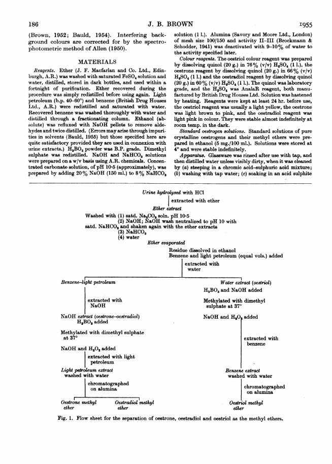
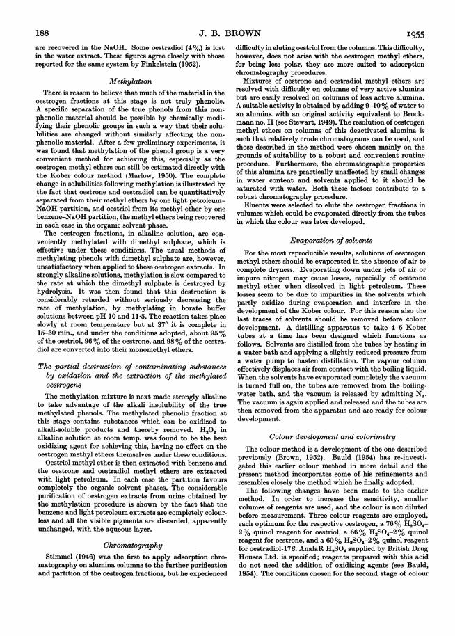
Fig. 1. Flow sheet for the separation of oestrone, oestradiol and oestriol as the methyl ethers.
186
ESTIMATION OF OESTROGENS IN URINEsolution (Na2SO3, approx. 0-2% acidified with H2SO4) todestroy traces of chromic acid which would otherwise bea harmful contaminant in the method, and (d) rinsingthoroughly with tap water, then distilled water, beforebeing dried.
Optical densities were measured in a Unicam S.P. 600spectrophotometer using 1 cm. glass cells.
EXPERIMENTAL
The extraction and purification stages are given first ingeneral, then in detail and are summarized in Fig. 1.
Hydroly8si of conjugated oestrogen8 in urineThe hydrolysis of conjugated oestrogens in urine is a
complex problem which is still being investigated. Atpresent, acid hydrolysis only has been studied since it fitsmore easily than enzymic hydrolysis into a routine method.Preliminary experiments to find conditions giving maxi-mum yields of oestrogens from urine showed that the bestresults were obtained by the method recommended byMarrian & Bauld (1951) namely, boiling the urine underreflux 1 hr. with 15 vol. conc. HCI/100 vol. urine. However,when oestriol, oestroneoroestradiol- 17,Bis addedto the urinebefore this acid treatment, from 10 to 20% of the addedoestrogen is lost during the hydrolysis. Furthermore, itseems that similar losses of endogenous oestrogens alsooccur. These losses are not prevented by the addition ofreducing agents as suggested by Rosenmund (1948) and vanBruggen (1948), and, at present, have to be accepted alongwith the other known losses occurring in the method.
Extraction with ether and removalof the acid fraction
Ether seems to be the most suitable solvent for extractingthe three oestrogens from aqueous solutions but even sothorough extraction is necessary to extract all of theoestriol.Many workers remove the acid fraction from the ether
extract of hydrolysed urine by shaking with NaHCO3solutions (Clayton, 1949). Cohen & Marrian (1934) passedCO2 into alkaline solutions containing the acid and phenolfractions from urine until the solutions were neutral tophenolphthalein (pH 9-0) and then extracted the oestrogenswith ether. Engel, Slaunwhite, Carter & Nathanson (1950),and Engel (1950) modified this procedure and showed that itis more efficient than any other described for removing theacid fraction. In this laboratory it was found that oestriolis easily extracted with ether from aqueous solutions ata pH as high as 10-5, provided the aqueous solutions aresaturated with Na,CO3 or NaHCO8 at that pH. In fact, thepartition coefficient of oestriol between ether and concen-trated carbonate solution of pH 10-5 was found to bepractically the same as between ether and saturatedNaHCO3. Apparently, even at this pH, ionic concentrationrather than pH determines the solubility of oestriol inaqueous solutions. Concentrated carbonate solution ofpH 10-5, which is much more effective as a washing agentthan NaHCO3, can therefore be used for removing the acidfraction from ether extracts without any appreciable loss ofoestriol.
Urine extracts contain substances which change tocoloured products when dissolved in alkali and cannot thenbe re-extracted with ether at a pH above 7. These sub-
stances, which are effectively removed from the oestrogenfraction in the procedures of Cohen & Marrian (1934) and ofEngel et al. (1950), are changed and removed in the presentmethod by shaking the ether extract directly with NaOHsolution after first washing out the acid fraction with conc.Na2CO3 solution ofpH 10-5. The NaOH, which also extractssome of the oestrogens, is then partly neutralized to pH 10,and the carbonate-ion concentration increased, by addingsaturated NaHCO3 solution. Shaking again with the etherlayer extracts the oestrogens back into the ether and leavesthe coloured substances in the aqueous layer which is thendiscarded. The ether is washed with saturated NaHCO3 toneutralize any alkali still present, and thus eliminateexcessive loss of oestriol in the subsequent water wash. Theether is then washed with a small amount ofwater to removethe bicarbonate.The efficacy ofthis treatment is illustrated in the following
experiments. When the oestrogens were subjected to theprocedure, 97% ofthe oestriol and 100% of the oestrone andoestradiol were recovered in the ether phase.An ether extract of hydrolysed male urine was divided
into four equal parts and extracted (1) and (2) with 8%sodium bicarbonate, (3) with conc. carbonate solution ofpH 10-5, (4) by the new procedure described above. Theether was evaporated, the residue taken up in toluene andextracted with NaOH. The alkali extracts of (2) and (3) werepartly neutralized to a pH of 9-9-5 and extracted with etheraccording to the procedure of Engel (1950), and those of(1) and (4) were acidified with HCl and extracted with ether.The ether extracts were evaporated and the residues wereheated with the 76% H2SO4 2% quinol reagent as foroestriol in the modified Kober colour reaction. Uncorrectedoptical densities at 516 m,u. of the colours produced by theurine contaminants still present were (1) simple NaHCO3wash 0-315; (2) Engel procedure 0-120; (3) modified Engelprocedure 0-097; (4) the new procedure 0-099. The new pro-cedure incorporating the carbonate and alkali washes istherefore even more efficient and more convenient forremoving acidic and alkali-unstable substances from urineextracts than the method described by Engel.
The separation of the phenotic fraction and ofoestriolfrom oestrone and oestradiol
It is possible to methylate the three oestrogens togetherand separate, the methyl ethers on a single chromatogram.However, better purification is 'achieved by dividing intoan oestriol fraction and an oestrone plus oestradiol fractionbefore methylation, and chromatographing these on twoseparate columns after methylation. This preliminarydivision is conveniently performed at the same time as theneutral and phenolic fractions are separated from each other.In this way, Clayton (personal communication) extractedoestriol from benzene with water and then extractedoestrone and oestradiol with NaOH. However, this methodis tedious as thorough extractions are required for quanti-tative results. The ease with which the oestrogens areextracted from benzene with aqueous solvents is increasedby diluting with light petroleum, a solvent in which theoestrogens are relatively insoluble (Doisy, Huffman,Thayer & Doisy, 1941). A mixture of equal parts of benzeneand light petroleum was found to be completely satisfactory.Using this and the extraction procedure described in themethod, 98% of the oestriol is recovered in the waterextract, and 96% of the oestradiol and 97% of the oestrone
Vol. 6o 187
J. B. BROWNare recovered in the NaOH. Some oestradiol (4%) is lostin the water extract. These figures agree closely with thosereported for the same system by Finkelstein (1952).
MethylationThere is reason to believe that much of the material in the
oestrogen fractions at this stage is not truly phenolic.A specific separation of the true phenols from this non-phenolic material should be possible by chemically modi-fying their phenolic groups in such a way that their solu-bilities are changed without similarly affecting the non-phenolic material. After a few preliminary experiments, itwas found that methylation of the phenol group is a veryconvenient method for achieving this, especially as theoestrogen methyl ethers can still be estimated directly withthe Kober colour method (Marlow, 1950). The completechange in solubilities following methylation is illustrated bythe fact that oestrone and oestradiol can be quantitativelyseparated from their methyl ethers by one light petroleum-NaOH partition, and oestriol from its methyl ether by onebenzene-NaOH partition, the methyl ethers being recoveredin each case in the organic solvent phase.The oestrogen fractions, in alkaline solution, are con-
veniently methylated with dimethyl sulphate, which iseffective under these conditions. The usual methods ofmethylating phenols with dimethyl sulphate are, however,unsatisfactory when applied to these oestrogen extracts. Instrongly alkaline solutions, methylation is slow compared tothe rate at which the dimethyl sulphate is destroyed byhydrolysis. It was then found that this destruction isconsiderably retarded without seriously decreasing therate of methylation, by methylating in borate buffersolutions between pH 10 and 11-5. The reaction takes placeslowly at room temperature but at 370 it is complete in15-30 min., and under the conditions adopted, about 95%of the oestriol, 96% of the oestrone, and 98% of the oestra-diol are converted into their monomethyl ethers.
The partial destruction of contaminating substancesby oxidation and the extraction of the methylatedoestrogensThe methylation mixture is next made strongly alkaline
to take advantage of the alkali insolubility of the truemethylated phenols. The methylated phenolic fraction atthis stage contains substances which can be oxidized toalkali-soluble products and thereby removed. H202 inalkaline solution at room temp. was found to be the bestoxidizing agent for achieving this, having no effect on theoestrogen methyl ethers themselves under these conditions.
Oestriol methyl ether is then extracted with benzene andthe oestrone and oestradiol methyl ethers are extractedwith light petroleum. In each case the partition favourscompletely the organic solvent phases. The considerablepurification of oestrogen extracts from urine obtained bythe methylation procedure is shown by the fact that thebenzene and light petroleum extracts are completely colour-less and all the visible pigments are discarded, apparentlyunchanged, with the aqueous layer.
ChromatographyStimmel (1946) was the first to apply adsorption chro-
matography on alumina columns to the further purificationand partition of the oestrogen fractions, but he experienced
difficulty in eluting oestriol from the columns. This difficulty,however, does not arise with the oestrogen methyl ethers,for being less polar, they are more suited to adsorptionchromatography procedures.
Mixtures of oestrone and oestradiol methyl ethers areresolved with difficulty on columns of very active aluminabut are easily resolved on columns of less active alumina.A suitable activity is obtained by adding 9-10% of water toan alumina with an original activity equivalent to Brock-mann no. II (see Stewart, 1949). The resolution of oestrogenmethyl ethers on columns of this deactivated alumina issuch that relatively crude chromatograms can be used, andthose described in the method were chosen mainly on thegrounds of suitability to a robust and convenient routineprocedure. Furthermore, the chromatographic propertiesof this alumina are practically unaffected by small changesin water content and solvents applied to it should besaturated with water. Both these factors contribute to arobust chromatography procedure.
Eluents were selected to elute the oestrogen fractions involumes which could be evaporated directly from the tubesin which the colour was later developed.
Evaporation of solventsFor the most reproducible results, solutions of oestrogen
methyl ethers should be evaporated in the absence of air tocomplete dryness. Evaporating down under jets of air orimpure nitrogen may cause losses, especially of oestronemethyl ether when dissolved in light petroleum. Theselosses seem to be due to impurities in the solvents whichpartly oxidize during evaporation and interfere in thedevelopment of the Kober colour. For this reason also thelast traces of solvents should be removed before colourdevelopment. A distilling apparatus to take 4-6 Kobertubes at a time has been designed which functions asfollows. Solvents are distilled from the tubes by heating ina water bath and applying a slightly reduced pressure froma water pump to hasten distillation. The vapour columneffectively displaces air from contact with the boiling liquid.When the solvents have evaporated completely the vacuumis turned full on, the tubes are removed from the boiling-water bath, and the vacuum is released by admitting N2.The vacuum is again applied and released and the tubes arethen removed from the apparatus and are ready for colourdevelopment.
Colour development and colorimetryThe colour method is a development of the one described
previously (Brown, 1952). Bauld (1954) has re-investi-gated this earlier colour method in more detail and thepresent method incorporates some of his refinements andresembles closely the method which he finally adopted.The following changes have been made to the earlier
method. In order to increase the sensitivity, smallervolumes of reagents are used, and the colour is not dilutedbefore measurement. Three colour reagents are employed,each optimum for the respective oestrogen, a 76% H2SO4-2% quinol reagent for oestriol, a 66% H2SO4-2% quinolreagent for oestrone, and a 60% H2SO4-2% quinol reagentfor oestradiol-17f. AnalaR H2SO4 supplied by British DrugHouses Ltd. is specified; reagents prepared with this aciddo not need the addition of oxidizing agents (see Bauld,1954). The conditions chosen for the second stage of colour
188 I955
ESTIMATION OF OESTROGENS IN URINE
development are an H2SO4 concentration of about 55% anda heating time of 10 min., neither of which is critical.Bauld's most important improvement to the colour methodis the addition of fresh quinol to the mature H2S04-quinolreagents immediately before colour development. This isconveniently done by adding the quinol to the oestrogenfractions before evaporating the solvents. As recommendedby Bauld, the Kober tubes are standardized to approxi-mately the same diameters.When the colour method is applied to pure oestrogens,
colours produced are stable for at least 12 hr. and absorblight maximally at the following wavelengths (uncorrected):oestriol and oestrone, 513m,u.; oestradiol-17,B, 514 mi.;oestriol and oestrone methyl ethers, 516 m,u.; oestradiol-17,Bmethyl ether, 518 mp. The molecular extinction coefficientsare very nearly the same for the free oestrogens as for theirmethyl ethers but exact comparison is not possible owing tothe small differences in the absorption maxima.
are collected without preservative and stored at 4'. If the24 hr. volume is less than 1200 ml., the specimen is dilutedto this volume with distilled water.
Hydrolysis and extractionUrine (200 ml.) is heated to boiling under a reflux
condenser. Conc. HCI (1 N, 30 ml.) is then added throughthe condenser and theurine-HCl mixture is boiled for 60min.and then cooled rapidly under running tap water. Thecooled hydrolysed urine is extracted once with 200 ml. andtwice with 100 ml. volumes of ether. The ether is thenextracted with concentrated carbonate solution, ofpH 10-5(80 ml.) (which is rejected), and then shaken thoroughlywith NaOH (20 ml. of 8%). The NaOH layer is not dis-carded but is partly neutralized by adding 8% NaHCO3solution (80 ml.) and shaken again with the ether layer. Theaqueous layer is then discarded. The ether is washed firstwith 8% NaHCO3 solution (20 ml.) and then with water1A.n.m. Thp wn.+ r iaQ ±0rainaVl nfL 0a0 Jnmrslptlavuv na jQakiv u.). iL p wVXlaw itXCsuaeuo as uopVsLysJ43PUOSSD1e
0-6 - Extraction of the phenolfractionsand methylation
0-5 -The ether solution is poured into a flask and the ether is
distilled just to dryness on a water bath. The flask is removedimmediately from the water bath, ethanol (1 ml.) is added
bO to dissolve the residue, since oestriol is not easily soluble inv 0-4 - benzene, and the flask is allowed to cool. Its contents are
transferred with benzene (25 ml.) to a separating funnel-n / / /containing light petroleum (25 ml.). The benzene-light, 0-3 petroleum solution is extracted with two 25 ml. volumes of
0/c) / water and then with two 25 ml. volumes of 1-6% NaOH.u The water extracts, which contain the oestriol fraction,
0-2 // / are added to a 100 ml. stoppered conical flask containing0-2 X H3BO3 (0-9 g.) and 0% NaOH (4 ml.). The NaOH extracts,which contain the oestrone and oestradiol fractions, areadded to a similar flask containing only H3BO3 (0-9 g.). The
0.1 two flasks are placed in a 370 water bath and dimethylsulphate (1 ml.) is added to each. The dimethyl sulphate ishandled cautiously, in a fume cupboard, and volumes are
a I a5ffi & measured by means of a safety pipette. The flasks are0 2 4 6 8 10 shaken until the H3BO3 and dimethyl sulphate have dis-
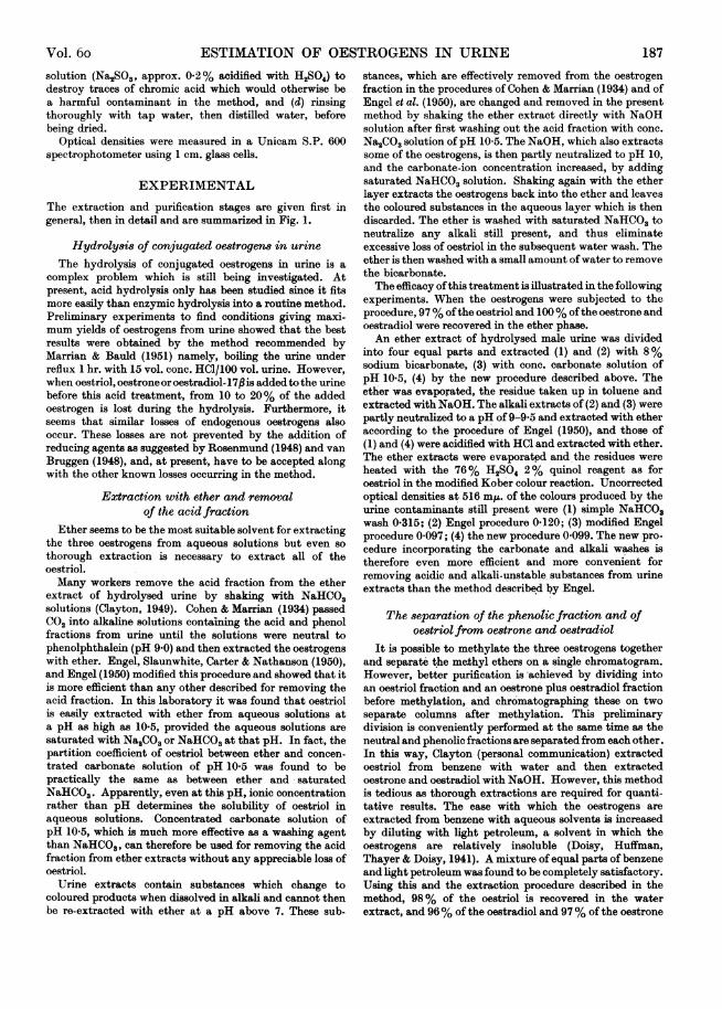
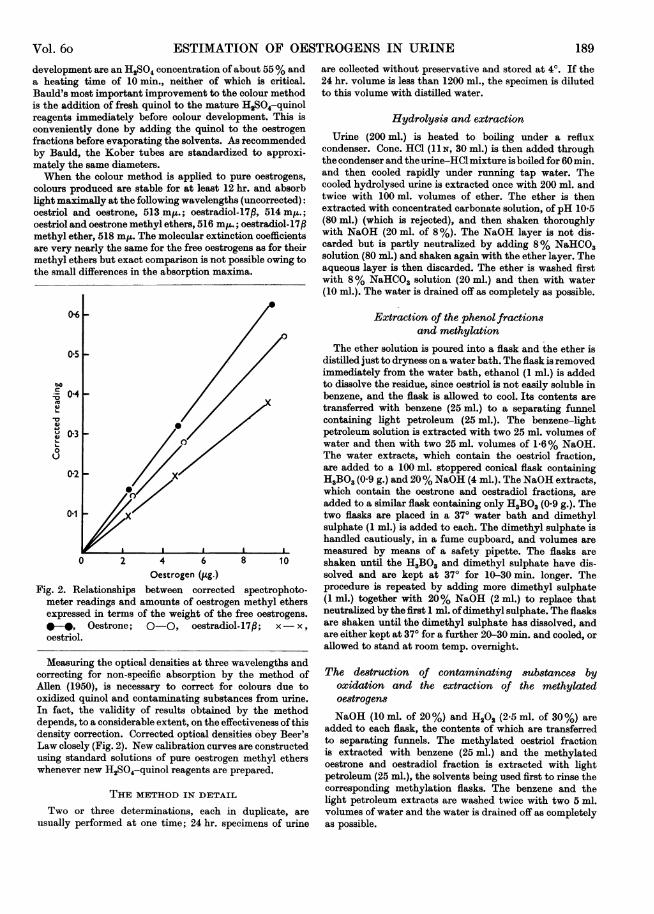
Oestrogen (Jug.) solved and are kept at 370 for 10-30 min. longer. TheFig. 2. Relationships between corrected spectrophoto- procedure is repeated by adding more dimethyl sulphate
meter readings and amounts of oestrogen methyl ethers (1 ml-) together with 20% NaOH (2 ml.) to replace thatexpressed in terms of the weight of the free oestrogens. neutralized by the first 1 ml. ofdimethyl sulphate. The flasks-*, Oestrone; 0-0, oestradiol-17p; x- x , are shaken until the dimethyl sulphate has dissolved, and
oestriol. are either kept at 370 for a further 20-30 min. and cooled, orallowed to stand at room temp. overnight.
Measuring the optical densities at three wavelengths andcorrecting for non-specific absorption by the method ofAllen (1950), is necessary to correct for colours due tooxidized quinol and contaminating substances from urine.In fact, the validity of results obtained by the methoddepends, to a considerable extent, on the effectiveness of thisdensity correction. Corrected optical densities obey Beer'sLaw closely (Fig. 2). New calibration curves are constructedusing standard solutions of pure oestrogen methyl etherswhenever new H2SO4-quinol reagents are prepared.
THE METHOD IN DETAIL
Two or three determinations, each in duplicate, areusually performed at one time; 24 hr. specimens of urine
The destruction of contaminating substances byoxidation and the extraction of the methylatedoestrogen8NaOH (10 ml. of 20%) and H202 (2-5 ml. of 30%) are
added to each flask, the contents of which are transferredto separating funnels. The methylated oestriol fractionis extracted with benzene (25 ml.) and the methylatedoestrone and oestradiol fraction is extracted with lightpetroleum (25 ml.), the solvents being used first to rinse thecorresponding methylation flasks. The benzene and thelight petroleum extracts are washed twice with two 5 ml.volumes of water and the water is drained off as completelyas possible.
Vol. 6o 189

J. B. BROWN
ChromatographyThe glass chromatogram tubes have an internal diameter
of 13 mm., a capacity of 40 ml. of solvent, a sealed-insintered glass support (porosity no. 3) for the aluminacolumn, and an interchangeable B 19 cone for connexionwith receiving flasks or tubes. Chromatograms are usuallyrun in groups of four or six. The rate of flow of solvents isadjusted to approximately 1 drop/2 sec. by applying slightsuction from a manifold through two-way stop cocks, bywhich means suction can be applied to or released fromindividual chromatogram tubes and their receiving flasks.A column is prepared by partly filling the chromatogramtube with benzene or light petroleum and then adding thealumina (2 g.), standardized as described later, in a thinstream so that entangled air frees itself during the passagethrough the solvent. When the alumina has settled, itssurface is levelled by tapping, and overlaid with abouta quarter of an inch of dry acid-ethanol washed sand toprotect it from disturbance during the addition of solvents.Solvents are sucked through to the level of the sand but nolower before another solvent is added.The methylated oestriol fraction in benzene is applied to
an alumina column prepared in benzene, taking care not totransfer any water to the column. The column is then elutedwith (a) 12 ml. of a mixture of 1.4% ethanol in benzene; theeffluent removes a urine pigment band and is discarded, and(b) 15 ml. of a mixture of 2.5% ethanol in benzene; theeffluent, containing all of the oestriol methyl ether, iscollected in a 6 x i in. Pyrex test tube with a standard B 19socket (Kober tube) in which the colour is later developed.The methylated oestrone and oestradiol fractions in light
petroleum are similarly applied to another alumina columnprepared in light petroleum. The column is eluted with(a) 12 ml. of a mixture of 25% benzene in light petroleum,the effluent being discarded; (b) 15 ml. of a mixture of 40%benzene in light petroleum; the effluent is collected intoa Kober tube and contains all of the oestrone methyl ether;(c) a further 12 ml. of the same mixture of 40% benzene inlight petroleum; the effluent being discarded; and (d) 12 ml.of benzene; the effluent is collected into a Kober tube andcontains all of the oestradiol methyl ether.
Preparation and 8tandardization of the alumina
Particular care is required in the standardization of thealumina used in this method. It is possible to alter either thevolumes and eluting powers of solvents for each new batchof deactivated alumina, but it is easier in practice to adjustthe alumina to a predetermined activity and keep the otherfactors constant. Alumina is deactivated by adding water(approx. 9.5 ml. per 100 g.), stirring to break down moistlumps, and shaking the mixture thoroughly until quitehomogeneous. Perceptible heat is evolved, and afterallowing to cool the activity is tested by preparing a 2 g.column in light petroleum and applying a solution ofoestrone methyl ether (lOug.) in light petroleum (25 ml.)which has been washed with water. The column is elutedwith 25% benzene in light petroleum in fractions and theoestrone methyl ether content ofeach fraction is determinedcolorimetrically. When the activity of the alumina iscorrect, oestrone methyl ether begins to appear in thel6th-20th ml. of eluate. If the alumina is too active, more
water is added, if not active enough, more active aluminais added until the required activity is obtained. The be-
I955haviours of the oestrogen methyl ethers on this aluminaalways conform closely to the elution pattern from which thechromatography procedure described in the method isderived. Small deviations from this pattern are sometimesfound with different batches of alumina and it is thennecessary to alter slightly the volumes of eluents used in themethod in order to ensure adequate safety factors in theblind cutting of fractions. The following procedures areundertaken with each fresh batch of alumina to check thispoint. Another column (2 g.) is prepared in light petroleum;a water-washed solution of oestrone and oestradiol methylethers (10 pg. of each) in light petroleum (25 ml.) is appliedto the column which is then eluted, first with 25% benzenein light petroleum (12 ml.), and then fractionally with 40%benzene in light petroleum. Oestrone methyl ether shouldbe completely eluted in the first 12 ml. of the 40% benzenein light petroleum and oestradiol methyl ether shouldbegin to be eluted when a total of about 30 ml. of eluate hasbeen collected. Another column is prepared and oestradiolmethyl ether similarly applied and eluted with (a) 12 ml. of25% benzene in light petroleum, (b) 27 ml. of 40% benzenein light petroleum, and then (c) fractionally with benzene.The oestradiol methyl ether should be eluted in the first8-10 ml. of benzene eluate. Likewise oestriol methyl etherin benzene (25 ml.) is washed with water and applied to a2 g. column prepared in benzene. When this column iseluted with 1.4% ethanol in benzene, oestriol methyl ethershould begin to be eluted in the 15th-17th ml. of eluate.Another similar column is eluted, first with 12 ml. of 1.4%ethanol in benzene, and then with 2-5% ethanol in benzenethe first 12 ml. of which should elute the oestriol methylether.The standardized alumina is stored in air-tight containers.
Caking sometimes occurs during storage but is easilybroken down by shaking.
Evaporation of 801vent8Quinol (4 mg.) in ethanolic solution (0-2 ml. of 2%, w/v)
and a small piece ofporous tile are added to each eluate in itsKober tube. Solutions are evaporated completely to drynessby heating in a water bath. Blank tubes containing quinolonly are prepared at the same time.
Colour development and colorimetryThe appropriate quinol-H2SO4 reagent (3 ml.) for the
particular oestrogen is added to the oestrogen fractions inKober tubes which are then heated for 20 min. in a boiling-water bath. The tubes are shaken twice during the first6 min. of heating. After heating, the tubes are cooled in abath of cold water. Water (1 ml.) is added to each oestrioltube, 0.5 ml. to each oestrone tube and 0-2 ml. to eachoestradiol tube; the tubes are shaken and reheated in theboiling-water bath for 10 min. They are then cooled again incold water for about 10 min. and optical densities aremeasured against similarly treated reagent blanks in thespectrophotometer at the following wavelengths: oestrioland oestrone fractions 480, 516 and 552 mIA.; oestradiolfraction 480, 518 and 556 mu.
Optical density readings (D) are corrected by applyingthe following formulae which are derived from Allen's(1950) formula multiplied by 2: oestriol and oestronecorrectedreadings = 2D5l6-(D480 +D.,); oestradiol correctedreading = 2D515-(D"0 + Ds5r). The amount of oestrogen
190
ESTIMATION OF OESTROGENS IN URINE
methyl ether present in each tube is found by applying thecorrected readings to the particular standard calibrationcurve prepared with the pure oestrogen methyl ether. Thisis then converted into the corresponding amount of freeoestrogen by multiplying by the ratio of the molecularweights. The 24 hr. excretion is calculated from this and the24 hr. urine volume.
RESULTS
Recoveries of oestrogen8 added to hydrolysed urineTo test the extraction, purification and colorimetricstages ofthe method a series ofrecovery experimentswere performed in which known amounts ofoestriol, oestrone and oestradiol- 17,B were added toportions of acid-hydrolysed 24 hr. male urines.Blank determinations were made at the same timeon the same urine. The amounts ofoestrogens added,calculated on the basis of a 24.hr. specimen to-gether with the percentages of these recovered aftersubtracting the urine blank values, are shown in
measuring differences in oestrogen concentrationsof the order of 4 ,g./24 hr. urine specimen.
Results from urines
Typical figures, including spectrophotometerreadings, obtained from the urine of a womanin the early proliferative phase of the menstrualcycle and from a normal man are shown in Table 2,to illustrate the order of results found in thesecases.The agreement obtained between duplicate
determinations is illustrated by the following figurestaken from 100 unselected consecutive duplicateanalysis performed by one worker. The seriesincludes results from three normal menstrualcycles and five recoveries from male urine where theoestrogen levels were between 0 and 40 jpg./day. Theaverage difference between duplicates in terms of24 hr. excretion was 0 3 ,ug. in the case of oestrone
Table 1. Recoveries of oestrogensfrom acid-hydrolysed male urine
Results are shown as the mean percentage recovery ± the standard deviation and are corrected for the endogenous blankvalues. Figures in parentheses refer to number of determinations.
Amounts added per 24 hr. urine (jig.)
OestriolOestroneOestradiol
4-788±10-8 (12)87±11*7 (11)80±5-5 (12)
25-3585±3-8 (11)84±5-2 (12)91±7-0 (11)
36-6083+3-0 (12)82±6-1 (10)86±6-6 (11)
Table 2. Typical resultsfrom urines containing small amounts of oestrogens
Oestriol 0'Oestrone 0Oestradiol 0
Duplicate determinations.
Optical densities measured_______________________ _ ^Corrected480 m,u. 516 or 518 m,. 552 or 556 m,. values
Case I. Normal male (1/6 of a 24 hr. specimen)*276 0-276 0-234 0*234 0-150 0-150 0-042 0.0*137 0-140 0-147 0-149 0*060 0-060 0 097 0.0'*235 0-261 0-195 0-214 0-134 0-149 0-021 0.0
CalculatedAA
Oestrogen OestrogenMg. pg./24 hr.
842198118
1.0 6*01-5 900-3 1-8
Case II. Normal female (early proliferative phase; 1/8-5 of a 24 hr. specimen)Oestriol 0-252 0-226 0-210 0-186 0-138 0-116 0 030 0 030 0 7Oestrone 0-128 0-096 0-119 0 090 0-071 0-048 0 039 0-036 0-6Oestradiol 0*212 0-196 0-169 0-152 0-124 0-109 0-002 -0-001 0
Table 1. Recoveries are between 80 and 90% even
at levels corresponding to 4Mpg. per day. Similarresults with a greater scatter, but not recorded here,were obtained in a small series at 2-5 pg./day. Thescatter of results is greatest at the lowest levels as
would be expected, for the colour due to addedo3strogen is then so slight that errors in instrumen-tation and in measuring the endogenous oestrogenblank, become large in comparison. These recoveryexperiments show that the method is reliable for
and oestradiol or 04 Mug. in the case of oestriol;95% of the duplicate measurements did not differby more than M0g. in the case of oestrone andoestradiol or 1-5 Mug. in the case of oestriol.The resolution into two components of the Kober
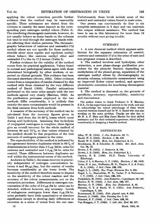
colour produced by an oestrone fraction from urineis shown in Fig. 3.The chromatographic behaviours of a number of
oestrogen methyl ethers and of the methylatedoestriol, oestrone and oestradiol fractions from
6-05-10
191Vol. 6o

J. B. BROWN
pooled luteal-phase urine are shown in Table 3.The oestrogen levels per litre of urine were: oestriol14 Htg.; oestrone 6 ,jg.; and oestradiol 1 jug.
DISCUSSION
The fractions obtained by applying the method tourine still contain impurities which interfere in thecolorimetric measurement of the oestrogens andhave a considerable effect on the specificity of the
0-20
0-16 _
,, 0-12-a
o 0-08
C_
004 _ /-\480 500 520 540 560
Wavelength (m,u.)
Fig. 3. Wavelength/absorption curves of Kober coloursproduced by A, the oestrone fraction from 1/5 of aman's 24 hr. urine, calculated by the correction formulato contain 1 4,ug. of oestrone methyl ether; B, 1-4 tg. ofpure oestrone methyl ether; C, the interfering colourscalculated by subtracting B from A.
method. However, a considerable amount ofevidence, none of which is completely conclusive,has been accumulated to show that the results aresignificant even when the urine contains only smallamounts of oestrogens.The red colour of the Kober reaction, with an
absorption maximum about 515 me., is highlyspecific for the natural oestrogens. However, manyof the impurities present in urine extracts produceyellow colours in the Kober reaction and thesemay form a considerable portion of the total colour,especially when the oestrogen concentration is low(see Table 2). For instance, in the case of maleurine, the optical densities at about 516 mu.contributed by the yellow colours as calculated bythe correction formula are about 0-9, 0-67 and 0-92of the total optical densities of the oestriol, oestroneand oestradiol fractions respectively. This is aserious objection, for it is necessary in these cases torely very much on the spectrophotometric methodfor correcting for this interference. The spectro-photometric correction is based on the assumptionthat the contaminating colours have linear wave-length/absorption curves in the region of theabsorption maximum of the oestrogen red colour.This is difficult to prove, but in all the cases wherethis point has been investigated colours producedwith urine extracts have been resolvable into twocomponents, one with the same absorption maxi-mum as the oestrogen Kober red colour, and theother with a linear wavelength/absorption curve inthis region of the spectrum (Fig. 3).The chromatographic behaviours of the urinary
Kober chromogens estimated as oestrogens after
Table 3. Elution of oe8trogen methyl ethers and urine fraction8 from alumina* columns
Results are expressed as the percentage present in each eluate and in the case of the urine fractions refer only to theKober chromogens estimated as oestrogens after applying the colour correction.
Eluate (ml.)
Benzene-light petroleum
25/75
12 4
40/60 BenzeneA
4 4 4 12 3 3 3 3 3- 28 58
- 64 343 68 28
9 36 43
14
21
12
3 55 35 7 -
2 72 24 211 61 28 -
1.4/98.611
t
Eluate (ml.) ethanol-benzene2.5/97.5
4 4 4 4 41 51 43 57 70 23
* Activity slightly greater than that specified in the method.
Methyl ethersOestroneOestradiol-17,EquilinEquileninOestradiol-17aUrine fraction
OestriolUrine fraction
192 I955
t Very pigmenteci fraction
Vol. 6o ESTIMATION OF OESTROGENS IN URINE 193
applying the colour correction provide furtherevidence that the method may be reasonablyspecific. These substances are eluted from thecolumns in exactly the same manner as the corre-sponding pure oestrogen methyl ethers (Table 3).The interfering chromogenic materials, however, donot usually behave as sharp bands on the columnsbut tend to trail through the oestrogen bands with-out affecting their elution patterns. The chromato-graphic behaviours of oestrone and oestradiol-17#methyl ethers are not specific for these methoxysteroids alone since equilin and equilenin methylethers behave like oestrone methyl ether andoestradiol-17 oc like its 17 P isomer (Table 3).
Further evidence for the validity of the methodcomes from its practical application. Values foundin normal and pathological states agree with thosedetermined biologically by other workers, or ex-pected on clinical grounds. This evidence has beendiscussed elsewhere (Brown, 1955). Other evidencecomes from a comparison ofresults obtained by thismethod with those obtained by the very differentmethod of Bauld (1955). Parallel estimationsperformed on the same urine sample with the twomethods agreed very closely (Marrian, 1955). Asthe purification processes employed in the twomethods differ considerably, it is unlikely thatexactly the same contaminants would be present inthe final extracts from both methods.A measure of the accuracy of the method can be
calculated from the recovery figures given inTable I and from the 10-20% losses which occurduring acid hydrolysis. Assuming that hydrolysisof conjugated oestrogens is complete, these figuresgive an overall recovery for the whole method ofbetween 60 and 75 %, so that values obtained bythe method should be this proportion of the trueamounts of oestrogens present in the urine.The reproducibility of the method is indicated by
the agreement between duplicates which in 95% ofdeterminations is better than 1 0 ,ug./24 hr. urine foroestrone and oestradiol and 1-5 ,ug./24 hr. urine foroestriol. These differences between duplicates arewell within the instrumental errors of the method.As shown in Table 1, the mean recovery is practic-
ally independent of oestrogen concentration be-tween 4 and 60 pg./day, but the scatter of resultsbecomes considerable at the lower levels. Thesensitivity of the method therefore seems to dependon the sensitivity of the colour reaction and theaccuracy of the colour measurements, not on theextraction procedure. Differences in oestrogen con-centration of the order of 2-5 pg./24 hr. urine can bedetected, without however, any accuracy. Levelsof endogenous oestrogens lower than 5 ,ug./24 hr.urine probably have little quantitative or absolutesignificance except in showing daily differences inexcretion in a series of urines from the one case.
Unfortunately these levels include most of theoestriol and oestradiol values found in male urine.One person can conveniently do four to six
complete estimations in 2 days and can maintainthis output without assistance. The method hasbeen in use in this laboratory for more than 18months without once giving trouble.
SUMMARY
1. A new chemical method which appears satis-factory for the separate estimation of oestriol,oestrone and oestradiol-17 p in the urine ofmen andnon-pregnant women is described.
2. The method involves acid hydrolysis, etherextraction, a new phase-change purification pro-cedure for the phenolic fraction depending onmethylation of the phenol group, separation of theoestrogen methyl ethers by chromatography onalumina columns, colorimetric measurement usingan improved Kober colour method, and spectro-photometric correction for interfering chromogenicmaterial.
3. The method is discussed on the grounds ofspecificity, accuracy, reproducibility, sensitivityand convenience.The author wishes to thank Professor G. F. Marrian,
F.R.S., for his supervision and interest in the work, and alsoto acknowledge the valued interchange of ideas whichoccurred between himself and Dr W. S. Bauld who was alsoworking on the same problem. Thanks are also due toMr H. A. F. Blair and Miss Janet Mackie for their skilledassistance and for their technical experience, which helpedconsiderably in designing a feasible method.
REFERENCESAllen, W. M. (1950). J. clin. Endocrin. 10, 71.Bauld, W. S. (1954). Biochem. J. 56, 426.Bauld, W. S. (1955). Mem. Soc. Endocrinol. 3, 11.Brockmann, H. & Schodder, H. (1941). Ber. dt8ch. chem.
Ge8. 74, 73.Brown, J. B. (1952). J. Endocrin. 8, 196.Brown, J. B. (1955). Mem. Soc. Endocrinol. 3, 1.Clayton, B. E. (1949). Ph.D. Thesis, University of
Edinburgh.Cohen, S. L. & Marrian, G. F. (1934). Biochem. J. 28, 1603.Doisy, E. A. jun., Huffmann, M. N., Thayer, S. A. & Doisy,
E. A. (1941). J. biol. Chem. 138, 283.Engel, L. L. (1950). Rec. Progr. Hormone Res. 5, 335.Engel, L. L., Slaunwhite, W. R., Carter, P. & Nathanson,
I. T. (1950). J. biol. (Chem. 185, 255.Finkelstein, M. (1952). Acta Endocrin., Copenhagen, 10, 149.Marlow, H. W. (1950). J. biol. chem. 183, 167.Marrian, G. F. (1955). Mem. Soc. Endocrinol. 3, 48.Marrian, G. F. & Bauld, W. S. (1951). Acta Endocrin.,
Copenhagen, 7, 240.Rosenmund, H. (1948). Helv. physiol. acta. 6, 349.Stewart, A. (1949). Disc. Faraday Soc. 7, 65.Stimmel, B. F. (1946). J. biol. Chem. 162, 99.Van Bruggen, J. T. (1948). J. Lab. clin. Med. 33, 207.
13 Bioch. 1955, 60