
Charcot–Marie–Tooth disease-associated mutant tRNA … · Charcot–Marie–Tooth...
6
Charcot–Marie–Tooth disease-associated mutant tRNA synthetases linked to altered dimer interface and neurite distribution defect Leslie A. Nangle, Wei Zhang, Wei Xie, Xiang-Lei Yang*, and Paul Schimmel* The Skaggs Institute for Chemical Biology and the Department of Molecular Biology, The Scripps Research Institute, BCC-379, 10550 North Torrey Pines Road, La Jolla, CA 92037 Contributed by Paul Schimmel, May 29, 2007 (sent for review May 21, 2007) Charcot–Marie–Tooth (CMT) diseases are the most common heri- table peripheral neuropathy. At least 10 different mutant alleles of GARS (the gene for glycyl-tRNA synthetase) have been reported to cause a dominant axonal form of CMT (type 2D). A unifying connection between these mutations and CMT has been unclear. Here, mapping mutations onto the recently determined crystal structure of human GlyRS showed them within a band encompass- ing both sides of the dimer interface, with two CMT-causing mutations being at sites that are complementary partners of a ‘‘kissing’’ contact across the dimer interface. The CMT phenotype is shown here to not correlate with aminoacylation activity. How- ever, most mutations affect dimer formation (to enhance or weaken). Seven CMT-causing variants and the wild-type protein were expressed in transfected neuroblastoma cells that sprout primitive neurites. Wild-type GlyRS distributed into the nascent neurites and was associated with normal neurite sprouting. In contrast, all mutant proteins were distribution-defective. Thus, CMT-causing mutations of GlyRS share a common defect in local- ization. This defect may be connected in some way to a change in the surfaces at the dimer interface. aminoacyl tRNA synthetase cellular localization crystal structure electrostatic surface potential inherited peripheral neuropathy D ominant mutations in human glycyl-tRNA synthetase (GlyRS) (1–6) and tyrosyl-tRNA synthetase (TyrRS) (7) are amongst the genetic causes of Charcot–Marie–Tooth (CMT) disease, the most common heritable peripheral neuropathy (8). The rationale for two members of the aminoacyl tRNA synthetase family of proteins being associated with CMT is not known. One hypothesis is that, in a way that is not understood, defects in translation lead to the disease phenotype (5). Indeed, in the case of TyrRS and GlyRS, activities of specific CMT-causing mutant GlyRSs and TyrRSs were reported to be deficient (5, 7). In contrast, however, in a mouse model of CMT, the disease-causing mutant GlyRS is fully active for aminoacylation (4). In addition, a mouse heterozy- gous for a loss-of-function Gars allele has a normal phenotype, even though the level of GlyRS activity in cell lysates is reduced by the expected two-fold (4). This observation has turned attention to the possibility that an alternative function of GlyRS and TyrRS, asso- ciated with neuronal homeostasis or development, is behind the CMT-connection. This possibility has been fostered by the growing awareness of the expanded functions of specific human tRNA synthetases, which appear to link translation to the systems biology of broad signaling pathways in higher organisms (9). In the case of the homodimeric human GlyRS, at least 10 dominant mutations have been annotated (1– 6). The mutations do not cluster together and, instead, scatter across the sequence in a way that suggests no obvious relationship between them. However, the recent determination of the 3D structure of human GlyRS affords an opportunity to now examine the spatial relationships between the sites of the mutations, and to see whether those relationships suggested a unifying theme. In that connection, a recent structure, and a functional analysis, of one mutant protein showed that the dimer interface was sensitive to a CMT-causing mutation that was itself distal to that interface (10). This observa- tion raised the possibility of interconnections within the structure of GlyRS that could, in principle, provide a rationale for the scattered locations of the various mutations that caused CMT. For example, we wanted to see how the mutations were positioned relative to the dimer interface. If those locations suggested the possibility of mutational effects on the interface, then that would provide motivation to examine experimentally the dimerization interaction. At the same time, the structure also gave us the opportunity to model and to understand the locations of the mutations relative to the active site and the tRNA binding interface. This information could provide the foundation and rationale for studying in more detail the relationship, if any, between disease and aminoacylation activity. Because TyrRS distributed strongly into sprouting neurites of neuroblastoma cells, and this selective local- ization is lost with mutant forms of TyrRS (7), we wanted to investigate GlyRS for the same phenomenon. The rationale was that if a neurite distribution pattern similar to that of TyrRS was seen, then effects of mutations on that distribution pattern might unify the various mutant proteins, and do so in a way that could relate to the dimerization interface or aminoacylation activity. Results Mapping of CMT-Causing Mutations. Human GlyRS is a homodimer with the monomer unit having 685 residues composed of an N-terminal appended WHEP-TRS domain (disordered in the crystal structure), a catalytic domain, and a C-terminal anticodon binding domain (10). The catalytic domain contains the character- istic three conserved motifs (1, 2, and 3) of class II tRNA syntheta- ses and, in addition, three insertions (I, II, and III) between the motifs. The 10 reported CMT-causing mutations are spread throughout the primary sequence of human GlyRS. (In the de- scription below, residues at positions associated with CMT-causing mutations are put in italic font. Residues on opposite subunits are distinguished by unprimed and primed designations.) When these mutations are placed on the structure, all of them concentrate around a band that is centered on the dimer interface (Fig. 1A). Eight (E71G, L129P, P234KY, G240R, I280F, H418R, D500N, G526R) are in the catalytic, and two (S581L and G598A) are in the anticodon binding domain (Fig. 1B). Except for D500N, all are within the conserved regions shared by human and T. thermophilus GlyRS. Interestingly, D500N, which lies in the disordered insertion Author contributions: L.A.N. and W.Z. contributed equally to this work; X.-L.Y. and P.S. designed research; L.A.N., W.Z., and W.X. performed research; L.A.N., W.Z., W.X., X.-L.Y., and P.S. analyzed data; and X.-L.Y. and P.S. wrote the paper. The authors declare no conflict of interest. Abbreviations: CMT, Charcot–Marie–Tooth disease; GlyRS, glycyl-tRNA synthetase; TyrRS, tyrosyl-tRNA synthetase. *To whom correspondence may be addressed. E-mail: [email protected] or [email protected]. © 2007 by The National Academy of Sciences of the USA www.pnas.orgcgidoi10.1073pnas.0705055104 PNAS July 3, 2007 vol. 104 no. 27 11239 –11244 BIOCHEMISTRY Downloaded by guest on March 21, 2020
Transcript of Charcot–Marie–Tooth disease-associated mutant tRNA … · Charcot–Marie–Tooth...

Charcot–Marie–Tooth disease-associated mutant tRNAsynthetases linked to altered dimer interface andneurite distribution defectLeslie A. Nangle, Wei Zhang, Wei Xie, Xiang-Lei Yang*, and Paul Schimmel*
The Skaggs Institute for Chemical Biology and the Department of Molecular Biology, The Scripps Research Institute, BCC-379,10550 North Torrey Pines Road, La Jolla, CA 92037
Contributed by Paul Schimmel, May 29, 2007 (sent for review May 21, 2007)
Charcot–Marie–Tooth (CMT) diseases are the most common heri-table peripheral neuropathy. At least 10 different mutant alleles ofGARS (the gene for glycyl-tRNA synthetase) have been reported tocause a dominant axonal form of CMT (type 2D). A unifyingconnection between these mutations and CMT has been unclear.Here, mapping mutations onto the recently determined crystalstructure of human GlyRS showed them within a band encompass-ing both sides of the dimer interface, with two CMT-causingmutations being at sites that are complementary partners of a‘‘kissing’’ contact across the dimer interface. The CMT phenotypeis shown here to not correlate with aminoacylation activity. How-ever, most mutations affect dimer formation (to enhance orweaken). Seven CMT-causing variants and the wild-type proteinwere expressed in transfected neuroblastoma cells that sproutprimitive neurites. Wild-type GlyRS distributed into the nascentneurites and was associated with normal neurite sprouting. Incontrast, all mutant proteins were distribution-defective. Thus,CMT-causing mutations of GlyRS share a common defect in local-ization. This defect may be connected in some way to a change inthe surfaces at the dimer interface.
aminoacyl tRNA synthetase � cellular localization � crystal structure �electrostatic surface potential � inherited peripheral neuropathy
Dominant mutations in human glycyl-tRNA synthetase (GlyRS)(1–6) and tyrosyl-tRNA synthetase (TyrRS) (7) are amongst
the genetic causes of Charcot–Marie–Tooth (CMT) disease, themost common heritable peripheral neuropathy (8). The rationalefor two members of the aminoacyl tRNA synthetase family ofproteins being associated with CMT is not known. One hypothesisis that, in a way that is not understood, defects in translation leadto the disease phenotype (5). Indeed, in the case of TyrRS andGlyRS, activities of specific CMT-causing mutant GlyRSs andTyrRSs were reported to be deficient (5, 7). In contrast, however,in a mouse model of CMT, the disease-causing mutant GlyRS isfully active for aminoacylation (4). In addition, a mouse heterozy-gous for a loss-of-function Gars allele has a normal phenotype, eventhough the level of GlyRS activity in cell lysates is reduced by theexpected two-fold (4). This observation has turned attention to thepossibility that an alternative function of GlyRS and TyrRS, asso-ciated with neuronal homeostasis or development, is behind theCMT-connection. This possibility has been fostered by the growingawareness of the expanded functions of specific human tRNAsynthetases, which appear to link translation to the systems biologyof broad signaling pathways in higher organisms (9).
In the case of the homodimeric human GlyRS, at least 10dominant mutations have been annotated (1–6). The mutations donot cluster together and, instead, scatter across the sequence in away that suggests no obvious relationship between them. However,the recent determination of the 3D structure of human GlyRSaffords an opportunity to now examine the spatial relationshipsbetween the sites of the mutations, and to see whether thoserelationships suggested a unifying theme. In that connection, arecent structure, and a functional analysis, of one mutant protein
showed that the dimer interface was sensitive to a CMT-causingmutation that was itself distal to that interface (10). This observa-tion raised the possibility of interconnections within the structureof GlyRS that could, in principle, provide a rationale for thescattered locations of the various mutations that caused CMT. Forexample, we wanted to see how the mutations were positionedrelative to the dimer interface. If those locations suggested thepossibility of mutational effects on the interface, then that wouldprovide motivation to examine experimentally the dimerizationinteraction. At the same time, the structure also gave us theopportunity to model and to understand the locations of themutations relative to the active site and the tRNA binding interface.This information could provide the foundation and rationale forstudying in more detail the relationship, if any, between disease andaminoacylation activity. Because TyrRS distributed strongly intosprouting neurites of neuroblastoma cells, and this selective local-ization is lost with mutant forms of TyrRS (7), we wanted toinvestigate GlyRS for the same phenomenon. The rationale wasthat if a neurite distribution pattern similar to that of TyrRS wasseen, then effects of mutations on that distribution pattern mightunify the various mutant proteins, and do so in a way that couldrelate to the dimerization interface or aminoacylation activity.
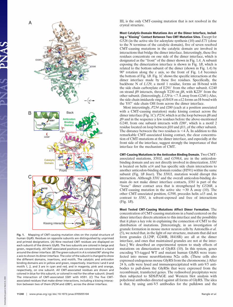
ResultsMapping of CMT-Causing Mutations. Human GlyRS is a homodimerwith the monomer unit having 685 residues composed of anN-terminal appended WHEP-TRS domain (disordered in thecrystal structure), a catalytic domain, and a C-terminal anticodonbinding domain (10). The catalytic domain contains the character-istic three conserved motifs (1, 2, and 3) of class II tRNA syntheta-ses and, in addition, three insertions (I, II, and III) between themotifs. The 10 reported CMT-causing mutations are spreadthroughout the primary sequence of human GlyRS. (In the de-scription below, residues at positions associated with CMT-causingmutations are put in italic font. Residues on opposite subunits aredistinguished by unprimed and primed designations.) When thesemutations are placed on the structure, all of them concentratearound a band that is centered on the dimer interface (Fig. 1A).Eight (E71G, L129P, P234KY, G240R, I280F, H418R, D500N,G526R) are in the catalytic, and two (S581L and G598A) are in theanticodon binding domain (Fig. 1B). Except for D500N, all arewithin the conserved regions shared by human and T. thermophilusGlyRS. Interestingly, D500N, which lies in the disordered insertion
Author contributions: L.A.N. and W.Z. contributed equally to this work; X.-L.Y. and P.S.designed research; L.A.N., W.Z., and W.X. performed research; L.A.N., W.Z., W.X., X.-L.Y.,and P.S. analyzed data; and X.-L.Y. and P.S. wrote the paper.
The authors declare no conflict of interest.
Abbreviations: CMT, Charcot–Marie–Tooth disease; GlyRS, glycyl-tRNA synthetase; TyrRS,tyrosyl-tRNA synthetase.
*To whom correspondence may be addressed. E-mail: [email protected] [email protected].
© 2007 by The National Academy of Sciences of the USA
www.pnas.org�cgi�doi�10.1073�pnas.0705055104 PNAS � July 3, 2007 � vol. 104 � no. 27 � 11239–11244
BIO
CHEM
ISTR
Y
Dow
nloa
ded
by g
uest
on
Mar
ch 2
1, 2
020
III, is the only CMT-causing mutation that is not resolved in thecrystal structure.
Most Catalytic-Domain Mutations Are at the Dimer Interface, Includ-ing a ‘‘Kissing’’ Contact Between Two CMT Mutation Sites. Except forG526 (in the active site for adenylate synthesis (10) and E71 (closeto the N terminus of the catalytic domain), five of seven resolvedCMT-causing mutations in the catalytic domain are involved ininteractions that bridge the dimer interface. Interestingly, these fiveresidues concentrate on one side of the dimer interface, which isdesignated as the ‘‘front’’ of the dimer shown in Fig. 1A. A subunitexposing the dimerization interface is shown in Fig. 1B, which isrelated to the bottom subunit of the dimer (shown in Fig. 1A) by90° rotation along the x axis, so the front of Fig. 1A becomesthe bottom of Fig. 1B. Fig. 1C shows the specific interactions at thedimer interface made by these five residues. Specifically, thebackbone N of L129, a motif 1 residue, forms an H-bond withthe side chain carboxylate of E291� from the other subunit. G240on strand �9 interacts, through T230 on �8, with K229� from theother subunit. (Interestingly, L129 is �7 Å away from G240.) Also,the side chain imidazole ring of H418 on �12 forms an H-bond withthe Y87� side chain OH from across the dimer interface.
Most interestingly, P234 and I280 (each at a position associatedwith a CMT-causing mutation) make kissing contact across thedimer interface (Fig. 1C): P234, which is at the loop between �8 and�9 and in the sequence a few residues before the above-mentionedG240, from one subunit interacts with I280�, which is a motif 2residue located on loop between �10 and �11, of the other subunit.The distance between the two residues is �4 Å. In addition to thisremarkable CMT-associated kissing contact, the clear concentra-tion of CMT-mutations at the dimer interface, and especially at thefront side of the interface, suggest strongly the importance of thatinterface for the mechanism of CMT.
CMT-Causing Mutations in the Anticodon Binding Domain. Two CMT-associated mutations, S581L and G598A, are in the anticodon-binding domain and are not directly involved in dimerization. S581is located on helix �14 and has specific side chain interactions toanother anticodon-binding domain residue (H591) within the samesubunit (Fig. 1B Inset). The S581L mutation would disrupt thisinteraction. Although S581 and the overall anticodon-binding do-main do not make dimer interface contacts, S581 is part of the‘‘loose’’ dimer contact area that is strengthened by G526R, aCMT-causing mutation in the active site �30 Å away (10). Theother CMT-associated position, G598, precedes helix �15 and, incontrast to S581, is solvent-exposed and free of interactions(Fig. 1B).
Most Tested CMT-Causing Mutations Affect Dimer Formation. Theconcentration of CMT-causing mutations in a band centered on thedimer interface directs attention to this interface and the possibilitythat it plays a key role in explaining the connection of CMT to thedistribution of mutations. [Interestingly, in an investigation ofgranule formation in mouse motor neuron cells by Antonellis et al.(5), we noted that, in the light of our structure, mutants that did notform granules (L129P, G240R, H418R) are all at the dimerinterface, and ones that maintained granules are not at the inter-face.] We described an experimental system to study effects ofmutations on dimerization of GlyRS (10). In this system, genesencoding V5-tagged WT and mutant human GlyRS were trans-fected into mouse neuroblastoma N2a cells. (These cells alsoexpressed endogenous mouse GlyRS from the chromosome.) After24 h, cells were lysed and immunoprecipitated with anti-V5 anti-bodies to pull-down the GlyRSs that were expressed from therecombinant, transfected genes. The redissolved precipitates werethen subjected to SDS/PAGE and Western blot analysis withpolyclonal antibodies directed against all forms of GlyRS. The ideais that, by using anti-V5 antibodies for the pulldown and the
E71I280
P234’G526
G598L129’
H418’
S581
H418
G240
L129
P234G526’
E71’
G598’
S581’
I280’
G240’
M128
L129
E291’
N274’
3.1 Å
2.9 Å 3.5 Å
A
I280'P2343.8 Å
K229’
T230
G240
2.9 Å
2.8 Å3.1 Å
T137Y87’
H418
2.6 Å
2.8 Å
C
B
G598’
S581’
L129
H418
I280’P234
G598
S581
E71
H418’
L129’
P234’
G526'
G240G240’
G526
I280
E71’
Anticodon Binding Domain
Catalytic Domain
Insertion II
N
C
Insertion I
Μ1
Μ3
Μ2
H591
S581
2.6 Å
Front
Back
90
Kissing interaction
Fig. 1. Mapping of CMT-causing mutation sites on the crystal structure ofhuman GlyRS. Residues on opposite subunits are distinguished by unprimedand primed designations. (A) Nine resolved CMT residues are displayed oneach subunit of the dimeric GlyRS. The two subunits are colored in beige andgreen, respectively. All CMT-associated positions are concentrated in a bandaround the dimer interface. (B) The green subunit in A is rotated 90° along thex axis to shown its dimer interface. The color of the subunit is changed to showthe different domains, insertions, and motifs. The catalytic and anticodonbinding domains are in yellow and green, respectively. Insertions I and II andmotifs 1, 2, and 3 are in cyan and red, and in magenta, pink and orange,respectively, on one subunit. All CMT-associated residues are shown andcolored in blue for this subunit, or colored in red for the other subunit. (Inset)The interaction of CMT-associated S581 with H591. (C) The five CMT-associated residues that make dimer interactions, including a kissing interac-tion between two of them (P234 and I280�), across the dimer interface.
11240 � www.pnas.org�cgi�doi�10.1073�pnas.0705055104 Nangle et al.
Dow
nloa
ded
by g
uest
on
Mar
ch 2
1, 2
020

anti-GlyRS antibodies for the Western blot analysis, any nativeGlyRS that ‘‘hybridized’’ in vivo with V5-tagged GlyRS would bedetected. The hybridization is possible to detect because of the extramass of the V5-tagged protein that enables it to be distinguishedfrom the native enzyme. Thus, the ‘‘in vivo’’ experiment wasdesigned to look at how CMT-causing mutations affect heterodimerformation between CMT-causing mutant proteins and WT GlyRS.This kind of approach is of particular interest because all CMT-causing mutations are dominant, that is, they express their effectswhen one GARS allele is mutant, whereas the other is the WT.
We investigated 7 of the 10 CMT-causing mutations for theirability to form heterodimers with the WT mouse GlyRS (95%sequence identity to human GlyRS) (Fig. 2A). (Among those, themutant enzyme, G526R, was described in ref. 10.) As a referencepoint, when cells were transfected with V5-tagged WT GlyRS,�14% of the transfected GlyRS was in the form of the heterodimer.This number was little changed by the P234KY or H418R mutations.However, the other five tested mutant genes expressed proteins thataffected dimer formation in one way or another. In particular,D500N, G526R, and S581L mutations strengthen the capacity toform heterodimers with 34%, 35%, and 47% of the transfectedGlyRS in the form of the heterodimer, respectively. In contrast, theL129P and G240R substitutions yield proteins that showed noevidence of heterodimer formation.
To validate the results we obtained from the cell-based in vivosystem, we picked two mutations, G240R and G526R. One isrepresentative of disrupted heterodimers (G240R), whereas theother had strengthened interactions (G526R), according to theabove-described in vivo system. These two were studied by analyt-ical ultracentrifugation along with WT GlyRS. The result of ultra-centrifugation analysis for G526R GlyRS was described in ref. 10and confirmed that the G526R substitution enhances homodimerinteraction (Fig. 2B Inset). In contrast, a major peak (S value �5.0)and a small broad peak (S value �7.0) was observed by sedimen-tation velocity analysis for G240R GlyRS (Fig. 2B), suggestingG240R GlyRS exists mainly as a monomer in the ultracentrifugationexperiment. Thus, with both analyses used to study subunit inter-actions, the same conclusions were reached.
From the structural perspective, it is not surprising that L129Pand G240R mutations disrupted dimer formation because, as
shown in Fig. 1C, both L129 and G240 are involved with dimer-ization contacts as internal residues having constraints that causeconformational rigidity and restriction. In contrast, P234 and H418,although interacting with residues from the other subunit, have asurface, rather than an internal, location and, therefore, are not aslikely to affect the stability of the dimer. In particular, P234 islocated on an exposed and potentially flexible loop (betweenstrands �8 and �9). Its kissing interaction with I280� across thedimer interface may be maintained in a different way with theP234KY mutation. H418 is located just before the disorderedinsertion III, and its cross-dimer partner Y87� is located on a longloop in between two �- strands (�1 and �2). Given the flexibility ofthis region, the H418R mutation may not disrupt the cross-dimerinteraction.
It is of particular interest that the three mutations that enhanceddimer formation (D500N, G526R, and S581L) are more distal to thedimer interface. This observation, made originally in studying theeffects of the G526R mutant protein (10), is reinforced now bythe new results with the D500N and S581L mutant enzymes. Thus,long-range conformational communication in human GlyRS trans-ports a local disturbance to a distant place. This long-range com-munication may explain, at least in part, why, within the band(centered on the dimer interface) where they are concentrated,CMT-causing mutations can perpetrate their effects from differentpositions.
CMT-Causing Mutations That Potentially Affect tRNA Interaction. Wesought to understand whether the CMT-causing mutations wouldaffect the interaction of GlyRS with its tRNA substrate. Pursuantto this objective, we generated a model for the tRNA complex.Among the several complexes of class II tRNA synthetases thathave been solved with bound tRNA, E. coli ThrRS has theconformation most close to our structure of human GlyRS. There-fore, using the E. coli ThrRS-tRNAThr complex (11) [Protein DataBank (PDB) ID code 1QF6], we superimposed the two catalyticdomains (one from ThrRS and one from human GlyRS) togenerate a model for GlyRS bound to tRNA (Fig. 3A). In thismodel, the tRNA fits onto the structure of human GlyRS, with theanticodon docking to the anticodon binding domain, and the3�-CCA acceptor terminus fitting into the active site. However,because insertion I makes some steric clashes with the acceptorstem at the 5� end of the docked tRNA, the conformation (or theorientation relative to the rest of the enzyme) of insertion I wouldmost probably be modified when tRNA is bound. Easily, insertionI could make a conformational change, because it is structurallyseparated from the catalytic domain, and is away from the dimerinterface (10). In the crystal, insertion I is making lattice contactswith the anticodon recognition domain, suggesting that the currentconformation of insertion I could be somewhat influenced bycrystal lattice interactions. In this connection, insertion I wascompletely disordered in the crystal structure of T. ThermophilusGlyRS (12, 13), presumably because of lack of both tRNA bindingand crystal lattice interactions.
Four of the nine resolved CMT-causing mutations are close tothe tRNA interface. These are E71, P234, I280, and G598 (Fig. 3A).[Also, the lone CMT-causing mutation (D500) that is located in thedisordered region of insertion III can potentially be involved intRNA recognition (10).] E71 lies inside the elbow of the L-shapedtRNA, and potentially makes contact with both the acceptor- andD-stems. G598 is close to the 5�-side of the anticodon. Interestingly,some of the tRNA interface is proximal to the dimer interface. Inthis connection, the kissing residues P234 and I280� at the dimerinterface also are located close to the 5�-end of the bound tRNA.Thus, in principle, mutations at positions 234 and 280 could affecteither or both of a dimer or tRNA interaction.
endogenous
A
B
transfected
WT L129P P234KY G240R H418R D500N G526R S581L
Fig. 2. Effect of CMT-causing mutations on dimer formation. (A) Immunopre-cipitation experiments showing that V5-tagged mutant GlyRSs “pulled down”variousamountsofendogenousGlyRS,presumablybyformingdimers.L129PandG240R GlyRS did not pull down any endogenous GlyRS, whereas D500N, G526R,and S581L GlyRSs pull down more endogenous GlyRS than did WT GlyRS. P234KYand H418R GlyRSs pulled down endogenous GlyRS in an amount similar to thatseen with WT GlyRS. (B) Analytical ultracentrifugation experiment showingG240R GlyRS exists mainly as a monomer, whereas more dimers are formed byG526R mutant than by WT GlyRS (Inset).
Nangle et al. PNAS � July 3, 2007 � vol. 104 � no. 27 � 11241
BIO
CHEM
ISTR
Y
Dow
nloa
ded
by g
uest
on
Mar
ch 2
1, 2
020
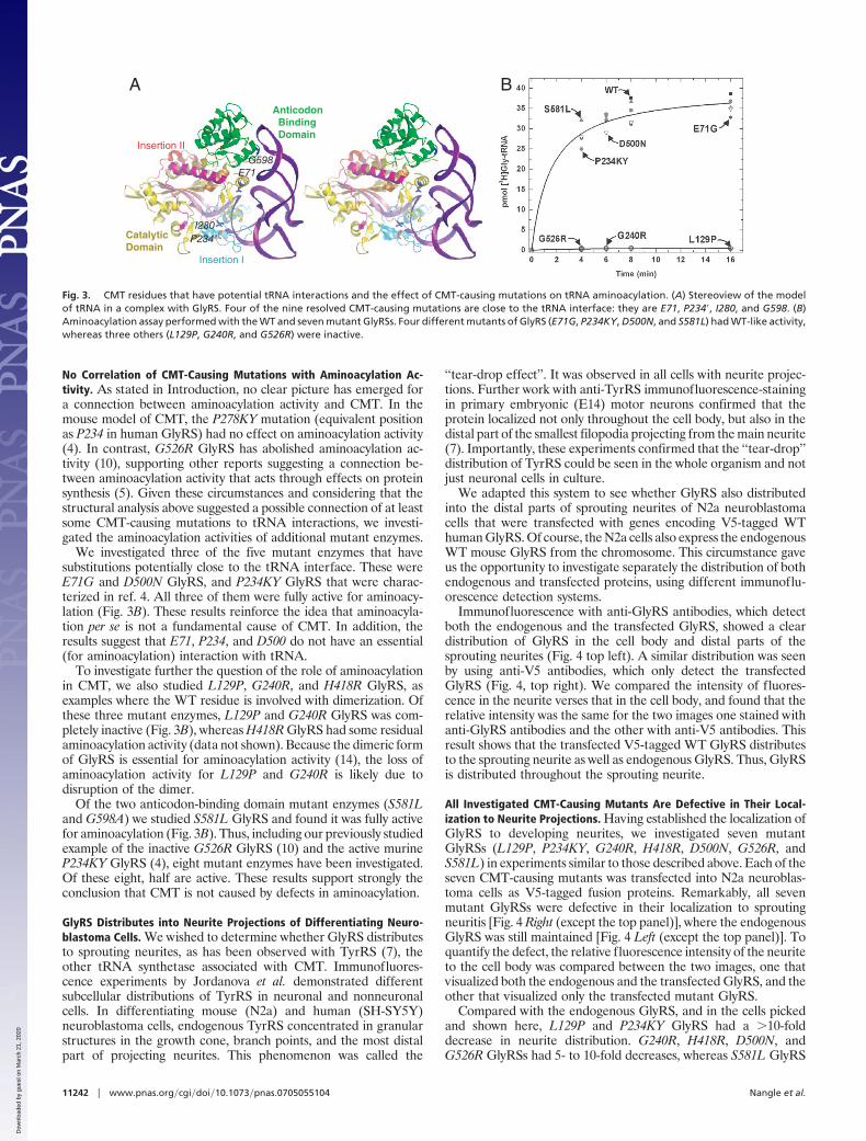
No Correlation of CMT-Causing Mutations with Aminoacylation Ac-tivity. As stated in Introduction, no clear picture has emerged fora connection between aminoacylation activity and CMT. In themouse model of CMT, the P278KY mutation (equivalent positionas P234 in human GlyRS) had no effect on aminoacylation activity(4). In contrast, G526R GlyRS has abolished aminoacylation ac-tivity (10), supporting other reports suggesting a connection be-tween aminoacylation activity that acts through effects on proteinsynthesis (5). Given these circumstances and considering that thestructural analysis above suggested a possible connection of at leastsome CMT-causing mutations to tRNA interactions, we investi-gated the aminoacylation activities of additional mutant enzymes.
We investigated three of the five mutant enzymes that havesubstitutions potentially close to the tRNA interface. These wereE71G and D500N GlyRS, and P234KY GlyRS that were charac-terized in ref. 4. All three of them were fully active for aminoacy-lation (Fig. 3B). These results reinforce the idea that aminoacyla-tion per se is not a fundamental cause of CMT. In addition, theresults suggest that E71, P234, and D500 do not have an essential(for aminoacylation) interaction with tRNA.
To investigate further the question of the role of aminoacylationin CMT, we also studied L129P, G240R, and H418R GlyRS, asexamples where the WT residue is involved with dimerization. Ofthese three mutant enzymes, L129P and G240R GlyRS was com-pletely inactive (Fig. 3B), whereas H418R GlyRS had some residualaminoacylation activity (data not shown). Because the dimeric formof GlyRS is essential for aminoacylation activity (14), the loss ofaminoacylation activity for L129P and G240R is likely due todisruption of the dimer.
Of the two anticodon-binding domain mutant enzymes (S581Land G598A) we studied S581L GlyRS and found it was fully activefor aminoacylation (Fig. 3B). Thus, including our previously studiedexample of the inactive G526R GlyRS (10) and the active murineP234KY GlyRS (4), eight mutant enzymes have been investigated.Of these eight, half are active. These results support strongly theconclusion that CMT is not caused by defects in aminoacylation.
GlyRS Distributes into Neurite Projections of Differentiating Neuro-blastoma Cells. We wished to determine whether GlyRS distributesto sprouting neurites, as has been observed with TyrRS (7), theother tRNA synthetase associated with CMT. Immunofluores-cence experiments by Jordanova et al. demonstrated differentsubcellular distributions of TyrRS in neuronal and nonneuronalcells. In differentiating mouse (N2a) and human (SH-SY5Y)neuroblastoma cells, endogenous TyrRS concentrated in granularstructures in the growth cone, branch points, and the most distalpart of projecting neurites. This phenomenon was called the
‘‘tear-drop effect’’. It was observed in all cells with neurite projec-tions. Further work with anti-TyrRS immunofluorescence-stainingin primary embryonic (E14) motor neurons confirmed that theprotein localized not only throughout the cell body, but also in thedistal part of the smallest filopodia projecting from the main neurite(7). Importantly, these experiments confirmed that the ‘‘tear-drop’’distribution of TyrRS could be seen in the whole organism and notjust neuronal cells in culture.
We adapted this system to see whether GlyRS also distributedinto the distal parts of sprouting neurites of N2a neuroblastomacells that were transfected with genes encoding V5-tagged WThuman GlyRS. Of course, the N2a cells also express the endogenousWT mouse GlyRS from the chromosome. This circumstance gaveus the opportunity to investigate separately the distribution of bothendogenous and transfected proteins, using different immunoflu-orescence detection systems.
Immunofluorescence with anti-GlyRS antibodies, which detectboth the endogenous and the transfected GlyRS, showed a cleardistribution of GlyRS in the cell body and distal parts of thesprouting neurites (Fig. 4 top left). A similar distribution was seenby using anti-V5 antibodies, which only detect the transfectedGlyRS (Fig. 4, top right). We compared the intensity of fluores-cence in the neurite verses that in the cell body, and found that therelative intensity was the same for the two images one stained withanti-GlyRS antibodies and the other with anti-V5 antibodies. Thisresult shows that the transfected V5-tagged WT GlyRS distributesto the sprouting neurite as well as endogenous GlyRS. Thus, GlyRSis distributed throughout the sprouting neurite.
All Investigated CMT-Causing Mutants Are Defective in Their Local-ization to Neurite Projections. Having established the localization ofGlyRS to developing neurites, we investigated seven mutantGlyRSs (L129P, P234KY, G240R, H418R, D500N, G526R, andS581L) in experiments similar to those described above. Each of theseven CMT-causing mutants was transfected into N2a neuroblas-toma cells as V5-tagged fusion proteins. Remarkably, all sevenmutant GlyRSs were defective in their localization to sproutingneuritis [Fig. 4 Right (except the top panel)], where the endogenousGlyRS was still maintained [Fig. 4 Left (except the top panel)]. Toquantify the defect, the relative fluorescence intensity of the neuriteto the cell body was compared between the two images, one thatvisualized both the endogenous and the transfected GlyRS, and theother that visualized only the transfected mutant GlyRS.
Compared with the endogenous GlyRS, and in the cells pickedand shown here, L129P and P234KY GlyRS had a �10-folddecrease in neurite distribution. G240R, H418R, D500N, andG526R GlyRSs had 5- to 10-fold decreases, whereas S581L GlyRS
A
G598E71
P234’I280
Anticodon Binding Domain
Catalytic Domain
Insertion II
Insertion I
B
Fig. 3. CMT residues that have potential tRNA interactions and the effect of CMT-causing mutations on tRNA aminoacylation. (A) Stereoview of the modelof tRNA in a complex with GlyRS. Four of the nine resolved CMT-causing mutations are close to the tRNA interface: they are E71, P234�, I280, and G598. (B)Aminoacylation assay performed with the WT and seven mutant GlyRSs. Four different mutants of GlyRS (E71G, P234KY, D500N, and S581L) had WT-like activity,whereas three others (L129P, G240R, and G526R) were inactive.
11242 � www.pnas.org�cgi�doi�10.1073�pnas.0705055104 Nangle et al.
Dow
nloa
ded
by g
uest
on
Mar
ch 2
1, 2
020

had a �2-fold decrease. Thus, although there is variability from cellto cell and mutant to mutant, all of the seven tested CMT-causingmutant enzyme had diminished or defective distributions into thenascent neurite projection.
DiscussionThe broad distribution of CMT-causing mutations on the primarysequence of GlyRS has long been puzzling. However, with ourrecently solved crystal structure, the apparent dispersion of themutations is diminished substantially. In particular, all nine CMT-associated positions, where the residues could be resolved in thestructure, are proximal to the dimerization interface and, in addi-tion, five are involved with subunit interactions across the dimerinterface. However, the effects of the CMT-causing mutations ondimer formation in vivo and in vitro show a distribution of effects,ranging from stronger dimer interactions (D500N, G526R, andS581L GlyRS) to completely disrupted subunit interactions (L129Pand G240R GlyRS).
L129P and G240R GlyRS are stable as monomers and could bedetected in transfected N2a neuroblastoma cells and purified asrecombinant proteins from bacteria. Thus, the monomer unit itselfcan fold and form a stable structure, without need for the dimerinteraction for stabilization. The stand-alone monomers may havea different, and maybe less tight, conformation than those in thedimer form, and the altered conformation could be the general
cause for the lost aminoacylation activity of the monomers. Inter-estingly, the homodimer of WT GlyRS is in a dynamic equilibriumwith the monomer (Fig. 2B), suggesting that the monomer form isnormally present at some level. Because the monomer is inactive foraminoacylation, a distinct biological role could be considered forthe monomer.
All CMT-causing mutations in GARS have a dominant pheno-type, which is typically associated with a gain-of-function. Het-erodimer formation between a WT and mutant subunit is one wayto acquire a new (in this case, pathological) function. However,because L129P and G240R mutant proteins are monomeric, they
A B
C D
E F
180 180
Fig. 5. Electrostatic surface potential of WT (A–D) and G526R (E and F) GlyRSs.The electrostatic surface is calculated by APBS (15), and rendered in blue and redto illustrate electrostatically positive and negative regions, respectively, in thespectrum ranging from �3 kBT/e to �3 kBT/e. (A) The dimer interface on thesubunit has an essentially neutral electrostatic distribution, with the tRNA bind-ing side (right side) more positively charged and, therefore, complementary tothe negatively charged tRNA backbone. The orientation of the subunit is thesame as in Fig. 1B. (B) The front of the dimer has the central band that is mostlypositive-charged. Five of the nine resolved CMT-associated residues are on orclose to the front surface in the central band. Outside the central band, a largepatch of negatively charged surface is located on each side across the dimer. Theorientation of the dimer is the same as in Fig. 1A. (C) The opposite side of thesubunit dimer interface shown in A. This side is exposed when a dimer is formed.[The orientation is the same as figure 1a of Xie et al. (10).] This side has a strikinglypolar charge distribution. In contrast to the positively charged (tRNA binding)face shown as the left side, the rest of the surface is, uniformly, negativelycharged, including one side of the anticodon-binding domain. Because insertionIII is disordered and, therefore, not included in the calculation of the electrostaticsurface potential, the charge distribution on the surface of the catalytic domaincan be different when this insertion is included. However, the positively chargedsurface of the central band in B is not likely to be affected by insertion III. (D) The‘‘back’’ of the dimer has two negatively charged lobes in the middle of the dimerinterface, each being part of the anticodon binding domain. Although the twodomains are close to each other in the dimer, they make no contact, possiblybecause of mutual repulsion. (E) Subunit dimer interface of G526R mutant GlyRS,which is similar to that of the WT shown in A. (F) Front dimer interface of G526Rdimer. An increase in positive charges in the center region is apparent, whencompared with the WT dimer shown in B.
α−GlyRS α−V5-tag
WT
L129P
P234KY
G240R
H418R
D500N
G526R
S581L
Ratio of relative intensity
1 : 1
>1 : 0.1
>1 : 0.1
1 : 0.2
1 : 0.1
1 : 0.2
1 : 0.2
1 : 0.6
Fig. 4. Immunofluorescence study in mouse neuroblastoma N2a cells showingthat all CMT-causing mutants are defective in neurite localization. Cells trans-fected with genes encoding WT or mutant GlyRSs are stained with anti-GlyRSantibody and with anti-V5 antibody, to investigate both endogenous and trans-fected proteins, respectively. Each antibody-staining image has two arrows, onepointing to the cell body, the other to a sprouting neurite. The relative fluores-cence intensity of the neurite to the cell body was compared between the twodifferent antibody stainings, and the ratio was calculated.
Nangle et al. PNAS � July 3, 2007 � vol. 104 � no. 27 � 11243
BIO
CHEM
ISTR
Y
Dow
nloa
ded
by g
uest
on
Mar
ch 2
1, 2
020

have less capacity to form heterodimers with the WT subunit andthereby ‘‘poison’’ aminoacylation or any other activity associatedspecifically with the dimer. Indeed, such heterodimers were notdetected in lysates of N2a cells transfected with genes for the L129Pand G240R mutant proteins (Fig. 2A). Related to this point, theCMT-associated D500N and S581L GlyRSs have stronger dimerinteractions and can form heterodimers. And yet, these mutantproteins are fully active for aminoacylation as homodimers and,therefore, are unlikely to have the ability to poison the WT protein,at least with respect to the aminoacylation activity. Thus, our dataprovide no support for the idea that subunit poisoning is causallyconnected to CMT arising from mutations in GlyRS.
Regarding the dimerization interface, most striking is the kissinginteraction between P234 and I280 and the association of CMT witha mutation of either of these residues. The a priori statisticallikelihood that ‘‘complementary’’ residues could randomly appearas sites of mutations for CMT is small. Therefore, that two residuesmake cross-subunit contacts, with each being the locus of a CMT-causing mutation, is powerful circumstantial evidence for a con-nection between CMT and the dimerization interface. Moreover,additional interconnections between CMT-causing mutations canbe seen. As an example, G526R has a long-range structural effecton another CMT-associated residue, i.e., S581. As a result of thislong-range effect, G526R GlyRS has a stronger dimer interaction(10). Interestingly, substituting the hydrophobic leucine for thehydrophilic serine, to give the CMT-causing S581L GlyRS, alsoprovides a stronger dimer interaction (Fig. 2A). Thus, whether oneconsiders direct close interactions, as seen in the kissing contacts,or whether indirect connections linking residues that are furtherapart are considered, a connection of CMT to the dimer interfaceis made.
The precise link between CMT and the dimerization interface isnot clear, however. The interface region of the dimer may becritically linked to CMT and, simultaneously or alternatively, theinterface itself (which is exposed in the monomer) may be thecritical feature. Regardless, all 5 CMT-associated positions thathave residues making dimer interactions are clustered on the frontside of the dimer interface (Fig. 1B). Interestingly, the dimerinterface (Fig. 5A), and the middle band of the front side of thedimer (Fig. 5B), where those five CMT residues are clustered,consistently have a neutral (white) to positively charged (blue)electrostatic surface. In contrast, the rest of the molecule haspeculiar and prominent large surfaces that are negatively charged(red) (Fig. 5 B–D). (The positively charged surface where CMT-residues are clustered only partially overlaps with the tRNA inter-face.) Among the 10 reported CMT-causing mutations, six have anet charge increase (E71G, P234KY, G240R, H418R, D500N, andG526R), whereas none has a charge decrease. This observationsuggests that there may be some sort of role for electrostaticinteractions in the function of GlyRS that is connected to neuro-degeneration and CMT. This role may be linked to, or in additionto, the role of the dimer interface. Although the G526R mutationstrengthens the dimer interface (10), it only had a subtle effect onthe electrostatic surface potential within the dimer interface (Fig.5E). However, remarkably, it causes an increase in positive chargedensity in the central region of the front side of the dimer (Fig. 5F),where many other CMT-associating residues are clustered.
Having ruled out a general role for aminoacylation (or appar-ently for protein synthesis) in CMT, the unknown function of GlyRSmay be specific to axon development, and is linked to its ability todistribute in nascent, sprouting neurites. As shown in Fig. 4, whereasWT GlyRS distributes into the sprouting neurites of N2a neuro-blastoma cells, all of the tested mutant enzymes were defective intheir distribution. This result demonstrates a function that iscommon to all CMT-associated mutant GlyRSs. Thus, if there is arole for the unusual electrostatic pattern seen in Fig. 5, then somepart of that electrostatic surface may interact with other partnersthat, in turn, affect the distribution of GlyRS into neurites andpossibly neurons.
Materials and MethodsImmunoprecipitation for Dimer Detection, Analytical Ultracentrifuga-tion, and Aminoacylation Assays. Those experiments were done asdescribed in ref. 10.
Cell Culture, Differentiation, Staining, and Fluorescence Microscopy.Mouse neuroblastoma cell line N2a was cultured in Eagle’s mini-mum essential medium, containing 2 mM L-glutamine, Earle’s BSS,1.5 g/liter sodium bicarbonate, 0.1 mM nonessential amino acids, 1.0mM sodium pyruvate and 10% FCS. N2a cell differentiation wasinduced by serum starvation (1% FCS) for 24 h. Cover glasses(0.17–0.25 mm thick with size of 18 mm; Fisher, Tustin, CA) weresterilized and placed in 12-well plates before cells were seeded. Cellswere fixed in 2% paraformaldehyde in PBS for 20 min, permeabil-ized in 0.2% Triton X-100 in PBS for 2 min, rinsed in PBS twice andthen blocked in 10% goat serum for 1 h. First, cells were incubatedwith monoclonal anti-V5 antibodies at a dilution of 1:200 for 1 h,followed by incubation with goat anti-mouse IgG conjugated withTexas Red (1:200 dilution) (Invitrogen, Carlsbad, CA) for 1 h.Polyclonal anti-GlyRS antibodies (in serum) were then added intocells at 1:100 dilution for 1 h followed by incubation with goatanti-rabbit IgG conjugated by FITC for 1 h (1:200 dilution) (VectorLaboratories, Burlingame, CA). (All antibodies were diluted in 2%normal goat serum.) Washings with PBS (3X) were done afterevery incubation with antibodies. Samples were counterstainedwith SlowFade Gold antifade reagent with DAPI (Invitrogen) andthen mounted on slides. Slides were visualized and analyzed undera Bio-Rad (Zeiss, Thornwood, NY) Radiance 2100 Rainbow laserscanning confocal microscope (LSCM) attached to a Nikon (To-kyo, Japan) TE2000-U microscope with infinity corrected optics.Fluorescence intensities were calculated by Adobe Photoshop(Adobe Systems, San Jose, CA).
Note Added in Proof. After submission of this paper, Chihara et al. (16)described a mutation in GlyRS in Drosophila melanogaster that affectedelaboration and stability of terminal arborization of axons and dendrites.The mutation (P98L) is also located at the dimer interface.
We thank Dr. Min Guo for help in solving and interpreting the crystalstructures of GlyRS and Dr. Robert Burgess (The Jackson Laboratory,Bar Harbor, ME) and Professor Dieter Soll (Yale University, NewHaven, CT) for helpful comments on the manuscript. This work wassupported by National Institutes of Health Grants GM 15539 and GM23562, and a grant from the National Foundation for Cancer Research.
1. Antonellis A, Ellsworth RE, Sambuughin N, Puls I, Abel A, Lee-Lin SQ, Jordanova A,Kremensky I, Christodoulou K, Middleton LT, et al. (2003) Am J Hum Genet 72:1293–1299.
2. Sivakumar K, Kyriakides T, Puls I, Nicholson GA, Funalot B, Antonellis A, Sambuughin N,Christodoulou K, Beggs JL, Zamba-Papanicolaou E, et al. (2005) Brain 128:2304–2314.
3. Del Bo R, Locatelli F, Corti S, Scarlato M, Ghezzi S, Prelle A, Fagiolari G, Moggio M, CarpoM, Bresolin N, Comi GP (2006) Neurology 66:752–754.
4. Seburn KL, Nangle LA, Cox GA, Schimmel P, Burgess RW (2006) Neuron 51:715–726.5. Antonellis A, Lee-Lin SQ, Wasterlain A, Leo P, Quezado M, Goldfarb LG, Myung K,
Burgess S, Fischbeck KH, Green ED (2006) J Neurosci 26:10397–10406.6. James PA, Cader MZ, Muntoni F, Childs AM, Crow YJ, Talbot K (2006) Neurology
67:1710–1712.7. Jordanova A, Irobi J, Thomas FP, Van Dijck P, Meerschaert K, Dewil M, Dierick I, Jacobs
A, De Vriendt E, Guergueltcheva V, et al. (2006) Nat Genet 38:197–202.
8. Skre H (1974) Clin Genet 6:98–118.9. Park SG, Ewalt KL, Kim S (2005) Trends Biochem Sci 30:569–574.
10. Xie W, Nangle LA, Zhang W, Schimmel P, Yang XL (2007) Proc Natl Acad Sci USA104:9976–9981.
11. Sankaranarayanan R, Dock-Bregeon AC, Romby P, Caillet J, Springer M, Rees B,Ehresmann C, Ehresmann B, Moras D (1999) Cell 97:371–381.
12. Arnez JG, Dock-Bregeon AC, Moras D (1999) J Mol Biol 286:1449–1459.13. Logan DT, Mazauric MH, Kern D, Moras D (1995) EMBO J 14:4156–4167.14. Kawakami M, Nishio K (1985) J Biochem (Tokyo) 98:177–186.15. Baker NA, Sept D, Joseph S, Holst MJ, McCammon JA (2001) Proc Natl Acad Sci
USA 98:10037–10041.16. Chihara T, Luginbuhl D, Luo L (2007) Nat Neurosci, 17529987.
11244 � www.pnas.org�cgi�doi�10.1073�pnas.0705055104 Nangle et al.
Dow
nloa
ded
by g
uest
on
Mar
ch 2
1, 2
020


