Characterization of the Human SHT,, Receptor Gene Promoter · Characterization of the Human SHT,,...
11
The Journal of Neuroscience, July 1995, 75(7): 4885-4895 Characterization of the Human SHT,, Receptor Gene Promoter Qin-shi Zhu, Kevin Chen, and Jean Chen Shih Department of Molecular Pharmacology and Toxicology, School of Pharmacy, University of Southern California, Los Angeles, California 90033 The regulation of 5-HT,, receptor (5HT,,R) expression has been implicated in a variety of pathological processes and has been shown to be extremely complicated and contro- versial. In order to understand the mechanisms of regula- tion of this receptor, it is important to characterize its pro- moter. In this report, the 5’ end of the human 5-HT,,R gene was cloned and characterized. Anchored PCR mapped mul- tiple transcription initiation sites at nucleotides -1157, -1137, -1127, and -496. Transfection of chimeric growth hormone plasmids containing various DNA fragments into 5-HTJt-positive human cell lines (SHSYQY, neuroblasto- ma; HeLa, cervix carcinoma) showed that the 0.74 kb HaellV Pvull fragment, which encompasses the initiation sites be- tween -1157 and -1127 and 5’ of the downstream initia- tion site (at -496), exhibited significant promoter activity. This promoter activity was not affected by the sequence upstream of the 0.74 kb fragment. The sequence down- stream (the 0.45 kb PvulVSmal fragment) strongly re- pressed this promoter activity, suggesting the presence of a silencer. Sequence analysis combined with gel retarda- tion and Dnase 1 footprinting assay identified multiple cis and trans elements for this fragment, including Spl, PEA3, cyclic AMP response element (CRE)-like sequence, and E-boxes. Two novel transcription factors have been de- tected by gel retardation and DNase 1 footprinting assay; one of them may be specific for human. The transcription factors and promoter activities were low in the negative cell line NCI-H460 (human lung large cell carcinoma). Interest- ingly, the 0.39 kb fragment, isolated from the 3’ end of the 0.74 kb fragment, exhibited the highest promoter activity. The possibility that this 0.39 kb fragment may be an alter- native promoter is discussed. These new data are essential for further study of the regulation of 5-HT,,R gene expres- sion. [Key words: serotonin, human 5-UT, receptor, promoter, human growth hormone, anchored PCR, gel retardation, DNase 1 footprinting, transcription factor, Spl, PEA3, CRE] Received Oct. 7, 1994; accepted Jan. 13, 1995. We thank Joseph Grimsby, Wu Yang, and Endi Wang for human and mouse 5-HT,,R genomic DNA clones and determination of part of the 5’ sequences; and Shi-dong Liao, Ya-xia Liu, and Hui-ren Liu for excellent technical assis- tance. This work was supported by Grant ROl MH37020, R37 MH39085 (Merit Award), and Research Scientist Award K05 MH00796 from the National In- stitute of Mental Health. Support from Boyd Welin Professorship is also ap- preciated. Correspondence should be addressed to Jean Cben Shih, Department of Mo- lecular Pharmacology and Toxicology, School of Pharmacy, University of Southern California, 1985 Zonal Avenue, Los Angeles, CA 90033. Copyright 0 1995 Society for Neuroscience 0270.6474/95/154885-11$05.00/O Serotonin (%hydroxytryptamine, 5HT) plays important roles in regulating diverse biological processes in nervous, cardiovas- cular, and gastrointestinal systems. Thesedifferent functions are presumablymediated through specific receptors. Physiological, biochemical, and pharmacological studies have defined at least 12 receptor subtypes, which molecular cloning has confirmed. All 5-HT receptorsare G-protein coupled, except 5-HT, recep- tor, which is a ligand-gated ion channel (for recent reviews, see Hoyer et al., 1994; Shih et al., 1994). It has been shown that 5-HT,, receptor (5-HT,,R) is ex- pressed in diverse tissues such as brain, vascular smooth mus- cles, and platelets. Like the 5-HT,, receptor, it mediates stimu- lation of inositol polyphosphate formation by serotonin (de Courcelles et al., 1985). This receptor is implicated in various processes such as smooth muscle contraction (Cohen et al., 1981; Leysen et al., 1984), aldosterone production (Matsuoka et al., 1984), and platelet aggregation (DeClerck et al., 1984), and is related to disorders such as migraine headaches (Humphrey et al., 1990), anxiety (Taylor, 1990), and mental depression (Meltzer and Lowy, 1987). The antidepressant mianserin (Sand- ers-Bush, 1990) and hallucinogenicdrugs (Pierce and Peroutka, 1989; Sadzotet al., 1989) have been shownto interact with this receptor. The cDNAs for 5-HT,,R in rat (Pritchett et al., 1988; Julius et al., 1990) hamster (Chambard, 1990), mouse(Yang et al., 1992), and human(Saltzmanet al., 1991)have beencloned.The human 5-HT,,R gene is located on chromosome 13q14q21 (Sparkeset al., 1991), consists of three exons separated by two introns, and spans over 20 kb (Chen et al., 1992). The regulation of 5-HT,, receptor hasbeen related to a num- ber of pathological and neurological states.Expression of this receptor in transfectedmouse fibroblast activates phospholipase C-signaling pathway and promotescellular transformation (Ju- lius et al., 1990). However, the mechanisms of the regulation are poorly understoodand controversial. Most G-protein-cou- pled receptors are downregulatedby agonistsand upregulated by antagonists. In contrast, 5-HT,, receptorsare downregulated by both agonists (e.g., 5-HT, DOI) and antagonists (e.g., mian- serin) (Blackshear et al., 1983; Sanders-Bush et al., 1987).While the downregulationby mianserin has beenobserved in animals, this phenomenon wasnot reproducible in studies using cell lines. Mianserin downregulation was observed in rat C6 glioma cells (Toth and Shenk, 1994), but not in rat Pll cells (Ferry et al., 1993), indicating that this paradoxical downregulation requires a certain signaltransductionpathway which may not exist in all cells. Toth and Shenk (1994) reported a decrease in 5-HT,, re- ceptor mRNA levels in C6 glioma cells after mianserintreat- ment, suggesting that the regulation is at the level of transcrip- tion. A mianserin response element has been implicated in the
Transcript of Characterization of the Human SHT,, Receptor Gene Promoter · Characterization of the Human SHT,,...

The Journal of Neuroscience, July 1995, 75(7): 4885-4895
Characterization of the Human SHT,, Receptor Gene Promoter
Qin-shi Zhu, Kevin Chen, and Jean Chen Shih
Department of Molecular Pharmacology and Toxicology, School of Pharmacy, University of Southern California, Los Angeles, California 90033
The regulation of 5-HT,, receptor (5HT,,R) expression has been implicated in a variety of pathological processes and has been shown to be extremely complicated and contro- versial. In order to understand the mechanisms of regula- tion of this receptor, it is important to characterize its pro- moter. In this report, the 5’ end of the human 5-HT,,R gene was cloned and characterized. Anchored PCR mapped mul- tiple transcription initiation sites at nucleotides -1157, -1137, -1127, and -496. Transfection of chimeric growth hormone plasmids containing various DNA fragments into 5-HTJt-positive human cell lines (SHSYQY, neuroblasto- ma; HeLa, cervix carcinoma) showed that the 0.74 kb HaellV Pvull fragment, which encompasses the initiation sites be- tween -1157 and -1127 and 5’ of the downstream initia- tion site (at -496), exhibited significant promoter activity. This promoter activity was not affected by the sequence upstream of the 0.74 kb fragment. The sequence down- stream (the 0.45 kb PvulVSmal fragment) strongly re- pressed this promoter activity, suggesting the presence of a silencer. Sequence analysis combined with gel retarda- tion and Dnase 1 footprinting assay identified multiple cis and trans elements for this fragment, including Spl, PEA3, cyclic AMP response element (CRE)-like sequence, and E-boxes. Two novel transcription factors have been de- tected by gel retardation and DNase 1 footprinting assay; one of them may be specific for human. The transcription factors and promoter activities were low in the negative cell line NCI-H460 (human lung large cell carcinoma). Interest- ingly, the 0.39 kb fragment, isolated from the 3’ end of the 0.74 kb fragment, exhibited the highest promoter activity. The possibility that this 0.39 kb fragment may be an alter- native promoter is discussed. These new data are essential for further study of the regulation of 5-HT,,R gene expres- sion.
[Key words: serotonin, human 5-UT, receptor, promoter, human growth hormone, anchored PCR, gel retardation, DNase 1 footprinting, transcription factor, Spl, PEA3, CRE]
Received Oct. 7, 1994; accepted Jan. 13, 1995.
We thank Joseph Grimsby, Wu Yang, and Endi Wang for human and mouse 5-HT,,R genomic DNA clones and determination of part of the 5’ sequences; and Shi-dong Liao, Ya-xia Liu, and Hui-ren Liu for excellent technical assis- tance. This work was supported by Grant ROl MH37020, R37 MH39085 (Merit Award), and Research Scientist Award K05 MH00796 from the National In- stitute of Mental Health. Support from Boyd Welin Professorship is also ap- preciated.
Correspondence should be addressed to Jean Cben Shih, Department of Mo- lecular Pharmacology and Toxicology, School of Pharmacy, University of Southern California, 1985 Zonal Avenue, Los Angeles, CA 90033. Copyright 0 1995 Society for Neuroscience 0270.6474/95/154885-11$05.00/O
Serotonin (%hydroxytryptamine, 5HT) plays important roles in regulating diverse biological processes in nervous, cardiovas- cular, and gastrointestinal systems. These different functions are presumably mediated through specific receptors. Physiological, biochemical, and pharmacological studies have defined at least 12 receptor subtypes, which molecular cloning has confirmed. All 5-HT receptors are G-protein coupled, except 5-HT, recep- tor, which is a ligand-gated ion channel (for recent reviews, see Hoyer et al., 1994; Shih et al., 1994).
It has been shown that 5-HT,, receptor (5-HT,,R) is ex- pressed in diverse tissues such as brain, vascular smooth mus- cles, and platelets. Like the 5-HT,, receptor, it mediates stimu- lation of inositol polyphosphate formation by serotonin (de Courcelles et al., 1985). This receptor is implicated in various processes such as smooth muscle contraction (Cohen et al., 1981; Leysen et al., 1984), aldosterone production (Matsuoka et al., 1984), and platelet aggregation (DeClerck et al., 1984), and is related to disorders such as migraine headaches (Humphrey et al., 1990), anxiety (Taylor, 1990), and mental depression (Meltzer and Lowy, 1987). The antidepressant mianserin (Sand- ers-Bush, 1990) and hallucinogenic drugs (Pierce and Peroutka, 1989; Sadzot et al., 1989) have been shown to interact with this receptor.
The cDNAs for 5-HT,,R in rat (Pritchett et al., 1988; Julius et al., 1990) hamster (Chambard, 1990), mouse (Yang et al., 1992), and human (Saltzman et al., 1991) have been cloned. The human 5-HT,,R gene is located on chromosome 13q14q21 (Sparkes et al., 1991), consists of three exons separated by two introns, and spans over 20 kb (Chen et al., 1992).
The regulation of 5-HT,, receptor has been related to a num- ber of pathological and neurological states. Expression of this receptor in transfected mouse fibroblast activates phospholipase C-signaling pathway and promotes cellular transformation (Ju- lius et al., 1990). However, the mechanisms of the regulation are poorly understood and controversial. Most G-protein-cou- pled receptors are downregulated by agonists and upregulated by antagonists. In contrast, 5-HT,, receptors are downregulated by both agonists (e.g., 5-HT, DOI) and antagonists (e.g., mian- serin) (Blackshear et al., 1983; Sanders-Bush et al., 1987). While the downregulation by mianserin has been observed in animals, this phenomenon was not reproducible in studies using cell lines. Mianserin downregulation was observed in rat C6 glioma cells (Toth and Shenk, 1994), but not in rat Pll cells (Ferry et al., 1993), indicating that this paradoxical downregulation requires a certain signal transduction pathway which may not exist in all cells. Toth and Shenk (1994) reported a decrease in 5-HT,, re- ceptor mRNA levels in C6 glioma cells after mianserin treat- ment, suggesting that the regulation is at the level of transcrip- tion. A mianserin response element has been implicated in the

4886 Zhu et al. * Promoter oi Human 5-HT,, Receptor Gene
upstream sequence of the basal promoter. On the contrary, no mRNA level change was observed in rats injected with mian- serin (Roth and Ciaranello, 1991). Recently, Du et al. (1994) reported that 5HT stimulated 5-HT,, receptor promoter activity in rat myometrial smooth muscle cells via the S-HT,, receptor. These controversial results suggest the existence of a complex regulatory mechanism.
In order to understand the mechanism for human 5-HT,,R gene expression, the 5’ flanking sequence of this gene has been characterized for the first time. We have defined transcription initiation sites between - 1127 and - 1157 by anchored PCR. Promoter activity was detected in a 0.76 kb HaeIIUPvuII frag- ment which encompasses these transcription initiation sites. Transcription factors Spl, PEA3, E-box binding proteins, and two novel transcription factors have been shown to interact with this fragment. We also detected a downstream initiation site (at -496) and found promoter activity in a DNA fragment 5’ of this site, which raised the possibility for an alternative promoter.
Materials and Methods
DNA cloning and sequencing. The DNA fragments used in this study were isolated from an 8.5 kb EcoRI-phage clone lSE-5, which contains exons 1 and 2 of human 5HT,,R gene, intron 1, part of intron 2, and about 3.7 kb sequence 5’ of the translation start codon ATG (Chen et al., 1992). All the 3.7 kb 5’ sequence has been determined by Applied Biosystems model ABI370a automated DNA sequencer, and manually by the dideoxy chain termination method (Sanger et al., 1977).
Three point eight kilobases of mouse 5-HT,,R genomic DNA 5’ of the translation initiation start codon ATG was mapped and sequenced from the 9 kb EcoRI fragment in the genomic clone MS2cos54-E9 (Yang et al., 1992).
The comparison between human and mouse 5’ sequences was facil- itated by the sequence analysis software of Genetics Computer Group (University of Wisconsin Biotechnology Center, Madison).
Serotonin-stimulated hydrolysis of phosphoinositol (PI). In order to select 5-HT,,R-positive and -negative cell lines, serotonin-stimulated PI hydrolysis was performed. The procedure used was a combination of the methods of Serra et al. (1986), Roth et al. (1986), and Kursar et al. (1992). Cells were seeded into 12-well dishes at a density of 4 X lo5 cells/well and grown in inositol-free DMEM with 10% dialyzed fetal bovine serum for 24 hr. Myo-3H-inositol was added to each well (1 mCi/ml) and the cells were grown for another 24 hr. Before the addition of serotonin, the cells were treated with serum-free DMEM containing 10 mM LiCl and 10 mu pargyline for 20 min. Twenty milliliters of the appropriate amount of serotonin was added and the samples were in- cubated at 37°C for 60 min. The reaction was terminated by aspiration of the medium followed by the addition of 1 ml ice-cold 75% methanol. The cells were scraped and the cell suspensions were transferred to ice- cold tubes containing 1 ml of chloroform and 0.5 ml of double-distilled water. The contents were mixed vigorously and then centrifuged at 2000 rpm at 4°C for 15 min. One milliliter of the upper aqueous phase was added to 2 ml of ice-cold double-distilled water and the mixture was applied onto a 10 ml AGl-X8 anion exchange column. The column was washed three times with 5 ml of double-distilled water, then washed twice with 5 mM sodium borate/50 mM sodium formate. The ‘H-inositol monophosphate was eluted with 4 ml of 0.2 M ammonium formate/O.l M formic acid. The eluate was mixed with 14 ml of scintillation cocktail (Budget-Solve, Research Products International) and counted for radio- activity.
Determination of transcription initiation sites by anchored PCR. In this experiment, a kit (5’ AmpliFINDER RACE kit) containing a human whole brain cDNA library from Clontech (Palo Alto, CA) was used in which a 35.mer oligonucleotide (anchor) was linked to the 3’ end of each cDNA molecule (5’-Race-ready cDNA). PCR was performed with a primer for the anchor and primers for human 5-HT,,R cDNA accord- ing to the procedure recommended by the manufacturer. Four nested primers for human 5-HT,,R cDNA were used. The most 3’ primer used (S21: 5’ CTGTAGAGCCTGGTGTCATC 3’) is complementary to the nucleotide +86 to +67 (the A of the translation start codon ATG is defined as +l). The second mimer (S387R: 5’ GCAATCAGAAA- CAGTGGGG 3’) was complementary to the nucleotide -441 to -459.
The third primer (S133R: 5’ AAGAGCTGAGCCACGTCCGC 3’) was complementary to the nucleotide -932 to -951. The most 5’ primer (S116R: 5’ GGGAAAGTAGGAAGAGCTG 3’) was complementary to the nucleotide -1083 to -1101. The PCR products were resolved on an agarose gel and the DNA bands were cloned into pT7Blue(R) vector (Novagen, Madison, WI) for sequence determination, using T7 DNA polymerase. The sequence obtained was compared with genomic DNA to define the 5’ ends of 5-HT,,R mRNA.
In order to map the 5’ end of 5-HT,,R mRNA in SHSY-5Y (human neuroblastoma) and HeLa (human cervix carcinoma) cells, polyA+ mRNA was isolated from these two cell lines with an mRNA isolation kit (version 1.2) from Invitroeen (San Diego, CA). These mRNA were reverse transcribed and ligated to the 35-bp anchor by single-strand ligation with T4 RNA ligase, as per the manufacturer’s instructions (Clontech). PCR and product sequence determination were performed as with anchor-ready cDNA, with the same nested primers mentioned above.
Promoter activity measurement. Various DNA fragments (see Fig. 1) were isolated from the 3.7 kb 5’ sequence of human 5-HT,,R gene and cloned into promoterless transient expression vector pOGH, which con- tains human growth hormone as the reporter gene (Selden et al., 1986). These constructs were then transfected into SHSY-SY, HeLa, NCI- H460, (human lung large cell carcinoma) and MLg (mouse lung fibro- blast) cells, using the low pH calcium phosphate coprecipitation method (Chen and Okayama, 1987). Promoter activity was assayed by measur- ing the human growth hormone secreted into medium by transfected cells with a kit from Nichols Institute Diagnostics (San Juan Capistrano, CA) as described previously (Zhu et al., 1992).
Nuclear extracts. Nuclear extracts from HeLa and NCI-H460 cells were prepared essentially according to published procedures (Dignam et al., 1983), except that the cells were ruptured in buffer A in a polytron (Brinkman Homogenizer, model pcu 11, Luzern, Switzerland) at posi- tion 3, as described previously (Zhu et al., 1992).
Gel retardation assay. DNA fragments from the promoter regions detected by transient transfection assay were labeled with 3ZP-dNTP and Klenow large fragment of DNA polymerase. Two and one-half micro- grams of poly[dA-dT].poly[dA-dT] and 0.25 pg of poly[dI-dC].poly[dI- dC] were preincubated with 3 pg of the nuclear extracts for 10 min at room temperature in a mixture of 19 ~1 containing 20 mM Hepes- NaOH, pH 7.8, 1 mM MgCl,, 50 mM NaCl, 0.5 mM EDTA, 0.5 mM DTT, and 10% glycerol. When indicated, 250-fold excess of Spl con- sensus oligonucleotides (Promega, Madison, WI) was also included to replace Spl binding to the DNA fragments. One microliter of end- labeled DNA (about 2 fmol) was added and the incubation was contin- ued for another 20 min at room temperature. After mixing with 2.5 pl of loading buffer (250 mM Tris-HCl, pH 7.8, 0.2% xylene cyanol, 0.2% bromophenyl blue, 40% glycerol), the mixture was loaded on a 5% nondenaturing acrylamide gel (acrylamide:bisacrylamide = 37.5: 1) in 90 mM boric acid, 0.1 mM EDTA prerun at 10 mA for 1 hr. Gel elec- trophoresis was carried out at 10 mA in the same buffer for 1.5 hr. The temperature was never above 30°C thus the protein-DNA complexes were not dissociated. The bands were visualized by autoradiography.
DNasel footprinting analysis. In this experiment, one end of the DNA fragment was labeled with ‘2P-dNTP Since the 0.74 kb HaeIIIl PvuII fraiment is too long for such an assay, subfragments (the 0.35 kb HaeIII/BamHI fragment and the 0.39 kb BamHI/PvuII fragment; the latter was further divided into the 0.24 kb BamHI/HinpI fragment and the 0.15 kb HinuI/PvuII fragment: see Figs. 3. 5) were isolated for this purpose. For the labeling o’f the 0.35 kb~HaeIII/BamHI fragment, the pOGH construct containing this fragment was digested with PstI (for the PstI site in the linker) and BamHI to isolate the 0.35 kb PstI/BamHI fragment. The sense strand was labeled at the BamHI site in a fill-in reaction with ‘2P-dATP and Klenow large fragment of DNA polymerase 1 in the presence of cold dGTP to prevent digestion of the BamHI site by the exonuclease activity of Klenow large fragment. Cold dCTP was also included to prevent labeling at the PstI site by exchange reaction. The antisense strand was labeled at the PstI site by an exchange reaction with ‘*P-dCTP and Klenow in the presence of cold dATP and dGTP to prevent further digestion at the PstI site and digestion at the BamHI site. In order to isolate the 0.24 and 0.15 kb fragments, the pOGH construct containing the 0.39 kb fragment was digested with BamHI to isolate a 0.39 kb BamHI/BamHI fragment (the 3’ BamHI site was from the polylinker). This fragment was then digested with HinpI to isolate the 0.24 kb BamHI/HinpI and 0.15 kb HinpI/BamHI fragments (see Fig. 5). These two fragments were labeled at the HinpI site with 32P-
The Journal of Neuroscience, July 1995, 75(7) 4887
Promoter activity (% of pXGH5 *SD)
I@- SHSY-5Y &La NCkH460 MLS E s 3.5 ND. 0.1 f 0.1 -
H s 3.0 ND. 0.1 * 0.1 - P s 2.0 ND. 0.1 + 0.1 -
pv 0.45 ND. 0.2 * 0.1 -
B B 1.40 2.6 f 0.1 1.9 f 0.4 0.3 t 0.4 0.1 f 0.1 R 0.70 4.2 k 1.0 3.0 f 0.1 -
Ea B 0.53 4.5 i 1.5 3.3 f 0.3 - IIaB 0.35 4.3 f 0.6 7.2 f 1.4 1.3 f 0.1 0.3 f 0.1 Ha Pv 0.74 4.5 f 0.3 3.3f1.7 -
Pv Pv 1.60 4.3 k 1.8 2.7 2 0.2 - Ha s 1.20 ND. 0.2 * 0.1 -
BPV 0.39 16.0 f 3.9 13.1 f 0.1 1.7 f 0.1 1 .o f 0.1
1 [email protected] u f30.0 1 @a0 cpll 2,055 75,367 32,656 17,315
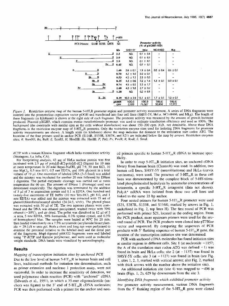
Figure 1. Restriction enzyme map of the human 5-HT,R promoter region and promoter activity measurement. A series of DNA fragments were inserted into the promoterless expression vector pOGH and transfected into four cell lines (SHSY-SY, HeLa, NCI-H460, and MLg). The length of these fragments (in kilobases) is shown at the right side of each fragment. The promoter activity was measured by the amount of growth hormone produced. Plasmid pXGH5, which contains mouse metallothionein promoter, was used to estimate transfection efficiency and used as 100%. The background (the constructs with similar cpm as the cells without transfection) was about 150-200 cpm. N.D., not detectable. Above these DNA fragments is the restriction enzyme map of 5-HT,,R promoter. Only the restriction enzyme sites used for isolating DNA fragments for promoter activity measurements are shown. A length scale (in kilobases) above the map indicates the distance to the translation start codon ATG. The locations of the four primers used in anchor PCR (S116R, S133R, S387R, and S21) are indicated below the map by arrows. Restriction enzymes sites: B, BamHI; Ba, Bali; E, EcoRI; H, HindIII; Ha, HaeIII; P, PstI; Pv, PvuII; R, RsaI; S, SmaI.
dCTP with a mutant Klenow fragment which lacks exonuclease activity (Stratagene, La Jolla, CA).
For footprinting analysis, 43 pg of HeLa nuclear protein was first incubated with 2.5 p,g of poly[dI-dC].poly[dI-dC] (Sigma) for 10 min at room temperature in 20 mM Hepes-NaOH, pH 7.9, 50 mM KCl, 10 mM MgCl,, b.5 mM DTT, 0.5 mM EDTA, and- 10% glycerol in a total volume of 19 ul. One microliter of labeled DNA (3-5 fmol) was added and the mix&e was incubated for another 20 min followed by DNase 1 digestion. The partial enzymatic cleavage was carried out at room temperature for 60 sec. The amount of DNase 1 (Promega) used was determined empirically. The digestion was terminated by the addition of 5 p,l of 3 M ammonium acetate and 0.1 M EDTA. One hundred and twenty-five microliters of TE buffer (10 mM Tris-HCl, pH 8.0, and 1 mM EDTA) was added and the mixture was extracted with 75 ml of phenol/chloroform/isoamyl alcohol (24:24: 1, v/v/v). The phenol phase was extracted with 50 ~1 of TE. The two aqueous phases were com- bined and the DNA was ethanol precipitated, washed twice with 70% ethanol (-2O”C), and air dried. The pellet was dissolved in 12 ~1 of 5 M urea, 5 mM EDTA, 50% formamide, 0.3% xylene cyanol, and 0.3% of bromoohenvl blue. The samples were heated at 90°C for 20 min. then loadid &mediately on a 7.s% acrylamide (acrylamide:bisacrylam- ide = 29: 1)/S M urea gel. Both a short and long run were performed to examine the proximal (relative to the labeled end) and the distal part of the fragments. MspI-digested pBR322 fragments were end labeled using jZP-dCTP and Klenow polymerase, and were used as molecular weight standards. DNA bands were visualized by autoradiography.
Results
Mapping of transcription initiation sites by anchored PCR Due to the low level of human S-HT,,R in human brain and cell lines, traditional methods for initiation site determination, such as primer extension and nuclease 1 protection assay, were not successful. In order to increase the sensitivity of detection, we used polymerase chain reaction (PCR) with “anchored” cDNA (Edwards et al., 1991), in which a 35mer oligonucleotide (an- chor) was ligated to the 3’ end of 5-HT,,R cDNA molecules; PCR was then performed with a primer for the anchor and nest-
ed primers specific to human 5-HT,,R cDNA to increase spec- ificity.
In order to map 5-HT,,R initiation sites, an anchored cDNA library from human brain (Clontech) was used. In addition, two human cell lines, SHSY-5Y (neuroblastoma) and HeLa (cervix carcinoma), were used. The presence of 5-HT,,R in these cell lines was demonstrated by the complete block of 5-HT-stimu- lated phosphoinositol hydrolysis by nanomolar concentrations of ketanserin, a specific 5-HT,,R antagonist (data not shown). PolyA+ mRNA were isolated from these two cell lines and linked to the same 35 bp anchor.
Four nested primers for human 5-HT,,R promoter were used (S21, S387R, S133R, and S116R; marked by arrows in Fig. 1, underlined in Fig. 2, top lines H). The first round of PCR was performed with primer S21, located in the coding region. From the PCR product, more upstream primers were used for the sec- ond round of PCR. The PCR products were cloned into pT7Blue vector and sequenced. By comparing the sequences of PCR products with 5’ flanking sequence of human 5-HT,,R gene, the location of the transcription initiation site was determined.
PCR with anchored cDNA molecules has found initiation sites at similar regions in different cells. Site 1 (at nucleotide - 1157; the A of the translation start codon ATG was defined +l) was found in brain and HeLa cells; site 2 (at - 1137) was found in SHSY-5Y cells; site 3 (at - 1127) was found in brain (see Fig. 1, sites 1, 2, 3, marked with vertical arrows; also Fig. 2, marked with thick arrows with the number above the initiation site).
An additional initiation site (site 4) was mapped to -496 in brain (Figs. 1, 2), 629 bp downstream from the site 3.
Identifying DNA fragments which exhibited promoter activity For promoter activity measurement, various DNA fragments from the 5’ flanking region of the 5-HT,,R gene were cloned
4888 Zhu et al. - Promoter of Human SHT,, Receptor Gene
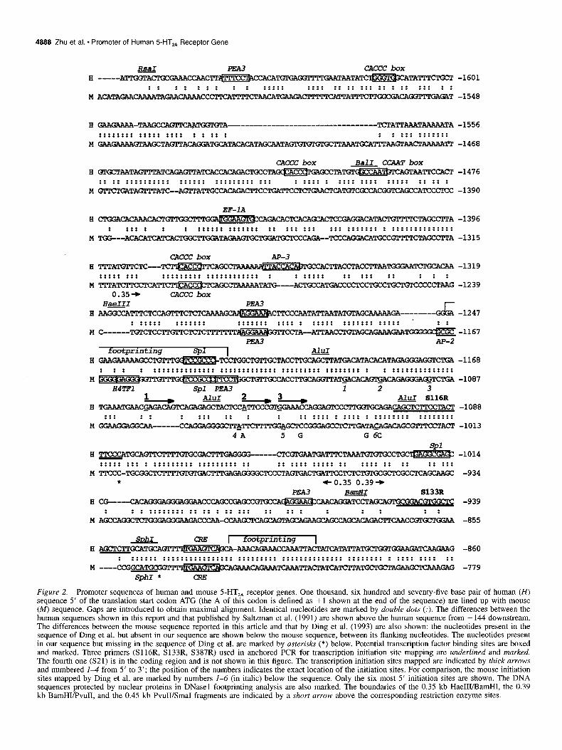
RsaI PEA3 CACCC box I3 -----ATTGGTACTGCC?W=ACTT~~@CcAcA~ cXMTlWUTAATATc=TAmWT -1601
: : :: ::: : : ::::: . . . . . . . . :::::::::::: ::: :: MACATAGAACAAAATAGAACAAAACCCTTCAmTCTAACATG TlTlTKATTATTTCTTGGCG GAT -1548
B
M
E
M
H
M
H
M
A
M
I3
M
H
M
E
M
B
M
E
M
GAAGAAAA-TAAGCCAQl'TCAA ~A----------------------------------T -1556 . . . . . . . . . . . . . . . . . . . . . . . . . . . . . . . . . . : : :: : : : ::: ::::::: GAAGAAAA6AAGCTA(3ITACAGGATGCATACACATAGCA TTAAATGCATTTAAQl'AACTAAAAA~ -1468
cAccc box CCAAT box Bali T~TA~A~~A~C~~~C~CTA~CC~~CTA~~~~~~~T -1476 :: :: :::::::::: :::::: ::::::::: ::: : :::: : :::: :::: ::::: :: :: : 6TC~TATATC--AdTATTGCCACAGACTTCCTGA -1390
EF-IA
cTGGAcAcAAAcAc-va CAGACACTCACA~CT TAGCCTl'A -1396 : ::: : : : :::::: ::::::: :: ::: ::: ::: ::::::: : ::::::::::::::
TGG---ACACATCATCACTGGCTn;OATAGAAC;n;CTGG TAGCClTA -1315
CACCC box m-3 TTTATQTTCTC---TC ~cc~~~~Ac~~~~C~ACCTACC~~~ TCTGCACAA -1319 . . . . . . . . . . . . . . . . . . . . . . . . . . . . . . . . . . . . . . . . . . . . . . . . . . . . . . . . . . : : ::::: :: ::: :: : : : 'M'TATCMCCTcATTc~~TcAGccT~TATG---ACTGccATGAcCCCTcCTGcCTGCTGTcCCCCTAAG -1239
0.35+ CACCC box H-I
G~A&Z&~~TT~~~AATATTAATA*AG~AAAAA~-------- AAGGccATTTcTccAGTTTcTcTc!AAAA G&i -1247 . : ::::: ::::::: . . . . . . . . . . . : . . . . . . . . . . . . . . . . . . . . . . . . . . . . . . . . . . . . . . . . . . . . . : :
C------TQrcTccTTGTTcTcTc-*-- CTA--ATTAA(?CT6AGCAGAAAGAA PEA3 -Yiz -1167
footprmtmg SPl I AluI GAAGAAAAAGCCTOrPTGOl~~~TACC~~~A~~TA~~TA~~~~ -1168 : :: : . . . . . . . . . . . . . . . . . . . . . . . . . . . . . . . . . . . . . . . . . . . . . . . . . . . . . . . . . . . . . . . . . . . . . . . . . . ..a... . . . . . . . . : : ::::::::::::::
TTGCAWl!TA~cAcA~CAGAGGGA@jTcTGA-1087 E4TFl Spl PEA3 1 2 3
1 D AluI 2 ) 3 * AluI S116R TGAAATGAACG~C~~CCATPCCC( !FXGXCAGAcAGcTcTTCCTACT -1088
::: : : : ::: :: : : . . . . . . . . :: :::: : :::: : . . . . . . . . . . . . . . . . . . . . . . . . GGAAGGAGGCAA------ccAGGAGGGGcm&m TTl'TG&GCTCCGGGAGCCTCTGATA~GACAGC GTTTCCTACT -1013
4A 5 G G 6C
spl TTcccATGcAGTTc-GAc- -----CTC-TGATTTCT-TGTGTGCCTGCWGGCW -1014 . . . . . . . . : . . . . . . . . . . . . . . . . . . . . . . . . . . . . . . . . . . . . . . . . . . . . . . . . :: :::: ::::: :: :::: :: :: :: ::: TTcCC-TGCGGCT GGGGCTCCCTAQl'GACTGATTccTcTCTGTGcGcTcGcC~GcAAGC -934
* co.35 0.39+
: : : :::::::: :: :: : :: ::: :: :: : : : : : : AGCCAGGCTCTGGGAGGGAAGACCCAA-CCAAGCTCAGCA -855
SDhI footprmtmg A~~~~~I--~-C-~AITA~TA~A~ BGATCAAGAAG -860
. . . . . . . . . . . . . . . . . . . . . . . . . . . . . . . . . . . . . . . . . . . . . . . . . . . . . . . . . . . . . . . . . . . . . . . . . . . . . . . . . . . . . . . . . . . . . . -779
Figure 2. Promoter sequences of human and mouse 5-HT,, receptor genes. One thousand, six hundred and seventy-five base pair of human (H) sequence 5’ of the translation start codon ATG (the A of this codon is defined as + 1 shown at the end of the sequence) are lined up with mouse (M) sequence. Gaps are introduced to obtain maximal alignment. Identical nucleotides are marked by double dots (:). The differences between the human sequences shown in this report and that published by Saltzman et al. (1991) are shown above the human sequence from -144 downstream. The differences between the mouse sequence reported in this article and that by Ding et al. (1993) are also shown: the nucleotides present in the sequence of Ding et al. but absent in our sequence are shown below the mouse sequence, between its flanking nucleotides. The nucleotides present in our sequence but missing in the sequence of Ding et al. are marked by asterisks (*) below. Potential transcription factor binding sites are boxed and marked. Three primers (Sll6R, Sl33R, S387R) used in anchored PCR for transcription initiation site mapping are underlined and marked. The fourth one (S21) is in the coding region and is not shown in this figure. The transcription initiation sites mapped are indicated by thick arrows and numbered I-$ from 5’ to 3’; the position of the numbers indicates the exact location of the initiation sites. For comparison, the mouse initiation sites mapped by Ding et al. are marked by numbers l-6 (in italic) below the sequence. Only the six most 5’ initiation sites are shown. The DNA sequences protected by nuclear proteins in DNasel footprinting analysis are also marked. The boundaries of the 0.35 kb Hae11103amH1, the 0.39 kb BamHWvuII, and the 0.45 kb PvuII/SmaI fragments are indicated by a short arrow above the corresponding restriction enzyme sites.

The Journal of Neuroscience, July 1995, 75(7) 4889
footprinting 1 E-box 1E-box H AGGGGACTCTACAC~~‘~~~~~~~~~~T~~~ TCJICATGTG!@lUGCT -780
. . . . . . . . . . . . . . . . . . . . . . . . : :: : :: :: :: : ::: :::::::: ::: : : ::::: M AGGGGATTCCACACTGGCCTAGTCACCATGCGACAGGCA---AGTCACA~TA-----------------GCGGACC!CT -719
E-box -I H GAAGPCA---AATOIPA6TCTCAn;CCGCPATATTTTATT TCATGCTTGGC -703
: :::: ::::::::::: :: ::: : :::: ::::::: :::::::::: : :: :: ::: . . . . . . ...* . . . . M GTC~ TGCTGTCCTTTACTGCTGTGGGA Tm'!rC!!t'TrC!~-'r'rAAAGTCAC!GCTTGGC! -640
PEA3 H -~cAlwrAATcA ~~~~C~T~~AC~~~C~CT
: ::: ::::: : . . . . . . . . . . . . . . . . . . . . . . . . . . : :::::::: :::::: ::: : . . . . . . . . M TAGAATGCCGl’CATTGCCGCG?vSAATTl’CTGA TQEGAAAGTTCTCTGCTGCGCTTCG-------GGATGCGTTTTCCT
+0.39*0.45+ PvuII E-box AP-4
H mATTAGCTAAGCAACATTATA~~~~C~~~~~~CT~~~~~T
:: ::: :: ::: : : ::: :::::::::::::::: :::: ::: :::: :::: ::: : M TTC~~~~CAGAGAATACAAGCn;AAATTCCTGT-----------
flpar * *
4, H ~~CC~~TACTATGGGATTAACACTG~
. . . . : :: ::: . . . . . . . . . . . . . . . . . . . . . . . . . . . . . . :: ::::::::::::::: :::::: : : : :
M -----AAAAAGO(TTTAACCC-TTATGATGGCATTAACAT Gl’GGGATTTTTAX’TGACTTCCTTAATTAATATAGAGG Erpar
83878 H ACA--------TCCCCACT-GTTTCTGATl’GCATGCTATTTTAATAATACTGTTGCTAAACTAGTACCATCGGCATAACC
:: . . . . . . . . :: :::: ::: :::::: :::::: : :::::::: : . . . . . . . : : . . . . . . . M GCACACAGCCCTCCCTCCTC~~~~~~C~ TGGCTGAACTCTTG
-623
-568
-543
-500
-464
-425
-393
-345
H AACAAAATGAaTATAm AAACAAGA6CCCAOTAOI)AT~~~~~G--~~A~T~C -316 ::: . . . . . . . . ::::: : :: : : : :: ::::::::::::: :: :::: :::::: :
M AACCC---GAGAATTGGCTGAAAGATTCTCACCGG?+-TACAAAACTTTTCTTCCTTAAC~GGAACACGl’TT~CTC -270
H CCGAACGC~ TCCn;CAACCTCTATGCT~~~~~CT-C~~~T -237 : ::: : : :: . . . . . . . . . . . . . . . . . . . . ::: : :: ::::::: :
M C----------------------------------AAATGCT -225
A AATAAAKCAAACAGTGGACTCT-CCTW TTOTGAAT-GAAGAAAACTTACAGCCACCACA(;rPTCAC -159 : : : . . . . . . . . . . ..*... . . . . : :::::: :: : : : : : : :: :: :::: :::::
M TACA--CCTGCCGCCGTGACTCTCCCTAGCACTGTGAAGCGA~TAATCAAGA~----CATCACACTTCTGl”AACTC -151
eo.45 ATTCGGGTGG TSmaI
H TCATTGTAATAATGGAA~CAAAAA TCCAGCCCCG~~A~~~~A -79 : : : . . . . . . . . . . . . . . . . : :: ::::: :::: :: :: ::: :: :::: : : :: :
M TTACT------ATGGAAGAGGAGA?%XAGCCAGAGGAC%CA~CAGGTCTACC-GCT~GCATGCCCTA~K~~ -78 SphI
G H CATCAAGQl’GAA TAWl’GAGCAGAAACTATAACCTGTTAGTCCTTCTACACCTCATCTGCTACAAGTTCTGGCTTAGAC&~ +3
: ::: ::::: :::: : . . . . . . . . . . . . : ::::: :: :::: ::::::::::::: ::: ::::::::::::
M AGTAAAGATGAATGGTG?b CCCCGGCTATGACT~~~~C~~~TA~C~C~~A~~ +3
Figure 2. Continued.
into promoterless expression vector pOGH in which human growth hormone was the reporter gene. These constructs were transfected into SHSY-5Y and HeLa cells (presence of 5-HT,,R), and NCI-H460 (human lung large cell carcinoma) cells (absence of 5-HT,,R). A mouse cell line MLg (lung fibro- blast), which expressed a high level of 5-HT,,R as demonstrated by a sevenfold increase in PI turnover when stimulated by se- rotonin, was used for comparison. Promoter activity was deter- mined by the production of the growth hormone and presented as the percentage of the activity of the control plasmid pXGH5,
which contains a powerful mouse metallothionein promoter, and was taken as 100%.
The 0.74 HaeIIUPvuII fragment (Fig. 1, fragment 0.74), which contains initiation sites l-3 and upstream the initiation site 4, exhibited promoter activity in 5-HT,,R-positive SHSY-5Y (4.5%, 92 cpm) and HeLa (3.3%, 2512 cpm) cells. The low cpm for the 0.74 kb fragment in SHSY-5Y cells was due to low transfection efficiency in these cells, as indicated by the low cpm of the control plasmid pXGH5 (2055 cpm) in SHSY-5Y cells compared with pXGH5 in HeLa cells (75,367 cpm). Inclusion

4890 Zhu et al. * Promoter of Human 5-HT,, Receptor Gene
of the upstream sequence (the 1.60 kb PvuII/PvuII fragment) had little effect (from 4.5% to 4.3% in SHSY-SY cells and from 3.3% to 2.7% in HeLa cells).
The 0.35 kb HaeIIUBamHI fragment, a subfragment isolated from the 0.74 kb fragment (Fig. 1, the fragment 0.35, also marked in Fig. 2 with small arrows at both ends) and containing initiation sites l-3, also exhibited promoter activity (4.3% in SHSY-5Y cells and 7.2% in HeLa cells). Inclusion of upstream sequences also had little effect (see fragments 0.53, 0.70, and 1.40). These results indicate that no enhancer was detected 5’ of the 0.74 or the 0.35 kb fragment.
When the sequence of the 0.74 kb fragment was extended to the SmaI site (the 1.20 kb HaeIII/SmaI fragment), a drastic drop +r promoter activity was seen (to 0.2% in HeLa cells and not detectable in SHSY-5Y cells). This result suggests that the 0.45 kb PvuIUSmaI fragment may contain a silencer for 5-HT,,R expression. The 0.45 kb fragment had little activity by itself (0.2% in HeLa cells and not detectable in SHSY-5Y cells), and all fragments which contain the 0.45 kb sequence (the 2.0 kb PstUSmaI, 3.0 kb HindIIUSmaI, and 3.5 kb EcoRVSmaI frag- ments) exhibited low promoter activity (0.1% in HeLa cells and not detectable in SHSY-5Y cells). The activities of these frag- ments also suggest that there was no enhancer in the 3.5 kb EcoRVSmaI fragment.
The 0.39 kb BamHUPvuII subfragment, isolated from the 3’ end of the 0.74 fragment (Fig. 1, fragment 0.39; Fig. 2, marked at both ends), exhibited the highest promoter activity in all four cell lines tested (16.0% in SHSY-5Y cells, 13.1% on HeLa cells, 1.7% in NCI-H460 cells, and 1 .O% in MLg cells). Since initia- tion site 4 was downstream of this fragment, this result suggests that there may be an alternative promoter in this fragment. This fragment contains cyclic AMP response element 5’ TGA- AGTCA 3’ (CRE, consensus sequence 5’ TGACGTCA 3’), a PEA3 site, and several E-boxes (Fig. 2, boxed and marked).
The validity of the observed promoter activity was supported by the experiments in which insignificant promoter activity was observed when the 0.35 kb (1.3%) and 0.39 kb (1.7%) fragments were transfected into the human 5-HT,,R-negative NCI-H460 cells. Also interesting, the promoter activities of these two frag- ments were very low in mouse cell line MLg (lung fibroblast) (Fig. l), which expresses high level of 5-HT,,R. These low pro- moter activities were not due to low transfection efficiency, be- cause the activity of pXGH5 was 32,656 cpm in NCI-H460 cells and 17,315 cpm in MLg cells (Fig. 1; compare cpm of pXGH5 in the four cell lines). Even though mouse 5-HT,,R transcription initiation sites are in the same region as human initiation sites l-3 (Fig. 2), this result implies differences in cis and trans el- ements between human and mouse 5-HT,,R promoters (see Dis- cussion).
Novel and known transcription factors bound to the 0.35 kb and the 0.39 kb fragment-gel retardation and DNase I protection assay
The promoter activity measurements have shown that the 0.74 kb HaeIII/PvuII fragment and its two subfragments, the 0.35 kb HaeIIUBamHI fragment and the 0.39 kb BamHI/PvuII fragment, exhibited promoter activity. In order to identify the transcription factors interacting with these fragments, gel retardation and DNase 1 protection analyses were performed. In gel retardation assay, nuclear proteins extracted from 5-HT,,R-positive (HeLa) cells were incubated with the 0.35 and 0.39 kb fragments end labeled with “*P-dNTP The protein-DNA complexes formed and
free DNA (with no protein bound) were resolved on a nonde- naturing polyacrylamide gel.
Lane 1 in Figure 3 shows strong protein-DNA complex bands in the 0.35 fragment, suggesting binding of multiple nuclear pro- teins. The intensity of all these bands was greatly reduced in the presence of unlabeled 0.35 kb fragment (data not shown), indi- cating that these bands were specific. Competition with Spl con- sensus oligonucleotides abolished most of the binding in the high-molecular-weight region (Fig. 3, lane 2). Since Spl has been known to form multiple bands at this region (Zhu et al., 1994), effective displacement by Spl oligonucleotides indicates that Spl may be one of the transcription factors. This result confirms the sequence analysis, which revealed two Spl binding sequences in the 0.35 kb fragment (Fig. 2, boxed and marked; Fig. 3A, triangles). There were two protein-DNA bands not dis- placeable by a 250-fold excess of Spl oligos (Fig. 3, lane 2, bands a, b), suggesting that there may be two non-Spl transcrip- tion factors interacting with the 0.35 kb fragment.
To further define the region responsible for the non-Spl bind- ing, the 0.35 kb fragment was digested with AluI to produce two subfragments (0.12 kb, 0.13 kb) which were used for gel retardation analysis (Fig. 3A). The 0.12 kb HaeIIYAluI fragment, the 5’ end of 0.35, contains one Spl binding sequence whereas the 0.35 kb fragment contains two. Thus, the Spl binding at a high-molecular-weight region was less in the 0.12 kb fragment than in the 0.35 kb fragment when the same amount of nuclear proteins was used (Fig. 3B, lanes 1, 3). In the presence of excess Spl oligos, there were two protein-DNA bands not displaceable, suggesting that there might be two non-Spl transcription factors binding to the 0.12 kb fragment (Fig. 3B, lane 4, bands a, b). These two non-Spl bands had similar mobilities (relative to the free DNA) on the gel as the two non-Spl bands in the 0.35 kb fragment (Fig. 3, lane l), so possibly the non-Spl bands in lanes 2 and 4 represent the same transcription factors.
Another fragment, the 0.13 kb AluI/BamHI fragment in the 3’ region of 0.35, also contains an Spl binding sequence (Fig. 3A). The presence of Spl binding was shown by the effective displacement of the higher-molecular-weight protein-DNA com- plex band by Spl oligonucleotides (Fig. 3B, compare lanes 5 and 6). Interestingly, only one non-Spl band (band a) was pres- ent (Fig. 3B, lane 6). Since both the 0.12 and the 0.13 kb frag- ments contain a PEA3 binding site (Fig. 2, boxed and marked; Fig. 3A, circles), and the position of band a in the gel was the same in both fragments 0.12 kb and 0.13 kb, these results sug- gest that band a may represent PEA3 binding. Thus, band b may be a novel transcription factor interacting with the 0.12 kb frag- ment.
To accurately define the DNA sequences interacting with tran- scription factors, DNasel footprinting experiments were per- formed in which the DNA sequence bound by protein was pro- tected from DNase 1 digestion, thus leaving a “bleached” area in the ladder produced by DNase 1 partial digestion. DNase 1 footprinting assay with the 0.35 kb fragment and HeLa nuclear extract showed a 30 bp protected sequence in the 0.12 kb part of the 0.35 kb fragment (from - 1248 to - 1219; Fig. 3A, marked under both the 0.35 and the 0.12 fragments; Fig. 2, marked by “footprinting”):
SPI 5’ GAGAAGAAAAAGCCTGTTTGGTCCGCCCTC 3’
This protected DNA sequence was longer than the 17-l 8 bp required for a single Spl site (Jones and Tjian, 1985) and the

The Journal of Neuroscience, July 1995, E(7) 4891
a
1 2 34 5 6
Figure 3. The restriction map (A), gel retardation (B) and DNasel footprinting analysis (C) of the 0.35 kb fragment. A, The restriction map of the 0.35 kb fragment and the subfragments used. The lengths of the fragments are shown at right side of each fragment (in kilobases). The cis elements are also shown: Spl, V; PEAS, 0; and the novel transcription factor binding site, n . The DNA sequence protected by nuclear proteins is indicated by a bracket below the map. Restriction enzyme sites: A, AluI; B, BamHI; Ha, HaeIII. B, Gel retardation assay. The 0.35 kb HaeIW BamHI (0.39, 0.12 kb HaeIIVAluI (0.12), and 0.13 kb AWBamHI fragments (0.13) were ?*P-end labeled and incubated with HeLa nuclear extract as described in Materials and Methods. Lane I shows the binding of nuclear proteins to the 0.35 fragment. Lane 2, The same amount of nuclear extract was used together with Spl consensus oligonucleotides to remove binding by the transcription factor Spl. Retarded protein bands not displaceable by Spl oligonucleotides are depicted as a and b. The same experiments were carried out for the 0.12 (lanes 3, 4) and 0.13 fragments (lanes 5, 6). C, DNasel footprinting analysis. The 0.35 HaeIII/BamHI fragment was isolated from its pOGH construct as a PstI/BamHI fragment, labeled at one end, and then partially digested by DNasel in the presence and absence of HeLa nuclear proteins. Competitive DNA (poly[dI-dC]) was used to remove nonspecific binding as described in Materials and Methods. Shown in this figure is the protection of the antisense strand labeled at the PstI site. Lane I shows DNasel partial digestion pattern in the absence (-) of nuclear protein. Lane 2 shows the DNA sequence protected by HeLa nuclear proteins (+), with the protected sequence shown.
Spl, site (in the reversed form CCGCCC) is located at the 3’ end (Fig. 3A, triangle in both 0.35 and 0.12 fragments). There- fore, it is likely that there is another protein binding site at the 5’ end. The protected sequence may be binding to a novel tran- scription factor, as the sequence has not been reported. The pres- ence of this novel factor was consistent with gel retardation ex- periments in which band b was detected in the 0.35 and the 0.12 kb fragments (Fig. 3B), but absent in the 0.13 kb fragment. In addition, this factor may be specific for human, because this sequence is not conserved in mouse (Fig. 2), rat (Du et al., 1994), or Chinese hamster (Chambard et al., 1990).
No retarded bands were observed when the same amount of NCI-H460 nuclear protein was used for gel retardation experi- ments (Fig. 4, lane 3). This finding is consistent with the obser- vation that there was no detectable 5-HT,,R activity and low promoter activity in this cell line. This result suggests that these factors may be essential for 5-HT,,R expression.
Gel retardation assay with the 0.39 kb fragment was not suc- cessful. The labeled DNA formed several bands in the absence of nuclear proteins and considerable DNA did not even enter the gel, possibly due to secondary structures in this fragment.
DNase 1 footprinting experiments with the subfragments iso-
laced from the 0.39 kb fragment (Fig. 5A, the 0.24 kb BamHV HinpI fragment and the 0.15 kb HinpI/PvuII fragment) detected two protein binding sequences. Lane 2 in Figure 5B shows that the protected sequence in the sense strand of the 0.24 kb frag- ment labeled at the HinpI site was
5’ AAAACAGAAACCAAATTAC 3’
This sequence was from nucleotide -891 to - 1109 (Fig. 5A, shaded square; Fig. 2, marked by “footprinting”), located im- mediately 3’ of the CRE-like sequence (Fig. 5A, boxed diamond; Fig. 2, boxed and marked). This sequence is novel, suggesting the presence of another novel transcription factor.
Figure 5B, lane 4 shows an additional protected sequence in the 0.24 kb fragment:
E-box 5’ GAATAACAAATGTATC 3’
It was from -810 to -795 and contains one of the four E-boxes (Fig. 5A, vertical bars; Fig. 2, boxed and marked; the protected E-box is marked by “footprinting”), suggesting that this E-box may be used for transcription of 5-HT,,R gene.

4892 Zhu et al. * Promoter of Human SHT,, Receptor Gene
1 2 3 Figure 4. Comparison of concentration of transcription factors in HeLa and NCI-H460 cells. The 32P-end-labeled 0.35 kb fragment was incubated with nuclear ,proteins extracted from HeLa (lane 2y and NCI- H460 (lane 3) cells as described in Materials and Methods. Lane I shows free DNA of the labeled 0.35 kb fragment, without addition of nuclear proteins.
Discussion
Our results show that multiple transcription initiation sites (sites l-3) are present in human 5-HT,,R gene. They are located from 1127 to 1157 bp upstream of the translation start codon ATG. These locations are similar to the initiation sites found in mouse (the most 5’ one at - 1111; Ding et al., 1993) and rat (the most 5’ one at - 1120; Du et al., 1994). Since no TATA box or ini- tiator (Inr; Smale and Baltimore, 1989) sequences were found in this region, multiple initiation sites are expected. On the other hand, the sequences following the initiation sites in these species are similar, suggesting that these sequences may be important. For example, the sequence following the most 5’ human initia- tion site (G, underlined) is similar to that of mouse initiation sites 1 and 2 (Ding et al., 1993):
human initiation site 1 5’ GAGACAGTCAGAGAG 3’ : : : : ::: :.::::
mouse initiation site 1 5’ GACACAGTGACAGAG 3’ : : : : :: ::.
mouse initiation site 2 5’ GACAGAGGGAGGTCT 3’
The sequence around human initiation sites 2 and 3 is similar to that of mouse initiation sites 4 and 5 (Ding et al., 1993), rat initiation site 1 (Du et al., 1994), and the HIP1 initiation se- quence in 3-phosphoglycerate kinase and osteonectin gene pro- moter (Means and Farnham, 1990):
human initiation site 2 and 3 5’ ATTCCCGTGGA 3’
mouse initiation site 4 and 5 5’ A_TTCTTTTGGA_ 3’ . . . . . . . . . . . . . .
rat initiation site 1 5’ ATTCTTCTGGA 3’ . . . . . . . . . . . . . .
HIP1 5’ ATTC---TGCA 3’
Another interesting phenomenon for the transcription initia- tion sites in both human and mouse is that the distances between these sites are very close to integral times of 10.5 bp (20 bp and 10 bp between human initiation sites 1 and 2, 2 and 3, respec- tively; 8 bp between mouse sites 1 and 2; 11 bp between sites 2 and 3; 31 bp between sites 3 and 4; 10 bp between sites 4 and 5; 22 bp between sites 5 and 6) required for one turn of B-type DNA (Wang, 1979). This suggests that these initiation sites are on the same side of the DNA. The significance of this phenomenon remains to be studied.
The 0.74 kb fragment encompasses human initiation sites l- 3, upstream initiation site 4, and exhibited significant promoter activity (Fig. 1). Since the upstream sequence had little effect on its promoter activity, it is suggested that the 0.74 kb fragment may contain sufficient cis elements for 5-HT,,R expression. Se- quence analysis, gel retardation, and footprinting experiments showed that multiple transcription factors may interact with this fragment. They are Spl, PEA3, E-box, and CRE binding pro- teins, and two novel transcription factors (Figs. 2, 3, 5).
A 30 bp protein binding area has been detected in the 0.74 kb fragment by DNase 1 footprinting experiments, 61 bp up- stream of the initiation sites l-3 (Figs. 2, 3), suggesting that this region may contain the key cis elements for 5-HT,,R expression. This region consists of as Spl binding sequence and a novel transcription factor binding sequence. The juxtaposition of these two binding sequences suggests that the bound Spl and the nov- el transcription factor may interact with each other, forming a more effective promoter structure. Comparison of this sequence with the corresponding sequence in mouse and rat 5-HT,,R pro- moter showed that the Spl site is conserved in all three species, but the sequence immediately 5’ of this Spl site is different. In mouse, there is an H4TFl binding sequence (5’ GGGGAGGG 3’) overlapped with an AP-2 binding sequence (Fig. 2, boxed and marked). In rat, there is an additional Spl sequence GGGCGGT (Du et al., 1994). The different transcription factors found in this region may explain the low promoter activity of the 0.35 kb human DNA fragment in mouse cells.
Gel retardation experiments (Fig. 4) have shown that NCI- H460 cells contained little, if any, of the transcription factors in HeLa cells, suggesting that the cellular concentration of the tran- scription factors may be important for 5-HT,,R promoter activ- ity. No other retarded band was seen on the gel, thus the low promoter activity observed in NCI-H460 cells was not due to the presence of repressors.
The presence of the downstream initiation site (site 4) in hu- man brain is interesting. It is 629 bp downstream of the initiation site 3. This site was not reported in mouse (Ding et al., 1993) and rat (Du et al., 1994), probably because all the probes used for nuclease protection assays and the PCR primer used for 5’ RACE analysis were upstream of the human initiation site 4, and thus this site could not be detected. Northern analysis with human brain RNA showed a major 5-HT,,R mRNA band (ap- proximately 4.5 kb) and a minor transcript about 600 bp smaller

The Journal of Neuroscience, July 1995, 75(7) 4893
A B Ip Fv
88 E u i-l 1 0.39
0 0.24
. 0.15
B 0.24 v, 0.24
1 2
(data not shown). A similar minor band about 400-500 bp short- er than the major 5-HT,,R mRNA band has also been seen in mouse brain RNA (Chen et al., 1992), and rat frontal cortex and smooth muscle mRNA (Julius et al., 1990; Rydelek-Fitzgerald et al., 1993; Du et al., 1994). Further, the highest promoter ac- tivity was detected upstream of this initiation site, but down- stream of initiation sites l-3 (Fig. 1). Similar high promoter activity has been detected in a corresponding mouse fragment (the 0.32 kb SphI/HpaI fragment, data not shown). Taken to- gether, these results suggest that the 0.39 kb fragment might contain an alternative promoter.
The 0.39 fragment contains a cyclic AMP response element (CR@-like sequence, three E-boxes, and a PEA3 site. In addi- tion, a new transcription factor binding sequence has been de- tected next to the CRE element by DNasel footprinting analysis (Fig. 5; also marked in Fig. 2). The factor bound to this site may interact with the CRE binding factor. The other protein binding sequence detected by footprinting experiments con- tained an E-box (Figs. 2, 5). E-boxes have been identified as enhancer elements of immunoglobulin genes (Church et al., 1985; Lenardo et al., 1987; Hagman et al., 1990) and many nonimmunoglobin genes (Buskin et al., 1989; Meister et al., 1989; Piette et al., 1990; Lin et al., 1991; Therrien and Drouin, 1993; etc.). The E-boxes are the binding site for a family of transcription factors containing a basic helix-loop-helix (bHLH) domain. However, none of the E-boxes found in human S-HT,,R
Figure 5. DNasel footprinting anal- ysis of the 0.39 kb fragment. A, Re- striction enzyme map of the 0.39 kb fragment and the DNA fragments used in footprinting experiments. The length of these fragments is shown at the right (in kilobases). The black dot at the end of each DNA fragment indicates the end labeled with 32P. The CRE-like se- quence is represented by +; the three E-boxes are marked by the smaller dark rectangles to the right. R shows the novel protein binding site detected by DNasel footprinting assay. The pro- tected regions are marked below the map. Restriction enzyme sites in the map: B, BamHI; Hp, HinpI; Pv, PvuII. B, Lanes I and 3 show different parts of the partial digestion pattern of the sense strand 0.24 kb fragment labeled at the HinpI site (see A for the map and labeling), in the absence of nuclear pro- teins (-). Lanes 2 and 4 show the vro- tected\ sequences by HeLa nuclear pro- teins ( t ), with the protected sequences shown. The E-box in the protected se- quence shown in lane 4 is boxed. The 0.15 fragment revealed no protected re- gion (not shown).
promoter are conserved in rodents (Fig. 2), indicating that these E-boxes may be a unique characteristic of the human 5-HT,,R gene. The structure of the 0.39 kb fragment is completely dif- ferent from that of the 0.35 kb fragment. If it indeed contains an alternative promoter, the regulation of 5-HT,,R gene expres- sion would be mediated by different mechanisms. Although the 0.39 kb fragment exhibited the highest promoter activity when tested alone (Fig. l), its activity is strongly repressed by the 0.35 kb fragment immediately upstream (Fig. 1; compare the 0.39 kb and the 0.74 kb fragment). This repressed activity of the 0.39 kb fragment may explain in part why the shorter transcript was only a minor one.
At this time, the possibility that site 4 may be an artifact due to premature termination cannot be excluded. Fold and stem search in the 5’ untranslated region showed that site 4 (under- lined) is located in the middle of a 13 bp stem:
GAT CGC 4 AATTGA 3’ G TAA TGGATGTATTTTT
G . * .
ATT : : : : ::::: :::
T ACCTACATAGAAA ATC A CCCAAT 5’
This stem may hinder the reverse transcriptase, resulting in ter- mination. Thus, whether the 0.39 kb fragment contains an alter- native promoter remains to be clarified.
The 5-HT,, receptor level is low in most cells. We have

4894 Zhu et al. l Promoter of Human 5-HT,, Receptor Gene
shown that the 0.45 kb PvuIVSmaI fragment, located in the 5’ untranslated region of human 5-HT,,R gene, significantly re- pressed reporter gene expression. This fragment contains 11 ATG sequences which may act as cryptic translation start sites, thus rendering the translation less efficient. In addition, this frag- ment contains a typical sequence for a TATA box, TATAAAA (Fig. 2, boxed), which might compete for the binding of TFIID for the assembly of transcription complex, thus lowering tran- scription efficiency. Moreover, the stems (such as the one shown above) and other secondary structures revealed in our folding studies (not shown) may influence mRNA stability or translation efficiency.
In summary, our results have shown that the 0.74 kb fragment contains the promoter for human 5-HT,,R gene. This fragment encompasses initiation sites l-3 and upstream of initiation site 4, and may interact with multiple transcription factors, including Spl, PEA3, E-box, and CRE binding proteins, and two novel transcription factors. One of them may be specific for human. The 0.45 kb fragment in the 5’ untranslated region may contain the silencer for the gene expression. The 0.39 kb fragment ex- hibited the highest promoter activity and was 5’ of a initiation site (-496), suggesting that there might be an alternative pro- moter. Further studies are needed to clarify this point.
References
Blackshear MA, Friedman RL, Sanders-Bush E (1983) Acute and chronic effects of serotonin (5HT) antagonists on serotonin binding sites. Naunvn Schmiedeberg’s Arch Pharmacol 324: 125-l 29.
Buskin JN, Gauschka SD (1389) Identification of amyocyte nuclear factor that binds to the muscle-specific enhancer of the mouse muscle creatine kinase. Mol Cell Biol 9:2627-2640.
Chambard JC, van Obberghen-Schilling E, Haslam RJ, Vouret V, Pouyssegur J (1990) Chinese hamster serotonin (5HT) type 2 recep- tor &DNk sequence. Nucleic Acids Res 18:5282. -- -
Chen C. Okamava H (1987) High-efficiencv transformation of mam- malian cells bi plasmid DNA.-Mol Cell giol 7:2745-2752.
Chen K, Yang W, Grimsby J, Shih JC (1992) The human 5-HT, re- ceptor is encoded by a multiple intron-exon gene. Mol Brain Res 14: 20-26.
Church GM, Ephrussi A, Gilbert W, Tonegawa S (1985) Cell-type- specific contacts to immunoglobulin enhancers in nuclei. Nature (Lond) 313:798-801.
Cohen ML, Fuller RW, Wiley KS (1981) Evidence for 5-HT, receptors mediating contraction in vascular smooth muscle. J Pharmacol Exp Ther 218:421425.
DeClerck F, Xhonnu B, Leysen J, Janssen PAJ (1984) Evidence for functional 5-HT, receptor sites on human blood platelets. Biochem Pharmacol 33:2807-2811.
de Courcelles D, Leysen J, DeClerk F, Van Belle H, Janssen P (1985) Evidence that phospholipid turnover is the signal transducing system coupled to serotonin S2 receptor sites. J Btil Chem 260:7603-7608.
Dignam JD, Lebovites RM, Roeder RG (1983) Accurate transcription initiation by RNA polymerase II in a soluble extract from isolated mammalian nuclei. Nucleic Acids Res 11: 1475-1489.
Ding G, Toth M, Zhou Y, Parks C, Hoffman BJ, Shenk T (1993) Glial cell-specific expression of the serotonin 2 receptor gene: selective rea&ation of a repressed promoter. Mol Brain kes 20: 181-191.
Du Y-L. Wilcox BD. Teitler M. Jefferv JJ (1994) Isolation and char- , \ I
acterization of the rat 5-hydroxytryptamine type 2 receptor promoter: constitutive and inducible activity in myometrial smooth muscle cells. Mol Pharmacol 45: 1125-l 13 1.
Edwards JBDM, Delort J, Mallet J (1991) Oligodeoxyribonucleotide ligation to single strand cDNAs: a new tool for cloning 5’ ends of mRNAs and for constructing cDNA libraries by in vitro amplifica- tion. Nucleic Acids Res 19:5227-5232.
Ferry RC, Unsworth CD, Molinoff PB (1993) Effects of agonists, par- tial agonists, and antagonists on the regulation of 5-hydroxytrypta- mine, receptors in Pl 1 cells. Mol Pharmacol 43:726-733.
Hagman J, Rudin CM, Haasch RD, Chaplin D, Storb U (1990) A novel
enhancer in the immunoglobulin lambda locus is duplicated and func- tionally independent of NFKB. Genes Dev 4:978-992.
Hoyer D, Clarke DE, Fozard JR, Hartig PR, Martin GR, Mylecharane EJ, Saxena PR, Humphrey PPA (1994) International union of phar- macology classification of receptors for 5-hydroxy-tryptamine (se- rotonin)..Pharmacol Rev 46: 1571203. - - - -
Humuhrev PPA. Feniuk W. Perren MJ. Beresford IJM. Skingle M (1490) a Serotonin and migraine. In: The neuropharmacology ‘bf se- rotonin (Whitaker-Azmitia PM, Peroutka SJ, eds), pp 587-600. New York: New York Academy of Sciences.
Jones KA, Tjian R (1985) Spl binds to promoter sequences and acti- vates herpes simplex virus “immediate-early” gene transcription in vitro. Nature 3 17: 179-l 82.
Julius D, Huang KN, Livelli T, Axe1 R, Jesse11 TM (1990) The 5-HT, receptor defines a family of structurally distinct but functionally con- served serotonin receptors. Proc Nat1 Acad Sci USA 87:928-932.
Kursar JD, Nelson DL, Wainscott DB, Cohen ML, Baez M (1992) Molecular cloning, functional expression, and pharmacological char- acterization of a novel serotonin receptor (5-hydroxytryptamine 2F) from rat stomach fundus. Pharmacology 42:549-557.
Lenardo M, Pierce JW, Baltimore D (1987) Protein-binding sites in Ig gene enhancers determine transcriptional activity and inducibility. Science 236:1573-1577.
Leysen JE, de Courcelles DC, De Clerck E Niemegeers JE, van Nueten JM (1984) SerotoninS2 receptor binding sites and functional cor- relates. Neuropharmacology 23:1493-1501.
Lin H, Yutzey KE, Konieczny SF (1991) Muscle-specific expression of the troponin-1 gene requires interactions between helix-loop-helix muscle regulatory factors and ubiquitous transcription factors. Mol Cell Biol 11:267-280.
Matsuoka H, Ishii M, Goto A, Sugimoto T (1985) Role of serotonin type 2 receptor in regulation of aldosterone production. Am J Physiol 2341234-238.
Means AL, Farnham PJ (1990) Transcription initiation from the dihy- drofolate reductase promoter is positioned by HIP1 binding at the initiation site. Mol Cell Biol 10:653-661.
Meister A, Weinrich SL, Nelson C, Rutter WJ (1989) The chymotryp- sin enhancer core. J Biol Chem 264:2074&2075 1.
Meltzer HY, Lowy MT (1987) The serotonin hypothesis of suppres- sion. In: Psychopharmacology: the third generation of progress (Meltzer HY, ed), pp 513-526. New York: Raven.
Pierce PA, Peroutka SJ (1989) Hallucinogenic drug interactions with neurotransmitter receptor binding sites in human cortex. Psychophar- macology 97: 118-122.
Piette J, Bessereau JL, Huchet M, Changeux JP (1990) Two adjacent MyoDl-binding sites regulate expression of the acetylcholine recep- tor alpha-subunit gene. Nature (Lond) 345:353-355.
Prichett DB, Bach AWJ, Wozny M, Taleb 0, Toso RD, Shih JC, See- burg PH (1988) Structure and functional expression of cloned rat serotonin 5HT-2 receptor. EMBO J 7:4135-4140.
Roth BL, Ciaranello RD (1991) Chronic mianserin treatment decreases 5-HT, receptor binding without altering 5-HT, receptor mRNA lev- els. Eur J Pharmacol 207:169-172.
Roth BL, Nakaki T, Chuang D-M, Costa E (1986) 5-Hydroxytrypta- mine 2 receptors coupled phospholipase C in rat aorta: modulation of phosphoinositide turnover by phorbol ester. J Pharmacol Exp Ther 238:480-485.
Rydelek-Fitzgerald L, Wilcox BD, Teitler M, Jeffrey JJ (1993) Sero- tonin-mediated 5-HT2 receptor gene regulation in rat myometrial smooth muscle cells. Mol Cell Endocrinol 92:253-259.
Sadzot B, Baraban JM, Glcnnon RA, Lyon RA, Leonhargt S, Jan C-R, Titeler M (1989) Hallucinogenic drug interactions at human brain 5-HT2 receptors: implications for treating LSD-induced hallucino- genesis. Psychopharmacology 98:495499.
Saltzman AG, Morse B, Whiteman MM, Ivanshchenko Y, Jaye M, Feld- er S (1991) Cloning of the human serotonin 5-HT, and 5-HT,, re- ceptor subtypes. Biochem Biophys Res Commun 181:1469-1478.
Sanders-Bush E (1990) Adaptive regulation of central serotonin recep- tors linked to phosphoinositol hydrolysis. Neuropsychopharmacology 3:411-416.
Sanders-Bush E, Breeding M, Roznoski M (1987) 5HT, binding sites after mianserin: comparison of loss of sites and brain levels of drug. Eur J Pharmacol 133:199-204.
Sanger F, Nicklen S, Coulson AR (1977) DNA sequencing with chain- terminating inhibitors. Proc Nat1 Acad Sci USA 74:5463-5467.

The Journal of Neuroscience, July 1995, 15(7) 4895
Selden RE Burke HK, Rowe ME, Goodman HM, Moore DD (1986) Human growth hormone as a reporter gene in regulation studies em- ploying transient gene expression. Mol Cell Biol 6:3 173-3 179.
Serra M, Smith TL, Yamamura HI (1986) Phorbol esters alter musca- rinic receptor binding and inhibit polyphosphoinositide breakdown. Biochem Biophys Res Commun 140:160-166.
Shih JC, Chen K, Gallaher TK (1994) Molecular biology of serotonin receptors. A basis for understanding and addressing brain function. In: Psychopharmacology: the fourth generation of progress (Bloom FE, Kupfer DJ, eds), in press. New York: Raven.
Smale ST, Baltimore D (1989) The “Initiator” as a transcription con- trol element. Cell 57: 103-l 13.
Sparkes RS, Lan N, Klisak I, Mohandas T, Diep A, Kojis T, Heinzmann C, Shih JC (1991) Assignment of a serotonin 5HT-2 receptor gene (HTR2) to human chromosome 13q14-q21 and mouse chromosome 14. Genomics 9:461-465.
Taylor DP (1990) Serotonin agents in anxiety. In: The neuropharma- cology of serotonin (Whitaker-Azmitia PM, Peroutka SJ, eds), pp 545-557. New York: New York Academy of Sciences.
Therrien M, Drouin J (1993) Cell-specific helix-loop-helix factor re- quired for pituitary expression of the pro-opiomelanocortin gene. Mol Cell Biol 13:2342-2353.
Toth M, Shenk T (1994) Antagonist-mediated down-regulation of 5-hy- droxytryptamine type 2 receptor gene expression: modulation of tran- scription. Mol Pharmacol 45:1095-l 100.
Wang JC (1979) Helical repeat of DNA in solution. Proc Nat1 Acad Sci USA 76:200-203.
Yang W, Chen K, Lan NC, Gallaher TK, Shih JC (1992) Gene structure and expression of the mouse 5-HT2 receptor. J Neurosci Res 33:196- 204.
Zhu QS, Grimsby J, Chen K, Shih JC (1992) Promoter organization and activity of human monoamine oxidase (MAO) A and B genes. J Neurosci 12:4437-4446.
Zhu QS, Chen K, Shih JC (1994) Bidirectional promoter of human monoamine oxidase A (MAO A) controlled by transcription factor Spl. J Neurosci 14:7393-7403.