CRIMEAN-CONGO HEMORRHAGIC FEVER (CCHF) By. Dr.Yasir Baloch L&DDDB Quetta

JOURNAL OF VIROLOGY, July 2002, p. 7263–7275 Vol. 76, No. 140022-538X/02/$04.00�0 DOI: 10.1128/JVI.76.14.7263–7275.2002
Characterization of the Glycoproteins of Crimean-Congo HemorrhagicFever Virus
Angela J. Sanchez, Martin J. Vincent, and Stuart T. Nichol*Special Pathogens Branch, Division of Viral and Rickettsial Diseases, Centers for Disease Control and Prevention,
Atlanta, Georgia 30333
Received 7 February 2002/Accepted 4 April 2002
Crimean-Congo hemorrhagic fever (CCHF) virus is the cause of an important tick-borne disease of humansthroughout regions of Africa, Europe, and Asia. Like other members of the genus Nairovirus, family Bunya-viridae, the CCHF virus M genome RNA segment encodes the virus glycoproteins. Sequence analysis of theCCHF virus (Matin strain) M RNA segment revealed one major open reading frame that potentially encodesa precursor polyprotein 1,689 amino acids (aa) in length. Comparison of the deduced amino acid sequences ofthe M-encoded polyproteins of Nigerian, Pakistani, and Chinese CCHF virus strains revealed two distinctprotein regions. The carboxyl-terminal 1,441 aa are relatively highly conserved (up to 8.4% identity difference),whereas the amino-terminal 243 to 248 aa are highly variable (up to 56.4% identity difference) and havemucin-like features, including a high serine, threonine, and proline content (up to 47.3%) and a potential forextensive O-glycosylation. Analysis of released virus revealed two major structural glycoproteins, G2 (37 kDa)and G1 (75 kDa). Virus protein analysis by various techniques, including pulse-chase analysis and/or reactivitywith CCHF virus-specific polyclonal and antipeptide antibodies, demonstrated that the 140-kDa (whichcontains the mucin-like region) and 85-kDa nonstructural proteins are the precursors of the mature G2 andG1 proteins, respectively. The amino termini of the CCHF virus (Matin strain) G2 and G1 proteins wereestablished by microsequencing to be equivalent to aa 525 and 1046, respectively, of the encoded polyproteinprecursor. The tetrapeptides RRLL and RKPL are immediately upstream of the cleavage site for mature G2and G1, respectively. These are completely conserved among the predicted polyprotein sequences of all theCCHF virus strains and closely resemble the tetrapeptides that represent the major cleavage recognition sitespresent in the glycoprotein precursors of arenaviruses, such as Lassa fever virus (RRLL) and Pichinde virus(RKLL). These results strongly suggest that CCHF viruses (and other members of the genus Nairovirus) likelyutilize the subtilase SKI-1/S1P-like cellular proteases for the major glycoprotein precursor cleavage events, ashas recently been demonstrated for the arenaviruses.
Crimean-Congo hemorrhagic fever (CCHF) viruses aremembers of the genus Nairovirus of the family Bunyaviridae.The nairoviruses are predominantly tick-borne viruses. Thetwo most important serogroups are the CCHF virus group,which includes CCHF and Hazara viruses, and the Nairobisheep disease group, which includes Nairobi sheep disease andDugbe viruses (21). CCHF virus was first described in the1940s, when more than 200 cases of severe hemorrhagic feveroccurred among agricultural workers in the Crimean peninsula(6). CCHF virus is now known to be present throughout muchof sub-Saharan Africa (33, 43), Bulgaria, the former Yugosla-via, northern Greece, the former Soviet Union (particularlythe former Central Asian republics), the Arabian peninsula,Iraq, Pakistan, and Xinjiang Province in northwest China.
CCHF case fatality is approximately 30%, with most deathsoccurring 5 to 14 days after onset of illness (37). Humanscommonly become infected by a bite from or contact withinfected ticks or by contact with blood or tissues of infectedlivestock. Most humans who become infected live or work inclose contact with livestock (sheep, goats, cattle, or ostriches)in areas where CCHF virus is endemic. Medical personnel can
also become infected following treatment of or surgery on apatient with unsuspected CCHF virus.
The geographic distribution of CCHF virus cases corre-sponds most closely with the distribution of Hyalomma ticks,suggesting their principal vector role. Although other ixodidticks can be infected, only some tick species of the Hyalomma,Dermacentor, and Rhipicephalus genera have been shown to becapable of transstadial transmission (i.e., passing the virus fromlarva to nymph to adult) of CCHF virus after feeding on aviremic host. Transovarial transmission (i.e., passage of virus tooffspring) of CCHF virus has also been shown to occur withsome of the tick species in these genera. Although virus canpersist in ticks, vertebrates are needed to provide blood mealsfor the ticks, and they can become infected and develop vire-mias capable of supporting virus transmission to uninfectedticks (12, 19, 32). A variety of livestock (sheep, goats, cattle,and ostriches), large wild herbivores, hares, and hedgehogs canbecome infected with CCHF virus, and in contrast to humaninfections, these infections generally result in inapparent orsubclinical disease (14, 30, 31, 36).
The RNA genome of members of the family Bunyaviridaeconsists of three negative-sense segments, S, M, and L, whichminimally encode the virus nucleocapsid, glycoproteins, and Lpolymerase proteins, respectively (28). The M segment gener-ally encodes two structural glycoproteins, G1 and G2. In ad-dition, members of the genera Bunyavirus, Phlebovirus, and
* Corresponding author. Mailing address: Special PathogensBranch, Mailstop G-14, Division of Viral and Rickettsial Diseases,Centers for Disease Control and Prevention, Atlanta, GA 30333.Phone: (404) 639-1115. Fax: (404) 639-1118. E-mail: [email protected].
7263
on April 10, 2019 by guest
http://jvi.asm.org/
Dow
nloaded from

Tospovirus also encode a nonstructural glycoprotein referredto as NSM. The glycoproteins of viruses of the genus Nairovirushave so far been poorly characterized. Hazara virus, a memberof the CCHF virus serogroup, was shown to possess threestructural glycoproteins of 84, 45, and 30 kDa (11). Similarly,Clo Mor virus (Sakhalin serogroup) appeared to contain threevirion-associated glycoproteins that were 90, 80, and 45 kDa(41, 42). However, like most members of the family Bunyaviri-dae, Qalyub virus (Qalyub serogroup) appeared to possess onlytwo structural glycoproteins, 75 and 40 kDa (7). The Clo Morand Qalyub viruses have also been shown to contain additionalglycoproteins in cell lysates which appear, by use of pulse-chaseanalysis, to be precursors of the mature viral glycoproteins.
The most extensively studied glycoproteins of the Nairovirusgenus are those of Dugbe virus (Nairobi sheep disease sero-group), in which two structural glycoproteins (73 and 35 kDa)were observed (5, 8, 20). Results obtained from N-terminalamino acid sequence analysis of the slower-migrating protein(G1) and experiments with antibodies targeting amino acidswithin the N-terminal portion of the predicted M segmentopen reading frame (ORF) polyprotein showed the order ofthe coding sequence to be G2 followed by G1. In addition, itwas noted that the N terminus of G1 was about 50 amino acids(aa) downstream of the nearest predicted signal sequence, afeature not seen in glycoproteins of viruses of other genera ofthe family Bunyaviridae. Like the Clo Mor and Qalyub viruses,the Dugbe virus M segment appears to generate at least oneprecursor protein. In virus-infected cells, 110- and 85-kDa pro-teins were shown to share epitopes with G1, but only the85-kDa protein demonstrated a precursor relationship to G1.In addition, a 70-kDa nonstructural protein was shown to pos-sess amino acids in common with G2 and may also be a pos-sible precursor protein. Within the family Bunyaviridae, suchglycoprotein precursor-product relationships appear to beunique to viruses of the Nairovirus genus.
Of the members of the Nairovirus genus, CCHF virus is themost pathogenic for humans. The virus glycoproteins likelyinfluence the vertebrate and tick host usage and cell tropism ofthe virus and the high pathogenicity of the virus during humaninfections. The purpose of this study was to identify the glyco-proteins encoded by the CCHF virus genome and understandtheir processing patterns. We have deduced the nucleotidesequence of the entire M genome RNA segment of the Matinstrain of CCHF virus (isolated from a patient in Pakistan),compared this sequence with those available in GenBank forall other viruses of the genus Nairovirus, and identified variousconserved motifs and features. These include CCHF virusstrain IbAr10200 (M. D. Parker, P. J. Glass, G. B. Jennings, R.Lofts, J. F. Smith, M. M. Miller, K. W. Spik, and R. Schoepp,unpublished data) and the more recent Chinese strainsBA8402 and BA66019 (23) and Dugbe virus (20). At the aminoterminus of the CCHF virus-encoded polyprotein, a highlyvariable mucin-like region was identified, a feature of nairovi-ruses that appears to be unique among other members of thefamily Bunyaviridae. The biosynthesis of glycoproteins in cellsinfected with CCHF virus strains Matin and IbAr10200 wasalso analyzed. In parallel, the complete M segment ORF ofCCHF virus strain IbAr10200 was cloned into an expressionvector, and the expression and processing of the glycoproteinswere analyzed and compared with those of CCHF virus-in-
fected cells. In addition, the exact N termini of both the G1 andG2 proteins were identified, and the experimentally deter-mined cleavage sites as well as predicted cleavage recognitionsites are discussed.
MATERIALS AND METHODS
Viruses, cell lines, and antibodies. CCHF virus strains IbAr10200 (originallyisolated from Hyalomma excavatum ticks from Sokoto, Nigeria, in 1966) andMatin (originally isolated from a patient in Pakistan in 1976) were used in thisstudy. Viruses were grown in Vero E6 or SW-13 cells. Cells were maintained inDulbecco’s modified Eagle medium (DMEM) supplemented with 2% fetal bo-vine serum and antimicrobial agents. All work with CCHF virus was conductedin the biosafety level 4 laboratory at the Centers for Disease Control andPrevention (CDC). SW-13 cells were used in transfection experiments for ex-pression of CCHF virus strain IbAr10200 glycoproteins. These cells were main-tained in DMEM supplemented with 10% fetal bovine serum. Vaccinia virusexpressing bacteriophage T7 RNA polymerase was grown in HeLa cells, andtiters were determined in CV-1 cells.
Hyperimmune mouse ascitic fluid (HMAF) raised against the CCHF virusIbAr10200 strain was provided by Thomas Ksiazek (CDC). Anthony Sanchez(CDC) provided antibodies to the mucin region of CCHF virus IbAr10200 strain.These antibodies were produced by replacing the mucin region of Ebola virus GP(inserted in an expression vector) with that of CCHF virus IbAr10200 strain andinjecting the plasmid DNA into mice. The exact region of the IbAr10200 strainglycoprotein ORF used in these vaccinations included aa 17 to 243, whicheliminated most of the predicted signal peptide at the N terminus and stoppedimmediately preceding the potential furin cleavage site RSKR (see Results).Monoclonal antibody against the nucleocapsid (N) protein of CCHF virusIbAr10200 strain was kindly provided by Jonathan Smith (U.S. Army MedicalResearch Institute of Infectious Diseases). Rabbit antipeptide antibodies weredesigned based on deduced amino acid sequences of various regions of theIbAr10200 strain polyprotein sequence and were generated under contract withResearch Genetics. These included antibodies to aa 540 to 551 in the G2 region(EIHGDNYGGPGD) and aa 1388 to 1399 in the G1 region (ETDYTKNFH-FHS) (see Results).
Analysis of the nucleotide sequence of CCHF virus Matin strain M segmentand comparison with that of other CCHF virus strains. In order to target the Msegment RNA termini of viruses belonging to the genus Nairovirus, initial prim-ers were designed on the basis of M segment sequences available in GenBank forother viruses in the genus Nairovirus. At this phase of the project, these includedCCHF virus IbAr10200 strain (U39455) and Dugbe virus (M94133). The primerdesigned to amplify the 5� (cDNA sense) termini was named NAIRO5 (5�-TCTCAAAGAIATACITGCGGCAC-3�), and the primer for the 3� termini wastermed NAIRO3 (5�-TCTCAAAGAIAIIGTGGCGGCA-3�), with I represent-ing inosine. RNA was isolated from Vero E6 cells infected with CCHF virusMatin strain with TriPure reagent (Boehringer Mannheim), purified with chlo-roform-isoamyl alcohol (24:1), and precipitated with isopropyl alcohol. Reversetranscription of the entire M RNA segment was performed with 5 �l of RNA,Nairovirus terminus primers (300 ng each), SuperScript II (Gibco-BRL) reversetranscriptase (RT; 200 U), 2 �l of 0.1 M dithiothreitol [DTT], 1 �l of a mix of thefour deoxynucleoside triphosphates (10 mM each ), 4 �l of 5� RT buffer, andRNasin (40 U) in a 20-�l reaction mixture. The reaction mixture was incubatedat 42°C for 2 h and then boiled for 3 min. PCR was done with 2 �l of the cDNA,2 �l of the mix of the four deoxynucleoside triphosphates (10 mM each), 10 �lof primers (10 pmol/�l), 10 �l of dimethyl sulfoxide, 10 �l of 10� buffer with 15mM MgCl2, and 2.6 U of Expand High-Fidelity polymerase (Boehringer Mann-heim) in a 100-�l reaction mixture.
PCR thermocycler conditions were used as recommended by the manufac-turer, with an annealing temperature of 55°C. PCR primers included the Nairo-virus terminus primers and internal primers designed from the CCHF virusIbAr10200 strain sequence. As more CCHF virus Matin strain sequence becameavailable, additional primers were designed and utilized. Nucleotide sequenceswere generated, aligned, and compared as described previously (27). Availablevirus M segment sequences used in comparisons included those for Dugbe virus(M94133), CCHF virus Nigerian strain IbAr10200, isolated from a tick (U39455),and CCHF virus Chinese strains BA8402, isolated from a tick (AF350449), andBA66019, isolated from a CCHF virus patient (AF350448).
Prediction servers provided by the Center for Biological Sequence Analysis,BioCentrum-DTU, Technical University of Denmark, were used to further an-alyze amino acid sequences. Prediction of transmembrane helices in proteins wasdone with TMHMM, version 2.0 (16). SignalP (version 1.1) allowed prediction of
7264 SANCHEZ ET AL. J. VIROL.
on April 10, 2019 by guest
http://jvi.asm.org/
Dow
nloaded from

signal sequence cleavage sites (22). Predictions of mucin-type GalNAc O-glyco-sylation sites were conducted with the NetOGlyc 2.0 prediction server (13).
Preparation and analysis of CCHF virus particles and infected cell proteins.Vero E6 or SW-13 cells were infected with CCHF virus IbAr10200 or Matinstrain at a multiplicity of infection of approximately 0.3 or less. The virus proteinswere radiolabeled approximately 17 to 24 h postinfection with 100 to 200 �Ci of[35S]cysteine or 100 �Ci of [3H]leucine (NEN) per ml for various labeling andchase periods, which are indicated in the figure legends. Cell monolayers, super-natants, and viral pellets were then harvested. Cell monolayers were lysed with1% Triton X-100 in TNE buffer (10 mM Tris-HCl [pH 7.5], 150 mM NaCl, 3 mMEDTA, 1 mM phenylmethylsulfonyl fluoride). Supernatant was harvested andanalyzed directly or used for virus pelleting. Virus was pelleted through a 20%sucrose cushion in SW41 or SW40 tubes at 30,000 rpm for 4 to 5 h at 4°C. Pelletwas resuspended in 1% sodium dodecyl sulfate (SDS) and either left untreatedor digested with endoglycosidase H (50 mU in sodium acetate buffer, pH 5.5) at37°C overnight (Boehringer Mannheim). Samples were loaded onto an SDS-10%polyacrylamide gel electrophoresis (PAGE) system as previously described (10).
Radiolabeled cell lysates and supernatants were used for radioimmunopre-cipitation analysis. Briefly, protein A-Sepharose CL-4B (Amersham PharmaciaBiotech) was incubated with antibody in radioimmunoprecipitation assay(RIPA) buffer (50 mM Tris-HCl [pH 7.5], 150 mM NaCl, 0.5% Triton X-100,0.5% deoxycholic acid, 0.1% sodium dodecyl sulfate) for 2 h at 4°C and thenradiolabeled virus samples were added and incubated for another 2 h. ProteinA-Sepharose beads were washed first in RIPA buffer, followed by buffer A (50mM Tris-HCl [pH 7.5], 0.5 M NaCl, 0.5% Triton X-100, 0.5% deoxycholic acid,0.1% SDS, 0.5% bovine serum albumin) and then buffer B (50 mM Tris-HCl [pH7.5], 0.5 M NaCl, 0.5% Triton X-100, 0.5% deoxycholic acid, 0.1% SDS). Sam-ples were loaded onto 10% NuPAGE Bis-Tris or 3 to 8% Tris-acetate precast gelsystems (Invitrogen).
For Western blot analysis, virus infections and harvesting of cell monolayersand supernatants were conducted in SW-13 cells as stated above except withoutthe addition of radiolabel. Supernatants were concentrated by vacuum-dry cen-trifugation. Samples were electrophoresed on 3 to 8% NuPAGE Tris-acetategels. Proteins were blotted onto nitrocellulose membranes with a semidry blot-ting system (Sigma). Membranes were probed with polyclonal antibodies specificfor the mucin region of CCHF virus IbAr10200 strain glycoprotein and antipep-tide antibodies generated against distinct peptide sequences within the deducedG1 and G2 protein sequences of the CCHF virus IbAr10200 strain. Peroxidase-labeled goat anti-rabbit and anti-mouse immunoglobulin G (Kirkegaard andPerry Laboratories), and 3,3�-diaminobenzidine were used to visualize the reac-tive proteins.
N-terminal sequencing of CCHF virus (Matin strain) proteins. The specificidentity of individual virus glycoprotein bands was analyzed by N-terminal se-quencing. Briefly, unlabeled virus pelleted from the supernatants of CCHF(Matin strain) virus-infected Vero E6 cells were subjected to SDS–10% PAGE,and the gel was then soaked in 3-cyclohexylamino-1-propanesulfonic acid(CAPS) transfer buffer (10 mM CAPS, 0.5 mM DTT, 10% methanol [pH 11.0]).A polyvinylidene difluoride membrane was rinsed in methanol and then soakedwith filter paper in CAPS buffer. Transfer of proteins to the membrane was donewith a semidry blotting apparatus (Sigma). The membrane was then soaked in 1mM DTT and stained with 0.1% Ponceau S containing 1 mM DTT in order tovisualize the protein bands. Excised protein bands were subjected to N-terminalsequencing, which was performed on a model 492 Procise protein sequencingsystem (Applied Biosystems/Perkin-Elmer Corporation) by using the Edmandegradation technique.
Cloning of CCHF virus IbAr10200 strain M segment ORF. Primers to amplifythe virus M segment ORF were designed on the basis of available CCHF virusIbAr10200 strain M segment sequence (U39455). In addition, SalI and KpnIrestriction sites were incorporated on the primers that would amplify the regionencoding the protein’s N terminus and C terminus, respectively. PCR productsrepresenting the two halves of the ORF were generated. The N-terminal andC-terminal PCR products were cloned into a pCR2.1 vector (Invitrogen), andthree independent clones of each were sequenced to verify correct insertion. Foreach half, all three clones were identical, but a small number of sequencedifferences relative to the reported CCHF virus IbAr10200 strain M segmentORF (U39455) were noted. The differences amounted to 10 nucleotides (nt) and8 aa substitutions in the compared ORF sequences, excluding the primer-derivedsequences, which contributed to the 23 nt and 25 nt at the beginning and end ofthe ORF, respectively.
For protein expression studies, the two cDNA halves were joined with a sharedNdeI restriction site present within the ORF sequence, and the full-length ORFinsert was subcloned into a pBluescript II KS vector (Stratagene) with theincorporated SalI and KpnI sites flanking the glycoprotein ORF. The ligated
plasmid was then transformed into Escherichia coli DH5� competent cells (LifeTechnologies). While growing the bacteria under normal conditions at 37°C, itwas found that the inserted gene appeared to be unstable, resulting in expressionof proteins of aberrant molecular masses. In order to maintain the stability of theplasmid, original plasmid DNA (pCR2.1) with the correct sequence was trans-formed into E. coli STBL2 competent cells (Life Technologies), following themanufacturer’s recommendations. Bacteria were grown at 30°C, and DNA wasisolated with DNA purification columns (Qiagen).
In order to characterize the features of the G1 protein, the region from nt 2880to the end of the glycoprotein ORF were amplified by primers G1F (5�-CACCATGGAAGTAAGTAAC-3�) and G1R (5�-GGTACCCTAGCCAATGTGTGTTTTTGT-3�). The resulting PCR products were cloned into a pCDNA3.1 Di-rectional Topo expression vector (Invitrogen). This construct utilized an ATGstart codon immediately preceding the closest upstream hydrophobic regionfrom the N terminus of G1 (see Results). This hydrophobic region was includedto function as the signal peptide for the G1 construct. STBL2 competent cellswere used for the transformation experiments and were grown at 30°C. A G1clone was obtained, and the nucleotide sequence was verified.
Transfection, radioimmunoprecipitation, and protein analyses. Transfectionand analyses of virus protein expression were performed as previously described(39). Briefly, SW-13 cells (5 � 105) were infected with vaccinia virus vTF7-3(multiplicity of infection � 5) for 60 min. Upon removal of the virus inoculum,DNA (5 �g) complexed with Lipofectamine Plus reagent (5 �g) (Life Technol-ogies) was added to the cells and incubated at 37°C in a CO2 incubator for 15 h.The cells were then placed in cysteine-free medium for 60 min, followed bylabeling with 100 �Ci of [35S]cysteine (New England Nuclear) for differentperiods. The label was removed, and the cells were further chased in DMEMcontaining 10% fetal bovine serum for the indicated time periods. The labeledcells were then lysed with RIPA buffer with 1 mM phenylmethylsulfonyl fluoride,and the clarified supernatants were immunoprecipitated with CCHF virus-spe-cific polyclonal or antipeptide antibodies. Protein A-Sepharose beads wereadded to the immune complexes and incubated for 3 to 15 h. After extensivewashing, the proteins were resolved with a 10% NuPAGE gel system (Invitro-gen) and analyzed by autoradiography.
Nucleotide sequence accession number. The CCHF virus IbAr10200 strainORF sequence represented in our clone has been deposited in GenBank underaccession number AF467768.
RESULTS
General features of the CCHF virus Matin strain M seg-ment sequence and comparison with other strains. The com-plete nucleotide sequence of the M segment of CCHF virusMatin strain was determined (GenBank accession no.AF467769) with the exception of the terminal primer regions(the first 23 nt at the 5� end [cDNA sense] and the last 22 nt atthe 3� end). The M segment of the Matin strain is 5,367 nt inlength (assuming no deletions or insertions in the primer re-gions) and encodes a polyprotein 1,689 aa in length. TheCCHF virus Matin strain M segment nucleotide and deducedamino acid sequences were compared with those available forother CCHF virus strains (Table 1). The Chinese strainBA88166 sequence (AF338470; B.-J. Ma, C.-S. Hang, and Q.Tang, unpublished data) was not included because it was vir-tually identical to that of strain BA8402.
The IbAr10200 strain M segment sequence was found to bethe most divergent of the CCHF virus strain sequences, withthe virus exhibiting sequence identity differences of 18.5 to19.3% within the M segment ORF nucleotide sequence and14.0 to 14.8% within the encoded amino acid sequence (Table1). The closest sequence relationship was seen between CCHFvirus Matin and BA8402 strains, with identity differences of7.0% in the M segment ORF nucleotide sequence and 6.2% inthe full-length encoded glycoprotein amino acid sequence. Incontrast, 62.9 to 63.4% difference in amino acid identity was
VOL. 76, 2002 CCHF VIRUS GLYCOPROTEINS 7265
on April 10, 2019 by guest
http://jvi.asm.org/
Dow
nloaded from

seen on comparison of the M segment-encoded CCHF viruspolyprotein with that of Dugbe virus.
The encoded polyprotein sequence appears to possess asignal peptide at the amino terminus, with signalase cleavagepredicted to occur between aa 24 and 25 (SEG-IH) for theCCHF virus Matin strain. The predicted cleavage sites for theother CCHF viruses are between aa 27 and 28 in BA8402(SHG-LS), aa 21 and 22 in BA66019 (LWS-LE), and aa 22 and23 in IbAr10200 (THG-SH). Five additional membrane-span-ning regions are predicted for the CCHF virus polyprotein(Fig. 1). The deduced Dugbe virus M segment-encodedpolyprotein is predicted to have a similar pattern of transmem-brane regions.
Further analysis of the CCHF virus Matin strain polyproteinpredicted 10 potential N-linked glycosylation sites, not includ-ing those present in predicted hydrophobic membrane-span-
ning domains or those predicted to be on the cytoplasmic sideof the membrane (Fig. 1). One of these sites contains anaspartic acid (NDT), which may reduce the likelihood of thissite being utilized. Six of the 10 sites are conserved among allthe CCHF virus strains. The entire CCHF virus Matin strain-encoded polyprotein contains a remarkable 79 cysteines, all ofwhich are conserved among all the CCHF viruses, suggestingextensive disulfide linkages.
More detailed comparison of the CCHF virus amino acidsequences revealed the N-terminal 243 to 248 aa to be highlyvariable, a feature also recently noted by Papa et al. (23). Thisvariability is quite striking when this region alone is comparedwith the remainder of the M segment-encoded polyprotein.For the variable region, the nucleotide differences observedamong the CCHF virus strains were 14.3 to 37.2% (Table 1).This translated to a surprisingly high (23.8 to 56.4%) difference
FIG. 1. Schematic representation of the glycoprotein ORF of CCHF virus Matin strain. SP represents the signal peptide predicted by theprediction server SignalP V1.1 (22). Cleavage of the signal peptide for the Matin strain was predicted to occur between aa 24 and 25 (SEG-IH)based on neural networks trained on eukaryotic data. The remaining five black bars represent transmembrane helices within the glycoprotein ORFwith the prediction server TMHMM 2.0 (16). With outside meaning the lumenal side, TMhelix meaning transmembrane helix, and inside meaningthe cytoplasmic side, the precise locations of the various regions of the Matin strain glycoprotein ORF predicted by the TMHMM server are asfollows: aa 1 to 701 (outside), aa 702 to 724 (TMhelix), aa 725 to 825 (inside), aa 826 to 848 (TMhelix), aa 849 to 857 (outside), aa 858 to 880(TMhelix), aa 881 to 973 (inside), aa 974 to 996 (TMhelix), aa 997 to 1599 (outside), aa 1600 to 1622 (TMhelix), and aa 1623 to 1689 (inside). Thetetrapeptide RRLL (aa 521 to 524) precedes the confirmed N terminus of G2, and RKPL (aa 1042 to 1045) precedes the confirmed N terminusof G1, and they are presumed to be cleavage recognition sites possibly used by the pyrolysin-like subtilase SKI-1/S1P. The asterisks denote potentialcleavage recognition sites: RSKR, aa 249 to 252 (furin) and RKLL, aa 809 to 812 (SKI-1). Note, however, that RKLL resides on the cytoplasmicside of the membrane. Y-shaped projections represent predicted N-linked glycosylation sites. The region of amino acid sequence included in anexpression construct of the G1 region of CCHF virus IbAr10200 strain is indicated (G1 plus the 79 aa upstream, which includes the nearestpredicted hydrophobic transmembrane domain to act as a signal peptide). Numbers below the bar graph are amino acid positions. Schematicdiagram not drawn to scale.
TABLE 1. Comparison of CCHF virus Matin strain glycoprotein sequence with that of other CCHF virus strainsa
Sequence Strain% Differences
Matin 10200 BA8402 BA66019
M segment ORF Matin 18.9 7.0 15.5(5,070 nt, 1,689 aa) 10200 14.0 19.3 18.5
BA8402 6.2 14.8 15.7BA66019 11.7 14.8 12.1
Variable region (N- Matin 35.3 14.3 26.9terminal 248 aa) 10200 53.5 37.2 34.2
BA8402 23.8 56.4 28.4BA66019 38.7 53.1 42.3
Conserved region Matin 16.1 5.7 13.5(C-terminal 1,441 aa) 10200 7.3 16.3 15.9
BA8402 3.2 7.8 13.5BA66019 7.0 8.4 6.9
a Sequence lengths are given relative to the Matin strain sequence. Values above the diagonal in each group are percent nucleotide (nt) differences in identity; valuesbelow the diagonals are percent amino acid (aa) differences in identity.
7266 SANCHEZ ET AL. J. VIROL.
on April 10, 2019 by guest
http://jvi.asm.org/
Dow
nloaded from
in amino acid identity for the encoded protein region. In con-trast, nucleotide differences for the remainder of the ORFwere 5.7 to 16.3%, with the encoded protein region displayingonly 3.2 to 8.4% difference in amino acid identity (Table 1).Analysis of the Dugbe virus M segment-encoded polyproteinrelative to that of the CCHF viruses indicated that Dugbe viruspossesses an equivalent highly variable region, although it isapproximately 155 aa shorter.
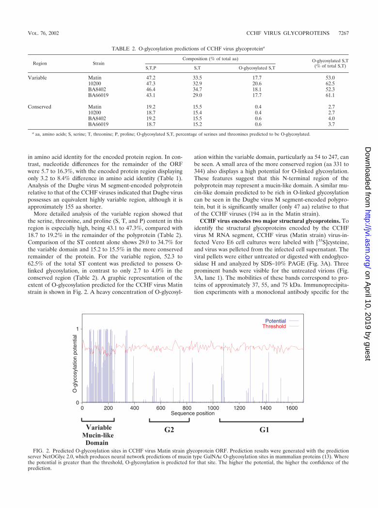
More detailed analysis of the variable region showed thatthe serine, threonine, and proline (S, T, and P) content in thisregion is especially high, being 43.1 to 47.3%, compared with18.7 to 19.2% in the remainder of the polyprotein (Table 2).Comparison of the ST content alone shows 29.0 to 34.7% forthe variable domain and 15.2 to 15.5% in the more conservedremainder of the protein. For the variable region, 52.3 to62.5% of the total ST content was predicted to possess O-linked glycosylation, in contrast to only 2.7 to 4.0% in theconserved region (Table 2). A graphic representation of theextent of O-glycosylation predicted for the CCHF virus Matinstrain is shown in Fig. 2. A heavy concentration of O-glycosyl-
ation within the variable domain, particularly aa 54 to 247, canbe seen. A small area of the more conserved region (aa 331 to344) also displays a high potential for O-linked glycosylation.These features suggest that this N-terminal region of thepolyprotein may represent a mucin-like domain. A similar mu-cin-like domain predicted to be rich in O-linked glycosylationcan be seen in the Dugbe virus M segment-encoded polypro-tein, but it is significantly smaller (only 47 aa) relative to thatof the CCHF viruses (194 aa in the Matin strain).
CCHF virus encodes two major structural glycoproteins. Toidentify the structural glycoproteins encoded by the CCHFvirus M RNA segment, CCHF virus (Matin strain) virus-in-fected Vero E6 cell cultures were labeled with [35S]cysteine,and virus was pelleted from the infected cell supernatant. Theviral pellets were either untreated or digested with endoglyco-sidase H and analyzed by SDS–10% PAGE (Fig. 3A). Threeprominent bands were visible for the untreated virions (Fig.3A, lane 1). The mobilities of these bands correspond to pro-teins of approximately 37, 55, and 75 kDa. Immunoprecipita-tion experiments with a monoclonal antibody specific for the
FIG. 2. Predicted O-glycosylation sites in CCHF virus Matin strain glycoprotein ORF. Prediction results were generated with the predictionserver NetOGlyc 2.0, which produces neural network predictions of mucin type GalNAc O-glycosylation sites in mammalian proteins (13). Wherethe potential is greater than the threshold, O-glycosylation is predicted for that site. The higher the potential, the higher the confidence of theprediction.
TABLE 2. O-glycosylation predictions of CCHF virus glycoproteina
Region StrainComposition (% of total aa) O-glycosylated S,T
(% of total S,T)S,T,P S,T O-glycosylated S,T
Variable Matin 47.2 33.5 17.7 53.010200 47.3 32.9 20.6 62.5BA8402 46.4 34.7 18.1 52.3BA66019 43.1 29.0 17.7 61.1
Conserved Matin 19.2 15.5 0.4 2.710200 18.7 15.4 0.4 2.7BA8402 19.2 15.5 0.6 4.0BA66019 18.7 15.2 0.6 3.7
a aa, amino acids; S, serine; T, threonine; P, proline; O-glycosylated S,T, percentage of serines and threonines predicted to be O-glycosylated.
VOL. 76, 2002 CCHF VIRUS GLYCOPROTEINS 7267
on April 10, 2019 by guest
http://jvi.asm.org/
Dow
nloaded from

CCHF virus N protein identified the 55-kDa protein as N (datanot shown). Consistent with this, the 55-kDa band did not shiftin mobility on treatment with endoglycosidase H (Fig. 3A, lane2). The two other prominent protein bands (37 and 75 kDa)showed a shift in migration when treated with endoglycosidaseH, suggesting that these were glycoproteins possessingN-linked glycosylation (Fig. 3A, lane 2). On digestion withendoglycosidase H, the approximately 37-kDa protein bandshifted to a faster-migrating, more diffuse band which ap-peared to contain two major forms. While the protein with thefaster mobility obviously possesses glycans sensitive to endogly-cosidase H digestion, the slower-migrating form may possesscomplex endoglycosidase H-resistant modifications (such asthose occurring in the medial Golgi). The 75-kDa proteinshifted to a faster-migrating form following digestion with en-doglycosidase H, again indicating the presence of high-man-nose or hybrid structures. In keeping with the nomenclature
used for the glycoproteins of other members of the familyBunyaviridae, the 37-kDa protein (faster migrating on PAGEanalysis) will be referred to as G2, and the 75-kDa protein(slower migrating) will be referred to as G1.
CCHF virus mature G2 and G1 glycoproteins are producedby proteolytic processing of the M segment-encoded polypro-tein. In order to identify the N terminus of the G2 and G1glycoproteins expressed by CCHF virus Matin strain, unla-beled virus pellets were prepared and proteins were separatedon SDS–10% PAGE and blotted onto a polyvinylidene diflu-oride membrane. The G2 and G1 proteins were visualized withPonceau S and excised for N-terminal sequencing analysis. TheN termini of the G2 and G1 proteins were determined to beginat aa 525 and 1046, respectively, relative to the sequence of theMatin strain M segment encoded polyprotein (shown diagram-matically in Fig. 1). Since the signal peptide for the polyproteinis predicted to be aa 1 through 24 and the N terminus of G2 is
FIG. 3. (A) Autoradiograph of CCHF virus (Matin strain) pellets grown in Vero E6 cells labeled overnight with [35S]cysteine and eitheruntreated or digested with endoglycosidase H. Virus samples were resolved on SDS–10% PAGE. (B) Lanes 1 and 2, autoradiograph of CCHF virus(IbAr10200 strain) virus-infected SW-13 cells and supernatant (Sup) labeled overnight with [35S]cysteine, immunoprecipitated with polyclonalHMAF against CCHF virus IbAr10200 strain, and resolved on a 10% NuPAGE gel system. Lanes 3 and 4, same as lanes 1 and 2 except sampleswere resolved on 3 to 8% NuPAGE. Lane 5, autoradiograph of supernatant from CCHF (Matin strain) virus-infected SW-13 cells labeled overnightwith [3H]leucine, immunoprecipitated as in lanes 1 to 4, and resolved on 10% NuPAGE.
7268 SANCHEZ ET AL. J. VIROL.
on April 10, 2019 by guest
http://jvi.asm.org/
Dow
nloaded from

500 aa downstream of the predicted signalase cleavage site,additional protease processing must be occurring to generatethe mature G2 protein (Fig. 1). Also, no other predicted mem-brane-spanning hydrophobic domains are present in this500-aa region of the polyprotein, indicating that the N termi-nus is not generated by an additional signalase cleavage event.Similarly, the N terminus of the mature G1 is located approx-imately 50 aa downstream of the nearest predicted membrane-spanning hydrophobic domain, suggesting that it is also notgenerated from a precursor protein by signalase cleavage (Fig.1). Analysis of the Dugbe virus M segment-encoded polypro-tein suggests a similar processing pattern for the Dugbe virusmature G1 and G2 proteins.
On further analysis of the protease cleavage sites that mustbe utilized to generate the N termini of the CCHF virus matureglycoproteins, it was noted that the tetrapeptides RRLL andRKPL immediately precede the G2 and G1 cleavage sites,respectively. In addition, these tetrapeptides are conserved inthe glycoprotein sequences of all the CCHF virus strains ex-amined to date. Interestingly, although the N terminus of theDugbe virus G2 has not been directly determined, by align-ment with the CCHF virus sequences, the Dugbe virus G2 Nterminus would be predicted to be at aa 375 and be immedi-ately preceded by the tetrapeptide RKLL. In addition, theDugbe virus G1 N terminus has been determined previously(20). It aligns well with the G1 N terminus of the CCHF virusand is also immediately preceded by the tetrapeptide RKLL.These data suggest that proteolytic cleavage events involved inthe generation of the N termini of the G2 and G1 proteins maybe common for viruses of the genus Nairovirus in general.More surprisingly, similar tetrapeptides have recently beenimplicated in the major protease cleavage events involved inthe processing of the arenavirus GPC glycoprotein precursorinto the mature GP1 and GP2 proteins. The tetrapeptidesRRLL and RKLL immediately precede the known GPC cleav-age sites of Lassa and Pichinde viruses, respectively (2, 18), andthe cellular protease subtilase SKI-1/S1P carries out the cleav-age, at least in the case of the RRLL site in Lassa virus (17, 38).In addition, Guanarito virus (another arenavirus), the cause ofVenezuelan hemorrhagic fever, possesses the tetrapeptideRKPL (identical to that seen in the CCHF virus glycoproteinprecursor) immediately prior to the predicted GPC cleavagesite (34). These conserved features suggest similarities in gly-coprotein processing between viruses of the genus Nairovirus,family Bunyaviridae, and viruses of the family Arenaviridae andimplicate SKI-1 or related proteases in the major glycoproteincleavage events for CCHF and Dugbe viruses.
The CCHF virus M segment-encoded polyprotein sequenceswere analyzed further for any other potential cleavage sites.The sequence RKLL was found in all of the CCHF virussequences analyzed (aa 809 to 812 for the Matin strain) at aposition in the polyprotein that could potentially be the Cterminus of G2 and result in a predicted G2 protein compa-rable in size to the 37-kDa G2 protein observed on PAGEanalysis (Fig. 1). However, no similar sequence was observed inthe comparable region of the Dugbe virus polyprotein se-quence. One additional potential cleavage site observed in allCCHF virus sequences was the sequence RSKR (aa 249 to 252for the Matin strain), a previously described furin proteasecleavage site (reviewed in reference 15). However, a similar
sequence was absent in the comparable region of the Dugbevirus polyprotein, and no data are available to suggest that thissite is utilized.
CCHF virus glycoproteins in infected cells and superna-tants. To identify potential CCHF virus glycoprotein precursorproteins present within infected cell cytoplasm as well as virusproteins that may be secreted from virus-infected cells, SW-13cells were infected with CCHF virus IbAr10200 strain andlabeled with [35S]cysteine. The viral proteins were then immu-noprecipitated with polyclonal HMAF raised against CCHFvirus IbAr10200 strain and visualized on 10% and 3 to 8%NuPAGE gel systems (Fig. 3B). As seen previously in [35S]cys-teine-labeled virions, the G1, N, and G2 proteins were clearlyseen in the infected cell supernatants (Fig. 3B, lanes 2 and 4).In addition, a 160-kDa protein was also seen (Fig. 3B, lane 4).
Two additional virus proteins of approximately 85 and 140kDa (referred to as P85 and P140, respectively) were observedwithin infected cells (Fig. 3B, lanes 1 and 3). While the 140-kDa and 160-kDa proteins have similar mobilities on a 10% gel(Fig. 3B, lanes 1 and 2), they can be distinguished as individualproteins on a 3 to 8% gel (Fig. 3B, lanes 3 and 4). The obser-vation of P85 and P140 in infected cells but not in virions orcell supernatants suggests that these proteins may be precursorproteins. Analysis of the CCHF virus glycoprotein amino acidsequences revealed that the variable mucin-like region (minusthe predicted signal peptide and assuming the furin recogni-tion site RSKR is cleaved) (Fig. 1) contains no cysteines. Basedon this observation, the CCHF virus Matin strain was labeledwith [3H]leucine instead of [35S]cysteine, and infected super-natants were immunoprecipitated with polyclonal HMAF tolook for any unique protein (structural or secreted) that wasnot observed previously. However, no additional proteins wereobserved, with only the P160, G1, N, and G2 proteins beingvisible (Fig. 3B, lane 5).
Processing of virus glycoproteins. To determine if the pro-teins present only in infected cells undergo any kind of pro-cessing, pulse-chase experiments were conducted. SW-13 cellswere infected with IbAr10200 strain virus, pulse-labeled with[35S]cysteine for 10 or 20 min, and then chased for 0.5, 1, 2, 4,or 6 h (Fig. 4A and B). The virus N protein was alreadydetectable in infected cells following the 10-min pulse. Both Nand P140 were detected by the 20-min pulse. The maximumamount of P140 was detectable during the 30-min and 1-hchases and then began to decrease. Similarly, P85 was detect-able as early as the 30-min chase, peaked at the 1-h chase timepoint, and then decreased to undetectable levels by the 4-hchase time point. Neither of these proteins was detectable inthe supernatant. These data are consistent with P140 and P85representing precursor proteins.
As P140 and P85 levels decreased, G2 and G1 protein levelsincreased, suggesting a precursor-product relationship. G2 andG1 were detectable in the infected cells at the 1-h chase timepoint and began to appear in the cell supernatant by 2 h ofchase. The P160 protein was detectable in the cells only at the2-h chase time point. P160 began to appear in the supernatantat the 4-h chase time point and increased by the 6-h chase timepoint. This suggests that the disappearance of P160 from theinfected cell and its subsequent appearance in the supernatantwere due to its release into the medium either in virions or asa secreted protein.
VOL. 76, 2002 CCHF VIRUS GLYCOPROTEINS 7269
on April 10, 2019 by guest
http://jvi.asm.org/
Dow
nloaded from

To further characterize the CCHF virus glycoproteins andtheir processing, an expression plasmid was constructed whichcontained the entire CCHF virus IbAr10200 strain M segmentORF inserted within pBluescript II KS. SW-13 cells weretransfected with the CCHF virus ORF clone and pulse-labeledwith [35S]cysteine for 20 min, followed by various lengths ofunlabeled chase periods. Following immunoprecipitation withpolyclonal HMAF raised against CCHF virus IbAr10200strain, the expressed virus proteins were analyzed on 3 to 8%gels (Fig. 4C). Results similar to those obtained with virus-infected cells were observed. P140 was detected as early as the
20-min pulse time point, with amounts beginning to decreasefollowing the 4-h chase. Also, P85 was detectable at the 20-minpulse time point, with amounts decreasing by 4 h of chase.These results are consistent with those obtained with CCHFvirus-infected cell lysates and supernatants and suggest thatP140 and P85 are glycoprotein precursors. G1 could be readilydetected in the 30-min chase and continued to increasethrough subsequent chase periods. G2 was more difficult tovisualize but could be detected after 3 and 4 h of chase.
To more precisely examine the relationship of P140 and P85to G1 and G2, a plasmid was constructed that would express
FIG. 4. (A and B) Autoradiographs of pulse-chase analyses of CCHF virus-infected cell cultures. SW-13 cells were infected with CCHF virusIbAr10200 strain, pulse-labeled with [35S]cysteine, and chased with unlabeled medium for the indicated time periods. Cell lysates (Cell) andsupernatants (Sup) were harvested, immunoprecipitated with polyclonal HMAF against CCHF virus IbAr10200 strain, and then resolved on a 3to 8% NuPAGE gel system. (C) Autoradiograph of pulse-chase analyses of cell lysates from transfected cell cultures. SW-13 cells were transfectedwith the CCHF virus IbAr10200 strain full-length glycoprotein ORF expression construct and an expression plasmid encoding for G1 plus theupstream hydrophobic region to function as a signal peptide. Cells were pulse-labeled with [35S]cysteine and chased for the indicated time periods.Cell lysates were harvested, immunoprecipitated with polyclonal HMAF against CCHF virus IbAr10200 strain, and resolved on a 10% NuPAGEsystem.
7270 SANCHEZ ET AL. J. VIROL.
on April 10, 2019 by guest
http://jvi.asm.org/
Dow
nloaded from

only the G1 coding region of the IbAr10200 strain plus the firsthydrophobic region upstream of the N-terminal cleavage sitefor G1 (Fig. 1). This region (approximately 50 aa upstream ofG1) was included to act as a signal peptide for the G1 protein.None of the ORF region that would encode G2 or anypolypeptides N terminal to G2 was included. When this G1clone was expressed in SW-13 cells, the P140 and G2 proteinswere no longer detected. However, both P85 and G1 proteinswere detected, suggesting that P140 is likely a precursor to G2,whereas P85 is a precursor of G1 (Fig. 4C). These resultsindicate that P85 may be processed from the larger M ORFpolyprotein by signalase cleavage following the hydrophobicdomain immediately upstream of G1, with subsequent cleav-age by a subtilase SKI-1-like protease to produce the matureG1.
Analysis with individual glycoprotein-specific antibodies.To verify the identities of the various individual glycoproteinsseen in CCHF virus-infected cell and supernatant prepara-tions, we used a variety of specific antibodies targeting variousregions of the virus M segment-encoded polyprotein. SW-13cells were either infected with the CCHF virus IbAr10200strain or transfected with the full-length IbAr10200 strain gly-coprotein clone and radiolabeled with [35S]cysteine. A G2-specific antipeptide antibody (generated against aa 540 to 551of the IbAr10200 strain ORF sequence) was used in immuno-precipitation assays of cell lysates. Autoradiographs of theprecipitated proteins resolved on a 10% NuPAGE gel showedreactivity of the G2 antipeptide to the 37-kDa G2 protein asexpected (Fig. 5A). In addition, the P140 protein was alsoprecipitated by the G2 antipeptide antibody.
To determine whether coprecipitation of G2 and P140 wasdue to the presence of G2 peptides within the P140 protein orif it was due simply to close interaction of P140 and G2,Western blot analysis was done, allowing separation of theproteins before reaction to the G2 antipeptide antibody. Un-labeled virus proteins for this experiment were generated byinfecting SW-13 cells with CCHF virus IbAr10200 strain andharvesting cell lysates. Results of this analysis on a 3 to 8%NuPAGE gel showed G2 antipeptide antibody reactivity toboth G2 and P140, confirming the presence of G2 peptides inboth G2 and P140 (Fig. 5B). These results, in combination withthe results obtained by pulse-chase analyses, strongly implicateP140 as a precursor protein to G2.
The same unlabeled CCHF virus (IbAr10200 strain) in-fected cell lysates were also analyzed by Western blot analysison a 3 to 8% gel with an antipeptide antibody generatedagainst aa 1388 to 1399 of the CCHF virus IbAr10200 strainglycoprotein sequence, which specifically targets G1. Resultsshowed reactivity to 75- and 85-kDa proteins similar to the75-kDa G1 and P85 observed in radioimmunoprecipitationassays of cell lysates, indicating that P85 contains G1 peptides(Fig. 5C). The pulse-chase results showing P85 processing intoG1, in addition to these Western blot results indicating G1sequence within P85, provide compelling evidence that P85 isa precursor protein of G1.
Polyclonal antibodies to the variable mucin-like region ofthe IbAr10200 strain of CCHF virus were generated in mice(see Materials and Methods). These antibodies were also usedin our Western blot studies of CCHF virus-infected cultures.Following infection of SW-13 cells with the IbAr10200 strain
virus, cell lysates and supernatants were harvested and ana-lyzed on a 3 to 8% gel with the antibody targeting the CCHFvirus IbAr10200 strain mucin region. Results showed reactivityof the antibody with the P140 protein in infected cells and theP160 protein in the supernatant, indicating the presence of themucin-like domain in both proteins (Fig. 5D). Our results alsorevealed a possible second mucin-associated protein with mo-bility corresponding to approximately 95 kDa (P95) in additionto P140 in infected cells. P95 may represent a cleavage productof P140, but further studies are needed to examine this possi-bility more thoroughly.
DISCUSSION
While the M segments and their encoded glycoproteins havebeen well characterized for viruses of other genera of thefamily Bunyaviridae, much less is known for the viruses of thegenus Nairovirus. In the case of CCHF virus, the difficulty ofworking with the virus and the containment conditions (bio-safety level 4) required have resulted in few studies beingcarried out even though the virus is an important cause ofhemorrhagic fever in humans throughout broad areas of Af-rica, Europe, and Asia. Here we present the complete nucle-otide sequence of the M segment of the Matin strain of CCHFvirus, which was originally isolated from a patient in Pakistanin 1976. Together with the recently available M segment se-quences for Nigerian and Chinese CCHF virus strains (23), thisnow provides insight into the extent of M segment and glyco-protein diversity of CCHF viruses over much of the geographicrange of the virus and the time span from 1965 to 1984. Highdiversity is evident, with up to 19.3% nucleotide sequencevariation and 14.8% amino acid differences observable amongthe virus M segment and glycoprotein precursor proteins, re-spectively. The presence of distinct virus genotypes in differentgeographic areas correlates well with observations based oncomparison of partial CCHF virus S segment nucleotide se-quences (25), although greater sequence diversity is observedin the M RNA segment and encoded proteins than in the Ssegment and nucleocapsid protein. This diversity, particularlyin the glycoprotein sequences, likely reflects the use of differ-ent principal vector tick hosts and virus-amplifying vertebratespecies in the virus life cycle in the different geographic re-gions. For instance, Hyalomma marginatum ticks are thoughtto play an important role in West Africa, whereas Hyalommaasiaticum is the implicated tick species in China (3, 37, 44).
The published data concerning the synthesis and processingof the glycoproteins of the viruses of the genus Nairovirusprovide a rather complex and inconsistent picture. Hazara andClo Mor viruses appear to possess three structural glycopro-teins encoded by their M genome RNA segments, while onlytwo glycoproteins have been described for Qalyub and Dugbeviruses, all with various electrophoretic mobilities (5, 7, 8, 11,20, 41, 42). One of the striking features revealed on analysis ofthe M segment-encoded polyprotein of the CCHF viruses wasthe presence of two distinct regions: a highly variable region,which includes the N-terminal 243 to 248 aa of the protein, anda relatively conserved region, which encompasses the remain-der of the protein. The variable N-terminal domain varied byup to approximately 37% at the nucleotide level and 56% atthe amino acid level. As indicated by the excess of amino acid
VOL. 76, 2002 CCHF VIRUS GLYCOPROTEINS 7271
on April 10, 2019 by guest
http://jvi.asm.org/
Dow
nloaded from

substitutions relative to nucleotide substitutions, most of thenucleotide changes are nonsynonymous, resulting in a rapidlyevolving variable region compared with the more conservedregion. The driving force behind this rapid evolution of CCHFvirus glycoproteins may be positive selection for the virus toevade the host immune system (9).
The similarities of this N-terminal variable region with hostcell mucins were also quite unexpected. The high percentage ofserines, threonines, and prolines (up to 47.3%), many of whichwere conserved among the CCHF virus strains, and the pre-dicted extensive O-glycosylation would indicate that the mu-cin-like feature must play some important structural or func-
tional role in the CCHF virus life cycle. The presence of asimilar mucin-like domain near the N terminus of the Dugbevirus M segment-encoded polyprotein, albeit much shorter (47aa) than that seen in CCHF viruses, suggests that this propertymay be conserved broadly among viruses of the genus Nairo-virus. No evidence of any similar mucin-like regions could beidentified on analysis of glycoprotein sequences of representa-tive members of the other four genera of the Bunyaviridaefamily, suggesting that this feature is unique to members of thegenus Nairovirus.
The function of this variable mucin-like region is currentlyunknown. There are no cysteine residues within this region
FIG. 5. (A) Autoradiograph of [35S]cysteine-labeled SW-13 cell lysates which were infected with CCHF virus IbAr10200 strain or transfectedwith the IbAr10200 full-length glycoprotein ORF construct and immunoprecipitated with G2 antipeptide. Antipeptide antibody was generatedagainst aa 540 to 551 of the IbAr10200 strain glycoprotein sequence and was therefore G2 specific. Immunoprecipitated proteins were resolvedon 10% NuPAGE. (B and C) Western blot analysis of cell lysates harvested from CCHF (IbAr10200 strain) virus-infected SW-13 cell cultures,resolved on 3 to 8% NuPAGE and reacted with antipeptide antibodies. Panel B shows cell lysates reacted with the same G2-specific antipeptideantibody used in panel A. Panel C shows cell lysates reacted with a G1-specific antipeptide antibody raised against aa 1388 to 1399 of the IbAr10200strain sequence. (D) Western blot analysis of cell lysates (cell) and supernatants (sup) harvested from SW-13 cells infected with CCHF virusIbAr10200 strain, resolved on 3 to 8% NuPAGE and reacted with a mucin-specific antibody. The mucin-specific antibody was generated by DNAimmunization of mice with a chimeric expression construct containing sequence from the variable mucin-like region of the IbAr10200 strain(without most of the predicted signal peptide and preceding the RSKR tetrapeptide).
7272 SANCHEZ ET AL. J. VIROL.
on April 10, 2019 by guest
http://jvi.asm.org/
Dow
nloaded from

(the N-terminal 243 to 248 aa, excluding the predicted signalpeptide), suggesting the lack any disulfide linkages. Mucins aredescribed as large glycoproteins with a high degree of O-gly-cosylation on serines and threonines (4, 35). This abundance ofO-glycosylation may offer carbohydrate heterogeneity, whichcould enable viruses to bind to a variety of molecules (1). Thepresence of many prolines within mucin-like sequences, whichcan induce �-turn conformations, probably allows close pack-ing of O-linked oligosaccharides. A large abundance of theseclosely packed O-linked sugars may offer protection from pro-teases. Curiously, the glycoprotein of Ebola virus, another hu-man pathogen causing hemorrhagic fever, also possesses avariable and highly O-glycosylated mucin-like region within themiddle third of its glycoprotein sequence (26). The mucin-likedomain of Ebola virus glycoprotein has been shown to be amajor determinant of pathogenicity, as expression of Ebolavirus glycoprotein in explanted human and porcine blood ves-sels caused massive endothelial cell loss and subsequent sub-stantial increase in vascular permeability, while deletion of themucin-like domain prevented these effects (45). It remains tobe seen if the mucin-like domain of CCHF virus has similarpathological functions.
The cumulative data presented here demonstrate thatCCHF virus possesses two major virion-associated glycopro-teins, G1 and G2, and that these are encoded by the virus MRNA segment in the order 5�-G2-G1-3�. This is consistent withearlier findings for Dugbe virus (20). The data for CCHF virus,together with that available for Dugbe virus, demonstrate thatglycoprotein synthesis and processing for the members of thegenus Nairovirus are quite distinct from those of members ofthe other family Bunyaviridae genera. For the viruses of theother genera, the mature G1 and G2 proteins are immediatelypreceded by a signal peptide sequence, and there is no evi-dence of a polyprotein precursor, suggesting that cotransla-tional cleavage of the G1 and G2 proteins of these virusesoccurs via a cellular signalase (28). In contrast, within theDugbe virus polyprotein precursor sequence, the only potentialsignal sequence for G1 is located about 50 aa upstream of theamino terminus of the mature G1 protein (20). The existenceof a precursor protein for G1 was supported by the identifica-tion of an 85-kDa protein that shared epitopes with G1 andwas found to be processed to G1 during pulse-chase studies ofDugbe virus-infected cells. In our study, we show that the Nterminus of the mature G1 protein of CCHF virus is alsolocated approximately 50 aa downstream of the closest poten-tial signal peptide hydrophobic region. Similarly, we were un-able to identify any obvious signal peptide immediately up-stream of the confirmed N terminus of the CCHF virus matureG2. Obviously, translocation of G2 into the endoplasmic retic-ulum lumen must be affected by a hydrophobic region that liesfurther upstream.
Virus protein analysis by various techniques, includingpulse-chase analysis and reactivity with CCHF virus-specificpolyclonal and antipeptide antibodies, demonstrated that theP140 (which contains the mucin-like region) and P85 nonstruc-tural proteins are the precursors of the mature G2 and G1proteins, respectively. P140 appears to represent the proteinregion from the mucin-like domain through the G2 region andpossibly to the cleavage site generating the N terminus of theP85 precursor of G1 (Fig. 1). A variety of lines of evidence,
particularly the synthesis of P85 by the G1 expression plasmid,suggest that P85 extends from about 50 aa upstream of themature G1 N terminus to the C terminus of G1 (presumably aa1689 relative to the Matin strain). The N terminus of P85 islikely generated from the polyprotein by cotranslational signa-lase cleavage prior to subsequent processing into the matureG1.
The N termini of the CCHF virus Matin strain G2 and G1proteins were determined by microsequencing to be equivalentto aa 525 and 1046 of the encoded polyprotein precursor,respectively. The tetrapeptides RRLL and RKPL are immedi-ately upstream of the N termini of mature G2 and G1, respec-tively, and were shown to be completely conserved among thepredicted polyprotein sequences of all the CCHF virus strains.In addition, Dugbe virus possesses the tetrapeptide RKLLimmediately upstream of the confirmed G1 cleavage site (20)and the predicted G2 cleavage site (aa 375). These tetrapep-tides are identical to or closely resemble the tetrapeptides thatrepresent the major cleavage recognition sites present in theglycoprotein precursors of arenaviruses, such as Lassa fevervirus (RRLL) and Pichinde virus (RKLL). The glycoproteinprecursor GPC of Lassa virus, another hemorrhagic fever vi-rus, is posttranslationally processed into GP1 and GP2 by pro-teolytic cleavage at a site preceded by the cleavage motifRRLL (18). Mutations in this tetrapeptide sequence inhibitedthe processing.
In addition, recent studies have shown that Lassa virus GPCcleavage occurs in the endoplasmic reticulum and is mediatedby SKI-1/S1P, a protease belonging to the pyrolysin branch ofsubtilases, following the RRLL sequence (17). This uniqueprotease plays a distinct role in cholesterol metabolism (24)and is involved in the generation of neuroactive peptides (29).The presence of the sequence RKLL immediately precedingthe Pichinde GPC cleavage site suggests that SKI-1 or a relatedprotease is also involved in this cleavage. In addition, theRKPL tetrapeptide motif seen in the CCHF virus glycoproteinprecursor also precedes the predicted GPC cleavage site inGuanarito virus, an arenavirus responsible for hemorrhagicfever in Venezuela (34). These results strongly suggest thatCCHF viruses (and other members of the genus Nairovirus)likely utilize the subtilase SKI-1/S1P-like cellular proteases forthe major glycoprotein precursor cleavage events that generatethe N termini of the G2 and G1 mature glycoproteins.
Since G2 is estimated to be approximately 37 kDa, based onPAGE mobility, and the N terminus is known to be located ataa 525 for CCHF virus Matin strain, the RKLL tetrapeptidelocated at aa 809 to 812 may represent the G2 protein Cterminus, which may also be generated by an SKI-1-like pro-tease cleavage event (Fig. 1). This tetrapeptide is conserved inall the CCHF virus strains, although not in the equivalentregion of the Dugbe virus sequence. However, if one examinesthe six potential membrane-spanning hydrophobic domains,beginning with the N-terminal signal peptide, the RKLL tet-rapeptide at aa 809 to 812 would be predicted to lie on thecytoplasmic side of the membrane, assuming the protein tran-sits the membrane at each of the hydrophobic domains (Fig. 1).If this model is correct, it is difficult to see how this tetrapep-tide could be a protease cleavage site. Further studies will beneeded to more precisely determine the C terminus of theCCHF virus G2 protein and the mechanism of its generation.
VOL. 76, 2002 CCHF VIRUS GLYCOPROTEINS 7273
on April 10, 2019 by guest
http://jvi.asm.org/
Dow
nloaded from
CCHF virus G1 protein probably extends from aa 1046 (forMatin strain) to the end of the glycoprotein ORF (Fig. 1), sincea predicted transmembrane domain is present at the C termi-nus to act as a membrane anchor, followed by an approxi-mately 65-aa cytoplasmic tail. Such a protein would be com-patible with the 75-kDa estimate that was determined on thebasis of the mobility of G1 on PAGE analysis.
Multiple sequence alignments of CCHF viruses also re-vealed the presence of a conserved tetrapeptide motif, RSKR,at the junction of the variable mucin-like region and the re-mainder of the glycoprotein. This motif conforms to the tet-rapeptide consensus sequence R-X-K/R-R recognized by furin,an enzyme belonging to the kexin family of subtilases andinvolved in the cleavage of a number of glycoproteins in theGolgi apparatus (reviewed in reference 15). Although furinactivation has been shown to occur for several viral glycopro-teins, including human immunodeficiency virus type 1, influ-enza virus, and Ebola virus (40), there is currently no evidencethat furin cleavage occurs at the RSKR site in the CCHF virusglycoprotein precursor. If furin cleavage did occur, the result-ing products (aa 25 to 252 [assuming the predicted signalpeptide is cleaved off] and aa 253 to 524, relative to the Matinstrain sequence) would likely be soluble proteins due to thelack of identifiable transmembrane domains. These may besecreted or bound to a membrane structural protein. However,additional experiments are in progress to further examine po-tential furin cleavage.
An additional protein, P160, was observed in virus-infectedcells and supernatants and found to react with CCHF virus(IbAr10200 strain) virus-specific polyclonal HMAF in immu-noprecipitation experiments and with mucin-specific antibod-ies in Western blot analyses. This may represent a more gly-cosylated form of the precursor protein P140, as additionalO-linked glycans are added late in the secretory pathway. How-ever, P160 may represent a distinct protein, since it would beunexpected to find a precursor protein in the supernatant.Further studies are under way to attempt to determine moreprecisely the origin of this protein.
The data presented here for CCHF virus together with ear-lier studies on other viruses of the genus Nairovirus show thattheir M segment polyprotein processing and mature G1 andG2 synthesis are much more complex than those for viruses ofthe other genera of the family Bunyaviridae. Our study alsouncovered unsuspected similarities between the glycoproteinsof CCHF virus and those of other hemorrhagic fever viruses.These included the identification of a glycoprotein mucin-likedomain similar to that present in Ebola virus and the likely useof SKI-1-like proteases for the major glycoprotein proteolyticprocessing events, similar to Lassa fever and Guanarito viruses.With the foundation provided by this study, further studies tomore precisely define and understand the complexity and sig-nificance of these elements of CCHF virus glycoproteinsshould be possible.
ACKNOWLEDGMENTS
We thank Anthony Sanchez for sharing the chimeric IbAr10200mucin antibody with us and for invaluable input at the initial stages ofthis project. We also thank Jonathan Smith for sharing his knowledgeand graciously providing us with monoclonal antibody and TamaraCrews of the Scientific Resources Program for protein microsequenc-ing.
REFERENCES
1. Brockhausen, I. 1995. Biosynthesis of O-glycans of the N-acetylgalac-tosamine-�-ser/thr linkage type, p. 201–259. In J. Montreuil, J. F. G. Vlieg-enthart, and H. Schachter (ed.), Glycoproteins. Elsevier Science, New York,N.Y.
2. Burns, J. W., and M. J. Buchmeier. 1993. Glycoproteins of the arenaviruses,p. 17–35. In M. S. Salvato (ed.), The arenaviridae. Plenum Press, New York,N.Y.
3. Camicas, J. L., J. P. Cornet, J. P. Gonzalez, M. L. Wilson, F. Adam, andH. G. Zeller. 1994. Crimean-Congo hemorrhagic fever in Senegal — presentstatus of the knowledge on the ecology of the CCHF virus. Bull. Soc. Pathol.Exot. 87:11–16.
4. Carraway, K. L., and S. R. Hull. 1991. Cell surface mucin-type glycoproteinsand mucin-like domains. Glycobiology 1:131–138.
5. Cash, P. 1985. Polypeptide synthesis of Dugbe virus, a member of theNairovirus genus of the Bunyaviridae. J. Gen. Virol. 66:141–148.
6. Chumakov, M. 1963. Study of viral haemorrhagic fevers. J. Hyg. Epidemiol.7:125–140.
7. Clerx, J. P. M., and D. H. L. Bishop. 1981. Qalyub virus, a member of thenewly proposed Nairovirus genus (Bunyaviridae). Virology 108:361–372.
8. El-Ghorr, A. A., A. C. Marriott, V. K. Ward, T. F. Booth, S. Higgs, E. A.Gould, and P. A. Nuttall. 1990. Characterization of Dugbe virus by biochem-ical and immunochemical procedures with monoclonal antibodies. Arch.Virol. 1990(Suppl. 1):169–179.
9. Endo, T., K. Ikeo, and T. Gojobori. 1996. Large-scale search for genes onwhich positive selection may operate. Mol. Biol. Evol. 13:685–690.
10. Feldmann, H., S. T. Nichol, H.-D. Klenk, C. J. Peters, and A. Sanchez. 1994.Characterization of filoviruses based on differences in structure and antige-nicity of the virion glycoprotein. Virology 199:469–473.
11. Foulke, R. S., R. R. Rosato, and G. R. French. 1981. Structural polypeptidesof Hazara virus. J. Gen. Virol. 53:169–172.
12. Gonzalez, J. P., J. L. Camicas, J. P. Cornet, O. Faye, and M. L. Wilson. 1992.Sexual and transovarian transmission of Crimean-Congo haemorrhagic fevervirus in Hyalomma truncatum ticks. Res. Virol. 143:23–28.
13. Hansen, J. E., O. Lund, N. Tolstrup, A. A. Gooley, K. L. Williams, and S.Brunak. 1998. NetOGlyc: prediction of mucin type O-glycosylation sitesbased on sequence context and surface accessibility. Glycoconj. J. 15:115–130.
14. Hoogstraal, H. 1979. The epidemiology of tick-borne Crimean-Congo hem-orrhagic fever in Asia, Europe, and Africa. J. Med. Entomol. 15:307–417.
15. Klenk, H.-D., and W. Garten. 1994. Host cell proteases controlling viruspathogenicity. Trends Microbiol. 2:39–43.
16. Krogh, A., B. Larsson, G. von Heijne, and E. L. L. Sonnhammer. 2001.Predicting transmembrane protein topology with a hidden Markov model:application to complete genomes. J. Mol. Biol. 305:567–580.
17. Lenz, O., J. ter Meulen, H.-D. Klenk, N. G. Seidah, and W. Garten. 2001.The Lassa virus glycoprotein precursor GP-C is proteolytically processed bysubtilase SKI-1/S1P. Proc. Natl. Acad. Sci. USA 98:12701–12705.
18. Lenz, O., J. ter Meulen, H. Feldmann, H.-D. Klenk, and W. Garten. 2000.Identification of a novel consensus sequence at the cleavage site of the Lassavirus glycoprotein. J. Virol. 74:11418–11421.
19. Logan, T. M., K. J. Linthicum, C. L. Bailey, D. M. Watts, D. J. Dohm, andJ. R. Moulton. 1990. Replication of Crimean-Congo hemorrhagic fever virusin four species of ixodid ticks (Acari) infected experimentally. J. Med. En-tomol. 27:537–542.
20. Marriott, A. C., A. A. El-Ghorr, and P. A. Nuttall. 1992. Dugbe Nairovirus MRNA: nucleotide sequence and coding strategy. Virology 190:606–615.
21. Nichol, S. T. 2001. Bunyaviruses, p.1603–1633. In D. M. Knipe and P. M.Howley (ed.), Fields virology, vol. 2. Lippincott Williams and Wilkins, Phil-adelphia, Pa.
22. Nielsen, H., S. Brunak, and G. von Heijne. 1999. Machine learning ap-proaches to the prediction of signal peptides and other protein sortingsignals. Protein Eng. 12:3–9.
23. Papa, A., B. Ma, S. Kouidou, Q. Tang, C. Hang, and A. Antoniadis. 2002.Genetic characterization of the M RNA segment of Crimean-Congo hem-orrhagic fever virus strains, China. Emerg. Infect. Dis. 8:50–53.
24. Rawson, R. B., D. Cheng, M. S. Brown, and J. L. Goldstein. 1998. Isolationof cholesterol-requiring mutant Chinese hamster ovary cells with defects incleavage of sterol regulatory element-binding proteins at site 1. J. Biol.Chem. 273:28261–28269.
25. Rodriguez, L. L., G. O. Maupin, T. G. Ksiazek, P. Rollin, A. S. Khan, T. F.Schwarz, R. S. Lofts, J. F. Smith, A. M. Noor, C. J. Peters, and S. T. Nichol.1997. Molecular investigation of a multisource outbreak of Crimean-Congohemorrhagic fever in the United Arab Emirates. Am. J. Trop. Med. Hyg.57:512–518.
26. Sanchez, A., A. S. Khan, S. R. Zaki, G. J. Nabel, T. G. Ksiazek, and C. J.Peters. 2001. Filoviridae: Marburg and Ebola viruses, p. 1279–1304. In D. M.Knipe and P. M. Howley (ed.), Fields virology, vol. 2. Lippincott Williamsand Wilkins, Philadelphia, Pa.
27. Sanchez, A. J., K. D. Abbott, and S. T. Nichol. 2001. Genetic identification
7274 SANCHEZ ET AL. J. VIROL.
on April 10, 2019 by guest
http://jvi.asm.org/
Dow
nloaded from

and characterization of Limestone Canyon virus, a unique Peromyscus-bornehantavirus. Virology 286:345–353.
28. Schmaljohn, C. S., and J. W. Hooper. 2001. Bunyaviridae: the viruses andtheir replication, p. 1581–1602. In D. M. Knipe and P. M. Howley (ed.),Fields virology, vol. 2. Lippincott Williams and Wilkins, Philadelphia, Pa.
29. Seidah, N. G., S. J. Mowla, J. Hamelin, A. M. Mamarbachi, S. Benjannet,B. B. Touré, A. Basak, J. S. Munzer, J. Marcinkiewicz, M. Zhong, J. C.Barale, C. Lazure, R. A. Murphy, M. Chretien, and M. Marcinkiewicz. 1999.Mammalian subtilase/kexin isozyme SKI-1: a widely expressed proproteinconvertase with a unique cleavage specificity and cellular localization. Proc.Natl. Acad. Sci. USA 96:1321–1326.
30. Shepherd, A. J., R. Swanepoel, P. A. Leman, and S. P. Shepherd. 1987. Fieldand laboratory investigation of Crimean-Congo haemorrhagic fever virus(Nairovirus, family Bunyaviridae) infection in birds. Trans. R. Soc. Trop.Med. Hyg. 81:1004–1007.
31. Shepherd, A. J., R. Swanepoel, S. P. Shepherd, G. M. McGillivray, and L. A.Searle. 1987. Antibody to Crimean-Congo hemorrhagic fever virus in wildmammals from southern Africa. Am. J. Trop. Med. Hyg. 36:133–142.
32. Shepherd, A. J., R. Swanepoel, S. P. Shepherd, P. A. Leman, and O. Mathee.1991. Viraemic transmission of Crimean-Congo haemorrhagic fever virus toticks. Epidemiol. Infect. 106:373–382.
33. Simpson, D. I., E. M. Knight, G. Courtois, M. C. Williams, M. P. Weinbren,and J. W. Kibukamusoke. 1967. Congo virus: a hitherto undescribed virusoccurring in Africa. I. Human isolations—clinical notes. East Afr. Med. J.44:86–92.
34. Spiropoulou, C. F., S. Kunz, P. E. Rollin, K. P. Campbell, and M. B. A.Oldstone. 2002. New World arenavirus clade C, but not clade A and B,viruses utilizes alpha-dystroglycan as its major receptor. J. Virol. 76:5140–5146.
35. Strous, G. J., and J. Dekker. 1992. Mucin-type glycoproteins. Crit. Rev.Biochem. Mol. Biol. 27:57–92.
36. Swanepoel, R., A. J. Shepherd, P. A. Leman, S. P. Shepherd, G. M. McGil-livray, M. J. Erasmus, L. A. Searle, and D. E. Gill. 1987. Epidemiologic and
clinical features of Crimean-Congo hemorrhagic fever in southern Africa.Am. J. Trop. Med. Hyg. 36:120–132.
37. Swanepoel, R. 1994. Crimean-Congo haemorrhagic fever, p. 723–729. InJ. A. W. Coetzer, G. R. Thomson, and R. C. Tustin (ed.), Infectious diseasesof livestock, with special reference to Southern Africa. Oxford UniversityPress, Cape Town, South Africa.
38. Touré, B. B., J. S. Munzer, A. Basak, S. Benjannet, J. Rochemont, C. Lazure,M. Chrétien, and N. G. Seidah. 2000. Biosynthesis and enzymatic character-ization of human SKI-1/S1P and the processing of its inhibitory prosegment.J. Biol. Chem. 275:2349–2358.
39. Vincent, M. J., N. U. Raja, and M. A. Jabbar. 1993. Human immunodefi-ciency virus type 1 Vpu protein induces degradation of chimeric envelopeglycoproteins bearing the cytoplasmic and anchor domains of CD4: role ofthe cytoplasmic domain in Vpu-induced degradation in the endoplasmicreticulum. J. Virol. 67:5538–5549.
40. Volchkov, V. E., H. Feldmann, V. A. Volchkova, and H.-D. Klenk. 1998.Processing of the Ebola virus glycoprotein by the proprotein convertasefurin. Proc. Natl. Acad. Sci. USA 95:5762–5767.
41. Watret, G. E., C. R. Pringle, and R. M. Elliott. 1985. Synthesis of bunyavirus-specific proteins in a continuous cell line (XTC-2) derived from Xenopuslaevis. J. Gen. Virol. 66:473–482.
42. Watret, G. E., and R. M. Elliott. 1985. The proteins and RNAs specified byClo Mor virus, a Scottish Nairovirus. J. Gen. Virol. 66:2513–2516.
43. Woodall, J. P., M. C. Williams, and D. I. Simpson. 1967. Congo virus: ahitherto undescribed virus occurring in Africa. II. Identification studies. EastAfr. Med. J. 44:93–98.
44. Yan, Y. C., L. X. Kong, L. Lee, Y. Q. Zhang, F. Li, B. J. Cai, and S. Y. Gao.1985. Characteristics of Crimean-Congo hemorrhagic fever virus (Xinjiangstrain) in China. Am. J. Trop. Med. Hyg. 34:1179–1182.
45. Yang, Z.-Y., H. J. Duckers, N. J. Sullivan, A. Sanchez, E. G. Nabel, and G. J.Nabel. 2000. Identification of the Ebola virus glycoprotein as the main viraldeterminant of vascular cell cytotoxicity and injury. Nat. Med. 6:886–889.
VOL. 76, 2002 CCHF VIRUS GLYCOPROTEINS 7275
on April 10, 2019 by guest
http://jvi.asm.org/
Dow
nloaded from