Characterisation of Absorbatox™ as a wound
175
Characterisation of Absorbatox™ as a wound healing agent by Khulekani Mncube BSc: Human Physiology BSc (Hons) Pharmacology Dissertation submitted in partial fulfilment of the requirements for the degree of: MSc with specialisation in Pharmacology April 2013 Faculty of Health Sciences Department of Pharmacology University of Pretoria Pretoria Supervisor: Dr A.D Cromarty © University of Pretoria
Transcript of Characterisation of Absorbatox™ as a wound
Characterisation of Absorbatoxtrade as a wound
healing agent
by
Khulekani Mncube BSc Human Physiology
BSc (Hons) Pharmacology
Dissertation submitted in partial fulfilment of the requirements for the degree of
MSc with specialisation in Pharmacology
April 2013
Faculty of Health Sciences Department of Pharmacology
University of Pretoria Pretoria
Supervisor Dr AD Cromarty
copycopy UUnniivveerrssiittyy ooff PPrreettoorriiaa
Page
ii
DEDICATION
I dedicate this dissertation to my Lord the risen Saviour Jesus Christ without Whom
my entire university career would not have been possible
ldquoAnd I am convinced and sure of this very thing that He Who began a good work in
you will continue until the day of Jesus Christ [right up to the time of His return]
developing [that good work] and perfecting and bringing it to full completion in yourdquo
- Philippians 16 (Amplified Bible)
Page
iii
ACKNOWLEDGEMENTS
bull I would like to express my gratitude to my supervisor Dr Duncan Cromarty for
lending his expertise and extending his support encouragement and advice
throughout my post-graduate studies
bull I would like to thank Professor Oppel BW Greeff for his support and words of
encouragement throughout my post-graduate studies
bull My sincere thanks goes to Dr Markus Stoeckli Dr Rocco Falchetto Dr Brendan
Prideaux Mr Gregory Morandi Dr Dieter Staab and the rest of the Analytical
Chemistry unit of Novartis Pharma AG (Basel Switzerland) for hosting me in their
lab as an intern their patience in training me in MALDI MSI their support and
encouragement even after my internship and also for helping me comfortably
integrate into their laboratory Thank you especially to Dr Stoeckli for teaching me
the importance of balance between life and work
bull I wish to thank Dr Marie-Claude Djidja for her help in troubleshooting my sample
preparation for the MALDI analysis and to Dr Melanie Ceci and Ms Nicole Ehrhard
for all their help in preparing my histological slides
bull My heartfelt gratitude goes out to Novartis Pharma AG especially to the Diversity
and Inclusion Team for their vision to create the Novartis Next Generation of
Scientists Internship and giving me the opportunity to be a part of this program
bull I am truly thankful to Dr Caprioli of Vanderbilt University for hosting me in his lab and
to Dr Erin Seeley and Mrs Jamie Allen for introducing me to and getting me
acquainted with MALDI mass spectrometry and popular American culture
bull I owe sincere thanks to the Chemistry Department of Tshwane University of
Technology and Professor Josef Heveling for hosting me in their lab My thanks go
Page
iv
out to Mr Joshua Malobela and Mr Benias Nyamunda for teaching me and helping
me perform my BET-nitrogen adsorption-desorption experiments
bull I am obliged to Mr Charles Noakes and Poretech for laser particle sizing analysis
work done on my samples and to Mr Noakes for his assistance in analysing the
results
bull I am very grateful to University of the Witwatersrand for facilitating my animal study
and especially to Dr Leith Meyer for performing the surgeries
bull I am thankful to Professor Francois Steffens and Mrs Joyce Jordaan for their
assistance in regards to statistical analysis and to Professor Steffens for patiently
explaining the statistical results to me
bull I wish to thank the Library staff of the Basic Medical Sciences Library University of
Pretoria for their assistance in acquiring research material
bull I wish to thank Mr Aaron Baloyi and Mr Solly Mahlangu of the Pathology Department
University of Pretoria for training me in preparing FFPE tissues
bull I am sincerely thankful to the NRF for financial support by way of a DAAD bursary
and KIC travel grant
bull I wish to thank my family friends and members of my church congregation for their
encouragement and prayers as well as Ms Nelsa Da Silva for providing me with a
home away from home in my final year of study
bull Finally my endless sincere and heartfelt gratitude to my parents and little sister for
their love emotional and spiritual support encouragement prayers and faith in my
ability to achieve I appreciate every sacrifice made on their part to ensure my
success God showed me great favour when He gave me you all Thank you
Page
v
TABLE OF CONTENTS
UNIVERSITY OF PRETORIA - DECLARATION vii
ABSTRACT viii
GLOSSARY OF ABBREVIATIONS x
LIST OF TABLES xvi
LIST OF FIGURES xvii
CHAPTER 1 INTRODUCTION 1
Literature review 1
Importance of Work 12
AIM 15
OBJECTIVES 15
CHAPTER 2 PHYSICAL CHARACTERISATION 16
INTRODUCTION 16
Nitrogen Gas Adsorption 16
Laser Particle Sizing and Size Distribution 21
MATERIALS AND METHODS 26
Nitrogen Adsorption 26
Laser Particle Sizing 27
RESULTS AND DISCUSSION 29
Nitrogen Adsorption 29
Laser Particle Sizing 35
CONCLUSION 37
CHAPTER 3 ANIMAL STUDY 38
INTRODUCTION 38
MATERIALS AND METHODS 41
Animals 41
Wound induction 43
Wound measurements 44
Statistical analysis 44
RESULTS 46
Page
vi
DISCUSSION 51
CHAPTER 4 MALDI-MSI 56
INTRODUCTION 56
MATERIALS AND METHODS 63
Formalin-Fixation Paraffin-Embedding (FFPE) 63
Mass Spectrometry 65
RESULTS AND DISCUSSION 73
CONCLUSION 88
CHAPTER 5 PROTEIN IDENTIFICATION 91
INTRODUCTION 91
MATERIALS AND METHODS 94
RESULTS AND DISCUSSION 96
Proteins identified on tissue from wound repair DAY THREE 98
Proteins identified on tissue from wound repair DAY SIX 101
Proteins identified on tissue from wound repair DAY SEVEN 103
Proteins identified on tissue from wound repair DAY NINE 106
Proteins identified on tissue from wound repair DAY THIRTEEN 108
Proteins identified on tissue from wound repair DAY SIXTEEN 109
CONCLUSION 112
CONCLUDING REMARKS 117
IMPROVEMENTS TO CURRENT STUDY 124
REFERENCES 126
APPENDIX Letters of ethical approval and statistical analysis 149
Page
vii
UNIVERSITY OF PRETORIA
FACULTY OF HEALTH SCIENCES
DEPARTMENT OF PHARMACOLOGY
I Khulekani Mncube
Student number 25150392
Subject of the work ldquoCharacterisation of Absorbatoxtrade as a wound healing
agentrdquo
Declaration
1 I understand what plagiarism entails and am aware of the University‟s policy in this
regard
2 I declare that this dissertation is my own original work Where someone else‟s
work was used (whether from a printed source the internet or any other source) due
acknowledgement was given and reference was made according to departmental
requirements
3 I did not make use of another student‟s previous work and submitted it as my own
4 I did not allow and will not allow anyone to copy my work with the intention of
presenting it as his or her own work
Signature __________________________________
Page
viii
ABSTRACT
Introduction Chronic wounds are a great burden to care-givers and patients alike
and are the main cause of many preventable amputations Such wounds are treated
with wound dressings but providing a wound environment that is conducive to proper
wound healing is not always possible with such dressings Absorbatoxtrade is a natural
zeolite that has been manipulated to increase its cationic exchange capacity and has
its main functionality as a potential wound healing agent in its strong capillary action
This quality enables the zeolite to absorb excess wound exudate and thus prevent
wound infection and maceration Absorbatoxtrade was characterised to determine its
effects on wound healing
Methods The physical characterisation of two grades of Absorbatoxtrade - granular
and micronised - was conducted using nitrogen adsorption to determine pore size
and surface area and laser particle sizing to determine the particle sizes of the
Absorbatoxtrade particles Full-thickness wounds of 8 x 8 mm were created on the
backs of pigs and treated with Absorbatoxtrade a positive and a negative control The
wound dimensions were measured and recorded The wounds were then excised on
selected days of each phase of wound healing and fixed in formalin The wound
sections were analysed by mass spectrometry imaging and abundant wound
proteins were identified from the tryptic digests using BLAST against the Swiss-Prot
database
Results The surface areas of the micronised and granular Absorbatoxtrade were 1443
and 1123 m2g respectively The micronised Absorbatoxtrade particle sizes ranged
between 08 microm to approximately 300 microm with an average pore diameter of 282 nm
The granular Absorbatoxtrade particle sizes ranged between 2 microm and 875 microm with
average pore diameters of 438 nm Absorbatoxtrade showed better wound healing by
delaying wound contraction and causing more rapid shallowing of the wound depths
compared to the negative control The difference observed in the wound healing
rates of the Absorbatoxtrade-treated and positive control groups were statistically
significant and the histological evaluations of the wounds treated with Absorbatoxtrade
showed wound closures that were associated with qualities that more closely
resembled normal healthy tissue than the positive control wounds The protein
Page
ix
activity in the trypsin-digested tissue including within the wound area and the
surrounding healthy tissue was successfully imaged using MALDI-MSI BLAST
software was used at an e-value of 30 to identify possible proteins from the tryptic
digests and were identified as proteins involved in wound healing
Discussion Micronised Absorbatoxtrade treated wounds showed more rapid healing
than the other treatments most likely due to the smaller particles and pores which
results in strong capillary action to absorb excess exudate Mass spectrometry
imaging allowed monitoring of the protein fluctuations that occur during wound
healing The proteins detected were then identified using BLAST and MASCOT
database comparison tools which identified that the abundant proteins detected by
mass spectrometry were not those typically observed in wound healing but rather
those involved in molecular aspects of wound healing like nerve regeneration cell
proliferation survival and migration
Keywords Nitrogen-adsorption Wound healing MALDI-TOF trypsin digestion
BLAST
Page
x
GLOSSARY OF ABBREVIATIONS
microgmL Microgram per millilitre
microm Micrometres
3D Three-dimensional
AESC Animal Ethics Screening Committee
AlO4 Aluminate
ANOVA Analysis of Variance
ASAP Accelerated Surface Area Porosity
AUCC Animal Use and Care Committee
BdB Broekhoff and de Boer
BET Brunauer Emmett and Teller
BJH Barrett Joyner and Halenda
BLAST Basic Local Alignment Search Tool
BLNK B-cell linker protein
BSA Bovine Serum Albumin
CAS Central Animal Service
CCD Charge-coupled device
CEC Cation exchange capacity
CHCA -cyano-4-hydroxycinnamic acid
Page
xi
CI Cranston and Inkley
cm3g Cubic centimetre per gram
CO2 Carbon dioxide
Cr Chromium
Da Dalton
DH Dollimore and Heal
DNA Deoxyribonucleic acid
dpi Dots per inch
EGF Epidermal growth factor
EGFR Epidermal growth factor receptor
EtOH Ethanol
EZS Electrical Zone Sensing
FFPE Formalin-Fixed Paraffin-Embedded
g Gram
H Hysteresis
HampE Haematoxylin and Eosin
HIVAIDS Human Immunodeficiency Virus Acquired Immunodeficiency
Syndrome
IL Interleukin
ITO Indium tin oxide
IUPAC International Union of Pure and Applied Chemistry
Page
xii
K Kelvin
kDa Kilodalton
KGF Keratinocyte growth factor
L Litre
lm Litres per minute
LS Light scattering
LSHU Liquid Sample Handling Unit
M Molecular weight of the adsorbate
mz Mass-to-charge ratio
m2g Square metre per gram
mA Milliampere
MALDI-MSI Matrix-assisted laser desorptionionisation mass spectrometry
imaging
MALDI-TOF Matrix-assisted laser desorptionionisation time-of-flight
MAPK Mitogen-activated protein kinase
MASCOT Modular Approach to Software Construction Operation and Test
mgkg Milligrams per kilogram
ml Millilitre
mm Millimetre
mM Millimolar
mmHg Millimetres of Mercury
Page
xiii
MMPs Matrix metalloproteinases
MS Mass spectrometer
MSMS Tandem mass spectrometry
NCBI National Centre for Biotechnology Information
NdYAG Neodymium-Doped Yttrium Aluminum Garnet
NF-κB Nuclear factor kappa-light-chain-enhancer of activated B cells
NH4CO3 Ammonium carbonate
NK Natural Killer
nm Nanometre
NP Nucleoprotein
ordmC Degree Celsius
PP0 Partial relative pressure
Paf1C Polymerase-associated factor 1 complex
PDGF Platelet-derived growth factor
PF4 Platelet factor 4
PMF Peptide mass fingerprinting
PSD Pore size distribution
PTMs Post-translational modifications
PVD Peripheral Vascular Disease
R Gas content
Page
xiv
RBP RNA-binding proteins
rk Radius (of the largest cylindrical capillary filled with condensate)
RNA Ribonucleic acid
ROI Region of interest
rp Radius into which condensation occurs
rRNA Ribosomal ribonucleic acid
s Second(s)
SEM Scanning electron microscope
SEM Standard error of the mean
SF Splicing factor
SiO4 Silicone tetraoxide
T Temperature
T-cell Thymus cell
TFs Transcription factors
TGF-β Transforming growth factor beta
Ti Titanium
TIMPs Tissue inhibitors of metalloproteinases
TLC Thin layer chromatography
TNF-α Tumour necrosis factor alpha
TPM Tropomysin
Page
xv
UV Ultraviolet
VEGF Vascular endothelial growth factor
Vm Monolayer capacity
vs Versus
Y Surface tension of the adsorbate
θ Contact angle
ρ Density of the adsorbate
Page
xvi
LIST OF TABLES
Table 1
Surface external and micropore areas of granular and micronised Absorbatoxtrade
Table 2
Total pore volumes and pore size distributions of granular and micronised
Absorbatoxtrade
Table 3
Wound width measurements (mm)
Table 4
Wound length measurements (mm)
Table 5
Wound depth measurements (mm)
Page
xvii
LIST OF FIGURES
Figure 1
Illustration of the wound healing process as well as the estimated time for which
each phase lasts (Broughton et al 2006)
Figure 2
Prevalence of Peripheral Vascular Disease (PVD) amongst diabetic patients in Africa
(Abbas and Archibald 2007)
Figure 3
Three-dimensional tetrahedral structure of a zeolite (Woodford 2012)
Figure 4
Graphical representation of methods used to determine surface area pore volume
and pore size distribution (Micromeriticsreg Instrument Corporation nd)
Figure 5
A) Low-angle light scattering from a particle larger than the light source wavelength
following the Fraunhofer theory B) A combination of high- and low-angle light
scattering from a particle slightly smaller than the light source wavelength in
accordance to the Mie theory (Young 2005)
Figure 6
Nitrogen adsorption-desorption isotherm for granular Absorbatoxtrade zeolite
Page
xviii
Figure 7
Nitrogen adsorption-desorption isotherm for micronised Absorbatoxtrade zeolite
Figure 8
The IUPAC classification of adsorption isotherms and hysteresis loops (Sing et al
1982)
Figure 9
Granular Absorbatoxtrade (left) and micronised Absorbatoxtrade (right)
Figure 10
Cumulative finer volume percent vs particle diameter (microm)
Figure 11
Pattern of wound treatments
Figure 12
Comparison of the effects of each treatment Absorbatoxtrade vs Negative control and
Cerdaktrade (positive control) on wound widths
Figure 13
Comparison of the effects of each treatment Absorbatoxtrade vs Negative control and
Cerdaktrade (positive control) on wound lengths
Page
xix
Figure 14
A line graph comparison of the wound contraction rates of each treatment group
Figure 15
Comparison of the effects of each treatment Absorbatoxtrade vs Negative control and
Cerdaktrade (positive control) on wound depths
Figure 16
Histological comparison of wound tissue treated with Cerdaktrade (positive control)
negative control and the two Absorbatoxtrade-treatments used in the study by
Oosthuizen et al (2009)
Figure 17
Graphical description of the process of the generation of ions and their subsequent
translation into ion-density distribution images (Seeley and Caprioli 2011)
Figure 18
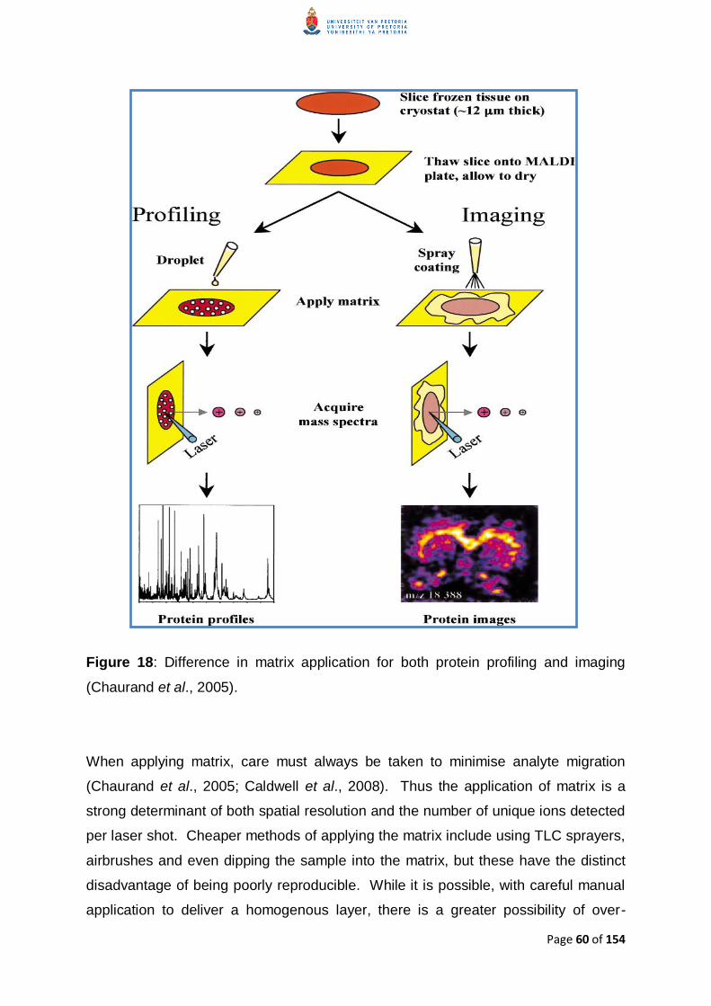
Difference in matrix application for both protein profiling and imaging (Chaurand et
al 2005)
Figure 19
Simplified scheme of formaldehyde reaction with proteins (a) Formaldehyde is
added to a protein with the formation of a reactive hydroxymethyl molecule (b)
Formation of an imine group (Schiffs base) (c) Methylene bridge formation between
a lysine residue (lysyl group) and nitrogen of a peptide linkage Formaldehyde is
depicted as methylene glycol formed by reaction with water (D‟Amico et al 2009)
Page
xx
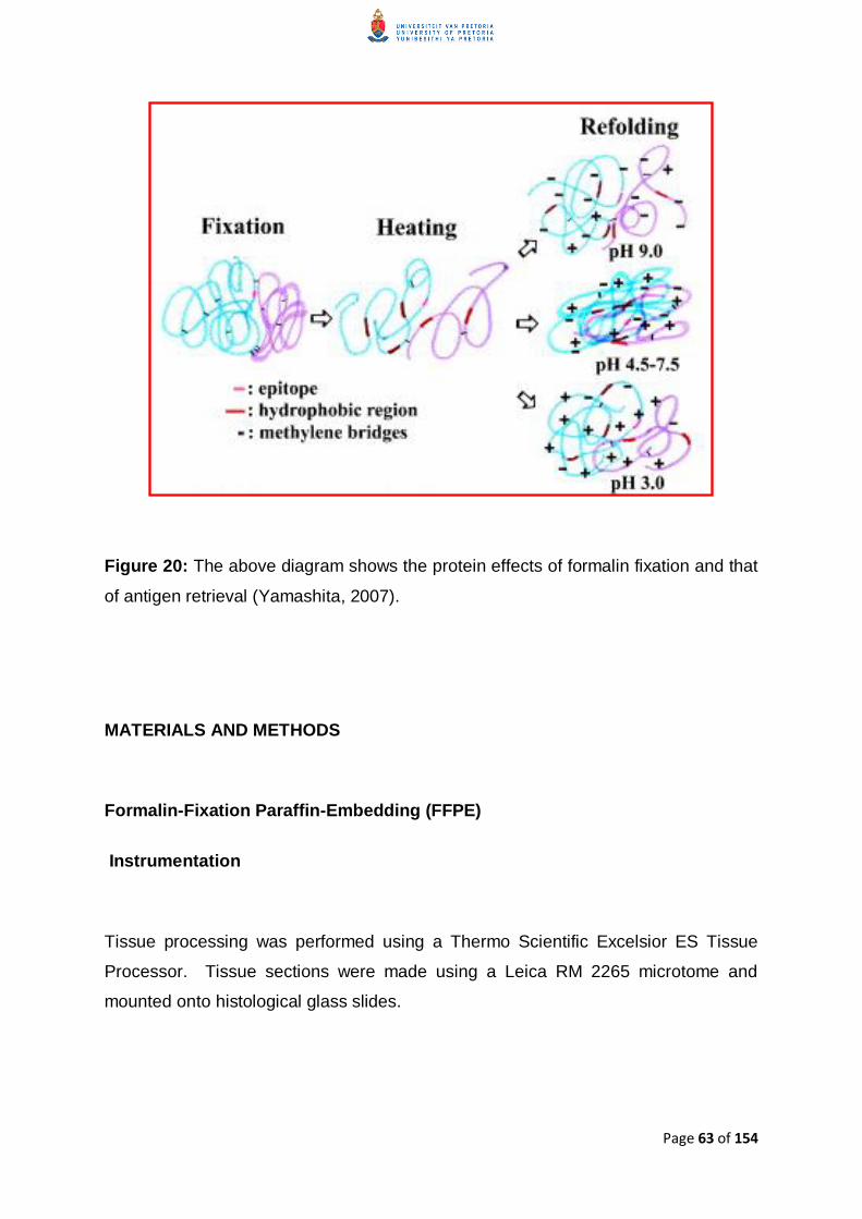
Figure 20
The above diagram shows the protein effects of formalin fixation and that of antigen
retrieval (Yamashita 2007)
Figure 21
Diagram of the embedded tissue and the approximate areas at which sections were
made for MALDI-MS analysis
Figure 22
Screen shot of the stacked mass spectra of the wound (blue) healthy (red) and the
CHCA matrix (green) as seen in TissueView 10
Figure 23
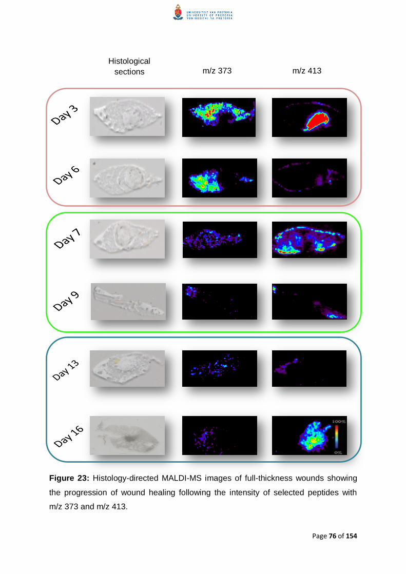
Histology-directed MALDI-MS images of full-thickness wounds showing the
progression of wound healing following the intensity of selected peptides with mz
373 and mz 413
Figure 24
Histology-directed MALDI-MS images of full-thickness wounds showing the
progression of wound healing following the intensity of selected peptides with mz
486 and mz 607
Figure 25
Histology-directed MALDI-MS images of full-thickness wounds showing the
progression of wound healing following the intensity of selected peptides with mz
644 and mz 795
Page
xxi
Figure 26
Histology-directed MALDI-MS images of full-thickness wounds showing the
progression of wound healing following the intensity of selected peptides with mz
833 and mz 1044
Figure 27
The figure above shows the ldquotop-downrdquo (right) and ldquobottom-uprdquo (left) methods that
can be used to identify and characterise proteins (Thiede et al 2005)
Figure 28
Graphical description of the workflow for collecting ldquobottom uprdquo proteomic data On-
tissue trypsin digestion resulted in tryptic digests which gave spectra that were fed
into databases to give positive identification of the proteins detected in the wound
tissue (adapted from Fenyouml 2000)
Figure 29
An output table of possible proteins identified from the data submitted to the BLAST
database
Figure 30
A graphical description illustrating the overlap seen in the wound healing process in
terms of the proteins detected in the animal models used in this study
Page 1 of 154
CHAPTER 1 INTRODUCTION
Literature review
ldquoHealing is a matter of time but it is sometimes also a matter of opportunityrdquo
ndash Hippocrates
Wounds result from either thermal or mechanical damage to the skin (Boateng et al
2008 Gibson and Schultz 2009) This damage interferes with the normal
anatomical and physiological function of the wounded area for example by
compromising the protective barrier against infection Wound healing is an important
and complex but necessary process that aims to restore the anatomical and
physiological integrity of the skin through a series of sequential but overlapping
phases These are the inflammatory proliferatory and remodelling or maturation
phases A healed wound is one that is completely covered by scab (Tian et al
2007) The process involves several mediators of inflammation many different
enzymes growth factors and several localised cell types like keratinocytes
fibroblasts and endothelial cells as well as immune cells (Diegelmann and Evans
2004 Blakytny and Jude 2006 Boateng et al 2008 Gibson and Schultz 2009)
When tissue injury is sustained the underlying blood vessels are damaged and
bleeding can occur Bleeding helps flush out foreign particles (Boateng et al 2008)
Blood flow brings platelets that undergo aggregation and release of important factors
that initiate the formation of a clot to discontinue the blood flow but also to facilitate
the release of a variety of cytokines which lead to vasodilation and increased
permeability of local blood vessels This is an important part of the maintenance of
Page 2 of 154
homeostasis and the beginning of the inflammatory phase (Singer and Clark 1999
Diegelmann and Evans 2004 Blakytny and Jude 2006)
The inflammatory phase begins immediately after the wound is sustained and lasts
three to four days during which protein-rich exudate is released into the wound to
promote vasodilation through the release of histamine and serotonin (Broughton et
al 2006 Boateng et al 2008) Vasodilation allows for the infiltration of neutrophils
which are drawn into the wounded tissue by locally released cytokines such as
interleukin-1 (IL-1) transforming growth factor beta (TGF-β) tumour necrosis factor
alpha (TNF-α) platelet factor (PF4) and bacterial by-products (Broughton et al
2006) The neutrophils not only play a phagocytic role in the wound area but also
condition the area for healing by releasing proteolytic enzymes to eliminate bacterial
debris and contaminants as well as damaged and necrotic tissue (Diegelmann and
Evans 2004 Broughton et al 2006 Boateng et al 2008 Sibbald and Woo 2008)
Macrophages start replacing the neutrophils as the process progresses and
dominate during the end stages of the inflammatory phase The macrophages
release platelet-derived growth factor (PDGF) vascular endothelial growth factor
(VEGF) and TGF-β all of which facilitate angiogenesis The presence of the
macrophages is also important for the transition into the proliferatory phase of the
wound healing process (Diegelmann and Evans 2004 Broughton et al 2006
Sibbald and Woo 2008 Kyriakides et al 2009)
The proliferatory phase occurs from approximately day four through to day fourteen
(Broughton et al 2006) and includes the major healing processes (Hunt et al
2000) During this period fibroblasts keratinocytes and endothelial cells are the
most dominant cells undergoing proliferation (Broughton et al 2006) Epithelial cells
on the edge of the wound begin to proliferate to create a new protective barrier
against fluid loss and potential infection (Singer and Clark 1999 Broughton et al
2006) There is also an increase in skin strength due to the appearance of newly
synthesised collagen by the fibroblasts (Boateng et al 2008) This cell proliferation
is stimulated by epidermal growth factor (EGF) and TGF-α produced by activated
platelets and macrophages (Broughton et al 2006) as well as keratinocytes
Page 3 of 154
(Diegelmann and Evans 2004) The secretion of mediators such as PDGF results
in the chemotaxis of neutrophils then macrophages (Diegelmann and Evans 2004)
PDGF is also a potent chemoattractant for monocytes fibroblasts and smooth
muscle cells and is associated with accelerated wound healing and enhanced tensile
strength across the wound (Ashraf et al 2009) TGF-β is a major mediator in wound
healing by regulating fibroblasts (Diegelmann and Evans 2004) It has a three-fold
effect on extracellular matrix deposition by increasing the overall production of matrix
by enhancing collagen synthesis (Diegelmann and Evans 2004 Broughton et al
2006) as well as that of proteoglycans and fibronectins (Diegelmann and Evans
2004) thus playing a role in scar formation (Ashraf et al 2009) decreasing the
production of matrix metalloproteinases (MMPs) and increasing the production of
tissue inhibitors of metalloproteinases (TIMPs) (Diegelmann and Evans 2004
Broughton et al 2006) TGF-β ndash especially TGF-β1 also plays an important role in
this stage of wound healing by acting as a potent chemoattractant for monocytes
macrophages neutrophils keratinocytes and fibroblasts TGF-β1 also induces the
release of other growth factors from these leukocytes (Singer and Clark 1999
Blakytny and Jude 2006) The MMPs are a family of zinc endopeptidases that are
responsible for degrading components of the extracellular matrix eliminating
damaged protein promoting movement of certain cellular components into the
centre of the wound destroying provisional extracellular matrix regulating the
activity of some growth factors and playing a role in angiogenesis (Osman et al
2002) The MMPs are divided into subgroups depending on their substrate The
MMPs that play a key role in wound healing are the collagenases (MMP-1 and -8)
and the gelatinases (MMP-2 and -9) (Muller et al 2008) Other mediators involved
in wound healing include the eicosanoids such as the prostaglandins which mediate
inflammation cytokines which regulate cellular activities and function and nitric
oxide ndash a physiologically active molecule released by macrophages (Diegelmann
and Evans 2004 Broughton et al 2006)
The maturation (remodelling) phase is the final stage of wound healing and can last
anything from a few days to two years (Broughton et al 2006 Boateng et al 2008)
depending on the size and depth of the wound tissue During this phase the pro-
collagen precursors synthesized during the proliferatory phase are converted from
Page 4 of 154
the immature but predominant collagen III to the mature organised collagen I
network that ensures a wound that is strong and resistant to sheer forces thus
limiting the chance of reopening (Diegelmann and Evans 2004 Broughton et al
2006 Sibbald and Woo 2008) This phase also results in the development of scar
tissue which is determined by the strength of the new epithelia (Hunt et al 2000
Boateng et al 2008)
Figure 1 Illustration of the wound healing process as well as the estimated time for
which each phase lasts (Broughton et al 2006)
Despite the complexity of the wound healing process its effectiveness can be
significantly affected by several factors such as arterial insufficiency growth factors
inadequate perfusion infection low oxygen tension neuropathy nutritional state
oedema as well as systemic factors and metabolic disorders (Hunt et al 2000)
(example diabetes) The presence of underlying physiological and biochemical
defects and imbalances such as those seen in diabetes often leads to impaired
Page 5 of 154
healing of wounds Muller et al (2008) state a ldquounifying pathophysiological
hypothesisrdquo supported by Blakytny et al (2005) Diegelmann and Evans (2004) and
Singer and Clark (1999) that in chronic wounds the wound healing process is
arrested in a state of inflammation This typically results in profuse neutrophil and
macrophage infiltration associated with the extensive discharge of reactive oxidative
species and degrading enzymes (proteases) and pro-inflammatory cytokines leading
to the formation of wounds that do not heal completely within the average 8-12
weeks andor are recurring (Blakytny and Jude 2006 Boateng et al 2008)
Diabetic ulcers are also prone to repeated infection owing to the inadequate
neutrophil and macrophage migration and impaired function (Singer and Clark 1999
Blakytny and Jude 2006 Hirsch et al 2008)
Chronic wounds are managed to prevent or treat infection that could lead to
amputation (Hunt et al 2000) In the case of diabetic patients Staphylococcus
aureus (S aureus) is the most abundant single isolate from diabetic wounds and
this often leads to the poor wound healing seen in these instances (Hirsch et al
2008)
In general treatment of wounds is done by debridement of necrotic tissue and
occlusive dressings (Hunt et al 2000) but this has not always been so The
treatment of wounds has evolved over centuries from the application of traditional
healing agents such as plants and animal fat to tissue engineered skin substitutes
(Boateng et al 2008) The use of some crude plants and animal fat however often
led to bacterial infection thus defeating the purpose of using a wound dressing
(Boateng et al 2008) Despite the development of skin substitutes wound
dressings are still the mainstay of chronic wound treatment (Singer and Clark 1999
Boateng et al 2008) although dressing such wounds appropriately is a challenge
The goal of wound dressings is to provide an environment that is conducive to
proper wound healing A dressing should readily absorb excessive wound exudate
so as to prevent bacterial overgrowth exudate leakage and maceration at the edges
of the wound yet should not cause excessive drying of the wound The rationale
behind this is that wound exudate helps facilitate the healing process by providing
Page 6 of 154
the wound bed with nutrients and assists in the migration of the epithelial cells
(Singer and Clark 1999 Sullivan et al 2001 Boateng et al 2008)
Systemic pharmacological agents can also be used in wound healing but these have
the potential to cause toxic side effects and therefore direct application of healing
agents to the wound is preferred (Boateng et al 2008)
Absorbatoxtrade embedded in the wound dressing helps to create a wound healing
environment ideal for rapid and positive wound healing and since it is a topical
wound treatment does not have the potential to cause systemic toxicity
AbsorbatoxTM is a synthetically enhanced hydrated aluminosilicate and a
clinoptilolite of the zeolite family which has limited natural activity but has been
subjected to a patented process that increases its cation exchange capacity (CEC)
to 5 times its original capacity Like all zeolites Absorbatoxtrade is composed of
aluminium oxygen and silicon in a tetrahedral arrangement resulting in pores and a
cage like structure since the tetrahedral units cannot fill the available space entirely
(Kaneko 1994) The three-dimensional arrangement of AlO4 and SiO4 tetrahedra in
Absorbatoxtrade enclosing a porous cage like complex (Dutta et al 2005) exemplifies
this The porous complex provides Absorbatoxtrade with specific physicochemical
properties including ion exchange capacity adsorbent nature size exclusion
framework and catalytic properties The advantage of Absorbatoxtrade in wound
healing lies in its ability to draw wound exudate out of the wound by strong capillary
forces but still keep the wound from becoming overly dry
With the aim of further investigating Absorbatoxtrade as a wound healing agent the
processed zeolite was subjected to physicochemical characterisation The
Brunauer Emmet and Teller (BET)-nitrogen adsorption-desorption experiments
were performed to determine the average surface area and total pore volume of two
Absorbatoxtrade samples of different physical dimensions ndash micronised (fine powder)
Page 7 of 154
and granular Laser particle sizing was used to determine the average particles
sizes of the same two Absorbatoxtrade samples
At present gas adsorption is the best and one of the more commonly used
adsorption methods to investigate the characteristics of porous materials such as
surface area total pore volume and pore size distribution (PSD) (Sing 1998
Choma et al 2002) such as Absorbatoxtrade The BET theory upon which the BET-
nitrogen adsorption-desorption experiments are based is that gas molecules
physically adsorb on a solid surface in layers infinitely that no interaction between
these layers exist and that the Langmuir theory can be applied to each layer The
resulting BET equation is used to calculate the surface area and pore volume of
solids by the physical adsorption of gas molecules (Brunauer et al 1938)
While knowledge of a material‟s surface area gives insight into how it will react with
another substance and is thus an important quality to investigate when
characterising porous materials (Bae et al 2010) it is in fact the pore size
distribution which is the strongest determinant of adsorption (Wang et al 2005)
The method of Barrett Joyner and Halenda (BJH) (Micromeriticsreg Instrument
Corporation nd) is used to determine pore size distribution from experimental
isotherms using the Kelvin model for pore filling (Micromeriticsreg Instrument
Corporation nd Choma et al 2002)
The BJH method of determining pore size distribution analyses the nitrogen
desorption isotherm at liquid nitrogen temperature based on the assumption that the
mechanisms of physical adsorption on the pore walls and capillary condensation in
the pore can be employed to establish equilibrium between the adsorbed and gas
phases during the process of desorption (Barrett et al 1951 Choma et al 2002)
Particle size plays an important role in many industries including the pharmaceutical
and mining industries (Micromeriticsreg nd) Numerous techniques exist to
Page 8 of 154
determine particle size (Bowen 2002) however light scattering techniques such as
laser particle sizing are more widely used due to their high levels of accuracy and
reproducibility and also that these analysis techniques are generally faster than
microscopy for example (Barth and Flippen 1995 De Ridder et al 2000
Vanderhallen et al 2002 Micromeriticsreg nd) Laser particle sizing uses the
principle of light scattering to determine particle size and light scattering instruments
are often based on the Mie theory The Mie theory is a mathematical-physical theory
of the scattering of electromagnetic radiation by isotropic spherical particles as a
function of the angle at which light is scattered at the point of interaction with the
particle The intensities of light at each scattering angle are measured and are
correlated to particle size (Webb 2004 Ferraris et al 2004)
Further exploration into a possible mechanism of action of Absorbatoxtrade included
actual examination of wound tissue from an animal study at the subcellular level
using a technique called matrix-assisted laser desorptionionisation mass
spectrometry imaging (MALDI MSI)
MALDI MSI is a soft-ionisation technique that has gained popularity in modern
proteomics (the analysis of proteins in a living system) (Liu et al 2010) largely
because it allows researchers to study biological processes extensively by the
systematic analysis of proteins expressed in a cell or tissue (Yates 1998) The
technique came as a result of many years of investigation into the use of lasers for
the soft ionisation of biomolecules (Caldwell et al 2008) such as proteins (Tarran et
al 2007) MALDI-time-of-flight-MS (MALDI-TOF-MS) has made possible the
sensitive detection and measurement of large intact proteins from tissue sections
and even plasma as they fluctuate during the various biological processes and even
stages of wound healing (Jemal and Xia 2006 Zhao et al 2006) In addition to
being able to measure the protein fluctuations in tissue MALDI MSI another
sensitive and robust technique allows researchers to image tissue by the
quantitation of proteins present in thin histological tissue sections MALDI MSI and
MALDI-TOF enable analysis of tissue sections by detecting the presence relative
concentration location and spatial orientation of small molecules and lipids in
Page 9 of 154
addition to proteins and other macromolecules in biological structures (Yates 1998
Aebersold and Goodlett 2001) This makes following the changes occurring during
physiological processes at molecular level possible which makes MALDI-TOF and
MALDI MSI each an ideal tool to investigate the biological effects that Absorbatoxtrade
has on wounds with respect to its ability to provide an environment that results in
improved wound healing
MALDI-MSI works as follows An organic material (termed matrix) is applied to a thin
(10 μm ndash 30 μm) tissue section that was previously mounted on to a plate and
dehydrated The plate upon which the section is mounted is introduced into a mass
spectrometer where a focused laser beam is pulsed along the surface resulting in
the ejection of ions from the tissue These ions are accelerated into a flight tube and
strike thereafter a detector at different times The lighter ions travel faster and thus
hit the detector sooner This difference in flight time is translated into spectra
Distribution images for specific ions are then extracted from the thousands of
acquired spectra
Ion-intensity distribution images despite being a graphical representation of the
spectra analysed do not offer information on the proteins present in the tissue For
this reason protein and peptide identification is necessary Protein identification is
typically performed using tandem mass spectrometry (MSMS) However the
identification of intact proteins ndash regarded as the ldquotop-downrdquo approach ndash raises some
serious challenges For this reason it is not uncommon to introduce an enzyme to
digest the protein into peptides and then measure the tandem mass spectra of the
peptides (Worley et al 1995 Resing and Ahn 2005 Xu and Ma 2006) From here
a number of methods can be employed to identify the proteins de novo sequencing
sequence tagging database searches and consensus of multiple search engines
(Xu and Ma 2006) While MALDI has made possible the analysis of significantly
large proteins the greatest impact still remains in the analysis of peptides generated
by proteolytic digestion A reason for this is while the mass accuracy of the MALDI
MS is high it is not sufficient to identify a protein with confidence based only on its
molecular weight (Veenstra et al 2006) It is thus the mass spectrum of the peptide
Page 10 of 154
fragments resulting from the use of a proteolytic enzyme such as trypsin that gives
the ldquopeptide mass fingerprintrdquo that is used to identify the proteins in a tissue sample
(Veenstra et al 2006)
Trypsin is the favoured enzyme for peptide mass fingerprinting as it is moderately
cheap extremely effective and produces tryptic digests of on average 8-10 amino
acids which are ideally suited for MS analysis (Thiede et al 2005) This is
exemplified by its use in almost all large-scale projects in mass spectrometry to give
more easily analysable peptides (Olsen et al 2004) Trypsin belongs to the serine
protease family and shows primary structural similarity to chymotrypsin The
enzymatic mechanism requires that the target amino acid be recognised in the
binding pocket of the trypsin The substrate binding pocket of the trypsin has a
negatively charged aspartate at the bottom of it and binds basic amino acids by
means of an ionic interaction The target amino acids for trypsin cleavage need to
have long side chains and be positively charged to permit this formation of the ionic
bonds Arginine and lysine both fulfil the criteria necessary for the ionic interaction
required for trypsin to work and it is at these regions that trypsin lyses unless either
the arginine or lysine is followed by a proline (Olsen et al 2004 Fremout et al
2010) The resultant peptides are characteristic specific and unique for each given
protein thus identifying the peptide means identifying the protein as a whole
Identifying the protein from the peptide fragments is termed ldquobottom-uprdquo or ldquoshot-
gunrdquo proteomics This method is also advantageous in terms of its sensitivity and
proteome coverage (Fremout et al 2010 Mallick and Kuster 2010) The peptides
identified by this method are then fed into a bioinformatics algorithm which allows for
the proteins to be identified after comparing a query sequence with a library or
database of sequences Two examples of bioinformatics algorithms that can be
used to identify the peptides are MASCOT and BLAST which correlate mass spectra
with peptides in sequence databases to return a positive identification MASCOT is
a probability-based search engine for peptide identification and peptide sequence
assignment (Olsen et al 2004 Weatherly et al 2005) BLAST is a similarity search
program that matches similar peptide sequences and either identifies the protein or
confirms a vague hit produced by MASCOT (Shevchenko et al 2001)
Page 11 of 154
The wound healing model used for the study is a Large White Female pig While
using a human model for the study would be ideal it is obvious that such a study
would raise serious ethical debate Other wound healing studies use rodents such
as rabbits or mice for such studies due to the ease of handling and the cost of
housing for these animals The main disadvantage of using rodents as models is
that they simply do not properly represent wound healing as seen in humans due to
he many differences between rodent and human skin profiles Porcine skin
however shows greater similarity to human skin than other species (Hirsch et al
2008 Velander et al 2008)
The main similarities between the skins of the two species with regards to wound
healing are those of the epidermis skin vascularisation and epidermal collagen
This similarity in structure was most evident in a study conducted by Moritz and
Henrique in 1946 during which skin sections from both humans and pigs treated with
benzidine were seen to be virtually indistinguishable (Vardaxis et al 1997)
With respect to wound healing porcine skin also heals similarly to human skin
Rodent skin heals mainly through contraction whereas both human and porcine
skins heal primarily through re-epithelialisation Another important aspect of pigs as
wound healing models is that pigs are also more closely representative of chronic
wounds in patients with serious illnesses since they too are placed under a great
deal of physiological stress with the induction of numerous wounds the regular and
repeated handling as well as the frequency of general anaesthesia (Sullivan et al
2001)
Page 12 of 154
Importance of Work
ldquoMy grandma lost both legs to diabetes my mama already lost her foot and now I got
diabetes I figured I would just lose my legs too I never knew there was anything I
could do to stop itrdquo
- Quote from a REACH focus group participant (1999)
According to Diabetes South Africa 4-6 million people in South Africa have diabetes
While a diagnosis of diabetes does not necessarily mean that one will develop a
chronic wound much less undergo an amputation because of it it is important to
note that diabetic patients are at a higher risk of developing peripheral vascular
disease ndash the primary cause of chronic wounds Figure 2 is a graph summarising
the high prevalence of peripheral vascular disease in diabetic patients in selected
African countries
In addition to diabetes HIVAIDS should also be considered in the development of
chronic wounds The development of a chronic wound in an HIVAIDS patient can
result from a simple cut from a non-sterile edge Because the HIVAIDS patients are
immune-compromised the immune response that is normally seen when a wound is
sustained may not occur or may not be quite as efficient as in a healthy person This
results in the bacteria that would ordinarily be eliminated by the neutrophils and
macrophages proliferating unchecked and causing a localised infection In such a
case it is not necessarily the infection that leads to the chronic wound but rather
that the absence of critical immune responses prevents release of chemotactic
cytokines and growth factors necessary for the wound to progress from the
inflammatory phase to the proliferatory phase and finally to be properly healed
According to the mid-year report of Statistics South Africa of 2011 166 of the
adult population in South Africa were living with HIV this is an increase of 117
million since 2001 with almost a quarter of these being due to new infections
Page 13 of 154
Figure 2 Prevalence of Peripheral Vascular Disease (PVD) amongst diabetic
patients in Africa (Abbas and Archibald 2007)
Both diabetes and HIVAIDS are on the increase not just in South Africa but
throughout the whole of Africa where chronic wounds are a considerable burden to
both patients and healthcare systems In Africa (Abbas and Archibald 2007) the
increasing number of diabetic patients and rate of HIV infection with their related
complications including poor wound healing are leading to numerous preventable
amputations These health problems are the driving force for this project in
investigating wound healing on the proteomic level where an understanding of the
differences in wound healing kinetics and phase-dependent proteomics could lead to
identifying targets for early interventions that could prevent exacerbation of wound
status to the point where drastic intervention measures are required
Determining the mechanism of action of Absorbatoxtradeas a wound healing agent
could greatly benefit South African patients The potential outcome of this research
is not only to achieve effective wound management which is much needed but also
8
41
21
12 10 15
54
0
10
20
30
40
50
60
70
80
90
100
South Africa Zambia Tanzania Ethiopia Sudan Malawi Nigeria
PV
D r
ate
(
)
Page 14 of 154
to create a marketable safe and effective wound dressing that will contribute to
healing of chronic wounds and thus the prevention of amputations and the reduction
in loss of employment due to disability
While it could be argued that disability does not equal loss of employment when the
number of people in South Africa whose only income is from manual labour (such as
mechanics construction workers miners) is considered it becomes very clear that
this research does indeed hold promise to decrease unemployment due to
preventable amputation and essentially protect the South African economy
The importance of this research is not only limited to Africa however since an
estimated 5 million patients in the United States suffer from chronic wounds
(Velander et al 2008) In both the African and American contexts this often results
in lost time from work and hefty health care costs Unfortunately these high
incidence and prevalence rates are not likely to decrease significantly any time soon
with increasing sedentary lifestyles and unhealthy diets and the resultant cases of
obesity a cause of type II diabetes Interestingly enough a parallel increase in the
incidence of foot complications in the diabetes populations has been documented
(Abbas and Archibald 2007)
To quote from a wound healing research group (Velander et al 2007) ldquoWe need
better ways to treat diabetic woundsrdquo
Page 15 of 154
AIM
To assess changes in wound tissue and possibly identify possible mechanisms by
which Absorbatoxtrade induces wound healing
OBJECTIVES
1 Physical characterisation
11 Analysis of nitrogen adsorption isotherms to determine surface area of the
Absorbatoxtrade samples as well as their average pore volumes and
distribution
12 Laser particle sizing to determine the average particle size of the
Absorbatoxtrade samples
2 Biological characterisation
21 Conduct an in vivo wound study using a porcine model from which tissue will
be excised for MALDI-MS analysis
22 Perform MALDI MSI of tissue sections of excised wound tissue at different
stages of the healing process to visualise fluctuation of proteins during the
different stages of wound healing
23 Identify the protein species found in the wound tissue samples at different
stages of wound healing
Page 16 of 154
CHAPTER 2 PHYSICAL CHARACTERISATION
INTRODUCTION
Nitrogen Gas Adsorption
Zeolites are naturally occurring hydrated minerals which were discovered and named
in 1756 by the Swedish mineralogist Baron Cronstedt and later defined by Smith
(1984) as aluminosilicate frameworks based on a three-dimensional arrangement of
AlO4 and SiO4 (Breck 1964 Smith 1984) Zeolites are formed in cavities in lava
flows and in plutonic rocks and are made up of a multiple tetrahedral structure
(Breck 1964) The resulting lattice encloses a network of cavities and channels
which provide the zeolites with a variety of physicochemical properties that include
ion exchange capacity a size exclusion frame catalytic properties as well as a large
internal surface area allowing for an adsorbent nature (Kaneko 1994 Sakintuna et
al 2003 Dutta et al 2005) The dimensions of the three-dimensional channels and
the pore sizes of the zeolites determine the size exclusion and catalytic activity of a
zeolite (Sakintuna et al 2003 Dutta et al 2005) The structure of the zeolites
possesses a negative charge which can contain a variety of cations such as sodium
calcium and magnesium (Breck 1964)
Absorbatoxtrade is a zeolite a specific member of the clinoptilolite family of zeolites and
thus possesses the characteristics of zeolites
At present gas adsorption is the best technique to assess total surface area of
adsorbent materials such as Absorbatoxtrade and one of the more commonly used
methods to investigate the characteristics of porous materials including surface area
total pore volume and pore size distribution (PSD) (Sing 1998 Choma et al 2002)
Both the physical structure (surface area) and chemical properties (cation type)
influence the gas adsorption characteristics of a zeolite (Burevski and Poceva 1994
Sing 1998 Choma et al 2002)
Page 17 of 154
Figure 3 Three-dimensional tetrahedral structure of a zeolite (Woodford 2012)
Surface area and pore volume
The surface area of a material affects the way that it interacts with another
substance and is thus an important parameter to investigate when characterising
porous materials (Bae et al 2010) As a rule the greater the surface area the
greater the amount of a different compound can interact with the porous material
The Brauner-Emmett-Teller (BET)-nitrogen absorption method of determining
surface area remains the favoured method of determining overall surface area of
porous materials (Sing 1998 Bowen 2002) The Langmuir theory determines
surface area based on monolayer coverage of a complete surface by the adsorbate
The BET theory expands on this theory by incorporating multilayer adsorption (Sing
1998 Bae 2010) The assumptions upon which the BET theory is based are that
gas molecules adsorb on a solid surface in an infinite number of layers and that no
interaction exists between the different adsorption layers (Brunauer et al 1938)
To determine surface area an adsorbate ndash most often nitrogen at a subzero
temperature of 77 K (Sing 1998) ndash is compressed in the presence of a solid in
Page 18 of 154
steadily increasing quantities After each quantity of adsorbate is applied the
pressure is allowed to equilibrate while the amount of nitrogen adsorbed to the
surface is computed The amount of nitrogen adsorbed at each temperature and
pressure defines the adsorption isotherm Using this defined isotherm the quantity
of gas required to form a single layer on the surface of the material being studied is
determined and with the area covered by the newly calculated amount of gas known
the overall surface area of the material can be calculated (Micromeriticsreg Instrument
Corporation nd)
The large surface area of zeolites is due to their highly porous nature The pore
dimensions play a vital role in the physicochemical properties of solid adsorbents
(Kaneko 1994) This is exemplified by the ability of Absorbatoxtrade to draw up and
hold wound exudate by strong capillary forces Thus the most essential data is with
regards to the pore dimensions of the Absorbatoxtrade
During the process of adsorption filling of the pores occurs The course of
adsorption is dependent on the pore volume available In the case of macropores
(pores that exceed 50 nm in width) pore filling takes place via multilayer formation
In the case of micropores (pores smaller than 2 nm in width) adsorption occurs via a
volume-filling process at a lower relative pressure In mesopores pores between 2
and 50 nm in width both adsorption mechanisms occur multilayer pore filling occurs
first which is then followed by volume-filling (Kaneko 1994 Jaroniec and Kaneko
1997 Sing 1998 Sakintuna et al 2003) While it was initially thought that capillary
condensation and (micropore) volume-filling are the same it is now widely accepted
that this is not the case (Sing 1998 Dutta et al 2005) The surface area of zeolites
is dependent on the pore number and pore volumes and since high porosity provides
a greater surface area a higher adsorptive capacity is expected for these materials
(Kusumaningtyas 2006)
Page 19 of 154
Pore size distribution
Pore size distribution is defined as ldquothe population of pores as a function of pore
widthrdquo (Kaneko 1994) The method of Barrett Joyner and Halenda (BJH) is used to
determine pore size distribution from experimental isotherms using the Kelvin model
for pore filling (Choma et al 2002 Micromeriticsreg Instrument Corporation nd) and
is only applicable to open pores assuming the cylindrical pore model (Barrett and
Joyner 1951 Allen 1997) The Kelvin equation-based method incorporates
methods put forward by Barret Joyner and Halenda (BJH) Dollimore and Heal
(DH) Cranston and Inkley (CI) and also Broekhoff and de Boer (BdB) (Choma et
al 2002) and takes into consideration statistical film thickness (t-curve) as a function
of relative pressure as well as the relationship between the evaporation
(condensation) pressure and the pore width (Choma et al 2002) This is
represented by the Kelvin capillary condensation equation (Roberts 1967)
where rk is the radius of the largest cylindrical capillary filled with condensate ldquoγrdquo
represents the surface tension of the adsorbate ldquoρrdquo is the density of the adsorbate
ldquoMrdquo is the molecular weight of the adsorbate θ is the contact angle between the
condensed phase and the surface of the solid and is usually assumed to be equal to
zero R is the gas constant and T is the temperature measured in Kelvin and
PP0 is the partial pressure
The thickness t of the adsorbate layer is considered in the equation used to
determine the pore radius ldquorkrdquo
Page 20 of 154
where rp indicates the radius into which condensation occurs at the required relative
pressure (Broekhoff and de Boer 1968 Yağşi 2004 Dutta et al 2005)
The BJH method of determining pore size distribution analyses the nitrogen
desorption isotherm at liquid nitrogen temperature based on the assumption that the
mechanisms of physical adsorption on the pore walls and capillary condensation in
the pore can be employed to establish equilibrium between the adsorbed and gas
phases during the process of desorption (Barrett and Joyner 1951 Choma et al
2002) During adsorption the quantity of nitrogen required to form a monolayer on a
surface is calculated as a function of gradually increased pressure of nitrogen gas at
a constant temperature of 77 K As the pressure increases the gas condenses first
in the smallest pores This continues until all the pores are filled with liquid This is
known as the point of saturation (Micromeriticsreg Instrument Corporation nd) The
reverse occurs in desorption The pressure is gradually reduced allowing for
capillary condensation or in other words for the nitrogen that had originally
saturated the open pores to now evaporate (Brunauer et al 1938 Barrett and
Joyner 1951 Kaneko 1994 Allen 1997 Dutta et al 2005) So both adsorption
and desorption isotherms are necessary to ascertain pore size distribution using the
BJH method
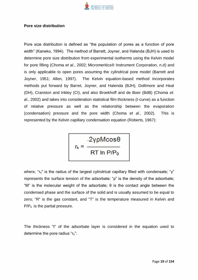
Figure 4 gives a concise description of the methods typically employed to determine
surface area pore volume and pore size distribution
Page 21 of 154
Figure 4 Graphical representation of methods used to determine surface area pore
volume and pore size distribution (Micromeriticsreg Instrument Corporation nd)
Laser Particle Sizing and Size Distribution
Webb (nd) cites McGraw Hill‟s Dictionary of Scientific and Technical Terms (Third
Edition) to define a particle as ldquoany relatively small subdivision of matter ranging in
diameter from a few angstroms to a few millimetresrdquo The size of a particle gives
important information on its behaviour such as performance as an abrasive
application in the case of cosmetics dissolution rate in the case of pharmaceuticals
and flow properties of fluid-cracking catalysts (Micromeriticsreg Saturn Digisizer reg II
nd)
Stage 4
A further increase in the gas
pressure will cause complete
coverage of the sample and fill
all the pores The BJH
calculation can be used to
determine pore diameter
volume and distribution
Stage 4
A further increase in the gas
pressure will cause complete
coverage of the sample and fill all
the pores The BJH calculation can
be used to determine pore
diameter volume and distribution
Stage 3
Further increasing gas
pressure will cause the
beginning of multi-layer
coverage Smaller pores
in the sample will fill first
BET equation is used to
calculate the surface area
Stage 2
As gas pressure increases
coverage of adsorbed
molecules increases to form
a monolayer (one molecule
thick)
Stage 1
Isolated sites on the
sample surface begin to
adsorb gas molecules at
low pressure
Page 22 of 154
Numerous techniques exist to determine particle size (Bowen 2002) such as
scanning electron microscopy (imaging) electrical zone sensing (Coulter principle)
sedimentation (centrifugation) and light interaction methods which include laser
particle sizing (laser diffraction) (Barth and Flippen 1995 Bowen 2002 Ferraris
2004 De Clerq 2004 Webb nd)
Scanning electron microscopy (SEM) is an analytical tool that uses a focused beam
of electrons to give a magnified image If the conditions are ideal this technique can
produce images with feature resolution at the nanometre level simultaneously giving
insight into both the morphology as well as the size distribution of the particles SEM
also gives information into any possible absence of homogeneity present in the
sample (Tsapatsis et al 1995 Bowen 2002 Ferraris 2004)
Sedimentation using either gravity or centrifugation is a very popular method based
on the application of Stokes‟ Law This law describes the terminal velocity of a
spherical particle settling in a fluid medium under the influence of a gravitational
force Sedimentation with centrifugation can result in a lower range of around 10 nm
and even further down towards a nanometre if an analytical ultracentrifuge is used
(Barth and Flippen 1995 Bowen 2002 Ferraris 2004 Webb nd)
Electrical zone sensing (EZS) is based on the Coulter principle This well-
established method works down to approximately 05 microm and allows for the
measurement of both the volume and number distribution In this technique the
suspended particles in an electrolyte at a highly dilute concentration are passed
through an orifice of a defined volume in an insulating wall between two electrodes
Voltage pulses proportional to the particle‟s volume are produced for each particle as
the particles enter the orifice (the sensing zone) The accumulation of pulses over a
period of time gives the particle size distribution EZS has also been used to
establish emulsion stability (Barth and Flippen 1995 Bowen 2002 Ferraris 2004
Webb nd)
Page 23 of 154
Laser diffraction measures the angular distribution of light scattered from a particle in
a dilute medium (Ferraris 2004)
While reliable information is obtainable from processing images acquired from using
direct methods such as microscopy (both light and electron) the methods are slow
and do not always yield statistically significant results (Vanderhallen et al 2000 De
Ridder et al 2002) Indirect methods such as laser diffraction are fast and highly
reproducible and are thus more widely used In addition to this light scattering
techniques are also useful for kinetic and in situ studies since they are often non-
destructive and non-perturbative to the species under investigation (Barth and
Flippen 1995 Vanderhallen et al 2000 De Ridder et al 2002)
Laser particle sizing uses the principle of light scattering to determine particle size
Light is defined as the energy residing in the narrow and very specific region of the
electromagnetic spectrum which depending on its interaction with matter can
produce various visual effects (Webb 2004) That is light can be reflected
refracted (bent) absorbed diffracted (slight deviation from its original path upon
interaction with matter) and even scattered (Ferraris 2004 Webb 2004) Scattered
light is made up of reflection refraction and diffraction and can thus be defined as a
change of the direction and intensity of a light beam when it strikes an object due to
the combined effects of reflection refraction and diffraction (Ferraris 2004 Webb
nd) Light scattering (LS) can either be dynamic or static Dynamic light scattering
(also known as quasi-elastic light scattering) uses the correlation of light intensity
variations and the Brownian movement of the particles to determine particle size
Techniques that rely on this type of light scattering vary widely depending on sample
concentration and conditions as well as environmental factors Static LS establishes
particle size using intensity characteristics of the scattering pattern at various angles
(Twomey et al 1994 Ferraris 2004 Webb nd)
During the 1800s many advances were made in understanding light scattering
Diverse theories were developed that related to particles of certain shapes and sizes
in a particular medium (Webb nd) In the 1970‟s these developments and
Page 24 of 154
knowledge resulted in the advent and commercialisation of the first laser light
scattering instruments (Micromeriticsreg Saturn Digisizer reg II nd)
These laser light scattering instruments use two fundamental methods of data
analysis the Mie and Fraunhofer theories
In 1906 Gustav Mie worked out a comprehensive mathematical-physical theory of
the scattering of electromagnetic radiation by isotropic spherical particles as a
function of the angle at which light is scattered at the point of interaction with the
particle When Mie developed the theory it could only be applied with boundary
conditions (i) only monochromatic light (light of a single colour or wavelength) is
considered (ii) the particle must be isotropic and (iii) spherical (iv) the incident light
is composed of plane waves (v) both scattering and absorption are considered (vi)
only static light is considered and (vii) no quantum effects such as Raman or
Doppler effects are considered (Ferraris 2004 Webb nd) Despite the constraints
of the Mie theory it can still be applied to determine particle size even if the particle
does not meet all the conditions since the current availability of computing software
means that particle-sizing instruments based on this theory are guided by expressed
conditions of Mie theory so as to construct a scattering pattern reducible by the Mie
theory (Webb nd)
Joseph Von Fraunhofer developed a theory that predicts the diffraction of light at the
edges of objects - in particular opaque objects The theory describes scattering
occurring when the particles investigated are far larger than the wavelength of the
light source in use The outcome of this low-angle light scatter is that light is
scattered off axis in a forward direction For this method the refractive index need
not be known since it is based only on the diffraction effect (Ferraris 2004 Young
2005 Webb nd) In contrast the general rule for the Mie theory is that the particles
are slightly smaller than the wavelength giving higher angles of refraction and a
broader range over which scattering occurs In this case forward (low-angle) scatter
is still predominant but the refractive index must be known (Ferraris 2004 Webb
nd)
Page 25 of 154
Figure 5 A) Low-angle light scattering from a particle larger than the light source
wavelength following the Fraunhofer theory B) A combination of high- and low-angle
light scattering from a particle slightly smaller than the light source wavelength in
accordance to the Mie theory (Young 2005)
The sizes of the zeolite particles were determined using laser light scattering
technology which gives high levels of resolution accuracy reproducibility and
repeatability The laser light scattering instrument used - a Micromeritics Saturn
DigiSizerreg - employs a laser diode and a modern charge-coupled device (CCD)
detector which allows a high-resolution digital representation of the light scattering
pattern to be captured Data reduction is then executed on the resulting information
using the Mie Theory (Micromeriticsreg Saturn Digisizer reg II nd)
A Fraunhofer Light
Scattering
B Mie Light Scattering
Page 26 of 154
MATERIALS AND METHODS
Nitrogen Adsorption
Instrumentation
A Micromeritics Accelerated Surface Area Porosity (ASAP) 2020reg was used to
conduct the nitrogen adsorption-desorption experiments This instrument is
equipped with two independent vacuum systems a vacuum system for sample
preparation and another for sample analysis which allows for the concurrent
preparation and analysis of samples without the one procedure interrupting the
other The ASAP 2020reg also has an elevator that raises and lowers the analysis
bath fluid Dewar automatically and a shield that encloses each Dewar as a means
of protection Instrument-grade nitrogen was used for the adsorption-desorption
study
Experimentation
Two Absorbatoxtrade samples were analysed a granular sample which looks like
sugar granules and the micronised sample which looks like talcum powder
Absorbatoxtrade is a hygroscopic compound ndash that is ndash it absorbs moisture from the
atmosphere when left exposed The Absorbatoxtrade was stored in a large sealed
bucket which was apparently not moisture tight Small samples of the zeolite
were removed from the bucket and placed in open petri dishes and placed in a
desiccator overnight The Absorbatoxtrade samples were then transferred to sealed
centrifuge tubes when not being used It was later observed that even in these
sealed tubes the zeolite was able to absorb moisture from the atmosphere With
this in mind both the zeolite samples were kept in a Petri dish in an oven at 85degC
overnight in an attempt to bdquodry‟ the samples before degassing During the sample
analysis process of degassing on the instrument the sample is heated and placed
under the vacuum to remove moisture and possible contaminants A mass of 25
Page 27 of 154
mg of granular or micronised Absorbatoxtrade was weighed out and each was
placed in its own tube A filler rod was placed in each tube to decrease the
volume within the tube and thus to accelerate degassing of the sample A rubber
stopper was used to plug the sample tube and the tube was attached to the
degassing port The sample tube was pushed into the degassing port fully and
secured A heating mantle was placed over the bulb of the sample tube and
secured using a mantle clip The initial degassing parameters (temperature
200degC and degassing time 3 hours) used were as in the method published by
Salama et al (2009) These parameters did not result in proper degassing as was
observed in the initial degassing analysis graphs Since Absorbatoxtrade had proven
to be highly hygroscopic before the failure to properly degas the sample was
attributed to high moisture content in the sample With this in mind both the
zeolite samples were kept in a Petri dish in an oven at 115degC for a period of three
days in an attempt to further bdquodry‟ the samples before degassing The initial
degassing parameters were then adjusted temperature pressure and degassing
times were increased to 300degC 100 mmHg and 12 hours respectively The
micronised samples were degassed under the same temperature and pressure
parameters but for an extended time of 24 hours
BET analysis was performed at 77 K and the BJH method applied to determine
pore diameters after which nitrogen-adsorption isotherm graphs were obtained
Laser Particle Sizing
Instrumentation
A Micromeritics Saturn DigiSizer 5200 VIIIreg was used to conduct the laser
particle sizing experiments This instrument has a sizing range of 01 microm to 1000
microm and is equipped with a liquid sample handling unit (LSHU) which has several
automated features one of which is a sample recirculation system that maintains
particle dispersion The unit is also equipped with an ultrasonic probe an auto-
Page 28 of 154
dilution function which monitors the concentration of the sample and adds liquid
as needed until optimum concentration is achieved as well as a device that
sprays residue from the reservoir walls so as to prevent sample carry-over
Filtered water was used for laser particle sizing experiments
Experimentation
A mass of 25 g of each Absorbatoxtrade sample (granular and micronised) was
suspended in water to a final concentration of 008 (granular) and 0002
(micronised) which gave an obscuration of 181 for the granular sample and
102 for the micronised sample Before analysis the samples were agitated
ultrasonically to disperse the sample and break up agglomerates After the
ultrasonic treatment at 60 intensity for 60 seconds for the granular Absorbatoxtrade
and 30 seconds for the micronised Absorbatoxtrade the samples were recirculated
for a period of 120 seconds to remove any air bubbles that may have formed in
the sample and tubing Recirculaton is necessary since bubbles also scatter light
and cannot be distinguished from solid particles by the system (Webb 2004)
The granular and micronised samples were pumped at a flow rate of 160 litres
per minute (lm) and 120 lm respectively through the sample cell past a laser
beam where the light scattering occurs The scatter pattern in both cases was
recorded by a 31 megapixel CCD detector and then used in the Mie theory
calculation to determine particle size distribution The refractive indices used
were (Re) 157 and (lm) 100 for the Absorbatoxtrade and 1331 for the water Water
temperature and viscosity was approximately 24degC and 0798 cp respectively
Page 29 of 154
RESULTS AND DISCUSSION
Nitrogen Adsorption
Initial characterisation of the Absorbatoxtrade zeolites was performed using nitrogen
adsorption-desorption isotherms This is a standard technique preferred for pore
size pore size distribution micropore volume and surface area determination
The surface area of both zeolite samples was assessed using isotherm data using
the method developed by Brunauer Emmett and Teller (BET) The BET method is
used for multilayer adsorption but is based on the Langmuir equation where
adsorption is limited to a monolayer After the analysis nitrogen adsorption-
desorption isotherm graphs of each Absorbatoxtrade sample were obtained
Figure 6 Nitrogen adsorption-desorption isotherm for granular Absorbatoxtrade zeolite
Page 30 of 154
Figure 7 Nitrogen adsorption-desorption isotherm for micronised Absorbatoxtrade
zeolite
In both cases the Absorbatoxtrade nitrogen adsorption-desorption isotherms have
similar shapes Isotherm shapes were originally defined by Brunauer et al (1940)
and classified into five groups and later reviewed and incorporated into a more
practical classification by the IUPAC which added an additional (sixth) isotherm
shape (Donohue and Aranovich 1998 Sing 1998 Storck et al 1998 Barton
1999)
Page 31 of 154
Figure 8 The IUPAC classification of adsorption isotherms and hysteresis loops
(Sing et al 1982)
Based on the isotherm classification (Figure 8) both the nitrogen adsorption-
desorption isotherms of the granular and micronised zeolites show similarity to Type
IV isotherms This type of isotherm is found with many mesoporous adsorbents
(Allen 1997) Literature states that zeolites are microporous materials (Kaneko
1994 Sakintuna et al 2003) and should thus exhibit Type I isotherms (Hodson
1998) However Absorbatoxtrade exhibits Type IV adsorption-desorption isotherms
which is in line with studies by Barton et al (1999) and Floquet et al (2003) that
report Type IV isotherm characteristics have been observed in tight-fitting molecules
in silicate materials At low pressures an absorbate layer forms on the pore surface
followed by sequential layers that eventually make up the multilayer The knee (the
first bend) in the adsorption isotherm is indicative of the point at which the
monolayer-multilayer forms (Sing et al 1982) A characteristic feature of this type of
isotherm is the hysteresis loop which is the non-overlapping region of the adsorption-
(a) Sorption isotherms (b) Hysteresis loops
Page 32 of 154
desorption branches (Figures 6 and 7) The loop exhibits different paths followed by
the adsorption and desorption isotherms The loop closes in both the Absorbatoxtrade
isotherms at different relative pressure ratios (PP0) the granular material at 012
and the micronised material at 02 The onset of the Type IV hysteresis loop marks
the start of capillary condensation in the mesopores (Sing et al 1982)
The adsorption isotherm is the quantitative description of the amount of adsorbate
adsorbed by a material (V) at a constant temperature at the adsorption pressure (P)
(Allen 1997 Barton et al 1999) The amount adsorbed is dependent on the
characteristics of the solid (adsorbent) porous vs non-porous and the pressure at
which adsorption occurs The amount of adsorbate involved in the adsorption can
be determined using a gravimetric or volumetric method after the sample is
degassed The ASAP 2020reg was set to measure the amount of adsorbate
volumetrically ndash that is the volume of gas adsorbed was determined by calculating
the amount of gas removed from the system by applying standard gas laws The
alternative method - the gravimetric method - assesses the increase in the weight of
the test material after adsorption The surface area is typically determined by
deducing the total adsorbate required to cover the test material with a monolayer of
adsorbate molecules This is termed the monolayer capacity (Vm) (Allen 1997)
Using the BET method the total surface area including the external and micropore
surface areas were determined for both the granular and micronised Absorbatoxtrade
samples These are expressed in Table 1 below
Page 33 of 154
Table 1 Surface external and micropore areas of granular and micronised
Absorbatoxtrade
SAMPLE DESCRIPTION GRANULAR
ABSORBATOXtrade
MICRONISED
ABSORBATOXtrade
BET Surface area (m2g) 1123 1443
t-Plot external surface
area (m2g) 495 461
t-Plot micropore area
(m2g) 628 982
When observing the two Absorbatoxtrade samples it is evident even at the
macroscopic level that the granular particles are larger than the micronised particles
(Figure 9) Even without further analysis the BET surface area values for the two
samples are in agreement with the surface area-to-volume ratio the larger particles
ndash the granular Absorbatoxtrade - have a smaller surface area than the smaller
micronised Absorbatoxtrade per unit mass
Figure 9 Granular Absorbatoxtrade (left) and micronised Absorbatoxtrade (right)
Page 34 of 154
The BJH method using the Kelvin equation is the most common way of determining
pore size distribution from nitrogen isotherms (Sing 1998) Pore size distribution is
obtained by decreasing the pressure at which adsorption occurred ie induce
desorption The starting point for this method is with an isotherm at saturated vapour
pressure when the pores are completely filled with nitrogen A slight decrease of the
pressure results in desorption of a measurable amount of the vapour and a residual
layer the thickness of which is used to calculate pore size distribution (Allen 1997)
Both Absorbatoxtrade samples exhibit Type IV desorption isotherms This isotherm
type is due to strong fluid-solid attractive interactions and gives insight into the
mesopore through its hysteresis loop Molecules form a liquid-like adsorbed layer
within the mesopore that has a meniscus the curvature of which is associated with
the Kelvin equation and is related to pore shape and size The hysteresis is
attributed to capillary cracks from which the adsorbate molecules (nitrogen in this
case) do not desorb as readily as they adsorb and gives an indication of the pore
filling or emptying mechanisms (Kaneko 1994 Allen 1997 Barton et al 1999)
The hysteresis loops in the isotherms of both samples are consistent with Type H3
hysteresis The adsorption isotherms are steep at saturation pressure (Allen 1997)
with asymptotic character not exhibiting limiting adsorption at high relative pressures
(08 ndash 10 mmHg) (Medioroz and Pajares 1987 Yu 2002 Yu et al 2004) while the
desorption isotherms slope are steep at high relative pressures and almost parallel
to the X-axis at intermediate and low relative pressures (02 ndash 08 mmHg) This type
of hysteresis is observed in open slit-shaped capillaries with parallel walls wide
pores and short necks and corresponds to a narrow size distribution of lamellar
mesopores (Yu et al 1996 Beacuterend et al 1995 Allen 1997) of non-uniform size
and shape (Leofanti et al 1998)
The pore volumes and pore size distributions were calculated using the BJH method
and are represented in Table 2
Page 35 of 154
Table 2 Total pore volumes and pore size distributions of granular and micronised
Absorbatoxtrade
SAMPLE DESCRIPTION
GRANULAR
ABSORBATOXtrade
MICRONISED
ABSORBATOXtrade
BET adsorption average
pore diameter (Angstrom) 43789 28212
t-Plot micropore volume
(cm3g) 001 003
BJH adsorption
cumulative volume of
pores (cm3g)
004 005
BJH desorption
cumulative volume of
pores (cm3g)
004 005
The values obtained from the BJH adsorption average pore diameter are in
agreement with the implication of the general shapes of the adsorption-desorption
isotherms of the Absorbatoxtrade samples The Type IV isotherms suggested that the
particles are mesoporous that is having a distribution of pore widths between 2 and
50 nm (Kaneko 1994 Jaroniec and Kaneko 1999 Barton et al 1999 Sing 2001
Sakintuna et al 2003) The granular Absorbatoxtrade has a pore diameter of
approximately 45 nm while the micronised Absorbatoxtrade has an average pore
diameter of 28 nm
Laser Particle Sizing
Laser particle sizing was employed to measure particle size distribution (PSD) in
both Absorbatoxtrade samples using a Micromeritics Saturn DigiSizerreg designed to
measure particle sizes in the range of 01 microm to 1000 microm (Webb nd
Page 36 of 154
Micromeriticsreg Saturn Digisizer reg II nd) The diluted sample suspensions were
introduced into the instrument and the particle sizes at each counting interval were
recorded The data referring to particle sizes and frequency with which each size
occurred were processed by the software
Figure 10 Cumulative finer volume percent vs particle diameter (microm)
Figure 10 (above) shows the distribution of diameters of the particles present in each
of the tested Absorbatoxtrade samples It is immediately evident when looking at the
graphs that the particles within the samples are not uniform in size This is in
agreement with the findings of the nitrogen adsorption-desorption experiments in
which the adsorption-desorption isotherms for both samples exhibited Type H3
hysteresis which corresponds to non-uniformly sized particles (Leofanti et al 1998)
Page 37 of 154
The granular Absorbatoxtrade particle sizes (green graph) range between 2 microm and
875 microm with the majority of the particles exceeding 90 microm in diameter These
particle sizes would have the capacity to accommodate the average pore diameters
of 438 nm as calculated using the BET method The micronised Absorbatoxtrade
particle sizes (red graph) are between 08 microm to approximately 300 microm This would
enclose the average pore diameter of 282 nm calculated using the BET method
As previously mentioned the micronised Absorbatoxtrade does have smaller particles
than the granular Absorbatoxtrade The light particle sizing gives more precise
measurements In addition to this the laser particle sizing results relate well with
those obtained from the nitrogen adsorption-desorption experiments and to the
surface area-to-volume ratios the larger particles of the granular Absorbatoxtrade have
a smaller total surface area while the smaller particle micronised Absorbatoxtrade has a
larger total surface area per unit mass
CONCLUSION
Absorbatoxtrade was characterised in terms of total surface areas and pore volumes
using the BET nitrogen adsorption-desorption method and particle size distribution
by laser particle sizing respectively It was established from the BET nitrogen
adsorption-desorption isotherms that both the granular and micronised Absorbatoxtrade
samples although different have mesoporous characteristics The results obtained
from the laser particle sizing experiments also related well to and supported some
characteristics identified by the nitrogen adsorption-desorption study by establishing
that the smaller particles have a larger surface area than the larger (granular)
particles and also showing non-uniformity in the particle sizes of the Absorbatoxtrade as
was suggested by the BET nitrogen adsorption-desorption isotherms
This work has not only culminated in the successful physical characterisation of the
Absorbatoxtrade zeolites but has also shown that the results from both BET nitrogen
adsorption-desorption and laser particle sizing experiments relate well to each other
Page 38 of 154
CHAPTER 3 ANIMAL STUDY
INTRODUCTION
The ability to closely predict the human biological response to a treatment or even
the efficacy of that treatment to combat a human disorder is the ultimate determinant
of a good in vivo model (Sullivan et al 2001) The choice of an animal model is also
dependent on various factors including the cost and ease of handling animal
availability as well as anatomical and physiological likeness to humans While small
mammals such as mice and rabbits easily meet the first three criteria and are often
used in wound healing studies they are significantly different structurally and
functionally More specifically humans heal mainly through re-epithelialisation
whereas these rodents heal largely through wound contraction (Sullivan et al 2001)
In addition to this rodents and even primates ndash despite their phylogenetic closeness
to humans ndash do not develop cheloids and hypertrophic scar formation is very rare as
these are not typical wound healing responses in these mammals (Wang et al
2001)
Pig skin on the other hand has been reported to be notably similar to human skin
both anatomically and physiologically (Moritz and Henriques 1946 Sullivan et al
2001 Wang et al 2001 Hirsch et al 2008 Velander et al 2008) This similarity
was especially noted in a study conducted by Moritz and Henrique in 1946 during
which skin sections from both humans and pigs treated with benzidine were seen to
be virtually indistinguishable from each other (Vardaxis et al 1997)
The main similarities between the skins of the two species with regards to wound
healing are those of the epidermis skin vascularisation and epidermal collagen
Human and pig epidermis are similarly thick ranging between 50 and 120 microm and 30
ndash 140 microm respectively depending on the location of the skin on the body (Vardaxis
et al 1997 Sullivan et al 2001) The porcine epidermis like that of man has
Page 39 of 154
irregularities in the contour of both the upper and lower surfaces of the epidermis
(Moritz and Henriques 1946) Total turn-over time of porcine epidermis is
approximately 30 days and is about 27-28 days in humans Vardaxis et al (1997)
also noted that the epidermis of both species has the same number of layers in both
the viable and the horny layers
The vascular organisation of porcine and human skin has a lower mid-epidermal
and a sub-epidermal vascular plexus These structures ndash while similarly organised ndash
differ in the density of their distribution More specifically the sub-epidermal plexus
of the pig dermis is less dense than the human sub-epidermal plexus The lower
region of the hair follicles and other appendages in the pig are not as well
vascularised as the hair follicles and glands in the human skin (Vardaxis et al 1997
Sullivan et al 2001)
Porcine collagen exhibits significant biochemical similarity to that of human collagen
This similarity is illustrated in the usage of porcine skin and porcine skin products
both as biological wound dressings and in xenografts in humans both of which
trigger minimal or no immune response in the human subject (Vardaxis et al 1997
Sullivan et al 2001)
An additional but somewhat trivial fact to further illustrate the similarity in porcine and
human skin pigs are the only animals that experience sunburn in the same way as
in humans (Vardaxis et al 1997)
The use of a porcine model also allows for the evaluation of the role and presence of
growth factors in wound healing given that the wound healing patterns are
physiologically comparable (Sullivan et al 2001)
Pigs can also more closely represent chronic wounds in patients with serious
illnesses since they too are placed under a great deal of physiological stress with the
Page 40 of 154
induction of numerous wounds the regular and repeated handling as well as the
frequency of general anaesthesia (Sullivan et al 2001)
Although using human subjects to study wound healing in humans would most likely
give more accurate results this would raise ethical issues as patients‟ wounds
cannot go untreated in order to serve as controls for experiments It would also be
difficult to find a large enough number of people with similar types of wounds that
could be compared to each other (Vardaxis et al 1997 Sullivan et al 2001)
Despite the many technological advances that have occurred such as the use of
tissue engineered skin substitutes the mainstay of wound treatment is still wound
dressings a crucial characteristic of which is to remove excess wound exudate while
retaining wound moisture (Boateng et al 2008) Boateng et al (2008) classify
wound dressings as traditional (eg synthetic bandages and gauzes) which are dry
and do not provide the moist wound environment conducive to proper wound
healing and modern dressings such as hydrogels and hydrocolloidal dressings The
hydrocolloidals are preferred in paediatric wound management because of the pain-
free removal of this adhesive dressing Hydrogels are active wound healing agents
suitable for cleansing dry and necrotic wounds This type of dressing is also
permeable to metabolites and is typically non-irritating (Boateng et al 2008
Papazoglou et al 2010)
In this study the efficacy of Absorbatoxtrade and Cerdaktrade wound dressings in the
treatment of full-thickness wounds were compared Full thickness wounds are
wounds causing damage to the epidermis the dermal layers and the underlying
subcutaneous fat and deeper tissues (Boateng et al 2008)
Cerdaktrade is described as a device consisting of a non-woven fabric sachet filled with
microporous ceramic granules The ceramic granules work by capillary action The
interstitial voids between the granules also lead to the absorption and retention of
wound exudate The anti-bacterial action of ceramic helps to clean the wound while
Page 41 of 154
maintaining optimal conditions for normal wound healing Cerdaktrade is designed to
treat various wound types from diabetic and pressure wounds to surgical wounds
(Cerdaktrade nd)
AbsorbatoxTM - a modified naturally occurring clinoptilolite of the zeolite family - is a
synthetically enhanced hydrated aluminosilicate mineral which has been put through
a patented process that increases its cation exchange capacity (CEC) to five times
its original capacity Absorbatoxtrade is at least as safe as the other naturally occurring
clinoptilolites since no evidence exists to suggest that clinoptilolites in general are
changed in structure or otherwise even in the gastrointestinal tract (Pond et al
1989) The insolubility of this compound further exemplifies its safety as a wound
healing agent Its activity is due to its adsorbing and binding properties
Absorbatoxtrade comprises of aluminium oxygen and silicon atoms in a tetrahedral
arrangement arranged in way that creates cage-like open pores since the tetrahedral
units cannot take up all the available space entirely (Kaneko 1994) The wound
healing functionality of Absorbatoxtrade lies in its ability to draw up wound exudate by
strong capillary forces but still keep the wound from becoming overly dry The use of
Absorbatoxtrade has been proven safe in both animals and humans (Absorbatoxtrade
np)
MATERIALS AND METHODS
Animals
The animal study was conducted at the University of the Witwatersrand (Medical
School) at the CAS (Central Animal Service) Two female Large White pigs with an
initial weight of 25ndash30 kg were used On arrival the pigs were set up in individual
smooth-walled pens with stainless-steel gates and allowed to acclimatize to their
new surroundings for 7 days before initiation of any experiments A supervising
Page 42 of 154
veterinarian was in charge of the study insofar as ensuring the general health and
well-being of the animals and performing biopsies
The study was conducted according to the guidelines set out in the national
standards document SANS 103862008 with ethical approval having been obtained
from the AESC (Animal Ethics Screening Committee) of the University of the
Witwatersrand (reference no 2008-47-04) and from the AUCC (Animal Use and
Care Committee) of the University of Pretoria (reference no H027-08)
The study was divided into three phases correlating to the three phases of wound
healing and conducted over a period of sixteen days
On day one twenty 8 mm diameter full thickness (skin) wounds were made along
the back of each pig (10 above each psoas muscle on either side of the vertebrae)
The pigs were under general anaesthesia and opioid-based analgesic therapy during
this procedure Experimental treatment was in the form of wound dressings which
consisted of a porous sachet containing one of two different granular sizes of
Absorbatoxtrade and attached to an adhesive strip applied directly to the skin with the
sachet in direct contact with the wound The sachets were covered with a thick
gauze pad and the entire area was stabilized with a stretchable porous adhesive
covering (OpSite) This made provision for the collection of the wound exudate The
control wounds were treated in the same way except that the Absorbatoxtrade sachets
were not applied
The treatments that were used were negative control (empty sachet) Absorbatoxtrade
micronised Absorbatoxtrade granular and Cerdaktrade Each pig served as its own
control and wounds were randomised to receive accurate results
Page 43 of 154
Wound induction
The animals were anaesthetised by injecting Dormicum (03 mgkg) and Anaket (11
mgkg) before wound induction An inhalation agent Isofluor was used to maintain
anaesthesia during surgery The pigs received Temgesic (03 - 06 ml) administered
post-operatively as an analgesic for pain management
After induction of anaesthesia the hair on the backs of the animals was shaved off
using an electric shaver and the skin was subsequently scrubbed with a non-
antibiotic-containing soap Wounds were induced using an 8 mm punch biopsy (full-
skin thickness) along the psoas muscle ridge Full-thickness wounds were created
through both the epidermis and the full thickness of the dermis revealing the
subcutaneous fat Twenty wounds were created on the psoas muscle ridges on the
first day of the study (Figure 11) Wound dressings were applied directly after
surgery During dressing changes the wound dimensions were measured by
measuring the edges across two diagonals in addition to the depth On selected
days specific wounds and surrounding tissue were completely excised with a narrow
region of healthy tissue as follows
Pig 1 Pig 2
Day 3 4 wounds one per treatment
Day 6 4 wounds one per treatment 4 wounds one per treatment
Day 7 4 wounds one per treatment
Day 9 4 wounds one per treatment 4 wounds one per treatment
Day 13 4 wounds one per treatment 4 wounds one per treatment
Day 16 4 remaining wounds 4 remaining wounds
All areas from which wounds were excised were sutured closed After surgery new
experimental dressings were applied to each experimental wound but not from
where biopsies had been taken as these areas had already been sutured closed
On the final day of the study the remaining experimental wounds were excised
Page 44 of 154
Some wounds were snap-frozen while others were fixed in 10 formalin
Wound measurements
Measurements of the wounds were taken to serve as indicators of wound closure
Wound lengths (head to tail direction) and widths (side to side direction) were
measured using a digital calliper and recorded This was an indication of the rate of
wound contraction A calibrated depth indicator was utilised to measure the depths
of each wound
Statistical analysis
Statistical analysis was conducted by an internal consultant of the Department of
Statistics The data analysis consisted of two-way Analysis of Variance (ANOVA)
and Repeated Measures of General Linear Model analysis to compare the effects of
the experimental treatments (Absorbatoxtrade vs Control ndash positive and negative) The
level of significance was set at 005
Page 45 of 154
Figure 11 Pattern of wound treatments
Absorbatoxtrade-treated wounds
Positive controls (Cerdaktrade)
Negative controls
11
12
13
14
15
16
17
18
19
20
1
2
3
4
5
6
7
8
10
9
Page 46 of 154
RESULTS
Figure 12 Comparison of the effects of each treatment Absorbatoxtrade vs Negative
control and Cerdaktrade (positive control) on wound widths Measurements expressed
as mean width (mm) plusmn SEM
All the wounds of the different treatment groups started at a symmetrical diameter of
8 mm The negative control wounds exhibit a more rapid decrease in the wound
width than the two experimental treatments The Absorbatoxtrade group demonstrated
a decrease in wound width very similar to the Cerdaktrade treated group used as a
positive control
1
00
20
40
60
80
100
Day 1 Day 2 Day 3 Day 6 Day 9 Day 13 Day 16
Wo
un
d w
idth
(m
m)
Recovery time
Wound width measurements
Negative control
Absorbatox
Positive control (Cerdak)
Page 47 of 154
Table 3 Wound width measurements (mm)
Average wound widths (mm) plusmn SEM
Treatment Day 1 Day 2 Day 3 Day 6 Day 9 Day 13 Day 16
Negative
control 80 plusmn 00 74 plusmn 04 73 plusmn 03 61 plusmn 08 42 plusmn 08 28 plusmn 08 13 plusmn 07
Absorbatox
trade 80 plusmn 00 76 plusmn 02 70 plusmn 02 60 plusmn 05 48 plusmn 06 36 plusmn 07 25 plusmn 07
Positive
control
(Cerdaktrade)
80 plusmn 00 76 plusmn 04 74 plusmn 03 60 plusmn 07 41 plusmn 07 33 plusmn 09 28 plusmn 09
The widths of the wounds were consistently lower in the Absorbatoxtrade-treated
groups than the positive control until Day 6 Thereafter the wound widths were
comparable between the two groups The difference between the wound widths on
Day 9 was shown to be statistical significant
Page 48 of 154
Figure 13 Comparison of the effects of each treatment Absorbatoxtrade vs Negative
control and Cerdaktrade (positive control) on wound lengths Measurements expressed
as mean length (mm) plusmn SEM
Table 4 Wound length measurements (mm)
Average wound lengths (mm) plusmn SEM
Treatment Day 1 Day 2 Day 3 Day 6 Day 9 Day 13 Day 16
Negative
control 80 plusmn 00 86 plusmn 01 83 plusmn 01 70 plusmn 08 45 plusmn 08 29 plusmn 08 24 plusmn 08
Absorbatoxtrade 80 plusmn 00 87 plusmn 01 81 plusmn 01 70 plusmn 05 53 plusmn 06 36 plusmn 07 36 plusmn 07
Positive
control
(Cerdaktrade)
80 plusmn 00 88 plusmn 01 86 plusmn 02 69 plusmn 08 46 plusmn 08 31 plusmn 09 31 plusmn 09
00
10
20
30
40
50
60
70
80
90
100
Day 1 Day 2 Day 3 Day 6 Day 9 Day 13 Day 16
Wo
un
d len
gth
(m
m)
Recovery time
Wound length measurements
Negative control
Absorbatox
Positive control (Cerdak)
Page 49 of 154
The wound lengths in the Absorbatoxtrade-treated group were consistently lower than
the positive control group until Day 6 and showed statistical significance in this
regard on Days 3 and 9 On Days 9 and 13 statistical significance was observed
between the negative control and Absorbatoxtrade-treated groups with the negative
control showing lower mean values for the wound lengths
The wound lengths and widths were taken into account together to determine the
rate of wound closure To better visualise this data a line graph of the values was
constructed See Figure 14
Figure 14 A line graph comparison of the wound contraction rates of each treatment
group
The faster wound closures seen in the negative control groups are indicative of
wound contraction A wound that closes faster may appear to point towards rapid
wound healing however despite the surface being closed the wound is weaker and
0
1
2
3
4
5
6
7
8
9
Day 1 Day 2 Day 3 Day 6 Day 9 Day 13 Day 16
Wo
un
d c
on
tracti
on
(m
m)
Recovery time
Wound contraction (length + width)
Negative control
Absorbatox
Positive control (Cerdak)
Page 50 of 154
more prone to re-opening under even slight tensile force since it has little
ldquofoundationrdquo
Figure 15 Comparison of the effects of each treatment Absorbatoxtrade vs Negative
control and Cerdaktrade (positive control) on wound depths Measurements expressed
as mean depth (mm) plusmn SEM
Table 5 Wound depth measurements (mm)
Average wound depths (mm) plusmn SEM
Treatment Day 1 Day 2 Day 3 Day 6 Day 9 Day 13 Day 16
Negative
control 53 plusmn 03 51 plusmn 03 44 plusmn 03 30 plusmn 06 13 plusmn 05 00 00
Absorbatox
trade 54 plusmn 03 51 plusmn 03 44 plusmn 03 18 plusmn 04 06 plusmn 03 00 00
Positive
control
(Cerdaktrade)
53 plusmn 03 50 plusmn 03 47 plusmn 02 29 plusmn 04 08 plusmn 03 00 00
00
10
20
30
40
50
60
Day 1 Day 2 Day 3 Day 6 Day 9 Day 13 Day 16
Wo
un
d d
ep
th (
mm
)
Recovery time
Wound depth measurements
Negative control
Absorbatox
Positive control (Cerdak)
Page 51 of 154
The wounds were of comparable depths when initially excised and varied slightly but
did not exceed 6 mm
From Table 5 it is evident that the Absorbatoxtrade-treated wounds started off with a
slightly greater depth than the negative- and positive-treated wounds Despite this
these wounds were shallower from the third day onwards the Absorbatoxtrade- treated
groups exhibited consistently lower wound depth measurements than the negative
control and Cerdaktrade (positive control) groups This suggests that the wounds
treated with Absorbatoxtrade heal more rapidly from the bottom as opposed to wound
contraction from the edges inwards This implies that Absorbatoxtrade promotes repair
in the lower layers of the dermis with new cell development and collagen deposition
characteristics that are reputed to lead to a stronger healed wound
The Absorbatoxtrade-treated groups showed statistically significant differences
compared to the negative and positive control groups between Days 3 and 9 On
Day 3 there was a 20 reduction in the mean wound depths in the Absorbatoxtrade
vs positive-controlled groups On Day 9 the Absorbatoxtrade-treated group showed
78 ndash 100 mean depth reduction vs the 72 ndash 90 mean depth reduction
observed at this time point for the positive control treatment
DISCUSSION
Although the study compared limited numbers of samples it appears that treatment
with Absorbatoxtrade resulted in a more rapid decrease of wound depths compared to
the positive (Cerdaktrade) and negative control treatment groups Furthermore less
wound contraction was observed in the Absorbatoxtrade treatment groups than in the
negative group and this wound contraction was although similar to the positive
control slightly slower in the Absorbatoxtrade group than in the positive control group
The wound depth decreases brought about by Absorbatoxtrade treatments were more
rapid than the control groups and these changes were statistically significant These
factors are positive indicators that Absorbatoxtrade brings about good wound healing
Page 52 of 154
During the proliferation phase of wound healing fibroblasts and epithelial cells
migrate into the wound to replace lost and damaged tissue (Boateng et al 2008)
Macrophages express PDGF and TNF-α and initiate the provisional formation of
extracellular matrix which contributes to a wound becoming shallower The
fibroblasts also synthesise proteoglycans and fibronectin to further contribute to
extracellular matrix generation A further function is to synthesise immature collagen
with the aim of increasing wound strength however at this stage there is also
increased epithelial thickening to help close the wound until sufficient collagen has
formed to bridge the wound (Broughton et al 2006 Schultz and Wysocki 2009
Guo and DiPietro 2010) The matrix contains latent TGF-β in the form of pro-TGF-β
which requires a protease to activate the inactive TGF-β for its effects to be brought
about The required protease is expressed by macrophages and fibroblasts The
activated TGF-β stimulates influx of fibroblasts and enhances collagen synthesis
(Broughton et al 2006 Schultz and Wysocki 2009)
Wound contraction resulting in surface wound closure (Broughton et al 2006 Guo
and DiPietro 2010) is brought about by macrophage-directed initiation of fibroblast
differentiation into contractile fibroblasts (myofibroblasts) triggered by PDGF and
TGF-β1 (Broughton et al 2006 Guo and DiPietro 2010)
The final phase of the healing process (remodelling) is characterised by the
formation of mature collagen under the influence of TGF-β causing the thickening of
the extracellular matrix as the immature collagen III seen in earlier stages of wound
healing is replaced with mature collagen I (Broughton et al 2006) TGF-β plays a
pivotal role in scar formation by stimulating the deposition of collagen elastin and
fibronectin in the extracellular matrix This protein expression as well as the
additional formation of cellular connective tissue and strengthening of the new
epithelium determines the characteristics of the final scar (Broughton et al 2006
Schultz and Wysocki 2009 Ashraf et al 2009)
While no examination of wound exudate or histological sections was conducted for
the present study a previous study conducted in the Department of Pharmacology
Page 53 of 154
University of Pretoria by Oosthuizen et al (2009) included histological evaluations of
cellular and extracellular matrix changes These histological evaluations showed
that the wound closures observed in the Absorbatoxtrade and Cerdaktrade groups
(positive control) were associated with the establishment of new epithelium with well-
defined basal layers normal collagen arrangement and neovascularisation (Figure
16)
These sought after healing characteristics were absent in the negative control
groups
Figure 16 Histological comparison of wound tissue treated with Cerdaktrade (positive
control) negative control and the two Absorbatoxtrade-treatments used in the study by
Oosthuizen et al (2009)
In the Oosthuizen et al (2009) study the inflammatory cytokines found in the wound
exudates retained within the wound dressings of the different treatment groups were
Page 54 of 154
IL-1β IL-6 IL-12 and TNF-α It was established that no significant differences or
obvious trends relating to the concentration of inflammatory cytokines existed
(Oosthuizen et al 2009) Concentrations of TGF-β were also assessed but no
significant differences were observed in the wound exudates retained from the
wound dressings of different treatments Nonetheless both studies showed
improved wound healing in the Absorbatoxtrade-treated groups than in the positive and
negative controls At the end of the wound exudates evaluations the researchers
hypothesised that the improved wound healing observed in the Absorbatoxtrade and
Cerdaktrade groups was probably because the cytokine-rich exudates ndash although
absorbed by the treatments in the wound dressings - still remained in contact with
the wound allowing them to continue their chemotactic functions
The hypothesis proposed by this study is in line with literature which suggests that
TGF-β is essential to wound healing due to its role in controlling cellular proliferation
and differentiation (Ashraf et al 2009) In fact a study cited by Ashraf et al (2009)
stated that topical application of TGF-β has been shown to increase tensile strength
and reverse steroid-induced healing impairment in rats Furthermore according to a
study conducted by Lin et al (1989) the individual factor found in the wound that
stimulated the greatest healing response was TGF-β which promoted a significant
increase in collagen synthesis and fibroblast proliferation These effects were seen
to be consistent with previous observations made in other wound healing models
(Lin et al 1989)
The cytokines that were detected in the previous study‟s investigation of wound
exudates have many different roles in the phase of inflammation IL-1 is released by
platelets that form the clot soon after a wound is sustained to prevent excessive
bleeding IL-1 also acts as a chemo-attractant for neutrophils stimulating neutrophil
adherence to the endothelial cells and thus represents the immune system‟s
bacteria-destroying (Broughton et al 2006) and dead tissue-removing trigger IL-12
is a T-cell stimulating factor and mediates the enhancement of the cytotoxic activity
of the natural killer (NK) cells (Arno et al 2011) TNF-α in concert with IL-1 induces
matrix-metalloproteinase transcription in macrophages and fibroblasts while IL-6 is
Page 55 of 154
released in response to IL-1 secretion and is responsible for recruiting macrophages
in the proliferatory phase of healing (Broughton et al 2006)
The differences observed between the Absorbatoxtrade-treated and the negative and
positive control groups showed statistical significance in the differences between
wound healing rates The graphs as well as the physical observations exhibit
improved and more rapid healing (however slight) in the Absorbatoxtrade groups
compared to the other groups The wound healing properties of the Absorbatoxtrade-
treated groups although comparable to that of the Cerdaktrade (positive control) group
did show superiority to those of the negative control group Despite this similarity
between the Absorbatoxtrade and positive control (Cerdaktrade) groups it should be
noted the particle sizes and internal pore sizes of Cerdaktrade are larger than those of
Absorbatoxtrade which give Absorbatoxtrade an advantage as the more comfortable
wound-healing device In addition to this perhaps in the case of comparing wound
dressings biological significance is an aspect that should also be considered the
Absorbatoxtrade wounds filled out then scarred sooner than the Cerdaktrade and
negative-control treatments which indicates improved wound healing and promises a
result of a strong wound once healed
In conclusion pig skin wounds were found to be a good model for the study of
excisional wound healing due to its similarity to human skin in many respects
Results from use of this animal model may be easily translated to the human
condition to compare different treatment options and for testing different approaches
in wound management The results observed during this study indicate that
Absorbatoxtrade could be used as a promising wound healing agent that results in
strong wounds once healed
Page 56 of 154
CHAPTER 4 MALDI-MSI
INTRODUCTION
Mass spectrometry is a powerful analytical technique that has both qualitative and
quantitative applications (El-Aneed et al 2009) The first mass spectrometer was
constructed in 1912 by Sir Joseph John Thomson of Cambridge University By
1913 Sir Thomson had demonstrated the value of this technique in the field of
analytical chemistry Since then mass spectrometry has developed from analysing
only inorganic molecules to biological macromolecules (El-Aneed et al 2009)
Mass spectrometry depends on the formation of gas-phase ions that are separated
according to their mass-to-charge ratio (mz) (El-Aneed et al 2009) Complex
biological compounds such as proteins however cannot be converted to the gas
phase without extensive degradation and significant destruction Thus the advent of
soft ionisation techniques that induced ionisation with limited fragmentation led to the
progression of proteomic studies MALDI MS ndash matrix-assisted laser
desorptionionisation mass spectrometry ndash is such a technique that resulted from
many years of investigation into the use of lasers for the ionisation of biomolecules
such as proteins (Tarran et al 2007 Caldwell et al 2008) The majority of the
energy from the laser is used to volatilise an added ionisable matrix instead of
degrading the proteins and other biomolecules within the sample (Jagtap and
Ambre 2005) MALDI MS has since gained massive popularity in modern
proteomics due to its versatility in numerous applications and has been used in
various extensive studies of biological processes and samples by the systematic
analysis of proteins expressed in a cell or tissue (Caprioli et al 1997 Yates 1998
El-Aneed et al 2009)
MALDI-TOF-MS was first introduced between 1987 and 1988 by Karas and Tanaka
who reported UV-laser desorption of bio-organic compounds with masses above 10
kDa (Karas et al 1987 Karas et al 1988 Tanaka et al 1988) It made possible
the sensitive detection and quantitation of proteins from tissue sections and even
Page 57 of 154
serum samples as they fluctuate during the various biological processes and also
during the stages of wound healing (Jemal and Xia 2006 Zhao et al 2006)
In addition to being able to measure the protein fluctuations in tissue MALDI MSI
(mass spectrometry imaging) as pioneered by Caprioli et al (1997) is a sensitive
and robust technique that allows researchers to image tissue by the spatial
quantitation of proteins present in histological sections MALDI MSI and MALDI-TOF
allow analysis of thin tissue sections including assessing relative concentration
location composition and spatial orientation of small molecules lipids proteins and
peptides in biological structures (Yates 1998 Aebersold and Goodlett 2001 Jagtap
and Ambre 2005 Chaurand et al 2005 Marshall et al 2010) This makes
following the changes occurring during physiological changes at molecular level
possible making it an ideal tool to investigate the biological effects that
Absorbatoxtrade has on wound tissue with respect to its ability to provide an
environment that results in improved wound healing
The basic components of the MALDI mass spectrometer are the ion source mass
analyser and detector The ion source is the compartment in which charged species
are produced from sample molecules The mass analyser part of the instrument is
where the ions are separated based on their mass-to-charge (mz) ratio This
isolation of ions is driven by an electric field across a flight tube that accelerates ions
with equivalent kinetic energy resulting in larger ions moving slower The detector
determines the abundance of each ion present and the mass depending on the order
in which the ions strike it (El-Aneed et al 2009 Murayama et al 2009)
MALDI MSI separates the ionised particles ejected from an intact tissue sample
treated with matrix according to differences in their mz ratios The data is then
merged from the multi-channel (mz) measurements of mass with a sample surface
position allowing for rapid mapping of the ionisable content of tissues (Caldwell et al
2008 Murayama et al 2009) The technique depends on an ultraviolet absorbing
matrix that easily ionises and transfers charge to the sample molecules MALDI
matrix is typically a small acidic aromatic compound selected for its ability to absorb
Page 58 of 154
wavelength of the irradiating laser and to transfer energy to closely associated
molecules (Aebersold and Goodlett 2001 Chaurand et al 2005) The matrix is
applied evenly to the tissue section on a conductive target plate and then irradiated
in the source using a laser to induce ionisation of the sample molecules which are
then analysed by MS to result in the spectral arrangement being plotted (Aebersold
and Goodlett 2001 Chaurand et al 2004 Chaurand et al 2005 Walch et al
2008 Goodwin et al 2008) The drying of the matrix-analyte combination results in
the formation of crystals which are subjected to a UV laser beam tuned to an
appropriate frequency which strikes the matrix-analyte combination The matrix
absorbs the UV laser impulse and since there is a high matrix to analyte ratio the
majority of the energy is absorbed by the matrix resulting in less direct irradiation of
the analyte The absorbed light energy results in the ejection of ions from the tissue
samples which are then accelerated down an evacuated flight tube of known
dimensions by a constant electrical field (Chaurand et al 2003) The lighter ions
arrive at the detector faster than the heavier ions and the times-of-flight are recorded
by a detector that produces a signal dependant on the number of impacts of each ion
group (Chaurand et al 2003 Jagtap and Ambre 2005) The data is generated from
successive laser shots to yield a TOF mass spectrum as a function of time and
translated into multiple mass spectra from the irradiated spot (Aebersold and
Goodlett 2001 Chaurand et al 2005 Jagtap and Ambre 2005 Zhao et al 2006)
The analysis of the ions gives insight into the composition of the tissue (El-Aneed et
al 2009) Scanning across a tissue section at defined geometrical co-ordinates
(Aebersold and Goodlett 2001 Zhao et al 2006) results in mass spectra which can
be compiled into a format in which the ions produce a map of the approximate
quantities of biomolecule of each mass detected (Chaurand et al 2005) which can
be shown on a colour-intensity scale at each sample co-ordinate location (Naono et
al 1997) The resultant MALDI image is to quote Leinweber et al (2009) a bdquothree-
dimensional (3D) array of data with the plane of the tissue section representing the
XY dimensions with thousands of mass spectra acquired across the surface of the
tissue representing the Z-dimensionrdquo This entire process can be summarised into a
graphical representation as seen in Figure 17
Page 59 of 154
Figure 17 Graphical description of the process of the generation of ions and their
subsequent translation into ion-density distribution images (Seeley and Caprioli
2011)
Protein profiling and imaging from thin tissue sections are fairly new MALDI
techniques (Chaurand et al 2005) These techniques involve the application of
matrix onto the tissue either as a uniform layer (as in imaging) or in specific areas
(as in profiling) shown in Figure 18 taken from Chaurand et al (2005) Figure 18
shows the general method followed to obtain mass spectra and ion density
distribution images
Page 60 of 154
Figure 18 Difference in matrix application for both protein profiling and imaging
(Chaurand et al 2005)
When applying matrix care must always be taken to minimise analyte migration
(Chaurand et al 2005 Caldwell et al 2008) Thus the application of matrix is a
strong determinant of both spatial resolution and the number of unique ions detected
per laser shot Cheaper methods of applying the matrix include using TLC sprayers
airbrushes and even dipping the sample into the matrix but these have the distinct
disadvantage of being poorly reproducible While it is possible with careful manual
application to deliver a homogenous layer there is a greater possibility of over-
Page 61 of 154
spraying a sample resulting in horizontal analyte migration and thus inaccurate
results Automated sprayers reduce the likelihood of such problems and have the
added advantage of increased reproducibility (Aebersold and Goodlett 2001
Caldwell et al 2008) The choice of matrix must also be optimal for the different
molecular classes being analysed (Walch et al 2008) It has been reported that
sinapinic acid provided the best signal for higher molecular weight proteins while α-
cyano-4-hydroxycinnamic acid is better suited to lower molecular weight peptides
when analysing tissue sections (Aebersold and Goodlett 2001)
To date the majority of proteomic tissue analysis with MALDI MSI involves the use
of snap-frozen tissue However since the standard method of tissue and organ
preservation employed by many pathology laboratories is formalin fixation
researchers have developed methods to analyse formalin-fixed paraffin-embedded
(FFPE) tissue The ability to analyse FFPE tissue is of great value since these
samples open the door to examine the clinical course of diseases and to use
repository stored tissue sections to identify specific protein abundance (Chaurand et
al 2006 Lemaire et al 2007 Groseclose et al 2008 Ronci et al 2008)
Formalin fixation causes cross-links between closely associated proteins in tissue
thus forming a meshwork that stabilizes the tissue mass and maintains morphology
While triglycerides are not affected formalin fixation could occur in the protein
portion of lipoproteins Formalin acts mainly on the amino groups more specifically
the lysyl residues of the proteins to form intra-and inter-molecular cross-links This
cross-linking results in the formation of aminomethylol groups that condense with
other functional groups to form (hydroxy)methylene bridges It is this aspect of
formalin fixation that results in the variably reversible damage to some antibody
binding sites (Werner et al 2000 Ronci et al 2008 Namimatsu et al 2005)
In the case of formalin fixation enzyme digestion or heat-induced epitope retrieval
may unmask masked sites The use of an antigen retrieval reagent such as citrate
buffer can be used for this purpose and has been shown to produce specific
immunostaining and epitope sensitivity equivalent to that attained in frozen (unfixed)
Page 62 of 154
tissue with more specific antigen localisation (Namimatsu et al 2005) Namimatsu
et al (2005) explains a possible mechanism of antigen retrieval (epitope retrieval)
formalin cross-links start off as imines that can react with citraconic anhydride to give
reversibly modified amines Hydrolysis of the adducts of citraconic anhydride and
amines occurring at pH 74 in hot water liberates the originally fixed amines
Below are the chemical explanations of the processes of formalin-fixation and
antigen retrieval respectively
Figure 19 Simplified scheme of formaldehyde reaction with proteins (a)
Formaldehyde is added to a protein with the formation of a reactive hydroxymethyl
molecule (b) Formation of an imine group (Schiffs base) (c) Methylene bridge
formation between a lysine residue (lysyl group) and nitrogen of a peptide linkage
Formaldehyde is depicted as methylene glycol formed by reaction with water
(D‟Amico et al 2009)
Page 63 of 154
Figure 20 The above diagram shows the protein effects of formalin fixation and that
of antigen retrieval (Yamashita 2007)
MATERIALS AND METHODS
Formalin-Fixation Paraffin-Embedding (FFPE)
Instrumentation
Tissue processing was performed using a Thermo Scientific Excelsior ES Tissue
Processor Tissue sections were made using a Leica RM 2265 microtome and
mounted onto histological glass slides
Page 64 of 154
Tissue preparation
The complete full thickness wounds and at least 3 mm of surrounding skin were
surgically excised from the backs of the pigs at selected recovery times while under
anaesthesia and placed individually in marked specimen containers filled with 10
buffered formalin solution The new wound was stitched closed treated with topical
antibiotic and a gauze pad and secured with OpSite breathable membrane The
excised samples were kept in the formalin solution until they were processed
appropriately for wax embedding
The tissue biopsies were transferred from their individual formalin-filled containers to
labelled histology cassettes and processed using a sequence of formalin solutions
of various percentages of ethanol and xylene in a Thermo Scientific Excelsior ES
Tissue Processor in preparation for wax-embedding The formalin fixing maintains
the tissue morphology the graded ethanol solutions dehydrated the tissue and the
xylene was used to serve as a clearing agent (Werner et al 2000) This tissue
preparation process was fully automated
Wax embedding
Each wound biopsy was placed into individual wax-embedding moulds and
orientated to provide the correct histological cut of the wound tissue A small
quantity of molten paraffin wax was poured into each mould The mould was briefly
placed on a cold plate to allow the lowest level of wax to partially set to ldquoholdrdquo the
biopsy The biopsy was then carefully orientated and positioned in the almost
ldquostickyrdquo wax in the mould The mould was then further filled around the biopsy
section with molten wax while the biopsy was held by the first layer of sticky wax
Each sample was labelled immediately after placing the cassette on the mould
Care was taken to not over cool the first layer of wax as this would cause cracking
when more hot wax was poured into the mould and result in broken or torn tissue
sections When the wax had set the moulds were carefully removed and the wax
blocks were stored at room temperature
Page 65 of 154
Sectioning
The wax blocks were placed on a cold plate at approximately -18degC This is because
cold wax is easier to section and does not fold over itself the way warmer wax does
Serial tissue sections were cut at a thickness of 10 microm using a Leica RM 2265
microtome using the manual hand wheel and mounted on histological glass slides
The slides were kept overnight in an oven at 37degC to allow them to dry and to ensure
better adherence to the glass slides
Mass Spectrometry
Sample preparation
To date the majority of proteomic tissue analysis and imaging by MALDI MSI
involves the use of snap-frozen tissue (Chaurand et al 2006 Lemaire et al 2007)
The use of FFPE tissue in MALDI MSI requires more sample preparation than does
frozen tissue Two processes are needed to be able to use FFPE for MALDI MSI
The first is to remove all the wax surrounding the tissue as wax can also ionise and
give a strong background signal in a MALDI spectrum The second process is that
of antigen retrieval to try reverse the protein cross-linkage before the sample can be
analysed A number of approaches were used to optimise sample preparation
methods as well as slide conductivity Some challenges arose and they along with
their solutions are presented
Challenge one Conductivity of slides
A conductive target plate must be used for MALDI-TOF-MS analysis of both
proteins and peptides from precipitated samples or tissue sections In most
cases a special indium tin oxide (ITO) coated slide is used to give high quality
mass spectra Since such slides were not available standard histological glass
slides were used The slides were rinsed with acetone then distilled water and
allowed to dry before being coated with gold using a gold sputter coater used for
Page 66 of 154
SEM sample preparation Histological glass slides (three at a time) were placed
in a gold sputter coater Argon gas was used to provide a source of ions for the
gold bombardment Gold deposition lasted for 30 seconds at 300 mA A study
conducted by Chaurand et al (2004) investigated the signal quality resulting from
the use of an ITO-coated slide versus a gold-coated slide The study
demonstrated no significant difference in signal intensity or resolution (Chaurand
et al 2004 Murayama et al 2009 Marshall et al 2010)
Outcome The histological tissue sections were immediately mounted onto the
gold-coated slides after sectioning which meant that these slides were also put
through the sample preparation procedures During the de-waxing procedures
small sections of the gold coating started lifting at the corners where the slides
were being handled Since the gold that peeled off immediately floated to the
surface and did not adhere to the tissue it was decided that the use of the slides
could continue During the process of antigen retrieval however the gold coating
came off as a sheet along with the tissue sample This caused damage to the
tissue sample in terms of folding over itself and breakage
A possible reason for this is that commercial gold-coated slides have an adhesion
layer of chromium (Cr) or titanium (Ti) under the gold layer If this layer of
adhesion is absent the gold delaminates from the glass during processes such as
sonication While the gold-coated slides used in this study were not exposed to
sonication the process of antigen retrieval delaminated the gold even without
considerable turbulence (Sigma-Aldrichreg nd) A possible reason for this is
vapour-deposited gold results in a weak bond between the gold film and glass
slide since the gold film adheres to the glass by mechanical means only and this
film is damaged by significant water exposure (Mosier-Boss and Lieberman 1999
Roth 1994) such as during antigen retrieval
Solution The challenge of conductivity was overcome by using conductive copper
tape (Hua et al 2007) Tissue sections were mounted on uncoated slides and
put through the sample preparation as described above A sample holder was
Page 67 of 154
custom made to allow a glass slide to be placed into it in such a way that the slide
would not protrude above the holder‟s border as this would also raise issues of the
incident angle of the laser not falling where it should and ions being ldquolostrdquo The
glass slides were cut to a size suitable for the custom-made holder using a glass
cutter and mounted onto the holder using 3Mtrade Copper Conductive Tape One
end of the tape was placed on the glass slide while the other end was stuck onto
the slide holder This was repeated on the opposite edge of the glass slide This
solution allowed for the required sample preparation treatments of the tissue
without the loss of tissue samples but also provided the necessary conductivity to
generate high quality spectra
Challenge two Trypsin digestion
Digestion of the sample with trypsin results in relatively short peptides that have
predictable C-terminal amino acids and for which mass data can be accurately
determined The resultant peptide fragments are usually in a specific pattern
(peptide map) which is used as a fingerprint of a known protein sequence (Caprioli
et al 1997 Murayama et al 2009 Oumlnnerfjord nd)
Method 1 A trypsin (Mass spectrometry grade Trypsin Gold (Promega)) solution
at a concentration of 005 microgmL was made up in 20 mM NH4CO3 The trypsin
solution was gently and evenly sprayed onto the entire tissue segment using an
airbrush (pneumatic nebuliser) The slide was then placed in a limited volume
incubation chamber which had a central platform upon which the slide could be
placed and surrounded by a small volume of water to create a humid environment
This was then placed in an oven at 37degC for a period of 2 hours
Outcome 1 At the end of the 2 hour period it was noticed that the tissue samples
were completely dry The slides were put though MSI analysis after the tissue
was sprayed with matrix Only matrix peaks could be observed This suggested
that the trypsin dried out on the tissue long before the 2 hour period was over and
thus no digestion had occurred
Page 68 of 154
This hypothesis was tested by repeating the ldquoincubationrdquo step using a bovine
serum albumin (BSA) standard that was spotted onto the plate and allowed to dry
After digestion using the same procedure the slide came out dry Matrix was
again sprayed onto the slide and the sample was analysed Again the matrix
peaks were observed No BSA peptides or trypsin peaks could be observed in
the MS results This confirmed that the trypsin digestion was not working and
probably due to rapid dehydration of the applied enzyme solution
Solution 1 Both a tissue sample and a BSA spotted slide were used in this part of
the trypsin digestion After the tissue and BSA were sprayed with trypsin the
samples were incubated in a CO2 incubator at 37degC and 90 relative humidity
with 5 CO2 for a period of 2 hours These conditions are also the optimal
physiological conditions of trypsin The samples were incubated in the same
incubation chamber as before to ensure humidity was maintained At the end of
incubation the samples were still moist MS analysis showed matrix some
trypsin as well as a few BSA peptide peaks However no significant peaks were
observed for the wound biopsy tissue
The modification of Djidja et al (2010) was then used This method used a very
high trypsin concentration of 20 microgmL since the previous concentration used was
too dilute for on-tissue digestion the previous concentration is used mainly for
detecting proteins already in solution This time the trypsin was made up in 40
mM NH4CO3
Outcome 2 The trypsin digestion according to the method of Djidja et al (2010)
resulted in peptide peaks for both the BSA spots and the wound biopsy tissue
samples without requiring further optimisation
Once these challenges were overcome the sample preparations continued as
necessary
Page 69 of 154
De-waxing (Deparaffinisation)
The tissue sections mounted onto the slides were put through a de-waxing process
following the in-house protocol of the Mass Spectrometry Research Centre of
Vanderbilt University (Tennessee USA) This process was not effective in clearing
the wax from the tissue and slides most likely because the tissue processing
methods and the type of tissue used in this study were different from those used at
Vanderbilt University The in-house protocol of Novartis Institutes for BioMedical
Research (Basel Switzerland) was then used The procedure was as follows the
de-waxing procedure was a sequence of two 15 minute washes in clean xylene
followed by a series of 5 minutes washes in 100 ethanol 85 ethanol and finally
70 ethanol Each solution was placed in a 76 cm x 57 cm x 3 cm staining dish
which was filled to cover the slides Four slides were treated at a time
Antigen retrieval
The purpose of this step is to reverse as best as possible the cross-links formed
during formalin fixation This was done using a method based largely on the in-
house protocol of the Mass Spectrometry Research Centre of Vanderbilt University
(Tennessee USA)
The slides were placed into a 10 mM Tris buffer solution at pH 9 for antigen retrieval
directly after the process of de-waxing The Vanderbilt protocol used a decloaking
chamber however waterbaths and microwaves have also been used for this
procedure (Namimatsu et al 2005) The antigen retrieval was performed in an
equivalent of a waterbath The slides were carefully placed upright in a 900 mL
beaker containing 900 mL Tris buffer at 60degC and the beaker was returned to an
oven set at 60degC for a period of 35 minutes to maintain the antigen retrieval solution
at a constant temperature At the end of the 35 minute period the beaker was set
on the bench to cool for about 5 minutes The slides were then washed five times
using a buffer exchange sequence where half the liquid is removed and replaced
with distilled water each time The final wash was 100 distilled water
Page 70 of 154
In cases where trypsin digestion did not immediately follow antigen retrieval the
slides were kept vertical to allow them to dry and placed in an uncharged desiccator
until they were used in the next procedure The dry slides were rehydrated by
placing them into distilled water for two minutes before continuing with trypsin
digestion
Trypsin digestion
Mass spectrometry grade Trypsin Gold (Promega) trypsin was used for the digestion
step of this component of the study The enzyme was purchased frozen and already
aliquoted into Eppendorf tubes They were stored at -20degC and only brought out
when needed The enzyme was resuspended in 20 mM NH4CO3 buffer before being
used for the digestion processes The application of the enzyme was done by
airbrushing which allowed a generous but even enzyme application After the tissue
samples were sprayed with trypsin they were placed in a limited volume incubation
chamber which contained a central platform upon which the slides were mounted
The platform was surrounded by a small volume of water to create a humid
environment during the 2 hour incubation The incubator conditions were 37degC with
5 CO2 at 90 relative humidity
Trypsin hydrolyses the protein on the C-terminal side of lysine and arginine unless
the following amino acid is a proline The advantage of this is that potentially at least
two protonation sites are created per peptide resulting in peptides that are easily
ionised These are detected as positive ions
Matrix application
Following the in situ digestion a solution of 10 mgmL of CHCA (α-Cyano-4-
hydroxycinnaminc acid) in a mixture of 505001 acetonitrilewatertrifluoroacetic
acid by volume was sprayed directly onto the tissue using a pneumatic nebuliser A
volume of 16 mL of matrix was used for four slides The edges of the glass slides
Page 71 of 154
were taped to a metal plate which was held upright by a magnetised board To
prevent making the sections too wet and thus cause molecular migration the matrix
was sprayed from top to bottom left to right This gave the matrix at the top of the
plate time to dry a bit while the glass slides near the bottom were sprayed The
same was true for the bottom slides when the top slides were being sprayed
Histological staining
For the interpretation of the MALDI-MS image correlating it to the histological data
obtained from a stained section gives valuable information and in some cases is
essential (Walch et al 2008) This is termed histological directed analysis (Cornett
et al 2007) This technique is applied to sequentially adjacent histological tissue
sections where one tissue section is stained with haematoxylin and eosin (HampE)
(purchased from Sigma-Aldrich) while the adjacent section is analysed using MALDI
MSI ((Cornett et al 2007 Walch et al 2008) The concept is that the limited
thickness of the tissue sections allows for the analysis of certain protein morphology
whose distribution spans up to about four sections allowing for a good comparison
between the stained tissue and the MALDI MSI analysed tissue section
A 1 stock solution of Eosin Y was made up in a 14 dilution of distilled water to
95 EtOH From this a working solution of 025 Eosin in 0005 glacial acetic
acid was made up Both solutions were stored at room temperature A volume of 50
g of Haematoxylin was dissolved in 1 L solution of 25 EtOH
The staining protocol was as follows 10 minutes in xylene (twice) rehydrate the
sections in two changes of 100 EtOH for 5 minutes each followed by 2 minutes in
95 EtOH then 2 minutes in 70 EtOH and a brief wash in distilled water The
sections were then stained by placing them in Haematoxylin for 8 minutes The
slides were then washed in water for a period of 10 minutes and then rinsed in
distilled water This was followed by a rinse (10 dips) in 95 EtOH and 1 minute in
Eosin Y to counterstain The sections were then dehydrated using the following
procedure 5 minutes in 95 EtOH and 5 minutes in 100 EtOH (twice) and
Page 72 of 154
cleared using two 5 minute rinses in xylene HampE stains the nuclei blue and the
cytoplasm pink A drop of xylene-based mounting medium was placed on the tissue
section and the coverslip was placed on the slide at an angle so as to prevent the
formation of air bubbles Care was also taken to not slide the coverslip along the
tissue to try straighten it since this would damage the tissue Once the slides were
properly dried the tissues were scanned at highest resolution (typically 1200 dpi) on
a flatbed scanner to provide histological images against which the ion-intensity
distribution images could be compared
Mass spectrometry analysis
In situ peptide analysis was performed on an AB SCIEX QSTARreg equipped with
Analyst QS 20 Software MALDI-MSI data were acquired in positive mode
operating with a NdYAG laser collecting full scan mass spectra from 100 to 2 000
Da Images were generated and reconstructed using TissueView 10 Software
MALDI-TOF mass calibration was performed using spots of BSA and cytochrome c
prior to MALDI-MSI analysis Peptide images were acquired at a spatial resolution
set at 250 microm with a laser repetition rate of 18 seconds per data point in a dynamic
pixel which allows for the ionisation of more material per laser activated spot The
laser power level was set at 40
Page 73 of 154
RESULTS AND DISCUSSION
The ability to image the protein species in the wound tissue using mass spectrometry
techniques has the potential to identify possible targets to improve current wound
healing technologies With regards to this study the potential to manipulate the
cationic selectivity of Absorbatoxtrade to remove mediators possibly compromising
wound healing or proteins to expedite wound healing now exists In this study full-
thickness wounds were created on the psoas ridge of the backs of pigs then excised
at different recovery times fixed and analysed using MALDI-MSI The wounds were
created by punch biopsy and then excised on different days during the wound
healing process with each day marking a bdquomilestone‟ of the healing process Tissue
samples were excised during the inflammatory proliferatory and remodelling
phases Serial sections of 10 microm thick were made of the top middle and bottom of
the wound as in Figure 21
Figure 21 Diagram of the embedded tissue and the approximate areas at which
sections were made for MALDI-MS analysis
Since matrix application was by pneumatic nebuliser peptide mass and matrix -
signals were observed throughout each tissue section Despite this certain peptide
masses were observed solely in the wound or in the healthy tissue at certain days
allowing for the progression of wound healing to be monitored
Cassette
Wax
Epidermal layer
Dermal layer
Page 74 of 154
Histologyndashdirected MALDI imaging was performed Two adjacent sections of the
sample tissue are used one for HampE staining and one for MALDI analysis This
allows for the combined mass spectral signal to be localised on the tissue
Because FFPE tissue as opposed to the conventional use of snap-frozen tissue was
analysed during this study the analyte masses observed at any point on the tissue
were derived from a peptide resulting from the tryptic digestion and not an entire
identified protein Thus for the remainder of this chapter the analyte masses will be
referred to by their mz number
The analyte masses were selected by drawing regions of interest (ROIs) on the
composite MSI scan of the tissue to indicate the wound the healthy tissue and the
matrix on the slide and then analysing them By stacking the y-axis of the intensities
of individual analyte masses the analyte masses specific to wounds andor the
healthy tissue were selected for ion-intensity distribution images An example of this
is shown in Figure 22
Figure 22 Screen shot of the stacked mass spectra of the wound (blue) healthy
(red) and the CHCA matrix (green) as seen in TissueView 10
Page 75 of 154
With the aim of ensuring that tissue section images could be accurately compared
each tissue section was corrected by subtracting the matrix background and
normalising for signal intensities to give an accurate representation of the protein
and non-protein species present in each tissue section
Each phase of wound healing is characterised by fluctuations of certain protein
species and the synthesis of new blood vessels nerve cells and epithelial cells in
addition to growth mediators for the tissue (Diegelmann and Evans 2004 Broughton
et al 2006 Boateng et al 2008 Sibbald and Woo 2008)
The inflammatory phase begins almost as soon as the wound is sustained and lasts
four to six days The proliferatory phase begins between the fourth and sixth day
and lasts until the thirteenth or fourteenth day The remodelling phase begins
around the thirteenth day after the wound is sustained and can last a few days
months and even years depending on the size and depth of the wound (Broughton
et al 2006 Boateng et al 2008) This phase also results in the development of
scar tissue the nature of which is determined by the strength of the new epithelia
(Hunt et al 2000 Boateng et al 2008) Full thickness wounds such as those
induced in the animals used in this study could remain in the remodelling phase for
a month or longer From the description of the wound healing sequence given
above it is clear that the phases of wound healing also overlap extensively
In Figure 23 below the wounds in the pink boxes are those wounds in the
inflammatory phase The wounds in the green boxes are in the proliferatory phase
and the blue boxes are those wounds predominantly in the remodelling phase
The wound sections were cut from approximately the same depth of the dermal layer
and were all from the micronised Absorbatoxtrade treatment group In each image the
signal intensities were normalised to show the most intense to the least intense
signals after subtracting the matrix signal background from within and around the
wound Many molecules detected during MALDI MSI had a selective distribution
matching to the geometry of the healing wound
Page 76 of 154
Figure 23 Histology-directed MALDI-MS images of full-thickness wounds showing
the progression of wound healing following the intensity of selected peptides with
mz 373 and mz 413
Histological
sections mz 373 mz 413
Page 77 of 154
Figure 24 Histology-directed MALDI-MS images of full-thickness wounds showing
the progression of wound healing following the intensity of selected peptides with
mz 486 and mz 607
Histological
sections mz 486 mz 607
Page 78 of 154
Figure 25 Histology-directed MALDI-MS images of full-thickness wounds showing
the progression of wound healing following the intensity of selected peptides with
mz 644 and mz 795
Histological
sections mz 644 mz 795
Page 79 of 154
Figure 26 Histology-directed MALDI-MS images of full-thickness wounds showing
the progression of wound healing following the intensity of selected peptides with
mz 833 and mz 1044
Histological
sections mz 833 mz 1044
Page 80 of 154
The method used for analysing the wound tissue involved trypsin digestion resulting
in peptides the spectra of which were run through protein databases to identify to
which proteins each peptide belonged Since few peptides were identified with this
technique no certain protein identifications could be made at this point therefore the
healing phases discussed in this chapter will be in relation to what is known from
literature about protein presence and function
Peptide of mz 373
The mass of this peptide would imply a peptide of only 3 amino acids if the terminal
amino acid is either lysine or arginine which can be assumed if it is a tryptic digest
product An alternative would be residues of about 8 amino acids making the same
assumption with respect to the terminal AA if the peptide is a doubly charged
peptide Normally MALDI ionisation gives singly charged ions only but it is known
that the smaller ions can be doubly charged A further question could be whether
this is an experimental artefact as the interference of low mass ions by the matrix is
a problem when using MALDI ionisation On Day 3 the wound is in the phase of
inflammation At this time a great deal of cellular and extracellular activity is
expected within and just around the wound The signs of inflammation (redness
very warm skin and pain) were seen in the same area and this peptide seems to
follow this same pattern Regions of high and moderate intensity of this peptide
were observed almost exclusively in the healthy surrounding tissue on Day 3
showing a steady decline but still clearly visible on Day 6
On Day 7 the wound would be in the proliferatory phase From Day 7 all the way
through to Day 16 very low intensity of this peptide was observed in the tissue Day
7 seems to show an even distribution of low-intensity peptide throughout the tissue
The peptide wanes on Day 9 when the wound is in the transition phase between
proliferation and remodelling The same intensity of the signal is observed during
the remodelling phase (Days 13 and 16) however the distribution of the peptide is
not as concentrated as during the inflammatory phase Thus it is very possible that
this peptide is involved mainly in the early stages of inflammation
Page 81 of 154
The inflammatory phase is largely characterised by vasodilation which also gives the
wound area the red appearance and warm-to-the-touch feeling Protein-rich exudate
enters the wound inducing vasodilation under the influence of histamine and
serotonin (Broughton et al 2006 Boateng et al 2008) The dilated blood vessels
facilitate the infiltration of pro-inflammatory cytokines such as interleukin-1 (IL-1)
tumour necrosis factor alpha (TNF-α) platelet factor (PF4) and transforming growth
factor beta (TGF-β) all of which contribute to attracting neutrophils which have a
major role in eliminating invading material and cell debris by phagocytosis
Peptide of mz 413
During the first few days of inflammation (Day 3) high-intensity of a peptide with mz
of 413 is observed within the area of the wound The peptide becomes considerably
less intense then completely disappears from the wound by Day 6 and is
concentrated only on the edges of the wound At this stage the wound is entering
the proliferatory phase The intensity of this peptide increases to about low-to-
moderate on Day 7 when the wound is expected to be in the proliferatory phase
More intense peptide signal is seen on the edges of the tissue The same intensity
of peptide is also detected on the right edge of the tissue on Day 9 The peptide
then diminishes at the beginning of the remodelling phase (Day 13) only to reappear
much stronger on Day 16 with the highest intensity appearing in the centre of the
wound The peptide now although more concentrated within the wound extends
beyond the border of the wound but does not exhibit a ldquotissue-widerdquo distribution
The location of this peptide coincides with the activity of and roles played by TNF-α
and TGF-β
TNF-α and TGF-β are present in the inflammatory phase as chemoattractants for
neutrophils but also play an important role in the remodelling phase by releasing
anti-inflammatory cytokines (Broughton et al 2006 Boateng et al 2008) Both
factors upregulate the inhibitory activity of TIMPs which in turn decreases the
extracellular matrix degradation by MMPs (Broughton et al 2006 Muller et al
2008)
Page 82 of 154
Peptide of mz 486
The intensity of a peptide with mz of 486 is high and most concentrated in the
wound on Day 3 The same intensity of peptide along with some moderate peptide
intensity with a wider distribution is observed in and just around the wound on Day 6
when the wound is approaching the end of the inflammatory phase On Day 7 a
weaker ldquotissue-widerdquo distribution of this peptide is seen A vertical side-view of the
same tissue in the late proliferatory phase Day 9 shows increased peptide in the
healthy tissue ldquocuppingrdquo the wound as it nears the end of the proliferatory phase At
the initiation of the remodelling phase a ldquotissue-widerdquo distribution of a moderate
concentration of peptide is seen As the remodelling phase advances (Day 16) the
peptide concentration decreases further and takes on a relatively random ldquotissue-
widerdquo distribution
The timing and the location of this peptide in the inflammatory and proliferatory
phases can be related to the function or presence of many other elements involved
in this transition phase of the wound healing process including macrophages TGF-
β TNF-α and IL-1 in both phases
Both interleukin-1 (IL-1) and tumour necrosis factor alpha (TNF-α) are initially
released by the platelets and later by neutrophils and monocytesmacrophages
Both these cytokines are chemo-attractants for neutrophils and stimulate the
adherence of the neutrophils to the endothelial cells The neutrophils play a
phagocytic role in the wound engulfing cell debris bacteria and other contaminants
that may have entered the wound when injury was sustained (Diegelmann and
Evans 2004 Broughton et al 2006 Boateng et al 2008 Sibbald and Woo 2008)
Macrophages are observed towards the end of the inflammatory phase and are
necessary for the wound to progress into the proliferation phase The macrophages
release a form of platelet-derived growth factor (PDGF) vascular endothelial growth
factor (VEGF) and transforming growth factor beta (TGF-β) to facilitate angiogenesis
which occurs during the proliferation phase (Diegelmann and Evans 2004
Broughton et al 2006 Sibbald and Woo 2008 Kyriakides et al 2009) The
macrophages - through phagocytosis - are also involved in clearing the combination
Page 83 of 154
of excessive or damaged extracellular matrix and debris that could alter the
proteome in and around these cells (Broughton et al 2006)
Peptide of mz 607
This peptide was found to be present in high intensity throughout the wound healing
timeline except for the last day measured the peptide was completely absent in the
late remodelling phase During the earlier phases of wound healing the peptide was
present mainly in the healthy tissue On Days 7 and 13 when the tissue is
transitioning from the inflammatory to the proliferatory and from the proliferatory to
the remodelling phase respectively the peptide maintained its intensity but was
more widely distributed throughout the tissue
While the peptide is present in the tissue up until the early phase of remodelling the
areas at which it concentrates differ from phase to phase which can be related to the
presence and role of the components involved at the different phases
During the inflammatory phase ndash Days 3 and 6 ndash the peptide is concentrated in the
healthy tissue This could be a result of the vasodilation in the areas immediately
surrounding the wound which facilitates the infiltration of numerous growth factors
due to the increased vascular permeability Another possibility is the movement of
neutrophils to other sites in the tissue through the extracellular matrix (Diegelmann
and Evans 2004 Broughton et al 2006)
During the proliferation phase the matrix is being laid down on macrophage initiation
through PDGF and TNF-α expression Macrophages also initiate epithelialisation by
inducing fibroblasts and keratinocytes proliferation with IL-1 and KGF-2 respectively
The fibroblasts cause keratinocytes proliferation and migration but these fibroblasts
also migrate into the wound and initiate the deposition of extracellular matrix The
keratinocytes at this stage express VEGF to direct angiogenesis towards the edge of
the wound (Singer and Clark 1999) while TGF-β stimulates the proliferation of
Page 84 of 154
fibroblasts of the tissue around the wounded area (Diegelmann and Evans 2004
Blakytny and Jude 2006 Broughton et al 2006) It is thus possible that the
intensity of the peptide in this area can be related to the presence and functions of
the discussed growth factors cytokines and cells
Peptide of mz 644
This peptide exhibited high-intensity in select areas of the healthy tissue during both
the inflammatory (Day 3 and 6) and early proliferatory phases (Day 7) In both
cases the peptide was more strongly present in the healthy tissue around the actual
wound although a wider distribution of the peptide was observed throughout the
tissue on Day 7 A vertical side-view of the tissue on Day 9 showed low peptide
abundance throughout the tissue with peptide only visible in healthy tissue away
from the edges of the wound On Day 13 a really small spot of high peptide
intensity was present within the wound This high peptide concentration was
however absent during the remodelling phase (Day 16) A low intensity of peptide
was observed in the healthy tissue and no peptide was observed in the wound area
The behaviour of this peptide can be related to the presence and activity of the
growth factors VEGF and PDGF cytokine IL-1 and macrophage cells which appear
and are active in the inflammatory and proliferatory phases IL-1 is released by
platelets and stimulates neutrophil adherence during inflammation The IL-1 also
attracts macrophages which release various chemoattractants which in turn recruit
additional macrophages The macrophages initiate provisional matrix through the
expression of PDGF in the later stages of inflammation as the wound transitions into
proliferation The VEGF is expressed by the keratinocytes which are stimulated by
the macrophages during angiogenesis towards the end of the proliferatory phase
(Broughton et al 2006)
Page 85 of 154
Peptide of mz 795
A peptide with mz of 795 was detected with a generally weak intensity during the
early phase of inflammation (Day 3) with two small areas of the healthy tissue
exhibiting the highest intensity The peptide increases greatly in intensity and
concentrated in the healthy tissue surrounding the wound during the
inflammatoryproliferatory transitional phase of the healing process (Day 6) The
distribution of the peptide at high intensity was more evenly spread throughout both
healthy as well as wound tissue during the proliferatory phase (Days 7 and 9) and
then again concentrated more around the wound during the early remodelling phase
(Day 13) although there appeared to less peptide present than on Day 6
Considering that this peptide was observed from the point of transition between the
inflammation and proliferation phases right up until early in the remodelling phase
(Days 6 to 13) as well as the location of peptide activity the distribution patterns of
the ions could be related to the presence function and effects of TGF-β TGF-β is
observed throughout these phases and thus the peptide activity observed here could
be as a result of TGF-β influence (Broughton et al 2006)
During the inflammatory phase TGF-β acts as a chemoattractant for monocytes
which infiltrate the tissue and become activated macrophages which will play a dual
role as phagocytes but also as secretors of growth factors PDGF and VEGF (Singer
and Clark 1999) TGF-β in conjunction with PDGF and the extracellular matrix
stimulates the fibroblasts of the tissue in the region of the wound to proliferate The
provisional extracellular matrix is gradually replaced with a collagenous matrix
presumably as a result of TGF-β stimulation (Singer and Clark 1999 Broughton et
al 2006) This growth factor is also responsible for the up-regulation of TIMP and
the synthesis of collagen I TIMPs inhibit MMPs thus increasing tissue adhesion
proteins Finally to ensure increased wound contractility TGF-β influences the
differentiation of fibroblasts into myofibroblasts (Singer and Clark 1999)
Page 86 of 154
Peptide of mz 833
The peptide with mz of 833 showed peculiar behaviour While the peptide was
present in different amounts but relatively equal intensities (moderate to high) in the
healthy regions of the tissue on most of the days in the of the wound healing
process it was completely absent on Days 9 and 16 The peptide was present in
healthy tissue adjacent to the wound area during the inflammatory phase (Day 3) but
seemed to migrate closer to the wound‟s edge on Day 6 The amount of peptide
present seemed to decrease on Day 7 and was present across the healthy and
wounded tissue at this time The peptide disappeared completely on Day 9 but
reappeared with a general distribution with the highest intensity in the centre of the
wound on Day 13 Moderate peptide intensity was present throughout the healthy
tissue surrounding the wound The peptide was again not detected on Day 16
The fact that the peptide appeared to no longer be present on certain days could be
due to experimental phenomenon or due to some kind of protein modification taking
place within the section representing the peptide of mz 833 In both cases it would
be expected that at least some background signal would be detectable yet the
background remained completely blank As the samples did show other peptides
from the same experimental runs it is unlikely to be due to a matrix related or
digestion related problem The same is true for experimental Day 16 for the peptide
of mz 795
Peptide of mz 1044
The peptide with mz of 1044 was present in high intensity in the healthy tissue
around the wound during early inflammation The peptide intensity increased in the
healthy tissue by Day 6 The distribution pattern changed on Day 7 when the
peptide remaining almost as intense as on the previous days spread throughout all
the tissue including the wound Towards the end of the proliferatory phase (Day 9)
the peptide had begun decreasing to a low-to-moderate intensity and was still
distributed across the entire tissue section on Day 13 with some small random areas
Page 87 of 154
of higher intensity with most areas of moderate intensity by early in the remodelling
phase Almost no peptide was observed in the tissue on Day 16 although a low
background could be seen remote from the wound in the healthy tissue
Peptides with mz 833 and 1044 show overlapping occurrence throughout the wound
healing process and will be discussed together However since the processes of
inflammation proliferation and remodelling have been discussed in the previous
discussions of peptide behaviour only the differences between the two images will
be considered in terms of the functions and presence of elements that can be related
to wound healing
On Day 9 ndash when the wound is going through proliferation ndash mz peptide 833 is
absent but some mz 1044 peptide is observed throughout the tissue with some
areas of intense peptide observed at the edge of the tissue A possible reason for
this is that this phase of healing sees the proliferation of endothelial cells and
fibroblasts as well as the migration of fibroblasts from the healthy tissue into the
wound to set in motion the deposition of the extracellular matrix (Broughton et al
2006) In addition to this epithelial cells on the edge of the wound begin to
proliferate to create a protective barrier against fluid loss and potential infection
(Singer and Clark 1999 Broughton et al 2006) A more intense (mz 1044) peptide
is observed throughout the tissue on Day 13 ndash the early days of the remodelling
phase ndash with regions of intense peptide being observed in disperse regions of the
healthy tissue and wound area A similar intensity of the peptide with mz 833 is
noticeable on this day too In both cases the species involved here could have a
role to play in the replacement of collagen III with the mature collagen I to ensure a
wound that is resistant to reopening under force or even the activity of TGF-β which
up-regulates TIMP so as to decrease MMP activity (Diegelmann and Evans 2004
Broughton et al 2006 Sibbald and Woo 2008) Equally likely in the case of mz
833 the concentration of intense peptide observed at the wound area could be
protein components of the scab that had formed by this day of the study in the
Absorbatoxtrade groups since this phase results in the formation of scar tissue (Hunt et
al 2000 Boateng et al 2008)
Page 88 of 154
In later stages of remodelling Day 16 TIMP activity is expected to be enhanced
under the direction of TGF-β A weak peptide of mz 1044 is noted and is a possible
indication of this (Broughton et al 2006)
CONCLUSION
Wound healing starts immediately after an injury is sustained and proceeds with
complex but well-organized interactions among various types of growth factors
cytokines tissues and cells in sequential overlapping phases that aim to restore
physiological and anatomical integrity (Lin et al 2003 Diegelmann and Evans
2004 Blaytny and Jude 2006 Boateng et al 2008 Gibson and Schultz 2009)
The object of MSI is to obtain information from tissue sections concerning the
localisation and identification of the proteins (and lipids) (Kaletaş et al 2009)
MALDI MS tissue imaging can generate data rich spectra however the ability to
successfully identify and pinpoint wound elements using this label-free imaging
technique is largely dependent on the methods and reagents used during sample
preparation (Kaletaş et al 2009 Van Remoortee et al 2010) For example if the
method used to apply the matrix is poor resulting in excessive matrix being applied
to the tissue the chance of tissue analyte migration increases significantly
(Aebersold and Goodlett 2001 Caldwell et al 2008) Equally important are the
wash steps put in place especially in the case of FFPE tissue to ensure that the
quality of the peptide signal obtained is not disturbed by that given by the wax
surrounding the tissue (Kaletaş et al 2009)
Aside from the need to incorporate effective wash steps the use of quality trypsin to
give representative tryptic peptides is necessary to give the added advantage of
increasing sensitivity of detection considering that a large number of the growth
factors are involved in the wound healing process but are present in very low
concentrations Also many of the highly abundant proteins have extremely high
Page 89 of 154
molecular mass (fibronectin has a mass of approximately 400 kDa) and some like
collagen do not cleave effectively with trypsin due to the sparse distribution of both
lysine and arginine residues In general low abundance proteins are difficult to
analyse using MALDI-TOF when there are proteins with abundance of greater than
100 times The digestion of high molecular weight proteins directly on tissues and
the subsequent image analysis of the resultant unique peptides enables
visualisation of their distribution throughout the tissue section (Kaletaş et al 2009)
but does require unique peptide masses to be able to confidently identify the protein
from which the peptide was released False positive results can easily be inferred by
incomplete data
Some complications in using this technique were highlighted when some time points
did not reveal peptides that were seen at all other time points and the reasons
cannot be easily pinpointed
The identification of proteins directly from tissue using MALDI-MSI following in situ
enzymatic digestion is a fairly new proteomic approach that has the potential to
bring about localisation and identification of potential wound protein targets (Djidja et
al 2009) such as over-active MMPs TIMPs or even inappropriately active or
inactive TGF-β Examination of the healing wound at multiple recovery intervals
after in situ enzyme digestion allowed for the observation of proteomic differences
and similarities These differences in protein distributions also showed in some
cases the difference between the wound area as well as the healthy tissue (Caldwell
et al 2008 Djidja et al 2010) The use of MALDI-MSI for this study not only
allowed for the monitoring of the wound healing processes but further cemented
what is currently known about the process the phases of wound healing most
certainly do overlap (Diegelmann and Evans 2004 Blaytny and Jude 2006
Boateng et al 2008 Gibson and Schultz 2009)
On the whole direct tissue analysis using MALDI-MSI is a powerful and valuable tool
for assessing protein (and non-protein) molecule location The correlation of this
information to the anatomical structures in tissue as seen in histology-directed
Page 90 of 154
analysis offers a great promise for better understanding of the biological processes
of wound healing (Cornett et al 2007 Walch et al 2008) And while more sample
preparation is required for the successful identification of proteins in FFPE tissue
with part of this preparation eliminating important lipid molecules that then cannot be
imaged this study opened the door to gain insight into the proteins at work within a
healing wound
Page 91 of 154
CHAPTER 5 PROTEIN IDENTIFICATION
INTRODUCTION
Proteomics is the study of the expressed protein complement of the genome of an
organism including the quantitation identification and characterisation of proteins
isolated from biological systems (O‟Malley 2002 Mallick and Kuster 2010) Two
strategies exist for protein identification ldquotop-downrdquo proteomics in which intact
proteins are analysed using mass spectrometry and ldquobottom-uprdquo proteomics in which
the peptides in proteolytic digests of proteins are analysed (Resing and Ahn 2005)
The analysis of intact proteins requires the acquisition of tandem mass spectra which
are matched against predicted fragments of all peptides present in a sequence
database and which correspond to the measured molecular masses (Baldwin 2004
Resing and Ahn 2005) The measured masses used in this method are obtained
from internallyndashfragmented peptides which result in ldquoinformativerdquo C- and N-terminal
peptide fragments (Nielsen et al 2005 Resing and Ahn 2005) ldquoBottom-uprdquo
(shotgun) proteomics was first publicized in a study that identified over 1400 proteins
including several low abundance proteins from Saccharomyces cerevisiae (Baldwin
2004 Habermann et al 2004 Olsen et al 2004 Resing and Ahn 2005) ldquoBottom-
uprdquo proteomics which includes peptide mass fingerprinting is the simplest method of
protein identification using MALDI-MS analysis The matrix-assisted laser
desorptionionisation time-of-flight mass spectrometry (MALDI-TOF-MS) based
peptide fingerprinting method entails proteolytic digestion of a sample with the
acquisition of one MS spectrum containing all the masses of the detected peptides
resulting from singly charged ions ([M+H]+) The combination of all these masses is
used as a fingerprint to search a database of theoretical protein cleavages (O‟Malley
2002 Baldwin 2004) Protein mass fingerprinting by MALDI-MS often fails to
identify low molecular mass proteins and protein fragments due to the limited
number of detectable unique peptides in these smaller proteins (Thiede et al 2005)
With the progression of proteomics and the improvement of the communication
network in biological research the importance of databases has proven itself
Page 92 of 154
remarkable The current quest of many research groups is to sequence all genes
and to make the information available on publically accessible databases (Rastogi et
al 2006) These databases are constructed by bioinformatics groups around the
world and are accessible at all times to add information or to perform searches
Bioinformatics is defined as ldquothe mathematical statistical and computing methods
that aim to solve biological problems using DNA and amino acid sequences and
related informationrdquo (Rastogi et al 2006)
There are a number of databases of different types that currently exist The
database types are the primary databases the composite databases and the
structural databases (Rastogi et al 2006) Primary databases contain over 300 000
protein sequences and serve as a repository for the raw data Composite databases
compile and filter sequence data from various primary databases to create combined
and non-redundant sets that are more comprehensive than the individual databases
They also include data on protein sequences from translated coding regions in DNA
sequence databases Structural databases pertain to 2D and 3D macromolecular
structures (Rastogi et al 2006)
MASCOT and BLAST are databases that can be used for peptide mass fragment
searches by correlating mass spectra of peptides in sequence databases to return a
positive identification and to identify homology between proteins respectively
The acronym MASCOT stands for modular approach to software construction
operation and test (Simpson and Jackson 1979 Simpson 1986) MASCOT is a
probability-based search engine for peptide identification and peptide sequence
assignment (Olsen et al 2004 Weatherly et al 2005) Every submitted tandem
mass spectrum is assigned a list of matching peptide sequences found in the
database accompanied by a score that indicates the statistical significance of the
match of the peptide to the submitted spectra The score is calculated as 10 x the
negative logarithm of the probability that the peptide identification is a chance event
The algorithm then determines a threshold ion score under which there is less than
Page 93 of 154
95 confidence in each individual peptide match (Nielsen et al 2005 Weatherly et
al 2005 Xu and Ma 2006)
BLAST ndash basic local alignment search tool ndash is one of the most commonly used
sequence analysis tools available in the public domain to identify unknown proteins
by sequence similarity to homologous proteins that are available in the database
(Habermann et al 2004 McGinnis and Madden 2004 Johnson et al 2005) The
database was developed by the National Centre for Biotechnology Information
(NCBI) in the USA BLAST is a sequence similarity search program designed for
speed with minimal sacrifice of sensitivity to distant sequence relationships BLAST
is capable of comparing nucleotide or protein queries to a database of nucleotide or
protein sequences (McGinnis and Madden 2004 Rastogi et al 2006) This search
engine is based on the Karlin-Altschul statistical theory which states that all scoring
schemes for which a positive score is possible and the expected score is negative
have implicit target frequencies (Olsen et al 2004) In addition to this BLAST can
perform alignments and provide information on the statistical significance of the
matches it reports This significance value is termed the ldquoe-valuerdquo The e-value or
expect value is a parameter that describes the number of hits that could be
expected by chance when searching a database of a certain size (McGinnis and
Madden 2004 Johnson et al 2008) The higher the e-value the lower the number
of significant hits in the case of a comparison a high e-value may give a large
number of possible proteins very few of which may be relevant to wound healing
Page 94 of 154
Figure 27 The figure above shows the ldquotop-downrdquo (right) and ldquobottom-uprdquo (left)
methods that can be used to identify and characterise proteins (Thiede et al 2005)
MATERIALS AND METHODS
Protein identification was performed using MASCOT (Matrix Science Inc website
httpwwwmatrixsciencecom) by submitting the peptide fingerprint data collected
from MALDI imaging of trypsinised histological wound slices in a query search
Within the MASCOT search engine the criteria were set up to include two possible
missed cleavages and the modifications allowed were histidinetryptophan oxidation
methionine oxidation and methionine deoxidation sodiation (C-terminal aspartic
acid and glutamic acid) as well as deamidation at the N-terminus and at glutamine
The following settings were also selected database ndash NCBI non-redundant
taxonomy ndash mammals peptide mass tolerance was set to 12 Da Only trypsin
digests were considered Setting the number of missed cleavages gives an
indication of how well the trypsin digestion is expected to have been and tests more
possibilities In other words setting the missed cleavages to zero simulates a limit
digest suggesting a perfect digestion in which all possible cleavages have taken
place leaving no uncleaved fragments The variable modifications refer to those
modifications that may or may not be present in the analysed sample including
Page 95 of 154
artefacts of sample handling such as oxidation as well as other modifications such
as the addition of cyanate from the alkaline decomposition of urea The presence or
absence of these modifications is usually not known to the researcher at the time of
the database submission The MASCOT default threshold values were used for the
search criteria In all the analyses the values were below the threshold and
therefore positive identification of the proteins within the tissue was not possible
using this search engine Since the MASCOT score was statistically unreliable all
acquired peptide sequences were further interpreted by the BLAST database to see
if a likely homology based match could be found
The acquired peptide sequences used in the MASCOT search were pasted into
BLAST The e-value was adjusted to 30 however most of the proteins identified fell
below an e-value of 10 The query was filtered for the taxonomy ie Sus scrofa All
other BLAST parameters were left at their default settings The spectra were then
matched to peptide sequences which either identified the protein or confirmed a hit
already identified using the MASCOT search
Figure 28 Graphical description of the workflow for collecting ldquobottom uprdquo proteomic
data On-tissue trypsin digestion resulted in tryptic digests which gave spectra that
were fed into databases to give positive identification of the proteins detected in the
wound tissue (adapted from Fenyouml 2000)
EFGHILMKSSLKED
WTQYCKYMVVMTY
CDRESSLYREWQRT
PGGILLKFGHILMKS
SLKEDWTQYCKYMV
VMTYCDRESSLYRE
WQRTPGGILLKDRE
EFGHILMK
LDK
EDWTQYCKY
MVVMT
KFGHIL
KDRE
GILL
Protein
Tryptic digest Experimental
mass spectrum
Page 96 of 154
RESULTS AND DISCUSSION
The idea of the protein identification part of the study was to identify target proteins
that were to be monitored for their changing concentrations the change in the active
enzymes and that of the extracellular matrix proteins with an optimistic hope of
being able to find unique peptides that were specifically associated with cytokines
growth factors and select proteins During wound healing it would be expected that
extracellular matrix proteins would be the most abundant proteins along with
collagen elastin laminin keratin fibronectin as well as intracellular proteins
especially actin to be found Other proteins that were expected to have been found
in fairly large concentrations were the matrix metalloproteinases which are reported
to be active in wounds during the initial inflammatory phase and during the
remodelling phase Intracellular proteins from the cells that are involved in the
different phases of wound healing would also be expected to be found in the different
areas of the wound tissue due to the high concentration of the cells involved This
would include the cells like platelets neutrophils macrophages fibroblasts
keratinocytes and specific cells found in the layers of dermis such as fat cells Due
to the fact that the tissue analysed was subjected to cross linking by formalin
exposure it was expected that several unusual protein or peptide calls would be
made when comparing the data to databases containing only unchanged protein
information Identification of proteins from several different animal species was
reported when the species was not limited to Sus scrofa in the FFPE tissue after
trypsinisation As this was seen during initial database submissions all of the
peptides identified between Days 3 to 16 of the wound healing process were limited
to Sus scrofa Despite this details of some of the porcine proteins could not be
found This is partially because the Swine Genome Sequencing Consortium
involved in sequencing the pig genome have not yet completed the pig genome
sequence analysis (Archibald et al 2010) For this reason some information for the
transcription factors etc found in the pig was related back to their human equivalent
proteins
Figure 29 is an example of the output of a BLAST search of a particular set of data
submitted for protein identification
Page 97 of 154
gi|229558899|gb|ACQ769031| homeodomain transcription factor Nanog-like protein
[Sus scrofa]
gi|154622684|gb|ABS835661| NANOG [Sus scrofa]
gi|194018700|ref|NP_0011234431|gi|90820134|gb|ABD988221| homeodomain
transcription factor Nanog [Sus scrofa]
gi|350586518|ref|XP_0033566502| PREDICTED hypothetical protein
LOC100623549 [Sus scrofa]
gi|311253626|ref|XP_0031256141| PREDICTED RNA-binding protein 12B [Sus
scrofa]
gi|350589828|ref|XP_0031310002| PREDICTED coiled-coil domain-containing
protein 70-like isoform 1 [Sus scrofa]
gi|311268009|ref|XP_0031318321| PREDICTED nuclear speckle splicing
regulatory protein 1-like [Sus scrofa]
Figure 29 An output table of possible proteins identified from the data submitted to
the BLAST database
Several high intensity peptides with their MS spectra were submitted to MASCOT
and BLAST databases for possible identification These peptides were selected
from the peptides with the highest signal intensities in an attempt to improve the
quality of the submitted MS data and to limit the effects of the background noise
This technique of using the most abundant signal intensities introduces a subjective
bias for the most abundant peptide which is not necessarily the most abundant
protein in the area of the sample being investigated Proteins identified were not
Page 98 of 154
subjected to false positive assessment but rather assessed from the point of their
potential function and whether this could fit the observation of these proteins in the
wound biopsies relative to the healing time The results of this are dealt with in the
chronological order of healing and a brief description of the functional characteristics
of each identified protein is given
Proteins identified on tissue from wound repair DAY THREE
Homeodomain transcription factor Nanog-like protein
A homeodomain is a protein structural domanin that encodes fo a homeobox
consisting of approximately 60 amino acid resides that make up a structure in which
three alpha-helices are connected This structure interacts with specific DNA
sequences in genes regulated by the homeotic gene (Gehring 1992 Billeter et al
1993)
The transcription factors (TFs) are proteins that bind to the promoter region of a
gene to regulate its rate of transcription (Barnes and Adcock 1998)
The homeodomain transcription factor for Nanog-like protein is a protein that is
involved in the regulation of gene expression of the protein NANOG
NANOG
NANOG is a multidomain protein with a conserved NK-2 type homeodomain It is
thought to have a role in controlling pluripotency and the differentiation of stem cells
These stem cells go through extensive proliferation but continue to remain prepared
for multilineage differentiation (Chambers et al 2003 Pan and Pei 2005 Camp et
al 2009) Pluripotency refers to the potential of a stem cell to differentiate into any
of the three germ layers endoderm mesoderm or the ectoderm (such as epidermal
Page 99 of 154
cells) (Arno et al 2011) NANOG has been proposed to play a key role in
maintaining stem cell pluripotency presumably by regulating the expression of genes
critical to stem cell renewal and differentiation (Chambers et al 2003 Pan and Pei
2005 Camp et al 2009 Zhu et al 2011) This presumption however is based on
in vitro studies (Camp et al 2009) and thus remains controversial
A study conducted by Camp et al (2009) identified the homologous genes of
mammalian NANOG in various teleost fish species They concluded that the primary
role of NANOG was in the regulation of embryonic stem cell proliferation (Chambers
et al 2003 Mitsui et al 2003 Camp et al 2009)
The presence of NANOG in the wound at such an early stage of repair shows that
despite all the processes of inflammation occurring at the time the tissue is
preparing to enter the proliferatory phase by employing this protein to regulate the
differentiation and proliferation of stem cells (and possibly other cell types) that will
be needed further down the line of the wound healing process NANOG is also
responsible for significantly up-regulating genes involved in the cell cycle DNA
replication as well as DNA damage repair (Han et al 2012)
Hypothetical protein LOC100623549
These are proteins that have been predicted from DNA sequence data but for which
no experimental evidence of their in vivo expression exists A hypothetical protein is
a consequence of a large open reading frame not having an analogue in the protein
database and thus cannot be related to proteins of known function or structure
(Zambarenski et al 1998 Eisenstein et al 2000)
The function of such a protein can be determined by comparing the amino acid
sequence of the open reading frame against all functionally assigned sequences in a
protein sequence database (Zambarenski et al 1998) If an extensive similarity
exists between the open reading frame and a functionally assigned sequence it is
assumed that the coded proteins share the same function This unfortunately is not
Page 100 of 154
true for as many as 62 of compared sequences (Zambarenski et al 1998) A
more sensitive and reliable method of defining the function of the hypothetical protein
is to employ structural genomics although its applicability has been brought into
question (Zambarenski et al 1998 Eisenstein et al 2000)
RNA-binding protein 12B
These proteins play key roles in the post-transcriptional control of RNAs The RNA
along with the RNA-binding proteins (RBPs) regulates the pattern of gene
expression during development Many of the RNA-binding proteins are essential
factors in the maintenance of the viability of germlines (Lee and Schedl 2006 Zhu et
al 2011) and early embryonic cells during their development They also have
essential functions in the development of tissues such as neuron muscle
hypodermis and excretory cells An additional role is played in the timing of
development The RBPs involved in somatic development have also been shown to
localise to nuclear speckles in the hypodermis and nervous system (Lee and Schedl
2006)
Coiled-coil domain-containing protein 70-like isoform 1
The coiled-coil domains are the motifs most commonly used by proteins for
oligomerisation They consist of amphipathic helices that are usually made up of a
4-3 pattern of hydrophobic and hydrophilic residues repeated in heptads (Leskov et
al 2003)
Nuclear speckle splicing regulatory protein 1-like
This protein regulates the splicing of nuclear speckles The nuclear speckles are
small sub-nuclear membraneless organelles also called the splicing factor (SF)
compartments or interchromatin granule clusters They are located in the
interchromatin regions of mammalian nucleoplasm and play a role in mitosis
Page 101 of 154
(Lamond and Spector 2003 Spector and Lamond 2011) While speckles are
present in all cells their levels are increased during cell division as compared to
when the cell is resting (Aacutelvarez et al 2003) It could thus be possible that speckles
in this stage are signalling the early progression from inflammation into proliferation
Proteins identified on tissue from wound repair DAY SIX
Two of the proteins identified within the wound tissue six days after initiation were
also present on Day 3 of the repair sequence Considering the functions of the RNA-
binding proteins and the coiled-coil domain-containing protein it is understandable
why they would still be present after six days
Protein SCAF11
This is a scaffold protein These proteins are molecules that bind to at least two
other signalling proteins They have an additional ability to regulate signal
transduction and sometimes to pinpoint signalling molecules associated with the
plasma membrane nucleus Golgi endosomes and mitochondria to specific areas of
a cell (Shaw and Filbert et al 2009) A number of golgin subfamily proteins were
also identified in the present study and may be closely associated with SCAF11 to
regulate the translocation of fibroblast-synthesised proteoglycans to the Golgi
apparatus for use in the proliferatory and remodelling phases (Blakytny and Jude
2006 Broughton et al 2006) The scaffold proteins could also serve to enhance
kinase specificity Modelling studies conducted by Shaw and Filbert (2009)
confirmed that a scaffold protein can bind mitogen-activated protein kinase (MAPK)
The activation of MAPKs resulted from membrane tears that occur during fibroblast-
collagen matrix remodelling (Broughton et al 2006) and together with other
kinases plays a role in cell migration that is initiated in response to extracellular
signals such as growth factors (many of which are present in the inflammatory
phase) (Enomoto et al 2005)
Page 102 of 154
Serinethreonine-protein kinase MRCK alpha-like
At Day six the wound is expected to be transitioning between the inflammation and
proliferation stages (Blakytny and Jude 2006) The proliferatory phase is
characterised by the migration of cells (such as fibroblasts) through the newly formed
extracellular matrix and into the wound where they lay down new extracellular matrix
develop new tissue and initiate angiogenesis (Blakytny and Jude 2006 Broughton
et al 2006 Boateng et al 2008 Arno et al 2011)
Cytoplasmic serinethreonine protein kinases appear to be activated by signal
transduction from a variety of cell-surface receptors (Soumlzeri et al 1992) The
serinethreonine kinase Akt is a well-known regulator of cell survival proliferation
and growth as well as angiogenesis and signal transduction (even at various stages
of muscle development) (Song et al 1993 Enomoto et al 2005) It has also been
shown to be vital for cell migration (Enomoto et al 2005)
The detection of this protein in wound tissue on Day 6 matches the changes in cell
function that are expected at this phase of wound healing in agreement with
literature (Enomoto et al 2005 Blakytny and Jude 2006 Broughton et al 2006
Boateng et al 2008 Arno et al 2011)
Myosin-Va
Myosin-Va is an actin-based motor molecule (Libby et al 2004 Desnos et al 2007
Wagner et al 2010) that transports from the endoplasmic reticulum into the dendritic
spines of Purkinje neurons (Wagner et al 2010) Myosin-Va may promote the
recruitment of secretory granules - which store hormones and neuropeptides - at the
release sites by driving the trafficking of these granules toward the plasma
membrane (Libby et al 2004 Desnos et al 2007) Neuropeptides stimulate cell
proliferation and also prompt growth factor production (Sibbald and Woo 2008)
Page 103 of 154
Proteins identified on tissue from wound repair DAY SEVEN
Data from Day seven did not show an overlap of protein identities observed on
previous days
Periaxin
Periaxin is required for the maintenance of myelin in the peripheral nervous system
It is expressed exclusively by myelinating Schwann cells Myelin is a multi-lamellar
structure that surrounds axons and increases axonal conduction velocity (Scherer et
al 1995 Gillespie et al 2000) It is possible that nerve regeneration in the wound
tissue was initiated by Day 7
B-cell linker protein (BLNK)
BLNK is an adaptor protein expressed in B cells and macrophages This protein
represents a central linker protein that links the B cell receptorndashassociated kinases
with a multitude of signalling pathways and may regulate B cell function and
development (Fu et al 1998 Wienands et al 2008) Research has shown that B
cells which produce antibodies against damaged tissue play a role in wound
healing (Boyce et al 2000 Nishio et al 2009) Nishio et al (2009) observed that
nude mice that had undergone splenectomy experienced delayed wound healing but
that transfer of enriched B cells resulted in accelerated wound healing in these mice
Boyce et al (2000) also observed a link between increased B lymphocytes and
healing in human wounds however the role of the cells in either case was not
defined
Page 104 of 154
Leucine-rich repeats and immunoglobulin-like domains 3 (LRIG)
LRIG behaves like a suppressor of receptor tyrosine kinases such as epidermal
growth factor receptor (EGFR) (Yi et al 2009) Epidermal growth factor (EGF) is
chemotactic and a potent mitogenic stimulant for epithelial cells endothelial cells
and fibroblasts (Blakytny and Jude 2006 Broughton et al 2006) LRIG has a direct
effect on cell growth and proliferation by inhibiting the continuation of both these
processes At this time (Day 7) the wound is expected to be in the proliferatory
phase The suppression of the effect of EGF could be a way of preventing further
migration of the epithelial cells and fibroblasts into the wound
Angiomotin like 2
Angiomotin is a protein of 675 amino acid residues that is expressed in human
endothelium The function of this protein is to regulate and increase the random
migration and directed migration of endothelial cells toward growth factors
(Troyanovsky et al 2001 Bratt et al 2005 Aase et al 2007)
It appears that while fibroblast and epithelial proliferation is being slowed or stopped
by the presence of LRIG the process of angiogenesis - the formation of new blood
vessels either through sprouting or intussusceptive growth (Bratt et al 2005) - is just
beginning This is an expected event during the proliferation phase of wound healing
(Blakytny and Jude 2006 Broughton et al 2006)
Testis-specific Y-encoded-like protein 2-like (TSPYL-2)
TSPYL-2 also known as differentially expressed nucleolar TGF-β1 target (DENTT)
is expressed in various tissues including normal human lung (Kandalaft et al 2008)
As a member of the super-family TSPYL TSPYL-2 plays a role in the regulation of
cell proliferation and meiotic differentiation (Martiacutenez et al 2002)
Page 105 of 154
Conserved oligomeric Golgi (COG) complex subunit 7
The COG complex is thought to operate in a pathway essential for the proper
structure and function of the Golgi apparatus The Golgi apparatus is central to the
modification sorting and packaging of newly synthesized protein (Presley et al
1998) including new proteins for cell secretion or use inside the cell (Presley et al
1998 Ungar et al 2005) The Golgi apparatus plays a role in proteoglycan
synthesis which is present in the extracellular matrix of animals (Cuberts et al 1994
Campbell 1996) The extracellular matrix is vital for proper wound healing as it
serves as the scaffold for mature collagen deposition (Broughton et al 2006)
FAS-associated factor 1
FAS-associated factor-1 (FAF1) is a FAS-binding pro-apoptotic protein that is
involved in down-regulating NF-κB activation and promoting apoptosis NF-κB is
activated by diverse stimuli including the pro-inflammatory cytokines TNF-α and IL-
1 (Park et al 2004 Blakytny and Jude 2006) The presence of this regulatory
protein at this stage of wound healing could indicate how a normal wound ensures
proper healing by arresting the inflammatory process when the invading material or
cell debris has been eliminated by down-regulating the NF-κB resulting from the
inflammatory cytokines
Protein phosphatase 1 (PP1)
Protein phosphatase 1 is a eukaryotic serinethreonine phosphatase responsible for
the regulation of a wide variety of cellular functions (Cohen 2002) PP1 is highly
enriched in dendritic spines and plays a role in both the regulation of ionic
conductance and long-term synaptic plasticity (Allen et al 1997) While no dendritic
spine proteins had been detected prior to Day 7 it is not to say that those proteins
were not present The BLAST searches were conducted at an e-value of 30
meaning that chance-hits were drastically decreased While this adjustment most
likely gives the most healing-relevant proteins there is a possibility that other
Page 106 of 154
relevant proteins were not identified Aside from this the presence of periaxin could
explain how PP1 fits into this stage of wound healing
TPM 3
TPM 3 is an entire gene This gene transcribes the tropomyosin alpha-3 chain A
tropomyosin beta chain was also identified on this day The tropomyosins are a
family of actin filament binding proteins The tropomyosins provide stability to the
filaments and regulate access of other actin-binding proteins (Lees-Miller and
Helfman 2005)
Proteins identified on tissue from wound repair DAY NINE
WD repeat-containing protein 87
The WD repeats are a conserved sequence motif that typically end in tryptophan and
aspartic acid (WD) and are usually found in all eukaryotes They have been linked to
gene expression RNA processing signal transduction and cell division (Yoon et al
2004) Their function in cell cycle regulation is to serve as a mitotic checkpoint
responsible for ensuring accurate chromosome segregation (Yoon et al 2004
Smith 2008) The occurrence of the WD repeat-containing protein during the
proliferatory phase is necessary not just for division but to ensure that the cells do
not enter anaphase if a misaligned chromosome is present (Yoon et al 2004) This
is probably important for the stem cells that later differentiate into epidermal cells
(Arno et al 2011)
Girdin
Girdin is a novel actin-binding Akt substrate that plays an important role in actin
organization and Akt-dependent cell motility in fibroblasts (Jiang et al 2008) Girdin
Page 107 of 154
is expressed ubiquitously and is vital in the formation of stress fibres and
lamellipodia (the cytoskeletal actin projections on the mobile edge of a motile cell)
and is essential for cell migration (Enomoto et al 2005 Jiang et al 2008)
Kinesin family member 16B
Members of this super-family are microtubule-associated motor proteins The
motors bind to microtubules and convert chemical energy supplied by ATP
hydrolysis into kinetic energy allowing these motors to move along the microtubules
(Hirokawa et al 1998) The kinesins play a part in cell division (Goshima and Vale
2005) particularly during pro-metaphase and metaphase During these cell division
phases the kinesins maintain the spindle length and slide the microtubules apart
within the spindles (Jiang et al 2008)
The detection of so many proteins involved in cell division on Day 9 is confirmation
that as literature explains (Jiang et al 2008) the wound is in the proliferatory
phase
Rho-associated protein kinase 2
Rho family members are regulators of actin re-arrangement and cell growth (Serra-
Pagegraves et al 1998) These small GTPases have been implicated in cytoskeletal
reorganisation to form stress fibres They have also been shown to function in the
mediation and co-ordination of cell migration and morphology in response to
initiation by growth factors and other extracellular signals (Matsui et al 1996
Enomoto et al 2005) Rho exerts a variety of actions in the cell including
transcriptional regulation by serum-response factor (Uehata et al 1997)
Page 108 of 154
ERC protein 2-like
This protein has been studied in humans however the BLAST search indicated that
this protein is found in Sus scrofa In humans the protein was thought to organise
the cytomatrix at the nerve terminals active zone (CAZ) The CAZ has been
implicated in defining the site of Ca2+-dependent exocytosis of neurotransmitter
molecules The ERC protein may also recruit liprin-alpha proteins to the CAZ
(Ohtsuka et al 2002)
The presence of this protein implies that neuronal activity is occurring in the wound
at this time This is possible since sensory nerves that innervate the skin are found
in contact with dermal and epidermal cells and can play a role in the wound healing
process by releasing a variety of neuropeptides (NP) such as substance P (Blakytny
and Jude 2006) Although the nerves in the full-thickness wound have obviously
sustained damage it should be kept in mind that a characteristic of the proliferatory
phase is the migration of epidermal cells (which according to Blakytny et al (2005)
may be in contact with these sensory nerves) into the wound area
Neurofilament heavy polypeptide was also detected in the wound on Day 9 and may
be related to the ERC protein
Proteins identified on tissue from wound repair DAY THIRTEEN
Once again zinc fingers proteins involved in RNA-binding as well as coiled-coil
domain-containing proteins and members of the kinesin family were identified in the
wound tissue This phase of the wound healing process has been referred to as
transitional phase since the wound is not expected to have fully completed the
proliferation phase therefore detecting proteins with strong influences and important
roles in both mitosis and cell migration are still expected
Page 109 of 154
Ankyrin repeat domain-containing protein 12-like
The ankyrin repeat is one of the most common modular proteinndashprotein interaction
motifs in nature and is also the most frequently observed amino acid motif in protein
databases This 33-amino acid residue motif has diverse functions including
transcription initiation cell cycle regulation cytoskeletal integrity and ion transport
amongst others It was first identified as the yeast cell cycle regulator and the
Drosophila signalling protein Notch Notch-family receptors mediate signals that
control the selection between alternative cell differentiation and this signal in primary
neurogenesis directs cells to adopt an epidermal fate as opposed to the default
state of neural differentiation (Royet et al 1998 Mosavi et al 2002 Mosavi et al
2004)
Proteins identified on tissue from wound repair DAY SIXTEEN
At this stage the wound is expected to be in the remodelling phase (Boateng et al
2008) yet there is an overlap seen in some of the proteins detected during this and
the inflammatory phases Detecting these proteins in the inflammatory phase
suggested that the wound was preparing to enter the proliferatory phase Detecting
these species during the remodelling phase is not unusual since the pro-collagen
precursors and immature but predominant collagen III synthesized during the
proliferatory phase are replaced by a mature organised collagen I network that
ensures that a wound is strong and resistant to force and reopening (Diegelmann
and Evans 2004 Broughton et al 2006 Sibbald and Woo 2008) The overlapping
protein species were the RNA-binding proteins hypothetical proteins as well as the
nuclear speckling splicing regulatory proteins
RNA polymerase-associated protein LEO1
RNA-polymerase 1 transcribes the rRNA gene to produce the majority of the
ribosome Since no information on the function of protein LEO 1 is currently
Page 110 of 154
available the RNA polymerase-associated factor 1 complex (Paf1C) was used for
functional comparison The RNA polymerase-associated factor 1 complex is a
transcription elongation factor with known roles in polymerase II transcription and
has been shown to have a role in maintaining the identity of the few pluripotent
embryonic stem cells present (Zhang et al 2010) Transcription factors and
proteins involved in the maintenance of pluripotency in stem cells were first detected
on Day 3 when the wound was preparing to enter the proliferatory phase The
presence of such proteins during the remodelling phase may be due to the continued
processes of angiogenesis and the stimulation of fibroblast dendritic network
swelling (Blakytny and Jude 2006) Wound contractility is also an aspect of healing
that is further reinforced during the remodelling phase To increase contractility
cells such as the fibroblasts must differentiate into myofibroblasts While this takes
place under the influence of TGF-β the presence of a protein that plays a role in
maintaining the pluripotency of stem cells suggests the possibility that perhaps some
stem cells can also undergo such differentiation (Blakytny and Jude 2006 Zhang et
al 2010)
Golgin subfamily A member 5-like
The members of the golgin family give identity and structure to the Golgi apparatus
and form part of the complex protein network at the Golgi membrane (Striegl et al
2009) The presence of the golgin during the proliferatory phase seems to be in
preparation for the remodelling phase to increase the overall thickness of the matrix
for strong repair The Golgi is involved in the production and packaging of the
extracellular matrix and is increased in size and function during highly functional
phases The thicker denser extracellular matrix expressed by fibroblasts results
from stronger collagen fibrils since the proteoglycans within it are replaced by the
more mature collagen type I The end result is a strong less pliant wound
(Broughton et al 2006) This member of the golgin subfamily was also identified on
Day 6 Fibroblasts present during the proliferatory phase (Day 6 is the expected
time of transition from inflammatory to proliferation phases) (Boateng et al 2008)
are also involved in the initial synthesis of proteoglycans to create the matrix
Page 111 of 154
CTTNBP2 N-terminal like
Cortactin-binding protein 2 is a neuron-specific F-actin associated protein that is
responsible for the regulation and maintenance of dendritic spinogenesis (Hsueh
2012) The dendritic spines are neuron-specific actin-rich subcellular structures at
the site of excitatory synapses (Nimchinsky et al 2002 Hsueh 2012)
The presence of proteins responsible for dendritic spinogenesis suggests neuro-
regeneration This is the regrowth or repair of nerve tissue by mechanisms such as
new neurons axons and synapses While nerve recovery can still occur in the case
of nerves being separated by a large gap ie punch biopsy it is more difficult to
achieve due to the muscle and skin damage This however does not exclude that
nerve regeneration is possible or taking place
Nucleoprotein TPR
The nucleoprotein TPR was identified on wound healing Day 16 A nucleoprotein is
a protein that is structurally associated with nucleic acid The TPR (translocated
promoter region) is a component of the TPR-MET fusion protein MET is a
membrane receptor normally expressed by epithelial cells and is essential in wound
healing (Grotegut 2006 Chmielowiec et al 2007)
Page 112 of 154
CONCLUSION
Wound healing is most certainly a complex continuum of overlapping events After
having indentified many proteins and defining their functions it is clear that the
phases of wound healing do in fact overlap greatly The overlap and continuum is
well represented in Figure 30
Figure 30 A graphical description illustrating the overlap seen in the wound healing
process in terms of the proteins detected in the animal models used in this study
Page 113 of 154
The identification of these proteins in the porcine wound tissue involved the use of
peptide mass fingerprinting (PMF) which compares the experimentally obtained
spectrum to theoretical spectra using a score to measure the degree of similarity A
high score for the theoretical spectrum is considered the best representative of the
experimental spectrum (McGinnis and Madden 2004 Johnson et al 2008) The
two databases used were MASCOT and BLAST MASCOT in many cases
identified the proteins as those belonging to other animal species completely
unrelated to Sus scrofa This was not completely unusual since the wound biopsies
had been fixed in formalin which introduces the structural modification of methylene
bridges (Lemaire et al 2007 Stauber et al 2008) The tryptic digests of the FFPE
tissue could have yielded peptides very different from a tryptic digest of a fresh
frozen sample
The use of proteomics allowed for the identification of the proteins detected in the
porcine wound samples While the proteins detected did not seem to play a direct
role in wound healing each had a function that can support an aspect of the wound
healing process That is many of the proteins identified play roles in DNA
replication cell migration and the transcription of genes that are necessary in the
formation of collagen nerve tissue as well as contractile fibroblasts All the proteins
identified were of porcine origin and while the most prominent growth factors in
wound healing were not detected in these samples of porcine wound tissues the
proteins detected have been discussed and their roles in the wound healing process
have been explained
During inflammation ndash immediately upon injury to around Day 6 - inflammatory
cytokines and immune cells come into play to mediate this process and to remove
foreign particles so as to prevent infection (Broughton et al 2006 Blakytny and
Jude 2006) While none of the literature referenced mention the proteins identified
in the present study when the characteristics of this phase are looked at more
closely the BLAST-identified proteins seem well-placed in this phase During
inflammation a fibrin clot is formed and serves as a scaffold for the invading immune
cells This clot also generates cellular signals to induce neutrophil response The
Page 114 of 154
matrix metalloproteinases (MMPs) in this healing phase clear damaged extracellular
matrix and facilitate cell migration (Blakytny and Jude 2006 Broughton et al 2006
Sibbald and Woo 2008) With regards to the current study it is the serinethreonine-
protein kinase MRCK alpha-like protein and protein SCAF 11 identified in this phase
that were shown to be the proteins involved in regulating cell growth and survival
cell migration and signal transduction
Proliferation begins around day four after the wound is sustained through to day 14
During this phase the main cells undergoing proliferation are the fibroblasts
keratinocytes and endothelial cells following signals initiated by growth factors
Angiogenesis in this phase is also directed by the keratinocytes (Broughton et al
2006 Boateng et al 2008) BLAST identified myosin-Va which recruits granules for
neuropeptide storage Neuropeptides prompt chemotaxis growth factor production
and stimulate cell proliferation (Libby et al 2004 Desnos et al 2007 Sibbald and
Woo 2008) Other proteins involved in cell proliferation are NANOG Rho-
associated protein kinase 2 WD repeat containing protein 87 and ankyrin repeat
domain-containing protein which are up-regulated at DNA replication level (Han et
al 2012) responsible for transcription regulation (Uehata et al 1997) involved in
cell division as a mitotic checkpoint (Yoon et al 2004 Smith 2008) and initiate
transcription and cell cycle regulation (Mosavi et al 2002) respectively NANOG
was identified during the inflammatory phase however as previously reported
(Blakytny and Jude 2006 Broughton et al 2006 Boateng et al 2008) and as was
observed in the study the wound healing phases overlap extensively Angiogenesis
is expected in this phase (Blakytny and Jude 2006 Broughton et al 2006 Boateng
et al 2008) Angiomotin like 2 protein identified in this phase and expressed by
endothelial cells regulates and increases endothelial migration (Troyanovsky et al
2001 Bratt et al 2005 Aase et al 2007)
The remodelling (maturation) phase is characterised by extracellular matrix
construction by continued synthesis and degradation of the extracellular matrix
constituents (Diegelmann and Evans 20004 Broughton et al 2006) The
strengthening of the new epithelium also occurs in this phase (Boateng et al 2008)
Page 115 of 154
Nucleoprotein TPR which is expressed by epithelial cells has been described as
being essential to wound healing (Grotegut 2006 Chmielowiec et al 2007) and was
identified during this phase When the matrix stiffens Rho-kinase dependent
myosin-light-chain phosphorylation mechanism of contraction is observed
(Broughton et al 2006) Rho-associated protein kinase was identified during the
proliferation phase which greatly overlapped with the remodelling phase Ankyrin
repeat domain-containing protein identified by BLAST plays a role in maintaining
cytoskeletal integrity (Uehata et al 1997) The acid also couples to G-proteins to
activate numerous signal transduction pathways initiated by GTPases such as Rho
(Broughton et al 2006)
This chapter discussed the proteins detected in the porcine wound tissue by MALDI-
MSI and this was achieved using the bottom-up proteomics approach and submitting
only the most intense peptide signals for database comparisons Despite the
unconventional use of FFPE samples thus requiring further tissue sample
preparation in this study several proteins that play a supportive role in cell
proliferation and wound healing were identified and did show a time based flux in
their concentration which could be matched to the wound healing phase when they
were identified
Several proteins were expected to have been found such as collagen fibronectin
and elastin but in retrospect these proteins although abundant do not cleave easily
with trypsin and this would account for the lack of peptides from these proteins in the
extracted peptide signal intensities There are a number of other proteins such as
the matrix metalloproteinases and actin that would be expected to be present and
found Initially this was seen as a failure in terms of the study but the insight into
the molecular events occurring during wound healing is valuable information and the
limitations of the use of FFPE treated samples was noted
A study conducted by Lemaire et al (2007) presented two methods for the direct
analysis of (dewaxed) FFPE tissue The first method ndash which utilises reactive matrix
Page 116 of 154
(24-dinitrophenylhydrazine) - is useful for tissues preserved in formalin for less than
one year (ideally 6 months) while the other can be used for FFPE tissue irrespective
of the length of fixation time The latter relies on repeated spotting of 2 microL of trypsin
onto the tissue every 10 minutes At the end of digestion the tissue was rinsed with
80 ethanol and then sprayed with matrix before MALDI analysis The use of
reactive matrix provided improved signal due to its ability to neutralise formalin
residues The Lemaire et al (2007) study concluded that after a year in formalin it is
impossible to obtain good signal by direct analysis The tissues used in the present
study were stored in formalin for approximately 10 months which could explain the
challenges experienced in obtaining good signals from some of the proteins that
were expected to be found
The identification of wound proteins from formalin-fixed paraffin-embedded tissue
analysed using MALDI-MSI was successfully done using probability-based search
engines Although the proteins that were expected to be observed were not
identified most likely due to the modifications leading to ldquofaultyrdquo trypsin digestion the
most intense signals identified proteins that can play a role in the wound healing
process
Page 117 of 154
CONCLUDING REMARKS
The original aim of this study was to investigate Absorbatoxtrade as a wound healing
agent to assess whether it can be used in effective treatment for chronic wounds
The study was divided into two branches physical characterisation of Absorbatoxtrade
which gave insight into the physical attributes as well as the characterisation of
healing wounds where the effects of Absorbatoxtrade on wounds where measured and
compared
The physical characteristics of Absorbatoxtrade (granular and micronised) were
determined to try understand its role as a wound healing agent prior to testing its
influence on wound tissue protein profiles
The physical characteristics of the surface areas and pore volumes were assessed
using the BET nitrogen adsorption-desorption technique and the particle size
distribution assessed by laser particle sizing The particle size differences of the
granular and micronised Absorbatoxtrade is visible at the macroscopic level and the
laser particle sizing results confirmed this in addition to determining the size
distribution Also the BET nitrogen adsorption-desorption indicated that the surface
area of the granular Absorbatoxtrade is significantly less than that of the micronised
Absorbatoxtrade When considering the surface-area to volume ratio it is clear that the
two methods employed to physically characterise the Absorbatoxtrade indirectly
confirmed each other‟s findings The BET nitrogen adsorption-desorption showed
Type IV isotherms with Type 3 hysteresis for both the granular and micronised
Absorbatoxtrade samples which indicate that the particles are mesoporous (2 - 50 nm)
and exhibit non-uniformity in particle size within each sample The laser particle
sizer showed a micronised Absorbatoxtrade particle range of 08 to 300 microm which is
large enough to accommodate the average pore volume of 28 nm as determined by
BET nitrogen adsorption-desorption The granular Absorbatoxtrade had a particle size
range of 2 ndash 875 microm which could easily accommodate the average pore volume of
45 nm
Page 118 of 154
Since the main functionality of the Absorbatoxtrade lies in its ability to draw up wound
exudate by strong capillary forces a very important physical characteristic of the
Absorbatoxtrade is in fact the pore size A smaller pore size results in stronger capillary
action which allows removal of excess wound exudate to provide an optimal wound
environment and to promote healing Granular and micronised Absorbatoxtrade were
used as the test treatments in the animal study and compared against a negative
control (a normal wound dressing) and a positive control (Cerdaktrade - commercially
available ceramic particle based adsorbent dressing)
The animal study was performed to use a wound healing model from which the
wounds would be analysed for rates of healing in terms of wound contraction and re-
epithelialisation indicated by wound dimensions including depth measurements The
use of a porcine wound healing model was based on the fact that the skin of the pig
closely resembles that of humans in anatomy and function with the only major
difference being the absence of sweat glands in porcine skin Wound healing in pigs
occurs primarily by re-epithelialisation which is the same process as in human skin
wounds
The difference between wound healing rates of the Absorbatoxtrade-treated and
Cerdaktrade-treated groups were statistically significant and it is important to note that
the histological evaluations of the wounds treated with Absorbatoxtrade showed wound
closures that are associated with the establishment of new epithelium with well-
defined basal layers normal collagen arrangement and neovascularisation that more
closely resembled normal healthy tissue than the Cerdaktrade-treated wounds
Measurements of the wound dimensions in the different treatment groups showed
signs of better wound healing in the Absorbatoxtrade treatment group For example
treatment with Absorbatoxtrade resulted in quicker decrease in wound depth with less
and slower wound contraction and reached scarring in a shorter period compared to
both negative as well as positive control groups A wound that heals primarily by
wound contraction is not as resistant to tensile force and re-opening as one that
heals mainly by re-epithelialisation Thus Absorbatoxtrade and the Cerdaktrade (positive
control) both improve the strength of a healed wound However unlike the
Page 119 of 154
micronised Absorbatoxtrade Cerdaktrade wound dressings are filled with ceramic spheres
with an average particle size of 22 mm pore widths ranging 03 ndash 5 microm and a
surface area of 6 m2g (Cerdaktrade nd) This suggests that Cerdaktrade has a weaker
capillary force than micronised Absorbatoxtrade
The positive results exhibited by Absorbatoxtrade are easily translatable to human skin
due to the similarity of human skin to the porcine skin model making it a promising
wound healing agent that can result in a stronger wound once completely healed In
light of the apparent improved wound healing based on the wound measurements
and histological assessment wound tissue from wounds treated with Absorbatoxtrade
were analysed using MALDI-MSI in an attempt to assess the presence of the
selected cytokines as observed in previous studies
MALDI-MSI was used to determine the presence of proteins in histological sections
of excised wound tissue biopsies at different stages of the wound healing process to
establish which proteins and non-protein species are present during the different
stages of wound healing This technique also allowed for the relative quantitation of
protein fluctuations in the tissue as the wound heals
Fresh-frozen tissue samples are typically used for MALDI-MS analysis however the
snap-frozen tissue samples for this study were lost due to shipping delays and
consequently the thawing and tissue decay which resulted in attempting the analysis
of formalin preserved tissue by MALDI-MSI This proved to be a challenging but not
impossible task to analyse formalin-fixed paraffin-embedded (FFPE) tissue and
required several added sample preparation steps than analysis of fresh snap-frozen
tissue The sample preparation required for successful imaging of FFPE tissue
unfortunately washes away almost all lipids during the dewaxing steps and thus only
the protein derived peptides could be imaged using this preserved tissue The most
important aspects to take into account when conducting MALDI analysis is to ensure
that target plate conductivity is maintained and that the proteins to be analysed are
within the range of instrument detection Both goals were achieved by using
Page 120 of 154
conductive copper tape and introducing an optimised trypsin digestion step to the
dewaxed samples during sample preparation Trypsin digestion cleaves the proteins
to produce peptides that range between 8-15 amino acids and approximately 1600
Da This peptide size is ideally suited for MALDI analysis as singly charged ions are
formed The first aspect of wound healing that was confirmed is that wound healing
does indeed begin immediately after a wound is sustained with the most intense
activity occurring in and immediately around the wound during the initial phase of
inflammation Some proteins involved in wound healing were successfully imaged
with respect to location and relative concentrations by means of ion intensity
distributions indicating the locations of the most activity in the wound These were
in agreement with literature about the sites of protein activity in the wound during the
various stages of healing and allowed monitoring of the healing process Using this
technology in conjunction with microscopy of the histological sections allowed for
the ions to be visually correlated to the anatomical structures in tissue located to
specific positions or areas in the tissue
Despite the loss of lipid molecules during the sample preparation achieving the
objective of detecting proteins in the wound confirmed the possibility of using
formalin-preserved tissue for visualising peptide fluctuations in healing wounds
The final objective was to identify the proteins of origin for selected high intensity
signal peptides identified by MALDI-MSI The approach used to achieve this was
ldquobottom-uprdquo proteomics which uses the proteolytic digests of proteins to identify
complete proteins based on peptide fragment fingerprinting and amino acid
sequence data The MASCOT and BLAST protein databases were used for this
assessment These databases compare the experimentally obtained spectra to
theoretical spectra for similarity of possible peptide masses and give a score as to
whether or not a protein can be identified BLAST was used to confirm the results
obtained by MASCOT since many of the MASCOT-identified proteins in the wound
tissue samples did not belong to Sus scrofa (the domestic pig) This is most likely
due to the tissue having undergone formalin fixation which formed cross-links
between the proteins Although the tissue samples were subjected to antigen
Page 121 of 154
retrieval formalin-fixation introduces methylene bridges between the amino groups
of the lysine chains which are not always reversible and since trypsin cleaves at
both arginine and lysine the tryptic digests could have yielded peptides very different
from a tryptic digest of a fresh frozen sample with no post-translational
modifications
BLAST identified the proteins as being of porcine origin The proteins identified
however did not seem to play a direct role in wound healing that is no collagen or
extracellular matrix proteins were identified (a possible side-effect of the formalin
preservation and subsequent trypsin digestion) however the function of the proteins
identified appear to play a role to various aspects of the wound healing process
especially cell proliferation and control
During inflammation inflammatory cytokines immune cells and metalloproteinases
come into play to mediate this process facilitate cell migration and remove foreign
particles so as to prevent infection A literature search found that the functions of the
serinethreonine-protein kinase MRCK alpha-like protein and protein SCAF 11
identified in this phase regulate cell growth and survival cell migration and signal
transduction
The NANOG Rho-associated protein kinase 2 WD repeat containing protein 87
and ankyrin repeat domain-containing protein identified are involved in up-regulating
DNA replication and mitosis while the angiomotin like 2 protein plays a role in
regulating endothelial cell migration Myosin-Va recruits granules for neuropeptide
storage and these neuropeptides stimulate cell proliferation During the proliferatory
phase fibroblast and endothelial cell proliferation is predominant Angiogenesis also
starts during this phase While the major proteins normally involved in these phases
were not identified it seems that the less acknowledged proteins also contribute to
the final status of a ldquohealedrdquo wound
Page 122 of 154
The same situation was found in the remodelling phase where collagen synthesis
continues and the epithelial cells are strengthened Nucleoprotein TPR ndash also
identified in this phase - contributes towards strengthening the new epithelial cells
(Grotegut 2006 Chmielowiec et al 2007)
This study shows that Absorbatoxtrade with its unique physical characteristics is a
promising wound healing agent that appears to accelerate wound healing by
encouraging cell proliferation and re-epithelialisation at a time when untreated full-
thickness wounds are still apparently caught in the inflammatory phase This was
confirmed by the proteins that were identified in the wounds In addition to this the
wounds although formalin-fixed could be successfully imaged using MALDI-MSI
and the wound healing process was easily monitored
During the planning of this study the ultimate achievement was envisioned as being
able to use MALDI-MSI to visualise and indentify the proteins most cited in literature
in the wound tissues obtained in this study The vision was to gain new insight into
wound healing The wound healing proteins most cited in literature were
unfortunately not identified What did happen however led to the enlightenment of
the physical aspects of Absorbatoxtrade which may have been responsible for the
remarkable wound healing observed in the pilot study conducted in 2009 by
Oosthuizen et al when Absorbatoxtrade was first tested as a wound healing agent
This study confirmed that pigs make a good model for the study of excisional wound
healing because of the similarities between porcine and human skin and because of
this the results from this animal model may be easily translated to the human
condition making Absorbatoxtrade a promising wound healing agent that will result in
strong wounds once healed Proteins that were never on the radar of intended
discovery were identified and this manifested the vision new insight into wound
healing was gained This study exposed some changes that occur in the wound on
the sub-cellular and to some extent the molecular level As Cooper et al (1999)
conveyed in an article about gaining new understanding into wound healing there is
a need to approach wound healing at both the cellular and molecular levels to help
Page 123 of 154
better wound management This research may have contributed towards moving in
that direction
Page 124 of 154
IMPROVEMENTS TO CURRENT STUDY
While all the objectives in the study were achieved there are some areas in which
improvements to the study could be made
To better evaluate how Absorbatoxtrade affects the wound healing process for
chronic wounds diabetic pig models could have been used Of course the
number of pigs would need to be increased accordingly Thus the animal study
design would have been
Diabetic pigs Healthy pigs
Two pigs Two pigs
20 wounds 20 wounds
Both pigs should have wounds treated with Absorbatoxtrade positive control and
negative control
MALDI MSI should have been conducted on the Absorbatoxtrade negative and
positive control wounds of exactly the same day post initiation Although this was
the original plan the destruction of the frozen tissue which contained the above-
mentioned treatments resulted in only Absorbatoxtrade treatments in formalin being
usable
Adding a technique such as liquid extraction surface analysis (LESA) coupled to
ESI tandem mass spectrometry to identify peptide amino acid sequences in
specific wound areas and healthy tissue areas would have improved the research
impact by giving more complete data including CID fragmentation confirming the
peptide sequences
Despite there being room for improvement in the research all the objectives were
met Laser particle sizing was added to the physical characterisation to gain
knowledge of the particle size distribution of the Absorbatoxtrade samples This in
turn worked to indirectly confirm the nitrogen adsorption findings
Page 125 of 154
The aim of this study was to assess changes in wound tissue and possibly identify a
mechanism by which Absorbatoxtrade improves wound healing This was achieved
MALDI-MSI was used to successfully image chronological changes occurring in the
tissue during wound healing process despite formalin-fixed tissue being used
Absorbatoxtrade was shown to seemingly accelerate wound healing by encouraging
cell proliferation and re-epithelialisation at a time when untreated full-thickness
wounds were still apparently caught in the inflammatory phase The proteins
identified and the times at which they were present confirmed this
Thus Absorbatoxtrade has been characterised as a wound healing agent both
physically and biologically
Page 126 of 154
REFERENCES
Aase K Ernkvist M Ebarasi L Jakobsson L Majumdar A Yi C Birot O
Ming Y Kvanta A and Edholm D (2007) Angiomotin regulates endothelial cell
migration during embryonic angiogenesis Genes and Development 21(16) 2055-
2068
Abbas Z and Archibald L (2007) The diabetic foot in sub-saharan africa A new
management paradigm Diabetic Foot 10(3) 128
Absorbatoxtrade (np) Absorbatoxtrade - Excecutive Summary [report] Pretoria p1-12
Aebersold R and Goodlett D R (2001) Mass spectrometry in proteomics
Chemical Reviews 101(2) 269-295
Allen P B Ouimet C C and Greengard P (1997) Spinophilin a novel protein
phosphatase 1 binding protein localized to dendritic spines Proceedings of the
National Academy of Sciences 94(18) 9956-9961
Allen T (1997) Particle size measurement (Fifth Ed ed) London Chapman and
Hall
Aacutelvarez M Estivill X and de la Luna S (2003) DYRK1A accumulates in splicing
speckles through a novel targeting signal and induces speckle disassembly Journal
of Cell Science 116(15) 3099-3107
Archibald AL Bolund L Churcher C Fredholm M Groenen MAM Harlizius
B Lee KT Milan D Rogers J and Rothschild MF (2010) Pig genome
sequence-analysis and publication strategy BMC Genomics 11(1) 438
Arno A Smith A H Blit P H Al Shehab M Gauglitz G G and Jeschke M
G (2011) Stem cell therapy A new treatment for burns Pharmaceuticals 4(10)
1355-1380
Page 127 of 154
Ashraf A Lee PH Kim K Zaporojan V Bonassar L Valentini R
Spangenberger A and Weinzweig J (2009) Effect of sustained-release PDGF and
TGF-beta on cyclophosphamide-induced impaired wound healing Plastic and
Reconstructive Surgery 124(4) 1118-1124
Bae Y S Yazaydin A O and Snurr R Q (2010) Evaluation of the BET method
for determining surface areas of MOFs and zeolites that contain ultra-micropores
Langmuir The ACS Journal of Surfaces and Colloids doi101021la100449z
Baldwin M A (2004) Protein identification by mass spectrometry issues to be
considered Molecular and Cellular Proteomics 3(1) 1-9
Barnes P and Adcock I (1998) Transcription factors and asthma European
Respiratory Journal 12(1) 221-234
Barrett E Joyner LG and Halenda PP (1951) The determination of pore volume
and area distributions in porous substances I computations from nitrogen
isotherms J Am Chem Soc 73(1)
Barth H G and Flippen R B (1995) Particle size analysis Analytical Chemistry
67(12) 257 ltlast_pagegt 272
Barton TJ Bull LM Klemperer WG Loy DA McEnaney B Misono M
Monson PA Pez G Scherer GW and Vartuli JC (1999) Tailored porous
materials Chemistry of Materials 11(10) 2633-2656
Beacuterend I Cases J Francois M Uriot J Michot L Masion A and Thomas F
(1995) Mechanism of adsorption and desorption of water vapor by homoionic
montmorillonites 2 the Li+ Na+ K+ Rb+ and Cs+-exchanged forms Clays and Clay
Minerals 43(3) 324-336
Billeter M Qian Y Q Otting G Muumlller M Gehring W and Wuumlthrich K (1993)
Determination of the nuclear magnetic resonance solution structure of an
antennapedia homeodomain-DNA complex Journal of Molecular Biology 234(4)
1084-1097
Page 128 of 154
Blakytny R and Jude E (2006) The molecular biology of chronic wounds and
delayed healing in diabetes Diabetic Medicine A Journal of the British Diabetic
Association 23(6) 594-608
Boateng J S Matthews K H Stevens H N and Eccleston G M (2008)
Wound healing dressings and drug delivery systems A review Journal of
Pharmaceutical Sciences 97(8) 2892-2923
Bowen P (2002) Particle size distribution measurement from millimeters to
nanometers and from rods to platelets Journal of Dispersion Science and
Technology 23(5) 631 -662
Boyce D Jones W Ruge F Harding K and Moore K (2000) The role of
lymphocytes in human dermal wound healing British Journal of Dermatology
143(1) 59-65
Bratt A Birot O Sinha I Veitonmaumlki N Aase K Ernkvist M and Holmgren L
(2005) Angiomotin regulates endothelial cell-cell junctions and cell motility Journal
of Biological Chemistry 280(41) 34859-34869
Breck D (1964) Crystalline molecular sieves Journal of Chemical Education
41(12) 678
Broekhoff J and de Boer J (1968) Studies on pore systems in catalysts XIII pore
distributions from the desorption branch of a nitrogen sorption isotherm in the case of
cylindrical pores B applications Journal of Catalysis 10(4) 377-390
Broughton G2nd Janis J E and Attinger C E (2006) The basic science of
wound healing Plastic and Reconstructive Surgery 117(7 Suppl) 12S-34S
Brunauer S Emmett P H and Teller Edward (1938) Adsorption of gases in
multimolecular layers J Am Chem Soc 60(2) 309
Burevski D and Poceva J (1994) Adsorption of nitrogen on nax canax and nay
zeolites Bulletin of the Chemists and Technologists of Macedonia 13(1) 15-18
Page 129 of 154
Caldwell R L Opalenik S R Davidson J M Caprioli R M and Nanney L B
(2008) Tissue profiling MALDI mass spectrometry reveals prominent calcium-
binding proteins in the proteome of regenerative MRL mouse wounds Wound Repair
and Regeneration 16(3) 442-449
Camp E Saacutenchez‐Saacutenchez AV Garciacutea‐Espantildea A DeSalle R Odqvist L
Enrique OConnor J and Mullor JL (2009) Nanog regulates proliferation during
early fish development Stem Cells 27(9) 2081-2091
Campbell N A (1996) Biology (4th Edition ed) Menlo Park CA The
BenjaminCummings Publishing Company
Caprioli R M Farmer T B and Gile J (1997) Molecular imaging of biological
samples Localization of peptides and proteins using MALDI-TOF MS Analytical
Chemistry 69(23) 4751-4760
Cerdak - the next dimension in wound care (nd) Technical Information [online]
Available at httpwwwcerdakcozatechinfoasp [Accessed 15 Apr 2012]
Chambers I Colby D Robertson M Nichols J Lee S Tweedie S and Smith
A (2003) Functional expression cloning of nanog a pluripotency sustaining factor in
embryonic stem cells Cell 113(5) 643-655
Chaurand P Schwartz S A Billheimer D Xu B J Crecelius A and Caprioli
R M (2004) Integrating histology and imaging mass spectrometry Analytical
Chemistry 76(4) 1145-1155
Chaurand P Schwartz S A Reyzer M L and Caprioli R M (2005) Imaging
mass spectrometry Principles and potentials Toxicologic Pathology 33(1) 92-101
Chaurand P Norris J L Cornett D S Mobley J A and Caprioli R M (2006)
New developments in profiling and imaging of proteins from tissue sections by
MALDI mass spectrometry Journal of Proteome Research 5(11) 2889-2900
Chmielowiec Jolanta Borowiak Malgorzata Morkel Markus Stradal Theresia
Munz Barbara Werner Sabine Wehland Juumlrgen Birchmeier Carmen and
Page 130 of 154
Birchmeier Walter (2007) c-met is essential for wound healing in the skin The
Journal of Cell Biology 177(1) 151-162
Choma J Jaroniec M Burakiewicz-Mortka W and Kloske M (2002) Critical
appraisal of classical methods for determination of mesopore size distributions of
MCM-41 materials Applied Surface Science 196(1ndash4) 216-223
Cohen P T (2002) Protein phosphatase 1ndashtargeted in many directions Journal of
Cell Science 115(2) 241-256
Cooper D M (1999) Wound healing New understandings Nurse Practitioner
Forum 10(2) 74-86
Cornett D S Reyzer M L Chaurand P and Caprioli R M (2007) MALDI
imaging mass spectrometry Molecular snapshots of biochemical systems Nature
Methods 4(10) 828-833
Cuberts B Bray D Lewis J Raff M Roberts K and and Watson J (1994)
Molecular biology of the cell New York Garland Science
DAmico F Skarmoutsou E and Stivala F (2009) State of the art in antigen
retrieval for immunohistochemistry Journal of Immunological Methods 341(1-2) 1-
18
De Clercq B Lant P A and Vanrolleghem P A (2004) Focused beam
reflectance technique for in situ particle sizing in wastewater treatment settling tanks
Journal of Chemical Technology and Biotechnology 79(6) 610-618
De Ridder F Deriemaeker L Coppens P and Finsy R (2000) Shape and size
determination by laser diffraction Feasibility of data analysis by physical modeling
Particle and Particle Systems Characterization 17(5-6) 195-205
Desnos C Huet S Fanget I Chapuis C Boumlttiger C Racine V Sibarita JB
Henry JP and Darchen F (2007) Myosin va mediates docking of secretory
granules at the plasma membrane The Journal of Neuroscience 27(39) 10636-
10645
Page 131 of 154
Diegelmann R F and Evans M C (2004) Wound healing An overview of acute
fibrotic and delayed healing Frontiers in Bioscience 9 283-289
Djidja MC Claude E Snel MF Francese S Scriven P Carolan V and
Clench MR (2010) Novel molecular tumour classification using MALDIndashmass
spectrometry imaging of tissue micro-array Analytical and Bioanalytical Chemistry
397(2) 587-601
Djidja M Francese S Loadman PM Sutton CW Scriven P Claude E Snel
MF Franck J Salzet M and Clench MR (2009) Detergent addition to tryptic
digests and ion mobility separation prior to MSMS improves peptide yield and
protein identification for in situ proteomic investigation of frozen and formalin‐fixed
paraffin‐embedded adenocarcinoma tissue sections Proteomics 9(10) 2750-
2763
Donohue M and Aranovich G (1998) Classification of gibbs adsorption isotherms
Advances in Colloid and Interface Science 76 137-152
Dufresne-Martin G Lemay J F Lavigne P and Klarskov K (2005) Peptide
mass fingerprinting by matrix-assisted laser desorption ionization mass spectrometry
of proteins detected by immunostaining on nitrocellulose Proteomics 5(1) 55-66
Dutta D Chatterjee S Ganguly B N and Pillai K T (2005) Structural aspects
of synthetic zeolite A comparative assay through positron annihilation and gas
adsorption methods Journal of Applied Physics 98(3) 1-7
Eisenstein E Gilliland GL Herzberg O Moult J Orban J Poljak RJ
Banerjei L Richardson D and Howard AJ (2000) Biological function made
crystal clear-annotation of hypothetical proteins via structural genomics Current
Opinion in Biotechnology 11(1) 25-30
El-Aneed A Cohen A and Banoub J (2009) Mass spectrometry review of the
basics Electrospray MALDI and commonly used mass analyzers Applied
Spectroscopy Reviews 44 210
Page 132 of 154
Enomoto A Murakami H Asai N Morone N Watanabe T Kawai K
Murakumo Y Usukura J Kaibuchi K and Takahashi M (2005) AktPKB
regulates actin organization and cell motility via GirdinAPE Developmental Cell
9(3) 389-402
Fenyouml D (2000) Identifying the proteome Software tools Current Opinion in
Biotechnology 11(4) 391-395
Ferraris C F Hackley V A and Avileacutes A I (2004) Measurement of particle size
distribution in portland cement powder Analysis of ASTM round robin studies
Cement Concrete and Aggregates 26(2) 1-11
Floquet N Coulomb J Weber G Bertrand O and Bellat J (2003) Structural
signatures of type IV isotherm steps Sorption of trichloroethene tetrachloroethene
and benzene in silicalite-I The Journal of Physical Chemistry B 107(3) 685-693
Fowler C B Evers D L O‟Leary T J and Mason J T (2011) Antigen retrieval
causes protein unfolding evidence for a linear epitope model of recovered
immunoreactivity Journal of Histochemistry and Cytochemistry 59(4) 366-381
Fremout W Dhaenens M Saverwyns S Sanyova J Vandenabeele P
Deforce D and Moens L (2010) Tryptic peptide analysis of protein binders in
works of art by liquid chromatography-tandem mass spectrometry Analytica Chimica
Acta 658(2) 156-162
Fu C Turck C W Kurosaki T and Chan A C (1998) BLNK A central linker
protein in B cell activation Immunity 9(1) 93-103
Gehring W J (1992) The homeobox in perspective Trends in Biochemical
Sciences 17(8) 277-280
George B (1986) Mascot 3 An informal introductory tutorial Software Engineering
Journal 1(3) 95
Gibson D and Schultz G (2009) Chronic wound diagnostic for matrix
metalloproteinase Wound Healing Southern Africa 2(2) 68
Page 133 of 154
Gillespie CS Sherman DL Fleetwood-Walker SM Cottrell DF Tait S
Garry EM Wallace VCJ Ure J Griffiths IR and Smith A (2000) Peripheral
demyelination and neuropathic pain behavior in periaxin-deficient mice Neuron
26(2) 523-531
Goodwin R J A Pennington S R and Pitt A R (2008) Protein and peptides in
pictures Imaging with MALDI mass spectrometry Proteomics 8(18) 3785-3800
Goshima G and Vale R D (2005) Cell cycle-dependent dynamics and regulation
of mitotic kinesins in drosophila S2 cells Molecular Biology of the Cell 16(8) 3896-
3907
Groseclose M R Massion P P Chaurand P and Caprioli R M (2008) High-
throughput proteomic analysis of formalin-fixed paraffin-embedded tissue
microarrays using MALDI imaging mass spectrometry Proteomics 8(18) 3715-
3724
Grotegut S (2006) The role of Hepatocyte Growth FactorScatter Factor in
Hepatoblastoma and Hepatocellular Carcinoma Progression PhD Universitaumlt Basel
Guo S and Dipietro L A (2010) Factors affecting wound healing Journal of
Dental Research 89(3) 219-229
Habermann B Oegema J Sunyaev S and Shevchenko A (2004) The power
and the limitations of cross-species protein identification by mass spectrometry-
driven sequence similarity searches Molecular and Cellular Proteomics 3(3) 238-
249
Han J Mistriotis P Lei P Wang D Liu S and Andreadis S T (2012) Nanog
reverses the effects of organismal aging on mesenchymal stem cell proliferation and
myogenic differentiation potential Stem Cells (Dayton Ohio) 30(12) 2746-2759
Hirokawa N Noda Y and Okada Y (1998) Kinesin and dynein superfamily
proteins in organelle transport and cell division Current Opinion in Cell Biology
10(1) 60-73
Page 134 of 154
Hirsch T Spielmann M Zuhaili B Koehler T Fossum M Steinau H Yao F
Steinstraesser L Onderdonk A and Eriksson E (2008) Enhanced susceptibility
to infections in a diabetic wound healing model BMC Surgery 8(1) 5
Hsueh Y P (2012) Neuron-specific regulation on F-actin cytoskeletons The role of
CTTNBP2 in dendritic spinogenesis and maintenance Communicative and
Integrative Biology 5(4) 334-336
Hua L Low T Y Meng W Chan-Park M B and Sze S K (2007) Novel
polymer composite to eliminate background matrix ions in matrix assisted laser
desorptionionization-mass spectrometry Analyst 132(12) 1223-1230
Hunt T K Hopf H and Hussain Z (2000) Physiology of wound healing
Advances in Skin and Wound Care 13(2 Suppl) 6-11
Jagtap R and Ambre A (2005) Overview literature on matrix assisted laser
desorption ionization mass spectroscopy (MALDI MS) Basics and its applications in
characterizing polymeric materials Bulletin of Materials Science 28(6) 515-528
Jaroniec M and Kaneko K (1997) Physicochemical foundations for
characterization of adsorbents by using high-resolution comparative plots Langmuir
13(24) 6589-6596
Jemal M and Xia Y Q (2006) LC-MS development strategies for quantitative
bioanalysis Current Drug Metabolism 7(5) 491-502
Jiang P Enomoto A Jijiwa M Kato T Hasegawa T Ishida M Sato T Asai
N Murakumo Y and Takahashi M (2008) An actin-binding protein girdin
regulates the motility of breast cancer cells Cancer Research 68(5) 1310-1318
Jimmy C Y Zhang L and Yu J (2002) Rapid synthesis of mesoporous TiO2
with high photocatalytic activity by ultrasound-induced agglomeration New Journal
of Chemistry 26(4) 416-420
Page 135 of 154
Johnson M Zaretskaya I Raytselis Y Merezhuk Y McGinnis S and Madden
T L (2008) NCBI BLAST A better web interface Nucleic Acids Research 36(suppl
2) W5-W9
Johnson R S Davis M T Taylor J A and Patterson S D (2005) Informatics
for protein identification by mass spectrometry Methods 35(3) 223-236
Kaletaş BK van der Wiel Ingrid M Stauber J Dekker LJ Guumlzel C Kros JM
Luider TM and Heeren R (2009) Sample preparation issues for tissue imaging by
imaging MS Proteomics 9(10) 2622-2633
Kandalaft L E Zudaire E Portal-Nuacutentildeez S Cuttitta F and Jakowlew S B
(2008) Differentially expressed nucleolar transforming growth factor-β1 target
(DENTT) exhibits an inhibitory role on tumorigenesis Carcinogenesis 29(6) 1282-
1289
Kaneko K (1994) Determination of pore size and pore size distribution 1
adsorbents and catalysts Journal of Membrane Science 96(1ndash2) 59-89
Karas M Bachmann D Bahr U and Hillenkamp F (1987) Matrix-assisted
ultraviolet laser desorption of non-volatile compounds International Journal of Mass
Spectrometry and Ion Processes 78 53-68
Karas M and Hillenkamp F (1988) Laser desorption ionization of proteins with
molecular masses exceeding 10000 daltons Analytical Chemistry 60(20) 2299-
2301
Kusumaningtyas R D (2006) Effect of zeolite 4a on water concentration in the
system of the esterification reaction of acetic acid with 1-butanol Indonesian Journal
of Chemistry 6(2) 132-137
Kyriakides TR Wulsin D Skokos EA Fleckman P Pirrone A Shipley JM
Senior RM and Bornstein P (2009) Mice that lack matrix metalloproteinase-9
display delayed wound healing associated with delayed reepithelization and
disordered collagen fibrillogenesis Matrix Biology 28(2) 65-73
Page 136 of 154
Lamond A I and Spector D L (2003) Nuclear speckles A model for nuclear
organelles Nature Reviews Molecular Cell Biology 4(8) 605-612
Lee M and Schedl T (2006) RNA-binding proteins WormBook ed [e-book] St
Louis MO The C elegans Research Community Wormbook
httpwwwwormbookorgchapterswww_RNAbindingproteinsRNAbindingproteinsht
ml [Accessed 2012]
Lees‐Miller J P and Helfman D M (2005) The molecular basis for tropomyosin
isoform diversity Bioessays 13(9) 429-437
Leinweber B D Tsaprailis G Monks T J and Lau S S (2009) Improved
MALDI-TOF imaging yields increased protein signals at high molecular mass
Journal of the American Society for Mass Spectrometry 20(1) 89-95
Lemaire R Desmons A Tabet J C Day R Salzet M and Fournier I (2007)
Direct analysis and MALDI imaging of formalin-fixed paraffin-embedded tissue
sections Journal of Proteome Research 6(4) 1295-1305
Leofanti G Padovan M Tozzola G and Venturelli B (1998) Surface area and
pore texture of catalysts Catalysis Today 41(1) 207-219
Leskov K S Klokov D Y Li J Kinsella T J and Boothman D A (2003)
Synthesis and functional analyses of nuclear clusterin a cell death protein The
Journal of Biological Chemistry 278(13) 11590-11600
Libby R T Lillo C Kitamoto J Williams D S and Steel K P (2004) Myosin
va is required for normal photoreceptor synaptic activity Journal of Cell Science
117(19) 4509-4515
Lin Z Q Kondo T Ishida Y Takayasu T and Mukaida N (2003) Essential
involvement of IL-6 in the skin wound-healing process as evidenced by delayed
wound healing in IL-6-deficient mice Journal of Leukocyte Biology 73(6) 713-721
Page 137 of 154
Liu X Wen F Yang J Chen L and Wei Y (2010) A review of current
applications of mass spectrometry for neuroproteomics in epilepsy Mass
Spectrometry Reviews 29(2) 197-246
Mallick P and Kuster B (2010) Proteomics A pragmatic perspective Nature
Biotechnology 28(7) 695-709
Marshall P Toteu-Djomte V Bareille P Perry H Brown G Baumert M and
Biggadike K (2010) Correlation of skin blanching and percutaneous absorption for
glucocorticoid receptor agonists by matrix-assisted laser desorption ionization mass
spectrometry imaging and liquid extraction surface analysis with nanoelectrospray
ionization mass spectrometry Analytical Chemistry 82(18) 7787-7794
Martiacutenez A Ozbun L L Angdisen J and Jakowlew S B (2002) Expression of
differentially expressed nucleolar transforming growth factor‐β1 target (DENTT) in
adult mouse tissues Developmental Dynamics 224(2) 186-199
Matsui T Amano M Yamamoto T Chihara K Nakafuku M Ito M Nakano T
Okawa K Iwamatsu A and Kaibuchi K (1996) Rho-associated kinase a novel
serinethreonine kinase as a putative target for small GTP binding protein rho The
EMBO Journal 15(9) 2208
McGinnis S and Madden T L (2004) BLAST At the core of a powerful and
diverse set of sequence analysis tools Nucleic Acids Research 32(suppl 2) W20-
W25
Mendioroz S Pajares J A Benito I Pesquera C Gonzaacutelez F and Blanco C
(1987) Texture evolution of montmorillonite under progressive acid treatment
Change from H3 to H2 type of hysteresis Langmuir 3(5) 676-681
Micromeriticsreg Instrument Corporation (nd) Gas Adsorption Theory [report]
Norcross Georgia Micromeriticsreg Instrument Corporation
Micromeriticsreg Saturn Digisizerreg II (nd) Saturn DigiSizerreg II High-Definition Digital
Particle Size Analyzer [e-book] Saturn DigiSizerreg II
Page 138 of 154
httpwwwinstrumentcomcnshowliteraturec12218pdf [Accessed November
2012]
Mitsui K Tokuzawa Y Itoh H Segawa K Murakami M Takahashi K
Maruyama M Maeda M and Yamanaka S (2003) The homeoprotein nanog is
required for maintenance of pluripotency in mouse epiblast and ES cells Cell
113(5) 631-642
Moritz A R and Henriques F C (1947) Studies of thermal injury II the relative
importance of time and surface temperature in the causation of cutaneous burns
The American Journal of Pathology 23(5) 695-720
Mosavi L K Cammett T J Desrosiers D C and Peng Z (2004) The ankyrin
repeat as molecular architecture for protein recognition Protein Science 13(6)
1435-1448
Mosavi L K Minor Jr D L and Peng Z (2002) Consensus-derived structural
determinants of the ankyrin repeat motif Proceedings of the National Academy of
Sciences 99(25) 16029-16034
Mosier-Boss P A and Lieberman S H (1999) Comparison of three methods to
improve adherence of thin gold films to glass substrates and their effect on the
SERS response Applied Spectroscopy 53(7) 862-873
Muller M Trocme C Lardy B Morel F Halimi S and Benhamou P Y (2008)
Matrix metalloproteinases and diabetic foot ulcers The ratio of MMP-1 to TIMP-1 is a
predictor of wound healing Diabetic Medicine 25(4) 419-426
Murayama C Kimura Y and Setou M (2009) Imaging mass spectrometry
Principle and application Biophysical Reviews 1(3) 131-139
Namimatsu S Ghazizadeh M and Sugisaki Y (2005) Reversing the effects of
formalin fixation with citraconic anhydride and heat A universal antigen retrieval
method Journal of Histochemistry and Cytochemistry 53(1) 3-11
Page 139 of 154
Nanney L B Caldwell R L Pollins A C Cardwell N L Opalenik S R and
Davidson J M (2006) Novel approaches for understanding the mechanisms of
wound repair Journal of Investigative DermatologySymposium Proceedings 11(1)
132-139
Naono H Hakuman M and Shiono T (1997) Analysis of nitrogen adsorption
isotherms for a series of porous silicas with uniform and cylindrical pores A new
method of calculating pore size distribution of pore radius 1-2 nm Journal of Colloid
and Interface Science 186(2) 360-368
Nielsen M L Savitski M M and Zubarev R A (2005) Improving protein
identification using complementary fragmentation techniques in fourier transform
mass spectrometry Molecular and Cellular Proteomics 4(6) 835-845
Nimchinsky E A Sabatini B L and Svoboda K (2002) Structure and function of
dendritic spines Annual Review of Physiology 64(1) 313-353
Nishio N Ito S Suzuki H and Isobe K (2009) Antibodies to wounded tissue
enhance cutaneous wound healing Immunology 128(3) 369-380
O‟Malley R (2002) Life‟s (more than) a BLAST The Biochemist 24 21-23
Ohtsuka T Takao-Rikitsu E Inoue E Inoue M Takeuchi M Matsubara K
Deguchi-Tawarada M Satoh K Morimoto K and Nakanishi H (2002) Cast a
novel protein of the cytomatrix at the active zone of synapses that forms a ternary
complex with RIM1 and munc13-1 The Journal of Cell Biology 158(3) 577-590
Olsen J V Ong S E and Mann M (2004) Trypsin cleaves exclusively C-
terminal to arginine and lysine residues Molecular and Cellular Proteomics 3(6)
608-614
Oumlnnerfjord P (nd) Mass identity of biomolecules by MALDI-TOF MS [e-book]
Lund Sweden p1-9 httpwwwdocincomp-273219794html [Accessed 12
October 2010]
Page 140 of 154
Oosthuizen M Bouwer W and Cromarty A D (2009) Comparative histological
and flow cytometric assay for wounds during healing Unpublished manuscript
Osman O Tortorella M Londei M and Quaratino S (2002) Expression of matrix
metalloproteinases and tissue inhibitors of metalloproteinases define the migratory
characteristics of human monocyte-derived dendritic cells Immunology 105 73
Pan G and Pei D (2005) The stem cell pluripotency factor NANOG activates
transcription with two unusually potent subdomains at its C terminus Journal of
Biological Chemistry 280(2) 1401-1407
Papazoglou E S Zubkov L Mao X Neidrauer M Rannou N and Weingarten
M S (2010) Image analysis of chronic wounds for determining the surface area
Wound Repair and Regeneration 18(4) 349-358
Park M Y Jang H D Lee S Y Lee K J and Kim E (2004) Fas-associated
factor-1 inhibits nuclear factor-κB (NF-κB) activity by interfering with nuclear
translocation of the RelA (p65) subunit of NF-κB Journal of Biological Chemistry
279(4) 2544-2549
Pond W Yen J and Crouse J (1989) Tissue mineral element content in swine
fed clinoptilolite Bulletin of Environmental Contamination and Toxicology 42(5)
735-742
Presley JF Smith C Hirschberg K Miller C Cole NB Zaal KJM and
Lippincott-Schwartz J (1998) Golgi membrane dynamics Molecular Biology of the
Cell 9(7) 1617-1626
Rastogi S C Mendiratta N and Rastogi P (2006) Bioinformatics Methods and
applications (Second ed) New Delhi Prentice-Hall of India
Record CJ Chaikuad A Rellos P Das S Pike ACW Fedorov O Marsden
BD Knapp S and Lee WH (2010) Structural comparison of human mammalian
ste20-like kinases PloS One 5(8) e11905
Page 141 of 154
Resing K A and Ahn N G (2005) Proteomics strategies for protein identification
FEBS Letters 579(4) 885-889
Roberts B (1967) A procedure for estimating pore volume and area distributions
from sorption isotherms Journal of Colloid and Interface Science 23(2) 266-273
Ronci M Bonanno E Colantoni A Pieroni L Di Ilio C Spagnoli LG Federici
G and Urbani A (2008) Protein unlocking procedures of formalin-fixed paraffin-
embedded tissues Application to MALDI-TOF imaging MS investigations
Proteomics 8(18) 3702-3714
Roth A (1994) Vacuum sealing techniques New York American Institute of
Physics
Royet J Bouwmeester T and Cohen S M (1998) Notchless encodes a novel
WD40-repeat-containing protein that modulates notch signaling activity The EMBO
Journal 17(24) 7351-7360
Sakintuna B Aktaş Z and Yuumlruumlm Y (2003) Synthesis of porous carbon materials
by carbonization in natural zeolite nanochannels PreprPap-AmChemSoc
DivFuel Chem 48(2) 614
Salama T M Ali I O Hanafy A I and Al-Meligy W M (2009) A novel
synthesis of NaA zeolite encapsulated iron (III) schiff base complex Photocatalytic
oxidation of direct blue-1 dye with hydrogen peroxide Materials Chemistry and
Physics 113(1) 159-165
Scherer S S Xu Y Bannerman P Sherman D L and Brophy P J (1995)
Periaxin expression in myelinating schwann cells Modulation by axon-glial
interactions and polarized localization during development Development 121(12)
4265-4273
Schultz G S and Wysocki A (2009) Interactions between extracellular matrix and
growth factors in wound healing Wound Repair and Regeneration 17(2) 153-162
Page 142 of 154
Seeley E H and Caprioli R M (2011) MALDI imaging mass spectrometry of
human tissue Method challenges and clinical perspectives Trends in Biotechnology
29(3) 136-143
Serra-Pagegraves C Medley Q G Tang M Hart A and Streuli M (1998) Liprins a
family of LAR transmembrane protein-tyrosine phosphatase-interacting proteins
Journal of Biological Chemistry 273(25) 15611-15620
Shaw A S and Filbert E L (2009) Scaffold proteins and immune-cell signalling
Nature Reviews Immunology 9(1) 47-56
Shevchenko A Sunyaev S Loboda A Shevchenko A Bork P and Ens W
(2001) Charting the proteomes of organisms with unsequenced genomes by
MALDI-quadrupole time-of-flight mass spectrometry and BLAST homology
searching Analytical Chemistry 73(9) 1917-1926
Sibbald RG and Woo K Y (2008) The biology of chronic foot ulcers in persons
with diabetes Diabetesmetabolism Research and Reviews 24(S1) S25-S30
Sigma-Aldrichreg (nd) Preparing Self-Assembled Monolayers (SAMs) A Step-by-
Step Guide for Solution-Based Self-Assembly Aldrichreg Technical Bulletins [report]
Milwaukee USA Sigma-Aldrichreg p1-3
Silverthorn D U (2003) Human physiology An integrated approach (3rd edition
ed) Benjamin-Cummings Publishing Company
Simpson H (1986) The MASCOT method Software Engineering Journal 1(3)
103-120
Simpson H and Jackson K (1979) Process synchronisation in MASCOT The
Computer Journal 22(4) 332-345
Sing K S W (1998) Adsorption methods for the characterization of porous
materials Advances in Colloid and Interface Science 76ndash77(0) 3-11
Page 143 of 154
Sing K Sing K Everett D Haul R Moscou L Pierotti R Rouquerol J and
Siemieniewska T (1982) Reporting physisorption data for gassolid systems Pure
and Appl Chem 54(11) 2201
Singer A J and Clark R A (1999) Cutaneous wound healing New England
Journal of Medicine 341(10) 738-746
Smith J (1984) Definition of a zeolite Zeolites 4(4) 309-310
Smith T F (2008) Diversity of WD-repeat proteins The Coronin Family of Proteins
20-30
Song W K Wang W Sato H Bielser D A and Kaufman S J (1993)
Expression of alpha 7 integrin cytoplasmic domains during skeletal muscle
development Alternate forms conformational change and homologies with
serinethreonine kinases and tyrosine phosphatases Journal of Cell Science 106(4)
1139-1152
Soumlzeri O Vollmer K Liyanage M Frith D Kour G Mark 3rd G and Stabel S
(1992) Activation of the c-raf protein kinase by protein kinase C phosphorylation
Oncogene 7(11) 2259
Spector D L and Lamond A I (2011) Nuclear speckles Cold Spring Harbor
Perspectives in Biology 3(2)
Storck S Bretinger H and Maier W F (1998) Characterization of micro-and
mesoporous solids by physisorption methods and pore-size analysis Applied
Catalysis A General 174(1) 137-146
Striegl H Roske Y Kuumlmmel D and Heinemann U (2009) Unusual armadillo
fold in the human general vesicular transport factor p115 PLoS One 4(2) e4656
Sullivan T P Eaglstein W H Davis S C and Mertz P (2001) The pig as a
model for human wound healing Wound Repair Regen 9(2) 66-76
Tanaka K Waki H Ido Y Akita S Yoshida Y Yoshida T and Matsuo T
(1988) Protein and polymer analyses up to mz 100 000 by laser ionization time-of-
Page 144 of 154
flight mass spectrometry Rapid Communications in Mass Spectrometry 2(8) 151-
153
Tarran SL Craft GE Valova V Robinson PJ Thomas G Markham R
Langlois NE and Vanezis P (2007) The use of proteomics to study wound
healing A preliminary study for forensic estimation of wound age Medicine Science
and the Law 47(2) 134-140
Thiede B Hohenwarter W Krah A Mattow J Schmid M Schmidt F and
Jungblut PR (2005) Peptide mass fingerprinting Methods (Duluth) 35(3) 237-
247
Thommes M (2010) Physical adsorption characterization of nanoporous materials
Chemie Ingenieur Technik 82(7) 1059-1073
Tian J Wong K K Ho C Lok C Yu W Che C Chiu J and Tam P K
(2007) Topical delivery of silver nanoparticles promotes wound healing
Chemmedchem 2(1) 129-136
Troyanovsky B Levchenko T Maringnsson G Matvijenko O and Holmgren L
(2001) Angiomotin an angiostatin binding protein that regulates endothelial cell
migration and tube formation The Journal of Cell Biology 152(6) 1247-1254
Tsapatsis M Lovallo M Okubo T Davis M E and Sadakata M (1995)
Characterization of zeolite L nanoclusters Chemistry of Materials 7(9) 1734-1741
Twomey T Mackay M Kuipers H and Thompson R (1994) In situ observation
of silicalite nucleation and growth A light-scattering study Zeolites 14(3) 162-168
Uehata M Ishizaki T Satoh H Ono T Kawahara T Morishita T Tamakawa
H Yamagami K Inui J and Maekawa M (1997) Calcium sensitization of smooth
muscle mediated by a rho-associated protein kinase in hypertension Nature
389(6654) 990-993
Page 145 of 154
Ungar D Oka T Vasile E Krieger M and Hughson F M (2005) Subunit
architecture of the conserved oligomeric golgi complex Journal of Biological
Chemistry 280(38) 32729-32735
Van Remoortere A Van Zeijl RJM Van den Oever N Franck J Longuespeacutee
R Wisztorski M Salzet M Deelder AM Fournier I and McDonnell LA (2010)
MALDI imaging and profiling MS of higher mass proteins from tissue Journal of the
American Society for Mass Spectrometry 21(11) 1922-1929
Vanderhallen F Deriemaeker L Manderick B and Finsy R (2002) Shape and
size determination by laser diffraction Parametric density estimation by neural
networks Particle and Particle Systems Characterization 19(2) 65-72
Vardaxis N J Brans T A Boon M E Kreis R W and Marres L M (1997)
Confocal laser scanning microscopy of porcine skin Implications for human wound
healing studies Journal of Anatomy 190(Pt 4) 601-611
Veenstra T Veenstra T and Yates J (2006) Mass spectrometry The foundation
of proteomics Proteomics for Biological DiscoveryJ Wiley and Sons 1-18
Velander P Theopold C Hirsch T Bleiziffer O Zuhaili B Fossum M Hoeller
D Gheerardyn R Chen M Visovatti S Svensson H Yao F and Eriksson E
(2008) Impaired wound healing in an acute diabetic pig model and the effects of
local hyperglycemia Wound Repair and Regeneration 16(2) 288-293
Wagner W Brenowitz S D and Hammer III J A (2010) Myosin-va transports
the endoplasmic reticulum into the dendritic spines of purkinje neurons Nature Cell
Biology 13(1) 40-48
Wagner S Coerper S Fricke J Hunt TK Hussain Z Elmlinger MW
Mueller JE and Becker HD (2003) Comparison of inflammatory and systemic
sources of growth factors in acute and chronic human wounds Wound Repair and
Regeneration 11(4) 253-260
Page 146 of 154
Walch A Rauser S Deininger S O and Hofler H (2008) MALDI imaging mass
spectrometry for direct tissue analysis A new frontier for molecular histology
Histochemistry and Cell Biology 130(3) 421-434
Wang C Li J Wang L and Sun X (2008) Influence of NaOH concentrations on
synthesis of pure-form zeolite A from fly ash using two-stage method Journal of
Hazardous Materials 155(1) 58-64
Wang J F Olson M E Reno C R Wright J B and Hart D A (2001) The pig
as a model for excisional skin wound healing Characterization of the molecular and
cellular biology and bacteriology of the healing process Comp Med 51(4) 341-348
Wang S Zhu Z H Coomes A Haghseresht F and Lu G Q (2005) The
physical and surface chemical characteristics of activated carbons and the
adsorption of methylene blue from wastewater Journal of Colloid and Interface
Science 284(2) 440-446
Weatherly D B Atwood J A Minning T A Cavola C Tarleton R L and
Orlando R (2005) A heuristic method for assigning a false-discovery rate for
protein identifications from mascot database search results Molecular and Cellular
Proteomics 4(6) 762-772
Webb P (nd) Interpretation of Particle Size Reported by Different Analytical
Techniques [e-book] Micromeritics Instrument Corporation
httpwwwmicromeriticscompdfmasinterpretation20of20particle20size20b
y20different20techniquespdf [Accessed 2012]
Webb P A (2004) Why Demand for the SediGraph Endures The microReport Vol
15 No 1 [report] Micromeriticsreg Instrument Corporation
Werner M Chott A Fabiano A and Battifora H (2000) Effect of formalin tissue
fixation and processing on immunohistochemistry The American Journal of Surgical
Pathology 24(7) 1016
Wienands J Schweikert J Wollscheid B Jumaa H Nielsen P J and Reth M
(1998) SLP-65 A new signaling component in B lymphocytes which requires
Page 147 of 154
expression of the antigen receptor for phosphorylation The Journal of Experimental
Medicine 188(4) 791-795
Woodford C (2012) Zeolites [image online] Available at
httpcdn4explainthatstuffcomzeolite-crystal-structurejpg [Accessed April 2013]
Worley K C Wiese B A and Smith R F (1995) BEAUTY An enhanced
BLAST-based search tool that integrates multiple biological information resources
into sequence similarity search results Genome Research 5(2) 173-184
Xu C and Ma B (2006) Software for computational peptide identification from MS-
MS data Drug Discovery Today 11(13-14) 595-600
Yağşi N (2004) Production and Characterisation of Activated Carbon from Apricot
Stones MSc The Middle East Technical University
Yamashita S (2007) Heat-induced antigen retrieval Mechanisms and application to
histochemistry Progress in Histochemistry and Cytochemistry 41(3) 141-200
Yates J R3rd (1998) Mass spectrometry and the age of the proteome Journal of
Mass Spectrometry 33(1) 1-19
Yi W Haapasalo H Holmlund C Jarvela S Raheem O Bergenheim AT
Hedman H and Henriksson R (2009) Expression of leucine-rich repeats and
immunoglobulin-like domains (LRIG) proteins in human ependymoma relates to
tumor location WHO grade and patient age Clinical Neuropathology 28(1) 21-27
Yoon YM Baek KH Jeong SJ Shin HJ Ha GH Jeon AH Hwang SG
Chun JS and Lee CW (2004) WD repeat-containing mitotic checkpoint proteins
act as transcriptional repressors during interphase FEBS Letters 575(1) 23-29
Young S (2005) Method Development and Validation for Measuring the Particle
Size Distribution of Pentaerythritol Tetranitrate (PETN) Powders
Yu J C Xu A Zhang L Song R and Wu L (2004) Synthesis and
characterization of porous magnesium hydroxide and oxide nanoplates The Journal
of Physical Chemistry B 108(1) 64-70
Page 148 of 154
Zarembinski TI Hung LW Mueller-Dieckmann HJ Kim KK Yokota H Kim
R and Kim SH (1998) Structure-based assignment of the biochemical function of
a hypothetical protein A test case of structural genomics Proceedings of the
National Academy of Sciences 95(26) 15189-15193
Zhang Y Smith AD Renfrow MB Schneider DA Zhang Y Smith IV AD
Renfrow MB and Schneider DA (2010) The RNA polymerase-associated factor 1
complex (Paf1C) directly increases the elongation rate of RNA polymerase I and is
required for efficient regulation of rRNA synthesis Journal of Biological Chemistry
285(19) 14152-14159
Zhao C Jiang H Smith D R Bruckenstein S and Wood T D (2006)
Integration of an on-line protein digestion microreactor to a nanoelectrospray emitter
for peptide mapping Analytical Biochemistry 359(2) 167-175
Zhu R Iacovino M Mahen E Kyba M and Matin A (2011) Transcripts that
associate with the RNA binding protein DEAD-END (DND1) in embryonic stem (ES)
cells BMC Molecular Biology 12(1) 37
Page 149 of 154
APPENDIX Letters of ethical approval and statistical analysis
Page 150 of 154
Page 151 of 154
Page 152 of 154
Page 153 of 154
Page 154 of 154

Page
ii
DEDICATION
I dedicate this dissertation to my Lord the risen Saviour Jesus Christ without Whom
my entire university career would not have been possible
ldquoAnd I am convinced and sure of this very thing that He Who began a good work in
you will continue until the day of Jesus Christ [right up to the time of His return]
developing [that good work] and perfecting and bringing it to full completion in yourdquo
- Philippians 16 (Amplified Bible)
Page
iii
ACKNOWLEDGEMENTS
bull I would like to express my gratitude to my supervisor Dr Duncan Cromarty for
lending his expertise and extending his support encouragement and advice
throughout my post-graduate studies
bull I would like to thank Professor Oppel BW Greeff for his support and words of
encouragement throughout my post-graduate studies
bull My sincere thanks goes to Dr Markus Stoeckli Dr Rocco Falchetto Dr Brendan
Prideaux Mr Gregory Morandi Dr Dieter Staab and the rest of the Analytical
Chemistry unit of Novartis Pharma AG (Basel Switzerland) for hosting me in their
lab as an intern their patience in training me in MALDI MSI their support and
encouragement even after my internship and also for helping me comfortably
integrate into their laboratory Thank you especially to Dr Stoeckli for teaching me
the importance of balance between life and work
bull I wish to thank Dr Marie-Claude Djidja for her help in troubleshooting my sample
preparation for the MALDI analysis and to Dr Melanie Ceci and Ms Nicole Ehrhard
for all their help in preparing my histological slides
bull My heartfelt gratitude goes out to Novartis Pharma AG especially to the Diversity
and Inclusion Team for their vision to create the Novartis Next Generation of
Scientists Internship and giving me the opportunity to be a part of this program
bull I am truly thankful to Dr Caprioli of Vanderbilt University for hosting me in his lab and
to Dr Erin Seeley and Mrs Jamie Allen for introducing me to and getting me
acquainted with MALDI mass spectrometry and popular American culture
bull I owe sincere thanks to the Chemistry Department of Tshwane University of
Technology and Professor Josef Heveling for hosting me in their lab My thanks go
Page
iv
out to Mr Joshua Malobela and Mr Benias Nyamunda for teaching me and helping
me perform my BET-nitrogen adsorption-desorption experiments
bull I am obliged to Mr Charles Noakes and Poretech for laser particle sizing analysis
work done on my samples and to Mr Noakes for his assistance in analysing the
results
bull I am very grateful to University of the Witwatersrand for facilitating my animal study
and especially to Dr Leith Meyer for performing the surgeries
bull I am thankful to Professor Francois Steffens and Mrs Joyce Jordaan for their
assistance in regards to statistical analysis and to Professor Steffens for patiently
explaining the statistical results to me
bull I wish to thank the Library staff of the Basic Medical Sciences Library University of
Pretoria for their assistance in acquiring research material
bull I wish to thank Mr Aaron Baloyi and Mr Solly Mahlangu of the Pathology Department
University of Pretoria for training me in preparing FFPE tissues
bull I am sincerely thankful to the NRF for financial support by way of a DAAD bursary
and KIC travel grant
bull I wish to thank my family friends and members of my church congregation for their
encouragement and prayers as well as Ms Nelsa Da Silva for providing me with a
home away from home in my final year of study
bull Finally my endless sincere and heartfelt gratitude to my parents and little sister for
their love emotional and spiritual support encouragement prayers and faith in my
ability to achieve I appreciate every sacrifice made on their part to ensure my
success God showed me great favour when He gave me you all Thank you
Page
v
TABLE OF CONTENTS
UNIVERSITY OF PRETORIA - DECLARATION vii
ABSTRACT viii
GLOSSARY OF ABBREVIATIONS x
LIST OF TABLES xvi
LIST OF FIGURES xvii
CHAPTER 1 INTRODUCTION 1
Literature review 1
Importance of Work 12
AIM 15
OBJECTIVES 15
CHAPTER 2 PHYSICAL CHARACTERISATION 16
INTRODUCTION 16
Nitrogen Gas Adsorption 16
Laser Particle Sizing and Size Distribution 21
MATERIALS AND METHODS 26
Nitrogen Adsorption 26
Laser Particle Sizing 27
RESULTS AND DISCUSSION 29
Nitrogen Adsorption 29
Laser Particle Sizing 35
CONCLUSION 37
CHAPTER 3 ANIMAL STUDY 38
INTRODUCTION 38
MATERIALS AND METHODS 41
Animals 41
Wound induction 43
Wound measurements 44
Statistical analysis 44
RESULTS 46
Page
vi
DISCUSSION 51
CHAPTER 4 MALDI-MSI 56
INTRODUCTION 56
MATERIALS AND METHODS 63
Formalin-Fixation Paraffin-Embedding (FFPE) 63
Mass Spectrometry 65
RESULTS AND DISCUSSION 73
CONCLUSION 88
CHAPTER 5 PROTEIN IDENTIFICATION 91
INTRODUCTION 91
MATERIALS AND METHODS 94
RESULTS AND DISCUSSION 96
Proteins identified on tissue from wound repair DAY THREE 98
Proteins identified on tissue from wound repair DAY SIX 101
Proteins identified on tissue from wound repair DAY SEVEN 103
Proteins identified on tissue from wound repair DAY NINE 106
Proteins identified on tissue from wound repair DAY THIRTEEN 108
Proteins identified on tissue from wound repair DAY SIXTEEN 109
CONCLUSION 112
CONCLUDING REMARKS 117
IMPROVEMENTS TO CURRENT STUDY 124
REFERENCES 126
APPENDIX Letters of ethical approval and statistical analysis 149
Page
vii
UNIVERSITY OF PRETORIA
FACULTY OF HEALTH SCIENCES
DEPARTMENT OF PHARMACOLOGY
I Khulekani Mncube
Student number 25150392
Subject of the work ldquoCharacterisation of Absorbatoxtrade as a wound healing
agentrdquo
Declaration
1 I understand what plagiarism entails and am aware of the University‟s policy in this
regard
2 I declare that this dissertation is my own original work Where someone else‟s
work was used (whether from a printed source the internet or any other source) due
acknowledgement was given and reference was made according to departmental
requirements
3 I did not make use of another student‟s previous work and submitted it as my own
4 I did not allow and will not allow anyone to copy my work with the intention of
presenting it as his or her own work
Signature __________________________________
Page
viii
ABSTRACT
Introduction Chronic wounds are a great burden to care-givers and patients alike
and are the main cause of many preventable amputations Such wounds are treated
with wound dressings but providing a wound environment that is conducive to proper
wound healing is not always possible with such dressings Absorbatoxtrade is a natural
zeolite that has been manipulated to increase its cationic exchange capacity and has
its main functionality as a potential wound healing agent in its strong capillary action
This quality enables the zeolite to absorb excess wound exudate and thus prevent
wound infection and maceration Absorbatoxtrade was characterised to determine its
effects on wound healing
Methods The physical characterisation of two grades of Absorbatoxtrade - granular
and micronised - was conducted using nitrogen adsorption to determine pore size
and surface area and laser particle sizing to determine the particle sizes of the
Absorbatoxtrade particles Full-thickness wounds of 8 x 8 mm were created on the
backs of pigs and treated with Absorbatoxtrade a positive and a negative control The
wound dimensions were measured and recorded The wounds were then excised on
selected days of each phase of wound healing and fixed in formalin The wound
sections were analysed by mass spectrometry imaging and abundant wound
proteins were identified from the tryptic digests using BLAST against the Swiss-Prot
database
Results The surface areas of the micronised and granular Absorbatoxtrade were 1443
and 1123 m2g respectively The micronised Absorbatoxtrade particle sizes ranged
between 08 microm to approximately 300 microm with an average pore diameter of 282 nm
The granular Absorbatoxtrade particle sizes ranged between 2 microm and 875 microm with
average pore diameters of 438 nm Absorbatoxtrade showed better wound healing by
delaying wound contraction and causing more rapid shallowing of the wound depths
compared to the negative control The difference observed in the wound healing
rates of the Absorbatoxtrade-treated and positive control groups were statistically
significant and the histological evaluations of the wounds treated with Absorbatoxtrade
showed wound closures that were associated with qualities that more closely
resembled normal healthy tissue than the positive control wounds The protein
Page
ix
activity in the trypsin-digested tissue including within the wound area and the
surrounding healthy tissue was successfully imaged using MALDI-MSI BLAST
software was used at an e-value of 30 to identify possible proteins from the tryptic
digests and were identified as proteins involved in wound healing
Discussion Micronised Absorbatoxtrade treated wounds showed more rapid healing
than the other treatments most likely due to the smaller particles and pores which
results in strong capillary action to absorb excess exudate Mass spectrometry
imaging allowed monitoring of the protein fluctuations that occur during wound
healing The proteins detected were then identified using BLAST and MASCOT
database comparison tools which identified that the abundant proteins detected by
mass spectrometry were not those typically observed in wound healing but rather
those involved in molecular aspects of wound healing like nerve regeneration cell
proliferation survival and migration
Keywords Nitrogen-adsorption Wound healing MALDI-TOF trypsin digestion
BLAST
Page
x
GLOSSARY OF ABBREVIATIONS
microgmL Microgram per millilitre
microm Micrometres
3D Three-dimensional
AESC Animal Ethics Screening Committee
AlO4 Aluminate
ANOVA Analysis of Variance
ASAP Accelerated Surface Area Porosity
AUCC Animal Use and Care Committee
BdB Broekhoff and de Boer
BET Brunauer Emmett and Teller
BJH Barrett Joyner and Halenda
BLAST Basic Local Alignment Search Tool
BLNK B-cell linker protein
BSA Bovine Serum Albumin
CAS Central Animal Service
CCD Charge-coupled device
CEC Cation exchange capacity
CHCA -cyano-4-hydroxycinnamic acid
Page
xi
CI Cranston and Inkley
cm3g Cubic centimetre per gram
CO2 Carbon dioxide
Cr Chromium
Da Dalton
DH Dollimore and Heal
DNA Deoxyribonucleic acid
dpi Dots per inch
EGF Epidermal growth factor
EGFR Epidermal growth factor receptor
EtOH Ethanol
EZS Electrical Zone Sensing
FFPE Formalin-Fixed Paraffin-Embedded
g Gram
H Hysteresis
HampE Haematoxylin and Eosin
HIVAIDS Human Immunodeficiency Virus Acquired Immunodeficiency
Syndrome
IL Interleukin
ITO Indium tin oxide
IUPAC International Union of Pure and Applied Chemistry
Page
xii
K Kelvin
kDa Kilodalton
KGF Keratinocyte growth factor
L Litre
lm Litres per minute
LS Light scattering
LSHU Liquid Sample Handling Unit
M Molecular weight of the adsorbate
mz Mass-to-charge ratio
m2g Square metre per gram
mA Milliampere
MALDI-MSI Matrix-assisted laser desorptionionisation mass spectrometry
imaging
MALDI-TOF Matrix-assisted laser desorptionionisation time-of-flight
MAPK Mitogen-activated protein kinase
MASCOT Modular Approach to Software Construction Operation and Test
mgkg Milligrams per kilogram
ml Millilitre
mm Millimetre
mM Millimolar
mmHg Millimetres of Mercury
Page
xiii
MMPs Matrix metalloproteinases
MS Mass spectrometer
MSMS Tandem mass spectrometry
NCBI National Centre for Biotechnology Information
NdYAG Neodymium-Doped Yttrium Aluminum Garnet
NF-κB Nuclear factor kappa-light-chain-enhancer of activated B cells
NH4CO3 Ammonium carbonate
NK Natural Killer
nm Nanometre
NP Nucleoprotein
ordmC Degree Celsius
PP0 Partial relative pressure
Paf1C Polymerase-associated factor 1 complex
PDGF Platelet-derived growth factor
PF4 Platelet factor 4
PMF Peptide mass fingerprinting
PSD Pore size distribution
PTMs Post-translational modifications
PVD Peripheral Vascular Disease
R Gas content
Page
xiv
RBP RNA-binding proteins
rk Radius (of the largest cylindrical capillary filled with condensate)
RNA Ribonucleic acid
ROI Region of interest
rp Radius into which condensation occurs
rRNA Ribosomal ribonucleic acid
s Second(s)
SEM Scanning electron microscope
SEM Standard error of the mean
SF Splicing factor
SiO4 Silicone tetraoxide
T Temperature
T-cell Thymus cell
TFs Transcription factors
TGF-β Transforming growth factor beta
Ti Titanium
TIMPs Tissue inhibitors of metalloproteinases
TLC Thin layer chromatography
TNF-α Tumour necrosis factor alpha
TPM Tropomysin
Page
xv
UV Ultraviolet
VEGF Vascular endothelial growth factor
Vm Monolayer capacity
vs Versus
Y Surface tension of the adsorbate
θ Contact angle
ρ Density of the adsorbate
Page
xvi
LIST OF TABLES
Table 1
Surface external and micropore areas of granular and micronised Absorbatoxtrade
Table 2
Total pore volumes and pore size distributions of granular and micronised
Absorbatoxtrade
Table 3
Wound width measurements (mm)
Table 4
Wound length measurements (mm)
Table 5
Wound depth measurements (mm)
Page
xvii
LIST OF FIGURES
Figure 1
Illustration of the wound healing process as well as the estimated time for which
each phase lasts (Broughton et al 2006)
Figure 2
Prevalence of Peripheral Vascular Disease (PVD) amongst diabetic patients in Africa
(Abbas and Archibald 2007)
Figure 3
Three-dimensional tetrahedral structure of a zeolite (Woodford 2012)
Figure 4
Graphical representation of methods used to determine surface area pore volume
and pore size distribution (Micromeriticsreg Instrument Corporation nd)
Figure 5
A) Low-angle light scattering from a particle larger than the light source wavelength
following the Fraunhofer theory B) A combination of high- and low-angle light
scattering from a particle slightly smaller than the light source wavelength in
accordance to the Mie theory (Young 2005)
Figure 6
Nitrogen adsorption-desorption isotherm for granular Absorbatoxtrade zeolite
Page
xviii
Figure 7
Nitrogen adsorption-desorption isotherm for micronised Absorbatoxtrade zeolite
Figure 8
The IUPAC classification of adsorption isotherms and hysteresis loops (Sing et al
1982)
Figure 9
Granular Absorbatoxtrade (left) and micronised Absorbatoxtrade (right)
Figure 10
Cumulative finer volume percent vs particle diameter (microm)
Figure 11
Pattern of wound treatments
Figure 12
Comparison of the effects of each treatment Absorbatoxtrade vs Negative control and
Cerdaktrade (positive control) on wound widths
Figure 13
Comparison of the effects of each treatment Absorbatoxtrade vs Negative control and
Cerdaktrade (positive control) on wound lengths
Page
xix
Figure 14
A line graph comparison of the wound contraction rates of each treatment group
Figure 15
Comparison of the effects of each treatment Absorbatoxtrade vs Negative control and
Cerdaktrade (positive control) on wound depths
Figure 16
Histological comparison of wound tissue treated with Cerdaktrade (positive control)
negative control and the two Absorbatoxtrade-treatments used in the study by
Oosthuizen et al (2009)
Figure 17
Graphical description of the process of the generation of ions and their subsequent
translation into ion-density distribution images (Seeley and Caprioli 2011)
Figure 18
Difference in matrix application for both protein profiling and imaging (Chaurand et
al 2005)
Figure 19
Simplified scheme of formaldehyde reaction with proteins (a) Formaldehyde is
added to a protein with the formation of a reactive hydroxymethyl molecule (b)
Formation of an imine group (Schiffs base) (c) Methylene bridge formation between
a lysine residue (lysyl group) and nitrogen of a peptide linkage Formaldehyde is
depicted as methylene glycol formed by reaction with water (D‟Amico et al 2009)
Page
xx
Figure 20
The above diagram shows the protein effects of formalin fixation and that of antigen
retrieval (Yamashita 2007)
Figure 21
Diagram of the embedded tissue and the approximate areas at which sections were
made for MALDI-MS analysis
Figure 22
Screen shot of the stacked mass spectra of the wound (blue) healthy (red) and the
CHCA matrix (green) as seen in TissueView 10
Figure 23
Histology-directed MALDI-MS images of full-thickness wounds showing the
progression of wound healing following the intensity of selected peptides with mz
373 and mz 413
Figure 24
Histology-directed MALDI-MS images of full-thickness wounds showing the
progression of wound healing following the intensity of selected peptides with mz
486 and mz 607
Figure 25
Histology-directed MALDI-MS images of full-thickness wounds showing the
progression of wound healing following the intensity of selected peptides with mz
644 and mz 795
Page
xxi
Figure 26
Histology-directed MALDI-MS images of full-thickness wounds showing the
progression of wound healing following the intensity of selected peptides with mz
833 and mz 1044
Figure 27
The figure above shows the ldquotop-downrdquo (right) and ldquobottom-uprdquo (left) methods that
can be used to identify and characterise proteins (Thiede et al 2005)
Figure 28
Graphical description of the workflow for collecting ldquobottom uprdquo proteomic data On-
tissue trypsin digestion resulted in tryptic digests which gave spectra that were fed
into databases to give positive identification of the proteins detected in the wound
tissue (adapted from Fenyouml 2000)
Figure 29
An output table of possible proteins identified from the data submitted to the BLAST
database
Figure 30
A graphical description illustrating the overlap seen in the wound healing process in
terms of the proteins detected in the animal models used in this study
Page 1 of 154
CHAPTER 1 INTRODUCTION
Literature review
ldquoHealing is a matter of time but it is sometimes also a matter of opportunityrdquo
ndash Hippocrates
Wounds result from either thermal or mechanical damage to the skin (Boateng et al
2008 Gibson and Schultz 2009) This damage interferes with the normal
anatomical and physiological function of the wounded area for example by
compromising the protective barrier against infection Wound healing is an important
and complex but necessary process that aims to restore the anatomical and
physiological integrity of the skin through a series of sequential but overlapping
phases These are the inflammatory proliferatory and remodelling or maturation
phases A healed wound is one that is completely covered by scab (Tian et al
2007) The process involves several mediators of inflammation many different
enzymes growth factors and several localised cell types like keratinocytes
fibroblasts and endothelial cells as well as immune cells (Diegelmann and Evans
2004 Blakytny and Jude 2006 Boateng et al 2008 Gibson and Schultz 2009)
When tissue injury is sustained the underlying blood vessels are damaged and
bleeding can occur Bleeding helps flush out foreign particles (Boateng et al 2008)
Blood flow brings platelets that undergo aggregation and release of important factors
that initiate the formation of a clot to discontinue the blood flow but also to facilitate
the release of a variety of cytokines which lead to vasodilation and increased
permeability of local blood vessels This is an important part of the maintenance of
Page 2 of 154
homeostasis and the beginning of the inflammatory phase (Singer and Clark 1999
Diegelmann and Evans 2004 Blakytny and Jude 2006)
The inflammatory phase begins immediately after the wound is sustained and lasts
three to four days during which protein-rich exudate is released into the wound to
promote vasodilation through the release of histamine and serotonin (Broughton et
al 2006 Boateng et al 2008) Vasodilation allows for the infiltration of neutrophils
which are drawn into the wounded tissue by locally released cytokines such as
interleukin-1 (IL-1) transforming growth factor beta (TGF-β) tumour necrosis factor
alpha (TNF-α) platelet factor (PF4) and bacterial by-products (Broughton et al
2006) The neutrophils not only play a phagocytic role in the wound area but also
condition the area for healing by releasing proteolytic enzymes to eliminate bacterial
debris and contaminants as well as damaged and necrotic tissue (Diegelmann and
Evans 2004 Broughton et al 2006 Boateng et al 2008 Sibbald and Woo 2008)
Macrophages start replacing the neutrophils as the process progresses and
dominate during the end stages of the inflammatory phase The macrophages
release platelet-derived growth factor (PDGF) vascular endothelial growth factor
(VEGF) and TGF-β all of which facilitate angiogenesis The presence of the
macrophages is also important for the transition into the proliferatory phase of the
wound healing process (Diegelmann and Evans 2004 Broughton et al 2006
Sibbald and Woo 2008 Kyriakides et al 2009)
The proliferatory phase occurs from approximately day four through to day fourteen
(Broughton et al 2006) and includes the major healing processes (Hunt et al
2000) During this period fibroblasts keratinocytes and endothelial cells are the
most dominant cells undergoing proliferation (Broughton et al 2006) Epithelial cells
on the edge of the wound begin to proliferate to create a new protective barrier
against fluid loss and potential infection (Singer and Clark 1999 Broughton et al
2006) There is also an increase in skin strength due to the appearance of newly
synthesised collagen by the fibroblasts (Boateng et al 2008) This cell proliferation
is stimulated by epidermal growth factor (EGF) and TGF-α produced by activated
platelets and macrophages (Broughton et al 2006) as well as keratinocytes
Page 3 of 154
(Diegelmann and Evans 2004) The secretion of mediators such as PDGF results
in the chemotaxis of neutrophils then macrophages (Diegelmann and Evans 2004)
PDGF is also a potent chemoattractant for monocytes fibroblasts and smooth
muscle cells and is associated with accelerated wound healing and enhanced tensile
strength across the wound (Ashraf et al 2009) TGF-β is a major mediator in wound
healing by regulating fibroblasts (Diegelmann and Evans 2004) It has a three-fold
effect on extracellular matrix deposition by increasing the overall production of matrix
by enhancing collagen synthesis (Diegelmann and Evans 2004 Broughton et al
2006) as well as that of proteoglycans and fibronectins (Diegelmann and Evans
2004) thus playing a role in scar formation (Ashraf et al 2009) decreasing the
production of matrix metalloproteinases (MMPs) and increasing the production of
tissue inhibitors of metalloproteinases (TIMPs) (Diegelmann and Evans 2004
Broughton et al 2006) TGF-β ndash especially TGF-β1 also plays an important role in
this stage of wound healing by acting as a potent chemoattractant for monocytes
macrophages neutrophils keratinocytes and fibroblasts TGF-β1 also induces the
release of other growth factors from these leukocytes (Singer and Clark 1999
Blakytny and Jude 2006) The MMPs are a family of zinc endopeptidases that are
responsible for degrading components of the extracellular matrix eliminating
damaged protein promoting movement of certain cellular components into the
centre of the wound destroying provisional extracellular matrix regulating the
activity of some growth factors and playing a role in angiogenesis (Osman et al
2002) The MMPs are divided into subgroups depending on their substrate The
MMPs that play a key role in wound healing are the collagenases (MMP-1 and -8)
and the gelatinases (MMP-2 and -9) (Muller et al 2008) Other mediators involved
in wound healing include the eicosanoids such as the prostaglandins which mediate
inflammation cytokines which regulate cellular activities and function and nitric
oxide ndash a physiologically active molecule released by macrophages (Diegelmann
and Evans 2004 Broughton et al 2006)
The maturation (remodelling) phase is the final stage of wound healing and can last
anything from a few days to two years (Broughton et al 2006 Boateng et al 2008)
depending on the size and depth of the wound tissue During this phase the pro-
collagen precursors synthesized during the proliferatory phase are converted from
Page 4 of 154
the immature but predominant collagen III to the mature organised collagen I
network that ensures a wound that is strong and resistant to sheer forces thus
limiting the chance of reopening (Diegelmann and Evans 2004 Broughton et al
2006 Sibbald and Woo 2008) This phase also results in the development of scar
tissue which is determined by the strength of the new epithelia (Hunt et al 2000
Boateng et al 2008)
Figure 1 Illustration of the wound healing process as well as the estimated time for
which each phase lasts (Broughton et al 2006)
Despite the complexity of the wound healing process its effectiveness can be
significantly affected by several factors such as arterial insufficiency growth factors
inadequate perfusion infection low oxygen tension neuropathy nutritional state
oedema as well as systemic factors and metabolic disorders (Hunt et al 2000)
(example diabetes) The presence of underlying physiological and biochemical
defects and imbalances such as those seen in diabetes often leads to impaired
Page 5 of 154
healing of wounds Muller et al (2008) state a ldquounifying pathophysiological
hypothesisrdquo supported by Blakytny et al (2005) Diegelmann and Evans (2004) and
Singer and Clark (1999) that in chronic wounds the wound healing process is
arrested in a state of inflammation This typically results in profuse neutrophil and
macrophage infiltration associated with the extensive discharge of reactive oxidative
species and degrading enzymes (proteases) and pro-inflammatory cytokines leading
to the formation of wounds that do not heal completely within the average 8-12
weeks andor are recurring (Blakytny and Jude 2006 Boateng et al 2008)
Diabetic ulcers are also prone to repeated infection owing to the inadequate
neutrophil and macrophage migration and impaired function (Singer and Clark 1999
Blakytny and Jude 2006 Hirsch et al 2008)
Chronic wounds are managed to prevent or treat infection that could lead to
amputation (Hunt et al 2000) In the case of diabetic patients Staphylococcus
aureus (S aureus) is the most abundant single isolate from diabetic wounds and
this often leads to the poor wound healing seen in these instances (Hirsch et al
2008)
In general treatment of wounds is done by debridement of necrotic tissue and
occlusive dressings (Hunt et al 2000) but this has not always been so The
treatment of wounds has evolved over centuries from the application of traditional
healing agents such as plants and animal fat to tissue engineered skin substitutes
(Boateng et al 2008) The use of some crude plants and animal fat however often
led to bacterial infection thus defeating the purpose of using a wound dressing
(Boateng et al 2008) Despite the development of skin substitutes wound
dressings are still the mainstay of chronic wound treatment (Singer and Clark 1999
Boateng et al 2008) although dressing such wounds appropriately is a challenge
The goal of wound dressings is to provide an environment that is conducive to
proper wound healing A dressing should readily absorb excessive wound exudate
so as to prevent bacterial overgrowth exudate leakage and maceration at the edges
of the wound yet should not cause excessive drying of the wound The rationale
behind this is that wound exudate helps facilitate the healing process by providing
Page 6 of 154
the wound bed with nutrients and assists in the migration of the epithelial cells
(Singer and Clark 1999 Sullivan et al 2001 Boateng et al 2008)
Systemic pharmacological agents can also be used in wound healing but these have
the potential to cause toxic side effects and therefore direct application of healing
agents to the wound is preferred (Boateng et al 2008)
Absorbatoxtrade embedded in the wound dressing helps to create a wound healing
environment ideal for rapid and positive wound healing and since it is a topical
wound treatment does not have the potential to cause systemic toxicity
AbsorbatoxTM is a synthetically enhanced hydrated aluminosilicate and a
clinoptilolite of the zeolite family which has limited natural activity but has been
subjected to a patented process that increases its cation exchange capacity (CEC)
to 5 times its original capacity Like all zeolites Absorbatoxtrade is composed of
aluminium oxygen and silicon in a tetrahedral arrangement resulting in pores and a
cage like structure since the tetrahedral units cannot fill the available space entirely
(Kaneko 1994) The three-dimensional arrangement of AlO4 and SiO4 tetrahedra in
Absorbatoxtrade enclosing a porous cage like complex (Dutta et al 2005) exemplifies
this The porous complex provides Absorbatoxtrade with specific physicochemical
properties including ion exchange capacity adsorbent nature size exclusion
framework and catalytic properties The advantage of Absorbatoxtrade in wound
healing lies in its ability to draw wound exudate out of the wound by strong capillary
forces but still keep the wound from becoming overly dry
With the aim of further investigating Absorbatoxtrade as a wound healing agent the
processed zeolite was subjected to physicochemical characterisation The
Brunauer Emmet and Teller (BET)-nitrogen adsorption-desorption experiments
were performed to determine the average surface area and total pore volume of two
Absorbatoxtrade samples of different physical dimensions ndash micronised (fine powder)
Page 7 of 154
and granular Laser particle sizing was used to determine the average particles
sizes of the same two Absorbatoxtrade samples
At present gas adsorption is the best and one of the more commonly used
adsorption methods to investigate the characteristics of porous materials such as
surface area total pore volume and pore size distribution (PSD) (Sing 1998
Choma et al 2002) such as Absorbatoxtrade The BET theory upon which the BET-
nitrogen adsorption-desorption experiments are based is that gas molecules
physically adsorb on a solid surface in layers infinitely that no interaction between
these layers exist and that the Langmuir theory can be applied to each layer The
resulting BET equation is used to calculate the surface area and pore volume of
solids by the physical adsorption of gas molecules (Brunauer et al 1938)
While knowledge of a material‟s surface area gives insight into how it will react with
another substance and is thus an important quality to investigate when
characterising porous materials (Bae et al 2010) it is in fact the pore size
distribution which is the strongest determinant of adsorption (Wang et al 2005)
The method of Barrett Joyner and Halenda (BJH) (Micromeriticsreg Instrument
Corporation nd) is used to determine pore size distribution from experimental
isotherms using the Kelvin model for pore filling (Micromeriticsreg Instrument
Corporation nd Choma et al 2002)
The BJH method of determining pore size distribution analyses the nitrogen
desorption isotherm at liquid nitrogen temperature based on the assumption that the
mechanisms of physical adsorption on the pore walls and capillary condensation in
the pore can be employed to establish equilibrium between the adsorbed and gas
phases during the process of desorption (Barrett et al 1951 Choma et al 2002)
Particle size plays an important role in many industries including the pharmaceutical
and mining industries (Micromeriticsreg nd) Numerous techniques exist to
Page 8 of 154
determine particle size (Bowen 2002) however light scattering techniques such as
laser particle sizing are more widely used due to their high levels of accuracy and
reproducibility and also that these analysis techniques are generally faster than
microscopy for example (Barth and Flippen 1995 De Ridder et al 2000
Vanderhallen et al 2002 Micromeriticsreg nd) Laser particle sizing uses the
principle of light scattering to determine particle size and light scattering instruments
are often based on the Mie theory The Mie theory is a mathematical-physical theory
of the scattering of electromagnetic radiation by isotropic spherical particles as a
function of the angle at which light is scattered at the point of interaction with the
particle The intensities of light at each scattering angle are measured and are
correlated to particle size (Webb 2004 Ferraris et al 2004)
Further exploration into a possible mechanism of action of Absorbatoxtrade included
actual examination of wound tissue from an animal study at the subcellular level
using a technique called matrix-assisted laser desorptionionisation mass
spectrometry imaging (MALDI MSI)
MALDI MSI is a soft-ionisation technique that has gained popularity in modern
proteomics (the analysis of proteins in a living system) (Liu et al 2010) largely
because it allows researchers to study biological processes extensively by the
systematic analysis of proteins expressed in a cell or tissue (Yates 1998) The
technique came as a result of many years of investigation into the use of lasers for
the soft ionisation of biomolecules (Caldwell et al 2008) such as proteins (Tarran et
al 2007) MALDI-time-of-flight-MS (MALDI-TOF-MS) has made possible the
sensitive detection and measurement of large intact proteins from tissue sections
and even plasma as they fluctuate during the various biological processes and even
stages of wound healing (Jemal and Xia 2006 Zhao et al 2006) In addition to
being able to measure the protein fluctuations in tissue MALDI MSI another
sensitive and robust technique allows researchers to image tissue by the
quantitation of proteins present in thin histological tissue sections MALDI MSI and
MALDI-TOF enable analysis of tissue sections by detecting the presence relative
concentration location and spatial orientation of small molecules and lipids in
Page 9 of 154
addition to proteins and other macromolecules in biological structures (Yates 1998
Aebersold and Goodlett 2001) This makes following the changes occurring during
physiological processes at molecular level possible which makes MALDI-TOF and
MALDI MSI each an ideal tool to investigate the biological effects that Absorbatoxtrade
has on wounds with respect to its ability to provide an environment that results in
improved wound healing
MALDI-MSI works as follows An organic material (termed matrix) is applied to a thin
(10 μm ndash 30 μm) tissue section that was previously mounted on to a plate and
dehydrated The plate upon which the section is mounted is introduced into a mass
spectrometer where a focused laser beam is pulsed along the surface resulting in
the ejection of ions from the tissue These ions are accelerated into a flight tube and
strike thereafter a detector at different times The lighter ions travel faster and thus
hit the detector sooner This difference in flight time is translated into spectra
Distribution images for specific ions are then extracted from the thousands of
acquired spectra
Ion-intensity distribution images despite being a graphical representation of the
spectra analysed do not offer information on the proteins present in the tissue For
this reason protein and peptide identification is necessary Protein identification is
typically performed using tandem mass spectrometry (MSMS) However the
identification of intact proteins ndash regarded as the ldquotop-downrdquo approach ndash raises some
serious challenges For this reason it is not uncommon to introduce an enzyme to
digest the protein into peptides and then measure the tandem mass spectra of the
peptides (Worley et al 1995 Resing and Ahn 2005 Xu and Ma 2006) From here
a number of methods can be employed to identify the proteins de novo sequencing
sequence tagging database searches and consensus of multiple search engines
(Xu and Ma 2006) While MALDI has made possible the analysis of significantly
large proteins the greatest impact still remains in the analysis of peptides generated
by proteolytic digestion A reason for this is while the mass accuracy of the MALDI
MS is high it is not sufficient to identify a protein with confidence based only on its
molecular weight (Veenstra et al 2006) It is thus the mass spectrum of the peptide
Page 10 of 154
fragments resulting from the use of a proteolytic enzyme such as trypsin that gives
the ldquopeptide mass fingerprintrdquo that is used to identify the proteins in a tissue sample
(Veenstra et al 2006)
Trypsin is the favoured enzyme for peptide mass fingerprinting as it is moderately
cheap extremely effective and produces tryptic digests of on average 8-10 amino
acids which are ideally suited for MS analysis (Thiede et al 2005) This is
exemplified by its use in almost all large-scale projects in mass spectrometry to give
more easily analysable peptides (Olsen et al 2004) Trypsin belongs to the serine
protease family and shows primary structural similarity to chymotrypsin The
enzymatic mechanism requires that the target amino acid be recognised in the
binding pocket of the trypsin The substrate binding pocket of the trypsin has a
negatively charged aspartate at the bottom of it and binds basic amino acids by
means of an ionic interaction The target amino acids for trypsin cleavage need to
have long side chains and be positively charged to permit this formation of the ionic
bonds Arginine and lysine both fulfil the criteria necessary for the ionic interaction
required for trypsin to work and it is at these regions that trypsin lyses unless either
the arginine or lysine is followed by a proline (Olsen et al 2004 Fremout et al
2010) The resultant peptides are characteristic specific and unique for each given
protein thus identifying the peptide means identifying the protein as a whole
Identifying the protein from the peptide fragments is termed ldquobottom-uprdquo or ldquoshot-
gunrdquo proteomics This method is also advantageous in terms of its sensitivity and
proteome coverage (Fremout et al 2010 Mallick and Kuster 2010) The peptides
identified by this method are then fed into a bioinformatics algorithm which allows for
the proteins to be identified after comparing a query sequence with a library or
database of sequences Two examples of bioinformatics algorithms that can be
used to identify the peptides are MASCOT and BLAST which correlate mass spectra
with peptides in sequence databases to return a positive identification MASCOT is
a probability-based search engine for peptide identification and peptide sequence
assignment (Olsen et al 2004 Weatherly et al 2005) BLAST is a similarity search
program that matches similar peptide sequences and either identifies the protein or
confirms a vague hit produced by MASCOT (Shevchenko et al 2001)
Page 11 of 154
The wound healing model used for the study is a Large White Female pig While
using a human model for the study would be ideal it is obvious that such a study
would raise serious ethical debate Other wound healing studies use rodents such
as rabbits or mice for such studies due to the ease of handling and the cost of
housing for these animals The main disadvantage of using rodents as models is
that they simply do not properly represent wound healing as seen in humans due to
he many differences between rodent and human skin profiles Porcine skin
however shows greater similarity to human skin than other species (Hirsch et al
2008 Velander et al 2008)
The main similarities between the skins of the two species with regards to wound
healing are those of the epidermis skin vascularisation and epidermal collagen
This similarity in structure was most evident in a study conducted by Moritz and
Henrique in 1946 during which skin sections from both humans and pigs treated with
benzidine were seen to be virtually indistinguishable (Vardaxis et al 1997)
With respect to wound healing porcine skin also heals similarly to human skin
Rodent skin heals mainly through contraction whereas both human and porcine
skins heal primarily through re-epithelialisation Another important aspect of pigs as
wound healing models is that pigs are also more closely representative of chronic
wounds in patients with serious illnesses since they too are placed under a great
deal of physiological stress with the induction of numerous wounds the regular and
repeated handling as well as the frequency of general anaesthesia (Sullivan et al
2001)
Page 12 of 154
Importance of Work
ldquoMy grandma lost both legs to diabetes my mama already lost her foot and now I got
diabetes I figured I would just lose my legs too I never knew there was anything I
could do to stop itrdquo
- Quote from a REACH focus group participant (1999)
According to Diabetes South Africa 4-6 million people in South Africa have diabetes
While a diagnosis of diabetes does not necessarily mean that one will develop a
chronic wound much less undergo an amputation because of it it is important to
note that diabetic patients are at a higher risk of developing peripheral vascular
disease ndash the primary cause of chronic wounds Figure 2 is a graph summarising
the high prevalence of peripheral vascular disease in diabetic patients in selected
African countries
In addition to diabetes HIVAIDS should also be considered in the development of
chronic wounds The development of a chronic wound in an HIVAIDS patient can
result from a simple cut from a non-sterile edge Because the HIVAIDS patients are
immune-compromised the immune response that is normally seen when a wound is
sustained may not occur or may not be quite as efficient as in a healthy person This
results in the bacteria that would ordinarily be eliminated by the neutrophils and
macrophages proliferating unchecked and causing a localised infection In such a
case it is not necessarily the infection that leads to the chronic wound but rather
that the absence of critical immune responses prevents release of chemotactic
cytokines and growth factors necessary for the wound to progress from the
inflammatory phase to the proliferatory phase and finally to be properly healed
According to the mid-year report of Statistics South Africa of 2011 166 of the
adult population in South Africa were living with HIV this is an increase of 117
million since 2001 with almost a quarter of these being due to new infections
Page 13 of 154
Figure 2 Prevalence of Peripheral Vascular Disease (PVD) amongst diabetic
patients in Africa (Abbas and Archibald 2007)
Both diabetes and HIVAIDS are on the increase not just in South Africa but
throughout the whole of Africa where chronic wounds are a considerable burden to
both patients and healthcare systems In Africa (Abbas and Archibald 2007) the
increasing number of diabetic patients and rate of HIV infection with their related
complications including poor wound healing are leading to numerous preventable
amputations These health problems are the driving force for this project in
investigating wound healing on the proteomic level where an understanding of the
differences in wound healing kinetics and phase-dependent proteomics could lead to
identifying targets for early interventions that could prevent exacerbation of wound
status to the point where drastic intervention measures are required
Determining the mechanism of action of Absorbatoxtradeas a wound healing agent
could greatly benefit South African patients The potential outcome of this research
is not only to achieve effective wound management which is much needed but also
8
41
21
12 10 15
54
0
10
20
30
40
50
60
70
80
90
100
South Africa Zambia Tanzania Ethiopia Sudan Malawi Nigeria
PV
D r
ate
(
)
Page 14 of 154
to create a marketable safe and effective wound dressing that will contribute to
healing of chronic wounds and thus the prevention of amputations and the reduction
in loss of employment due to disability
While it could be argued that disability does not equal loss of employment when the
number of people in South Africa whose only income is from manual labour (such as
mechanics construction workers miners) is considered it becomes very clear that
this research does indeed hold promise to decrease unemployment due to
preventable amputation and essentially protect the South African economy
The importance of this research is not only limited to Africa however since an
estimated 5 million patients in the United States suffer from chronic wounds
(Velander et al 2008) In both the African and American contexts this often results
in lost time from work and hefty health care costs Unfortunately these high
incidence and prevalence rates are not likely to decrease significantly any time soon
with increasing sedentary lifestyles and unhealthy diets and the resultant cases of
obesity a cause of type II diabetes Interestingly enough a parallel increase in the
incidence of foot complications in the diabetes populations has been documented
(Abbas and Archibald 2007)
To quote from a wound healing research group (Velander et al 2007) ldquoWe need
better ways to treat diabetic woundsrdquo
Page 15 of 154
AIM
To assess changes in wound tissue and possibly identify possible mechanisms by
which Absorbatoxtrade induces wound healing
OBJECTIVES
1 Physical characterisation
11 Analysis of nitrogen adsorption isotherms to determine surface area of the
Absorbatoxtrade samples as well as their average pore volumes and
distribution
12 Laser particle sizing to determine the average particle size of the
Absorbatoxtrade samples
2 Biological characterisation
21 Conduct an in vivo wound study using a porcine model from which tissue will
be excised for MALDI-MS analysis
22 Perform MALDI MSI of tissue sections of excised wound tissue at different
stages of the healing process to visualise fluctuation of proteins during the
different stages of wound healing
23 Identify the protein species found in the wound tissue samples at different
stages of wound healing
Page 16 of 154
CHAPTER 2 PHYSICAL CHARACTERISATION
INTRODUCTION
Nitrogen Gas Adsorption
Zeolites are naturally occurring hydrated minerals which were discovered and named
in 1756 by the Swedish mineralogist Baron Cronstedt and later defined by Smith
(1984) as aluminosilicate frameworks based on a three-dimensional arrangement of
AlO4 and SiO4 (Breck 1964 Smith 1984) Zeolites are formed in cavities in lava
flows and in plutonic rocks and are made up of a multiple tetrahedral structure
(Breck 1964) The resulting lattice encloses a network of cavities and channels
which provide the zeolites with a variety of physicochemical properties that include
ion exchange capacity a size exclusion frame catalytic properties as well as a large
internal surface area allowing for an adsorbent nature (Kaneko 1994 Sakintuna et
al 2003 Dutta et al 2005) The dimensions of the three-dimensional channels and
the pore sizes of the zeolites determine the size exclusion and catalytic activity of a
zeolite (Sakintuna et al 2003 Dutta et al 2005) The structure of the zeolites
possesses a negative charge which can contain a variety of cations such as sodium
calcium and magnesium (Breck 1964)
Absorbatoxtrade is a zeolite a specific member of the clinoptilolite family of zeolites and
thus possesses the characteristics of zeolites
At present gas adsorption is the best technique to assess total surface area of
adsorbent materials such as Absorbatoxtrade and one of the more commonly used
methods to investigate the characteristics of porous materials including surface area
total pore volume and pore size distribution (PSD) (Sing 1998 Choma et al 2002)
Both the physical structure (surface area) and chemical properties (cation type)
influence the gas adsorption characteristics of a zeolite (Burevski and Poceva 1994
Sing 1998 Choma et al 2002)
Page 17 of 154
Figure 3 Three-dimensional tetrahedral structure of a zeolite (Woodford 2012)
Surface area and pore volume
The surface area of a material affects the way that it interacts with another
substance and is thus an important parameter to investigate when characterising
porous materials (Bae et al 2010) As a rule the greater the surface area the
greater the amount of a different compound can interact with the porous material
The Brauner-Emmett-Teller (BET)-nitrogen absorption method of determining
surface area remains the favoured method of determining overall surface area of
porous materials (Sing 1998 Bowen 2002) The Langmuir theory determines
surface area based on monolayer coverage of a complete surface by the adsorbate
The BET theory expands on this theory by incorporating multilayer adsorption (Sing
1998 Bae 2010) The assumptions upon which the BET theory is based are that
gas molecules adsorb on a solid surface in an infinite number of layers and that no
interaction exists between the different adsorption layers (Brunauer et al 1938)
To determine surface area an adsorbate ndash most often nitrogen at a subzero
temperature of 77 K (Sing 1998) ndash is compressed in the presence of a solid in
Page 18 of 154
steadily increasing quantities After each quantity of adsorbate is applied the
pressure is allowed to equilibrate while the amount of nitrogen adsorbed to the
surface is computed The amount of nitrogen adsorbed at each temperature and
pressure defines the adsorption isotherm Using this defined isotherm the quantity
of gas required to form a single layer on the surface of the material being studied is
determined and with the area covered by the newly calculated amount of gas known
the overall surface area of the material can be calculated (Micromeriticsreg Instrument
Corporation nd)
The large surface area of zeolites is due to their highly porous nature The pore
dimensions play a vital role in the physicochemical properties of solid adsorbents
(Kaneko 1994) This is exemplified by the ability of Absorbatoxtrade to draw up and
hold wound exudate by strong capillary forces Thus the most essential data is with
regards to the pore dimensions of the Absorbatoxtrade
During the process of adsorption filling of the pores occurs The course of
adsorption is dependent on the pore volume available In the case of macropores
(pores that exceed 50 nm in width) pore filling takes place via multilayer formation
In the case of micropores (pores smaller than 2 nm in width) adsorption occurs via a
volume-filling process at a lower relative pressure In mesopores pores between 2
and 50 nm in width both adsorption mechanisms occur multilayer pore filling occurs
first which is then followed by volume-filling (Kaneko 1994 Jaroniec and Kaneko
1997 Sing 1998 Sakintuna et al 2003) While it was initially thought that capillary
condensation and (micropore) volume-filling are the same it is now widely accepted
that this is not the case (Sing 1998 Dutta et al 2005) The surface area of zeolites
is dependent on the pore number and pore volumes and since high porosity provides
a greater surface area a higher adsorptive capacity is expected for these materials
(Kusumaningtyas 2006)
Page 19 of 154
Pore size distribution
Pore size distribution is defined as ldquothe population of pores as a function of pore
widthrdquo (Kaneko 1994) The method of Barrett Joyner and Halenda (BJH) is used to
determine pore size distribution from experimental isotherms using the Kelvin model
for pore filling (Choma et al 2002 Micromeriticsreg Instrument Corporation nd) and
is only applicable to open pores assuming the cylindrical pore model (Barrett and
Joyner 1951 Allen 1997) The Kelvin equation-based method incorporates
methods put forward by Barret Joyner and Halenda (BJH) Dollimore and Heal
(DH) Cranston and Inkley (CI) and also Broekhoff and de Boer (BdB) (Choma et
al 2002) and takes into consideration statistical film thickness (t-curve) as a function
of relative pressure as well as the relationship between the evaporation
(condensation) pressure and the pore width (Choma et al 2002) This is
represented by the Kelvin capillary condensation equation (Roberts 1967)
where rk is the radius of the largest cylindrical capillary filled with condensate ldquoγrdquo
represents the surface tension of the adsorbate ldquoρrdquo is the density of the adsorbate
ldquoMrdquo is the molecular weight of the adsorbate θ is the contact angle between the
condensed phase and the surface of the solid and is usually assumed to be equal to
zero R is the gas constant and T is the temperature measured in Kelvin and
PP0 is the partial pressure
The thickness t of the adsorbate layer is considered in the equation used to
determine the pore radius ldquorkrdquo
Page 20 of 154
where rp indicates the radius into which condensation occurs at the required relative
pressure (Broekhoff and de Boer 1968 Yağşi 2004 Dutta et al 2005)
The BJH method of determining pore size distribution analyses the nitrogen
desorption isotherm at liquid nitrogen temperature based on the assumption that the
mechanisms of physical adsorption on the pore walls and capillary condensation in
the pore can be employed to establish equilibrium between the adsorbed and gas
phases during the process of desorption (Barrett and Joyner 1951 Choma et al
2002) During adsorption the quantity of nitrogen required to form a monolayer on a
surface is calculated as a function of gradually increased pressure of nitrogen gas at
a constant temperature of 77 K As the pressure increases the gas condenses first
in the smallest pores This continues until all the pores are filled with liquid This is
known as the point of saturation (Micromeriticsreg Instrument Corporation nd) The
reverse occurs in desorption The pressure is gradually reduced allowing for
capillary condensation or in other words for the nitrogen that had originally
saturated the open pores to now evaporate (Brunauer et al 1938 Barrett and
Joyner 1951 Kaneko 1994 Allen 1997 Dutta et al 2005) So both adsorption
and desorption isotherms are necessary to ascertain pore size distribution using the
BJH method
Figure 4 gives a concise description of the methods typically employed to determine
surface area pore volume and pore size distribution
Page 21 of 154
Figure 4 Graphical representation of methods used to determine surface area pore
volume and pore size distribution (Micromeriticsreg Instrument Corporation nd)
Laser Particle Sizing and Size Distribution
Webb (nd) cites McGraw Hill‟s Dictionary of Scientific and Technical Terms (Third
Edition) to define a particle as ldquoany relatively small subdivision of matter ranging in
diameter from a few angstroms to a few millimetresrdquo The size of a particle gives
important information on its behaviour such as performance as an abrasive
application in the case of cosmetics dissolution rate in the case of pharmaceuticals
and flow properties of fluid-cracking catalysts (Micromeriticsreg Saturn Digisizer reg II
nd)
Stage 4
A further increase in the gas
pressure will cause complete
coverage of the sample and fill
all the pores The BJH
calculation can be used to
determine pore diameter
volume and distribution
Stage 4
A further increase in the gas
pressure will cause complete
coverage of the sample and fill all
the pores The BJH calculation can
be used to determine pore
diameter volume and distribution
Stage 3
Further increasing gas
pressure will cause the
beginning of multi-layer
coverage Smaller pores
in the sample will fill first
BET equation is used to
calculate the surface area
Stage 2
As gas pressure increases
coverage of adsorbed
molecules increases to form
a monolayer (one molecule
thick)
Stage 1
Isolated sites on the
sample surface begin to
adsorb gas molecules at
low pressure
Page 22 of 154
Numerous techniques exist to determine particle size (Bowen 2002) such as
scanning electron microscopy (imaging) electrical zone sensing (Coulter principle)
sedimentation (centrifugation) and light interaction methods which include laser
particle sizing (laser diffraction) (Barth and Flippen 1995 Bowen 2002 Ferraris
2004 De Clerq 2004 Webb nd)
Scanning electron microscopy (SEM) is an analytical tool that uses a focused beam
of electrons to give a magnified image If the conditions are ideal this technique can
produce images with feature resolution at the nanometre level simultaneously giving
insight into both the morphology as well as the size distribution of the particles SEM
also gives information into any possible absence of homogeneity present in the
sample (Tsapatsis et al 1995 Bowen 2002 Ferraris 2004)
Sedimentation using either gravity or centrifugation is a very popular method based
on the application of Stokes‟ Law This law describes the terminal velocity of a
spherical particle settling in a fluid medium under the influence of a gravitational
force Sedimentation with centrifugation can result in a lower range of around 10 nm
and even further down towards a nanometre if an analytical ultracentrifuge is used
(Barth and Flippen 1995 Bowen 2002 Ferraris 2004 Webb nd)
Electrical zone sensing (EZS) is based on the Coulter principle This well-
established method works down to approximately 05 microm and allows for the
measurement of both the volume and number distribution In this technique the
suspended particles in an electrolyte at a highly dilute concentration are passed
through an orifice of a defined volume in an insulating wall between two electrodes
Voltage pulses proportional to the particle‟s volume are produced for each particle as
the particles enter the orifice (the sensing zone) The accumulation of pulses over a
period of time gives the particle size distribution EZS has also been used to
establish emulsion stability (Barth and Flippen 1995 Bowen 2002 Ferraris 2004
Webb nd)
Page 23 of 154
Laser diffraction measures the angular distribution of light scattered from a particle in
a dilute medium (Ferraris 2004)
While reliable information is obtainable from processing images acquired from using
direct methods such as microscopy (both light and electron) the methods are slow
and do not always yield statistically significant results (Vanderhallen et al 2000 De
Ridder et al 2002) Indirect methods such as laser diffraction are fast and highly
reproducible and are thus more widely used In addition to this light scattering
techniques are also useful for kinetic and in situ studies since they are often non-
destructive and non-perturbative to the species under investigation (Barth and
Flippen 1995 Vanderhallen et al 2000 De Ridder et al 2002)
Laser particle sizing uses the principle of light scattering to determine particle size
Light is defined as the energy residing in the narrow and very specific region of the
electromagnetic spectrum which depending on its interaction with matter can
produce various visual effects (Webb 2004) That is light can be reflected
refracted (bent) absorbed diffracted (slight deviation from its original path upon
interaction with matter) and even scattered (Ferraris 2004 Webb 2004) Scattered
light is made up of reflection refraction and diffraction and can thus be defined as a
change of the direction and intensity of a light beam when it strikes an object due to
the combined effects of reflection refraction and diffraction (Ferraris 2004 Webb
nd) Light scattering (LS) can either be dynamic or static Dynamic light scattering
(also known as quasi-elastic light scattering) uses the correlation of light intensity
variations and the Brownian movement of the particles to determine particle size
Techniques that rely on this type of light scattering vary widely depending on sample
concentration and conditions as well as environmental factors Static LS establishes
particle size using intensity characteristics of the scattering pattern at various angles
(Twomey et al 1994 Ferraris 2004 Webb nd)
During the 1800s many advances were made in understanding light scattering
Diverse theories were developed that related to particles of certain shapes and sizes
in a particular medium (Webb nd) In the 1970‟s these developments and
Page 24 of 154
knowledge resulted in the advent and commercialisation of the first laser light
scattering instruments (Micromeriticsreg Saturn Digisizer reg II nd)
These laser light scattering instruments use two fundamental methods of data
analysis the Mie and Fraunhofer theories
In 1906 Gustav Mie worked out a comprehensive mathematical-physical theory of
the scattering of electromagnetic radiation by isotropic spherical particles as a
function of the angle at which light is scattered at the point of interaction with the
particle When Mie developed the theory it could only be applied with boundary
conditions (i) only monochromatic light (light of a single colour or wavelength) is
considered (ii) the particle must be isotropic and (iii) spherical (iv) the incident light
is composed of plane waves (v) both scattering and absorption are considered (vi)
only static light is considered and (vii) no quantum effects such as Raman or
Doppler effects are considered (Ferraris 2004 Webb nd) Despite the constraints
of the Mie theory it can still be applied to determine particle size even if the particle
does not meet all the conditions since the current availability of computing software
means that particle-sizing instruments based on this theory are guided by expressed
conditions of Mie theory so as to construct a scattering pattern reducible by the Mie
theory (Webb nd)
Joseph Von Fraunhofer developed a theory that predicts the diffraction of light at the
edges of objects - in particular opaque objects The theory describes scattering
occurring when the particles investigated are far larger than the wavelength of the
light source in use The outcome of this low-angle light scatter is that light is
scattered off axis in a forward direction For this method the refractive index need
not be known since it is based only on the diffraction effect (Ferraris 2004 Young
2005 Webb nd) In contrast the general rule for the Mie theory is that the particles
are slightly smaller than the wavelength giving higher angles of refraction and a
broader range over which scattering occurs In this case forward (low-angle) scatter
is still predominant but the refractive index must be known (Ferraris 2004 Webb
nd)
Page 25 of 154
Figure 5 A) Low-angle light scattering from a particle larger than the light source
wavelength following the Fraunhofer theory B) A combination of high- and low-angle
light scattering from a particle slightly smaller than the light source wavelength in
accordance to the Mie theory (Young 2005)
The sizes of the zeolite particles were determined using laser light scattering
technology which gives high levels of resolution accuracy reproducibility and
repeatability The laser light scattering instrument used - a Micromeritics Saturn
DigiSizerreg - employs a laser diode and a modern charge-coupled device (CCD)
detector which allows a high-resolution digital representation of the light scattering
pattern to be captured Data reduction is then executed on the resulting information
using the Mie Theory (Micromeriticsreg Saturn Digisizer reg II nd)
A Fraunhofer Light
Scattering
B Mie Light Scattering
Page 26 of 154
MATERIALS AND METHODS
Nitrogen Adsorption
Instrumentation
A Micromeritics Accelerated Surface Area Porosity (ASAP) 2020reg was used to
conduct the nitrogen adsorption-desorption experiments This instrument is
equipped with two independent vacuum systems a vacuum system for sample
preparation and another for sample analysis which allows for the concurrent
preparation and analysis of samples without the one procedure interrupting the
other The ASAP 2020reg also has an elevator that raises and lowers the analysis
bath fluid Dewar automatically and a shield that encloses each Dewar as a means
of protection Instrument-grade nitrogen was used for the adsorption-desorption
study
Experimentation
Two Absorbatoxtrade samples were analysed a granular sample which looks like
sugar granules and the micronised sample which looks like talcum powder
Absorbatoxtrade is a hygroscopic compound ndash that is ndash it absorbs moisture from the
atmosphere when left exposed The Absorbatoxtrade was stored in a large sealed
bucket which was apparently not moisture tight Small samples of the zeolite
were removed from the bucket and placed in open petri dishes and placed in a
desiccator overnight The Absorbatoxtrade samples were then transferred to sealed
centrifuge tubes when not being used It was later observed that even in these
sealed tubes the zeolite was able to absorb moisture from the atmosphere With
this in mind both the zeolite samples were kept in a Petri dish in an oven at 85degC
overnight in an attempt to bdquodry‟ the samples before degassing During the sample
analysis process of degassing on the instrument the sample is heated and placed
under the vacuum to remove moisture and possible contaminants A mass of 25
Page 27 of 154
mg of granular or micronised Absorbatoxtrade was weighed out and each was
placed in its own tube A filler rod was placed in each tube to decrease the
volume within the tube and thus to accelerate degassing of the sample A rubber
stopper was used to plug the sample tube and the tube was attached to the
degassing port The sample tube was pushed into the degassing port fully and
secured A heating mantle was placed over the bulb of the sample tube and
secured using a mantle clip The initial degassing parameters (temperature
200degC and degassing time 3 hours) used were as in the method published by
Salama et al (2009) These parameters did not result in proper degassing as was
observed in the initial degassing analysis graphs Since Absorbatoxtrade had proven
to be highly hygroscopic before the failure to properly degas the sample was
attributed to high moisture content in the sample With this in mind both the
zeolite samples were kept in a Petri dish in an oven at 115degC for a period of three
days in an attempt to further bdquodry‟ the samples before degassing The initial
degassing parameters were then adjusted temperature pressure and degassing
times were increased to 300degC 100 mmHg and 12 hours respectively The
micronised samples were degassed under the same temperature and pressure
parameters but for an extended time of 24 hours
BET analysis was performed at 77 K and the BJH method applied to determine
pore diameters after which nitrogen-adsorption isotherm graphs were obtained
Laser Particle Sizing
Instrumentation
A Micromeritics Saturn DigiSizer 5200 VIIIreg was used to conduct the laser
particle sizing experiments This instrument has a sizing range of 01 microm to 1000
microm and is equipped with a liquid sample handling unit (LSHU) which has several
automated features one of which is a sample recirculation system that maintains
particle dispersion The unit is also equipped with an ultrasonic probe an auto-
Page 28 of 154
dilution function which monitors the concentration of the sample and adds liquid
as needed until optimum concentration is achieved as well as a device that
sprays residue from the reservoir walls so as to prevent sample carry-over
Filtered water was used for laser particle sizing experiments
Experimentation
A mass of 25 g of each Absorbatoxtrade sample (granular and micronised) was
suspended in water to a final concentration of 008 (granular) and 0002
(micronised) which gave an obscuration of 181 for the granular sample and
102 for the micronised sample Before analysis the samples were agitated
ultrasonically to disperse the sample and break up agglomerates After the
ultrasonic treatment at 60 intensity for 60 seconds for the granular Absorbatoxtrade
and 30 seconds for the micronised Absorbatoxtrade the samples were recirculated
for a period of 120 seconds to remove any air bubbles that may have formed in
the sample and tubing Recirculaton is necessary since bubbles also scatter light
and cannot be distinguished from solid particles by the system (Webb 2004)
The granular and micronised samples were pumped at a flow rate of 160 litres
per minute (lm) and 120 lm respectively through the sample cell past a laser
beam where the light scattering occurs The scatter pattern in both cases was
recorded by a 31 megapixel CCD detector and then used in the Mie theory
calculation to determine particle size distribution The refractive indices used
were (Re) 157 and (lm) 100 for the Absorbatoxtrade and 1331 for the water Water
temperature and viscosity was approximately 24degC and 0798 cp respectively
Page 29 of 154
RESULTS AND DISCUSSION
Nitrogen Adsorption
Initial characterisation of the Absorbatoxtrade zeolites was performed using nitrogen
adsorption-desorption isotherms This is a standard technique preferred for pore
size pore size distribution micropore volume and surface area determination
The surface area of both zeolite samples was assessed using isotherm data using
the method developed by Brunauer Emmett and Teller (BET) The BET method is
used for multilayer adsorption but is based on the Langmuir equation where
adsorption is limited to a monolayer After the analysis nitrogen adsorption-
desorption isotherm graphs of each Absorbatoxtrade sample were obtained
Figure 6 Nitrogen adsorption-desorption isotherm for granular Absorbatoxtrade zeolite
Page 30 of 154
Figure 7 Nitrogen adsorption-desorption isotherm for micronised Absorbatoxtrade
zeolite
In both cases the Absorbatoxtrade nitrogen adsorption-desorption isotherms have
similar shapes Isotherm shapes were originally defined by Brunauer et al (1940)
and classified into five groups and later reviewed and incorporated into a more
practical classification by the IUPAC which added an additional (sixth) isotherm
shape (Donohue and Aranovich 1998 Sing 1998 Storck et al 1998 Barton
1999)
Page 31 of 154
Figure 8 The IUPAC classification of adsorption isotherms and hysteresis loops
(Sing et al 1982)
Based on the isotherm classification (Figure 8) both the nitrogen adsorption-
desorption isotherms of the granular and micronised zeolites show similarity to Type
IV isotherms This type of isotherm is found with many mesoporous adsorbents
(Allen 1997) Literature states that zeolites are microporous materials (Kaneko
1994 Sakintuna et al 2003) and should thus exhibit Type I isotherms (Hodson
1998) However Absorbatoxtrade exhibits Type IV adsorption-desorption isotherms
which is in line with studies by Barton et al (1999) and Floquet et al (2003) that
report Type IV isotherm characteristics have been observed in tight-fitting molecules
in silicate materials At low pressures an absorbate layer forms on the pore surface
followed by sequential layers that eventually make up the multilayer The knee (the
first bend) in the adsorption isotherm is indicative of the point at which the
monolayer-multilayer forms (Sing et al 1982) A characteristic feature of this type of
isotherm is the hysteresis loop which is the non-overlapping region of the adsorption-
(a) Sorption isotherms (b) Hysteresis loops
Page 32 of 154
desorption branches (Figures 6 and 7) The loop exhibits different paths followed by
the adsorption and desorption isotherms The loop closes in both the Absorbatoxtrade
isotherms at different relative pressure ratios (PP0) the granular material at 012
and the micronised material at 02 The onset of the Type IV hysteresis loop marks
the start of capillary condensation in the mesopores (Sing et al 1982)
The adsorption isotherm is the quantitative description of the amount of adsorbate
adsorbed by a material (V) at a constant temperature at the adsorption pressure (P)
(Allen 1997 Barton et al 1999) The amount adsorbed is dependent on the
characteristics of the solid (adsorbent) porous vs non-porous and the pressure at
which adsorption occurs The amount of adsorbate involved in the adsorption can
be determined using a gravimetric or volumetric method after the sample is
degassed The ASAP 2020reg was set to measure the amount of adsorbate
volumetrically ndash that is the volume of gas adsorbed was determined by calculating
the amount of gas removed from the system by applying standard gas laws The
alternative method - the gravimetric method - assesses the increase in the weight of
the test material after adsorption The surface area is typically determined by
deducing the total adsorbate required to cover the test material with a monolayer of
adsorbate molecules This is termed the monolayer capacity (Vm) (Allen 1997)
Using the BET method the total surface area including the external and micropore
surface areas were determined for both the granular and micronised Absorbatoxtrade
samples These are expressed in Table 1 below
Page 33 of 154
Table 1 Surface external and micropore areas of granular and micronised
Absorbatoxtrade
SAMPLE DESCRIPTION GRANULAR
ABSORBATOXtrade
MICRONISED
ABSORBATOXtrade
BET Surface area (m2g) 1123 1443
t-Plot external surface
area (m2g) 495 461
t-Plot micropore area
(m2g) 628 982
When observing the two Absorbatoxtrade samples it is evident even at the
macroscopic level that the granular particles are larger than the micronised particles
(Figure 9) Even without further analysis the BET surface area values for the two
samples are in agreement with the surface area-to-volume ratio the larger particles
ndash the granular Absorbatoxtrade - have a smaller surface area than the smaller
micronised Absorbatoxtrade per unit mass
Figure 9 Granular Absorbatoxtrade (left) and micronised Absorbatoxtrade (right)
Page 34 of 154
The BJH method using the Kelvin equation is the most common way of determining
pore size distribution from nitrogen isotherms (Sing 1998) Pore size distribution is
obtained by decreasing the pressure at which adsorption occurred ie induce
desorption The starting point for this method is with an isotherm at saturated vapour
pressure when the pores are completely filled with nitrogen A slight decrease of the
pressure results in desorption of a measurable amount of the vapour and a residual
layer the thickness of which is used to calculate pore size distribution (Allen 1997)
Both Absorbatoxtrade samples exhibit Type IV desorption isotherms This isotherm
type is due to strong fluid-solid attractive interactions and gives insight into the
mesopore through its hysteresis loop Molecules form a liquid-like adsorbed layer
within the mesopore that has a meniscus the curvature of which is associated with
the Kelvin equation and is related to pore shape and size The hysteresis is
attributed to capillary cracks from which the adsorbate molecules (nitrogen in this
case) do not desorb as readily as they adsorb and gives an indication of the pore
filling or emptying mechanisms (Kaneko 1994 Allen 1997 Barton et al 1999)
The hysteresis loops in the isotherms of both samples are consistent with Type H3
hysteresis The adsorption isotherms are steep at saturation pressure (Allen 1997)
with asymptotic character not exhibiting limiting adsorption at high relative pressures
(08 ndash 10 mmHg) (Medioroz and Pajares 1987 Yu 2002 Yu et al 2004) while the
desorption isotherms slope are steep at high relative pressures and almost parallel
to the X-axis at intermediate and low relative pressures (02 ndash 08 mmHg) This type
of hysteresis is observed in open slit-shaped capillaries with parallel walls wide
pores and short necks and corresponds to a narrow size distribution of lamellar
mesopores (Yu et al 1996 Beacuterend et al 1995 Allen 1997) of non-uniform size
and shape (Leofanti et al 1998)
The pore volumes and pore size distributions were calculated using the BJH method
and are represented in Table 2
Page 35 of 154
Table 2 Total pore volumes and pore size distributions of granular and micronised
Absorbatoxtrade
SAMPLE DESCRIPTION
GRANULAR
ABSORBATOXtrade
MICRONISED
ABSORBATOXtrade
BET adsorption average
pore diameter (Angstrom) 43789 28212
t-Plot micropore volume
(cm3g) 001 003
BJH adsorption
cumulative volume of
pores (cm3g)
004 005
BJH desorption
cumulative volume of
pores (cm3g)
004 005
The values obtained from the BJH adsorption average pore diameter are in
agreement with the implication of the general shapes of the adsorption-desorption
isotherms of the Absorbatoxtrade samples The Type IV isotherms suggested that the
particles are mesoporous that is having a distribution of pore widths between 2 and
50 nm (Kaneko 1994 Jaroniec and Kaneko 1999 Barton et al 1999 Sing 2001
Sakintuna et al 2003) The granular Absorbatoxtrade has a pore diameter of
approximately 45 nm while the micronised Absorbatoxtrade has an average pore
diameter of 28 nm
Laser Particle Sizing
Laser particle sizing was employed to measure particle size distribution (PSD) in
both Absorbatoxtrade samples using a Micromeritics Saturn DigiSizerreg designed to
measure particle sizes in the range of 01 microm to 1000 microm (Webb nd
Page 36 of 154
Micromeriticsreg Saturn Digisizer reg II nd) The diluted sample suspensions were
introduced into the instrument and the particle sizes at each counting interval were
recorded The data referring to particle sizes and frequency with which each size
occurred were processed by the software
Figure 10 Cumulative finer volume percent vs particle diameter (microm)
Figure 10 (above) shows the distribution of diameters of the particles present in each
of the tested Absorbatoxtrade samples It is immediately evident when looking at the
graphs that the particles within the samples are not uniform in size This is in
agreement with the findings of the nitrogen adsorption-desorption experiments in
which the adsorption-desorption isotherms for both samples exhibited Type H3
hysteresis which corresponds to non-uniformly sized particles (Leofanti et al 1998)
Page 37 of 154
The granular Absorbatoxtrade particle sizes (green graph) range between 2 microm and
875 microm with the majority of the particles exceeding 90 microm in diameter These
particle sizes would have the capacity to accommodate the average pore diameters
of 438 nm as calculated using the BET method The micronised Absorbatoxtrade
particle sizes (red graph) are between 08 microm to approximately 300 microm This would
enclose the average pore diameter of 282 nm calculated using the BET method
As previously mentioned the micronised Absorbatoxtrade does have smaller particles
than the granular Absorbatoxtrade The light particle sizing gives more precise
measurements In addition to this the laser particle sizing results relate well with
those obtained from the nitrogen adsorption-desorption experiments and to the
surface area-to-volume ratios the larger particles of the granular Absorbatoxtrade have
a smaller total surface area while the smaller particle micronised Absorbatoxtrade has a
larger total surface area per unit mass
CONCLUSION
Absorbatoxtrade was characterised in terms of total surface areas and pore volumes
using the BET nitrogen adsorption-desorption method and particle size distribution
by laser particle sizing respectively It was established from the BET nitrogen
adsorption-desorption isotherms that both the granular and micronised Absorbatoxtrade
samples although different have mesoporous characteristics The results obtained
from the laser particle sizing experiments also related well to and supported some
characteristics identified by the nitrogen adsorption-desorption study by establishing
that the smaller particles have a larger surface area than the larger (granular)
particles and also showing non-uniformity in the particle sizes of the Absorbatoxtrade as
was suggested by the BET nitrogen adsorption-desorption isotherms
This work has not only culminated in the successful physical characterisation of the
Absorbatoxtrade zeolites but has also shown that the results from both BET nitrogen
adsorption-desorption and laser particle sizing experiments relate well to each other
Page 38 of 154
CHAPTER 3 ANIMAL STUDY
INTRODUCTION
The ability to closely predict the human biological response to a treatment or even
the efficacy of that treatment to combat a human disorder is the ultimate determinant
of a good in vivo model (Sullivan et al 2001) The choice of an animal model is also
dependent on various factors including the cost and ease of handling animal
availability as well as anatomical and physiological likeness to humans While small
mammals such as mice and rabbits easily meet the first three criteria and are often
used in wound healing studies they are significantly different structurally and
functionally More specifically humans heal mainly through re-epithelialisation
whereas these rodents heal largely through wound contraction (Sullivan et al 2001)
In addition to this rodents and even primates ndash despite their phylogenetic closeness
to humans ndash do not develop cheloids and hypertrophic scar formation is very rare as
these are not typical wound healing responses in these mammals (Wang et al
2001)
Pig skin on the other hand has been reported to be notably similar to human skin
both anatomically and physiologically (Moritz and Henriques 1946 Sullivan et al
2001 Wang et al 2001 Hirsch et al 2008 Velander et al 2008) This similarity
was especially noted in a study conducted by Moritz and Henrique in 1946 during
which skin sections from both humans and pigs treated with benzidine were seen to
be virtually indistinguishable from each other (Vardaxis et al 1997)
The main similarities between the skins of the two species with regards to wound
healing are those of the epidermis skin vascularisation and epidermal collagen
Human and pig epidermis are similarly thick ranging between 50 and 120 microm and 30
ndash 140 microm respectively depending on the location of the skin on the body (Vardaxis
et al 1997 Sullivan et al 2001) The porcine epidermis like that of man has
Page 39 of 154
irregularities in the contour of both the upper and lower surfaces of the epidermis
(Moritz and Henriques 1946) Total turn-over time of porcine epidermis is
approximately 30 days and is about 27-28 days in humans Vardaxis et al (1997)
also noted that the epidermis of both species has the same number of layers in both
the viable and the horny layers
The vascular organisation of porcine and human skin has a lower mid-epidermal
and a sub-epidermal vascular plexus These structures ndash while similarly organised ndash
differ in the density of their distribution More specifically the sub-epidermal plexus
of the pig dermis is less dense than the human sub-epidermal plexus The lower
region of the hair follicles and other appendages in the pig are not as well
vascularised as the hair follicles and glands in the human skin (Vardaxis et al 1997
Sullivan et al 2001)
Porcine collagen exhibits significant biochemical similarity to that of human collagen
This similarity is illustrated in the usage of porcine skin and porcine skin products
both as biological wound dressings and in xenografts in humans both of which
trigger minimal or no immune response in the human subject (Vardaxis et al 1997
Sullivan et al 2001)
An additional but somewhat trivial fact to further illustrate the similarity in porcine and
human skin pigs are the only animals that experience sunburn in the same way as
in humans (Vardaxis et al 1997)
The use of a porcine model also allows for the evaluation of the role and presence of
growth factors in wound healing given that the wound healing patterns are
physiologically comparable (Sullivan et al 2001)
Pigs can also more closely represent chronic wounds in patients with serious
illnesses since they too are placed under a great deal of physiological stress with the
Page 40 of 154
induction of numerous wounds the regular and repeated handling as well as the
frequency of general anaesthesia (Sullivan et al 2001)
Although using human subjects to study wound healing in humans would most likely
give more accurate results this would raise ethical issues as patients‟ wounds
cannot go untreated in order to serve as controls for experiments It would also be
difficult to find a large enough number of people with similar types of wounds that
could be compared to each other (Vardaxis et al 1997 Sullivan et al 2001)
Despite the many technological advances that have occurred such as the use of
tissue engineered skin substitutes the mainstay of wound treatment is still wound
dressings a crucial characteristic of which is to remove excess wound exudate while
retaining wound moisture (Boateng et al 2008) Boateng et al (2008) classify
wound dressings as traditional (eg synthetic bandages and gauzes) which are dry
and do not provide the moist wound environment conducive to proper wound
healing and modern dressings such as hydrogels and hydrocolloidal dressings The
hydrocolloidals are preferred in paediatric wound management because of the pain-
free removal of this adhesive dressing Hydrogels are active wound healing agents
suitable for cleansing dry and necrotic wounds This type of dressing is also
permeable to metabolites and is typically non-irritating (Boateng et al 2008
Papazoglou et al 2010)
In this study the efficacy of Absorbatoxtrade and Cerdaktrade wound dressings in the
treatment of full-thickness wounds were compared Full thickness wounds are
wounds causing damage to the epidermis the dermal layers and the underlying
subcutaneous fat and deeper tissues (Boateng et al 2008)
Cerdaktrade is described as a device consisting of a non-woven fabric sachet filled with
microporous ceramic granules The ceramic granules work by capillary action The
interstitial voids between the granules also lead to the absorption and retention of
wound exudate The anti-bacterial action of ceramic helps to clean the wound while
Page 41 of 154
maintaining optimal conditions for normal wound healing Cerdaktrade is designed to
treat various wound types from diabetic and pressure wounds to surgical wounds
(Cerdaktrade nd)
AbsorbatoxTM - a modified naturally occurring clinoptilolite of the zeolite family - is a
synthetically enhanced hydrated aluminosilicate mineral which has been put through
a patented process that increases its cation exchange capacity (CEC) to five times
its original capacity Absorbatoxtrade is at least as safe as the other naturally occurring
clinoptilolites since no evidence exists to suggest that clinoptilolites in general are
changed in structure or otherwise even in the gastrointestinal tract (Pond et al
1989) The insolubility of this compound further exemplifies its safety as a wound
healing agent Its activity is due to its adsorbing and binding properties
Absorbatoxtrade comprises of aluminium oxygen and silicon atoms in a tetrahedral
arrangement arranged in way that creates cage-like open pores since the tetrahedral
units cannot take up all the available space entirely (Kaneko 1994) The wound
healing functionality of Absorbatoxtrade lies in its ability to draw up wound exudate by
strong capillary forces but still keep the wound from becoming overly dry The use of
Absorbatoxtrade has been proven safe in both animals and humans (Absorbatoxtrade
np)
MATERIALS AND METHODS
Animals
The animal study was conducted at the University of the Witwatersrand (Medical
School) at the CAS (Central Animal Service) Two female Large White pigs with an
initial weight of 25ndash30 kg were used On arrival the pigs were set up in individual
smooth-walled pens with stainless-steel gates and allowed to acclimatize to their
new surroundings for 7 days before initiation of any experiments A supervising
Page 42 of 154
veterinarian was in charge of the study insofar as ensuring the general health and
well-being of the animals and performing biopsies
The study was conducted according to the guidelines set out in the national
standards document SANS 103862008 with ethical approval having been obtained
from the AESC (Animal Ethics Screening Committee) of the University of the
Witwatersrand (reference no 2008-47-04) and from the AUCC (Animal Use and
Care Committee) of the University of Pretoria (reference no H027-08)
The study was divided into three phases correlating to the three phases of wound
healing and conducted over a period of sixteen days
On day one twenty 8 mm diameter full thickness (skin) wounds were made along
the back of each pig (10 above each psoas muscle on either side of the vertebrae)
The pigs were under general anaesthesia and opioid-based analgesic therapy during
this procedure Experimental treatment was in the form of wound dressings which
consisted of a porous sachet containing one of two different granular sizes of
Absorbatoxtrade and attached to an adhesive strip applied directly to the skin with the
sachet in direct contact with the wound The sachets were covered with a thick
gauze pad and the entire area was stabilized with a stretchable porous adhesive
covering (OpSite) This made provision for the collection of the wound exudate The
control wounds were treated in the same way except that the Absorbatoxtrade sachets
were not applied
The treatments that were used were negative control (empty sachet) Absorbatoxtrade
micronised Absorbatoxtrade granular and Cerdaktrade Each pig served as its own
control and wounds were randomised to receive accurate results
Page 43 of 154
Wound induction
The animals were anaesthetised by injecting Dormicum (03 mgkg) and Anaket (11
mgkg) before wound induction An inhalation agent Isofluor was used to maintain
anaesthesia during surgery The pigs received Temgesic (03 - 06 ml) administered
post-operatively as an analgesic for pain management
After induction of anaesthesia the hair on the backs of the animals was shaved off
using an electric shaver and the skin was subsequently scrubbed with a non-
antibiotic-containing soap Wounds were induced using an 8 mm punch biopsy (full-
skin thickness) along the psoas muscle ridge Full-thickness wounds were created
through both the epidermis and the full thickness of the dermis revealing the
subcutaneous fat Twenty wounds were created on the psoas muscle ridges on the
first day of the study (Figure 11) Wound dressings were applied directly after
surgery During dressing changes the wound dimensions were measured by
measuring the edges across two diagonals in addition to the depth On selected
days specific wounds and surrounding tissue were completely excised with a narrow
region of healthy tissue as follows
Pig 1 Pig 2
Day 3 4 wounds one per treatment
Day 6 4 wounds one per treatment 4 wounds one per treatment
Day 7 4 wounds one per treatment
Day 9 4 wounds one per treatment 4 wounds one per treatment
Day 13 4 wounds one per treatment 4 wounds one per treatment
Day 16 4 remaining wounds 4 remaining wounds
All areas from which wounds were excised were sutured closed After surgery new
experimental dressings were applied to each experimental wound but not from
where biopsies had been taken as these areas had already been sutured closed
On the final day of the study the remaining experimental wounds were excised
Page 44 of 154
Some wounds were snap-frozen while others were fixed in 10 formalin
Wound measurements
Measurements of the wounds were taken to serve as indicators of wound closure
Wound lengths (head to tail direction) and widths (side to side direction) were
measured using a digital calliper and recorded This was an indication of the rate of
wound contraction A calibrated depth indicator was utilised to measure the depths
of each wound
Statistical analysis
Statistical analysis was conducted by an internal consultant of the Department of
Statistics The data analysis consisted of two-way Analysis of Variance (ANOVA)
and Repeated Measures of General Linear Model analysis to compare the effects of
the experimental treatments (Absorbatoxtrade vs Control ndash positive and negative) The
level of significance was set at 005
Page 45 of 154
Figure 11 Pattern of wound treatments
Absorbatoxtrade-treated wounds
Positive controls (Cerdaktrade)
Negative controls
11
12
13
14
15
16
17
18
19
20
1
2
3
4
5
6
7
8
10
9
Page 46 of 154
RESULTS
Figure 12 Comparison of the effects of each treatment Absorbatoxtrade vs Negative
control and Cerdaktrade (positive control) on wound widths Measurements expressed
as mean width (mm) plusmn SEM
All the wounds of the different treatment groups started at a symmetrical diameter of
8 mm The negative control wounds exhibit a more rapid decrease in the wound
width than the two experimental treatments The Absorbatoxtrade group demonstrated
a decrease in wound width very similar to the Cerdaktrade treated group used as a
positive control
1
00
20
40
60
80
100
Day 1 Day 2 Day 3 Day 6 Day 9 Day 13 Day 16
Wo
un
d w
idth
(m
m)
Recovery time
Wound width measurements
Negative control
Absorbatox
Positive control (Cerdak)
Page 47 of 154
Table 3 Wound width measurements (mm)
Average wound widths (mm) plusmn SEM
Treatment Day 1 Day 2 Day 3 Day 6 Day 9 Day 13 Day 16
Negative
control 80 plusmn 00 74 plusmn 04 73 plusmn 03 61 plusmn 08 42 plusmn 08 28 plusmn 08 13 plusmn 07
Absorbatox
trade 80 plusmn 00 76 plusmn 02 70 plusmn 02 60 plusmn 05 48 plusmn 06 36 plusmn 07 25 plusmn 07
Positive
control
(Cerdaktrade)
80 plusmn 00 76 plusmn 04 74 plusmn 03 60 plusmn 07 41 plusmn 07 33 plusmn 09 28 plusmn 09
The widths of the wounds were consistently lower in the Absorbatoxtrade-treated
groups than the positive control until Day 6 Thereafter the wound widths were
comparable between the two groups The difference between the wound widths on
Day 9 was shown to be statistical significant
Page 48 of 154
Figure 13 Comparison of the effects of each treatment Absorbatoxtrade vs Negative
control and Cerdaktrade (positive control) on wound lengths Measurements expressed
as mean length (mm) plusmn SEM
Table 4 Wound length measurements (mm)
Average wound lengths (mm) plusmn SEM
Treatment Day 1 Day 2 Day 3 Day 6 Day 9 Day 13 Day 16
Negative
control 80 plusmn 00 86 plusmn 01 83 plusmn 01 70 plusmn 08 45 plusmn 08 29 plusmn 08 24 plusmn 08
Absorbatoxtrade 80 plusmn 00 87 plusmn 01 81 plusmn 01 70 plusmn 05 53 plusmn 06 36 plusmn 07 36 plusmn 07
Positive
control
(Cerdaktrade)
80 plusmn 00 88 plusmn 01 86 plusmn 02 69 plusmn 08 46 plusmn 08 31 plusmn 09 31 plusmn 09
00
10
20
30
40
50
60
70
80
90
100
Day 1 Day 2 Day 3 Day 6 Day 9 Day 13 Day 16
Wo
un
d len
gth
(m
m)
Recovery time
Wound length measurements
Negative control
Absorbatox
Positive control (Cerdak)
Page 49 of 154
The wound lengths in the Absorbatoxtrade-treated group were consistently lower than
the positive control group until Day 6 and showed statistical significance in this
regard on Days 3 and 9 On Days 9 and 13 statistical significance was observed
between the negative control and Absorbatoxtrade-treated groups with the negative
control showing lower mean values for the wound lengths
The wound lengths and widths were taken into account together to determine the
rate of wound closure To better visualise this data a line graph of the values was
constructed See Figure 14
Figure 14 A line graph comparison of the wound contraction rates of each treatment
group
The faster wound closures seen in the negative control groups are indicative of
wound contraction A wound that closes faster may appear to point towards rapid
wound healing however despite the surface being closed the wound is weaker and
0
1
2
3
4
5
6
7
8
9
Day 1 Day 2 Day 3 Day 6 Day 9 Day 13 Day 16
Wo
un
d c
on
tracti
on
(m
m)
Recovery time
Wound contraction (length + width)
Negative control
Absorbatox
Positive control (Cerdak)
Page 50 of 154
more prone to re-opening under even slight tensile force since it has little
ldquofoundationrdquo
Figure 15 Comparison of the effects of each treatment Absorbatoxtrade vs Negative
control and Cerdaktrade (positive control) on wound depths Measurements expressed
as mean depth (mm) plusmn SEM
Table 5 Wound depth measurements (mm)
Average wound depths (mm) plusmn SEM
Treatment Day 1 Day 2 Day 3 Day 6 Day 9 Day 13 Day 16
Negative
control 53 plusmn 03 51 plusmn 03 44 plusmn 03 30 plusmn 06 13 plusmn 05 00 00
Absorbatox
trade 54 plusmn 03 51 plusmn 03 44 plusmn 03 18 plusmn 04 06 plusmn 03 00 00
Positive
control
(Cerdaktrade)
53 plusmn 03 50 plusmn 03 47 plusmn 02 29 plusmn 04 08 plusmn 03 00 00
00
10
20
30
40
50
60
Day 1 Day 2 Day 3 Day 6 Day 9 Day 13 Day 16
Wo
un
d d
ep
th (
mm
)
Recovery time
Wound depth measurements
Negative control
Absorbatox
Positive control (Cerdak)
Page 51 of 154
The wounds were of comparable depths when initially excised and varied slightly but
did not exceed 6 mm
From Table 5 it is evident that the Absorbatoxtrade-treated wounds started off with a
slightly greater depth than the negative- and positive-treated wounds Despite this
these wounds were shallower from the third day onwards the Absorbatoxtrade- treated
groups exhibited consistently lower wound depth measurements than the negative
control and Cerdaktrade (positive control) groups This suggests that the wounds
treated with Absorbatoxtrade heal more rapidly from the bottom as opposed to wound
contraction from the edges inwards This implies that Absorbatoxtrade promotes repair
in the lower layers of the dermis with new cell development and collagen deposition
characteristics that are reputed to lead to a stronger healed wound
The Absorbatoxtrade-treated groups showed statistically significant differences
compared to the negative and positive control groups between Days 3 and 9 On
Day 3 there was a 20 reduction in the mean wound depths in the Absorbatoxtrade
vs positive-controlled groups On Day 9 the Absorbatoxtrade-treated group showed
78 ndash 100 mean depth reduction vs the 72 ndash 90 mean depth reduction
observed at this time point for the positive control treatment
DISCUSSION
Although the study compared limited numbers of samples it appears that treatment
with Absorbatoxtrade resulted in a more rapid decrease of wound depths compared to
the positive (Cerdaktrade) and negative control treatment groups Furthermore less
wound contraction was observed in the Absorbatoxtrade treatment groups than in the
negative group and this wound contraction was although similar to the positive
control slightly slower in the Absorbatoxtrade group than in the positive control group
The wound depth decreases brought about by Absorbatoxtrade treatments were more
rapid than the control groups and these changes were statistically significant These
factors are positive indicators that Absorbatoxtrade brings about good wound healing
Page 52 of 154
During the proliferation phase of wound healing fibroblasts and epithelial cells
migrate into the wound to replace lost and damaged tissue (Boateng et al 2008)
Macrophages express PDGF and TNF-α and initiate the provisional formation of
extracellular matrix which contributes to a wound becoming shallower The
fibroblasts also synthesise proteoglycans and fibronectin to further contribute to
extracellular matrix generation A further function is to synthesise immature collagen
with the aim of increasing wound strength however at this stage there is also
increased epithelial thickening to help close the wound until sufficient collagen has
formed to bridge the wound (Broughton et al 2006 Schultz and Wysocki 2009
Guo and DiPietro 2010) The matrix contains latent TGF-β in the form of pro-TGF-β
which requires a protease to activate the inactive TGF-β for its effects to be brought
about The required protease is expressed by macrophages and fibroblasts The
activated TGF-β stimulates influx of fibroblasts and enhances collagen synthesis
(Broughton et al 2006 Schultz and Wysocki 2009)
Wound contraction resulting in surface wound closure (Broughton et al 2006 Guo
and DiPietro 2010) is brought about by macrophage-directed initiation of fibroblast
differentiation into contractile fibroblasts (myofibroblasts) triggered by PDGF and
TGF-β1 (Broughton et al 2006 Guo and DiPietro 2010)
The final phase of the healing process (remodelling) is characterised by the
formation of mature collagen under the influence of TGF-β causing the thickening of
the extracellular matrix as the immature collagen III seen in earlier stages of wound
healing is replaced with mature collagen I (Broughton et al 2006) TGF-β plays a
pivotal role in scar formation by stimulating the deposition of collagen elastin and
fibronectin in the extracellular matrix This protein expression as well as the
additional formation of cellular connective tissue and strengthening of the new
epithelium determines the characteristics of the final scar (Broughton et al 2006
Schultz and Wysocki 2009 Ashraf et al 2009)
While no examination of wound exudate or histological sections was conducted for
the present study a previous study conducted in the Department of Pharmacology
Page 53 of 154
University of Pretoria by Oosthuizen et al (2009) included histological evaluations of
cellular and extracellular matrix changes These histological evaluations showed
that the wound closures observed in the Absorbatoxtrade and Cerdaktrade groups
(positive control) were associated with the establishment of new epithelium with well-
defined basal layers normal collagen arrangement and neovascularisation (Figure
16)
These sought after healing characteristics were absent in the negative control
groups
Figure 16 Histological comparison of wound tissue treated with Cerdaktrade (positive
control) negative control and the two Absorbatoxtrade-treatments used in the study by
Oosthuizen et al (2009)
In the Oosthuizen et al (2009) study the inflammatory cytokines found in the wound
exudates retained within the wound dressings of the different treatment groups were
Page 54 of 154
IL-1β IL-6 IL-12 and TNF-α It was established that no significant differences or
obvious trends relating to the concentration of inflammatory cytokines existed
(Oosthuizen et al 2009) Concentrations of TGF-β were also assessed but no
significant differences were observed in the wound exudates retained from the
wound dressings of different treatments Nonetheless both studies showed
improved wound healing in the Absorbatoxtrade-treated groups than in the positive and
negative controls At the end of the wound exudates evaluations the researchers
hypothesised that the improved wound healing observed in the Absorbatoxtrade and
Cerdaktrade groups was probably because the cytokine-rich exudates ndash although
absorbed by the treatments in the wound dressings - still remained in contact with
the wound allowing them to continue their chemotactic functions
The hypothesis proposed by this study is in line with literature which suggests that
TGF-β is essential to wound healing due to its role in controlling cellular proliferation
and differentiation (Ashraf et al 2009) In fact a study cited by Ashraf et al (2009)
stated that topical application of TGF-β has been shown to increase tensile strength
and reverse steroid-induced healing impairment in rats Furthermore according to a
study conducted by Lin et al (1989) the individual factor found in the wound that
stimulated the greatest healing response was TGF-β which promoted a significant
increase in collagen synthesis and fibroblast proliferation These effects were seen
to be consistent with previous observations made in other wound healing models
(Lin et al 1989)
The cytokines that were detected in the previous study‟s investigation of wound
exudates have many different roles in the phase of inflammation IL-1 is released by
platelets that form the clot soon after a wound is sustained to prevent excessive
bleeding IL-1 also acts as a chemo-attractant for neutrophils stimulating neutrophil
adherence to the endothelial cells and thus represents the immune system‟s
bacteria-destroying (Broughton et al 2006) and dead tissue-removing trigger IL-12
is a T-cell stimulating factor and mediates the enhancement of the cytotoxic activity
of the natural killer (NK) cells (Arno et al 2011) TNF-α in concert with IL-1 induces
matrix-metalloproteinase transcription in macrophages and fibroblasts while IL-6 is
Page 55 of 154
released in response to IL-1 secretion and is responsible for recruiting macrophages
in the proliferatory phase of healing (Broughton et al 2006)
The differences observed between the Absorbatoxtrade-treated and the negative and
positive control groups showed statistical significance in the differences between
wound healing rates The graphs as well as the physical observations exhibit
improved and more rapid healing (however slight) in the Absorbatoxtrade groups
compared to the other groups The wound healing properties of the Absorbatoxtrade-
treated groups although comparable to that of the Cerdaktrade (positive control) group
did show superiority to those of the negative control group Despite this similarity
between the Absorbatoxtrade and positive control (Cerdaktrade) groups it should be
noted the particle sizes and internal pore sizes of Cerdaktrade are larger than those of
Absorbatoxtrade which give Absorbatoxtrade an advantage as the more comfortable
wound-healing device In addition to this perhaps in the case of comparing wound
dressings biological significance is an aspect that should also be considered the
Absorbatoxtrade wounds filled out then scarred sooner than the Cerdaktrade and
negative-control treatments which indicates improved wound healing and promises a
result of a strong wound once healed
In conclusion pig skin wounds were found to be a good model for the study of
excisional wound healing due to its similarity to human skin in many respects
Results from use of this animal model may be easily translated to the human
condition to compare different treatment options and for testing different approaches
in wound management The results observed during this study indicate that
Absorbatoxtrade could be used as a promising wound healing agent that results in
strong wounds once healed
Page 56 of 154
CHAPTER 4 MALDI-MSI
INTRODUCTION
Mass spectrometry is a powerful analytical technique that has both qualitative and
quantitative applications (El-Aneed et al 2009) The first mass spectrometer was
constructed in 1912 by Sir Joseph John Thomson of Cambridge University By
1913 Sir Thomson had demonstrated the value of this technique in the field of
analytical chemistry Since then mass spectrometry has developed from analysing
only inorganic molecules to biological macromolecules (El-Aneed et al 2009)
Mass spectrometry depends on the formation of gas-phase ions that are separated
according to their mass-to-charge ratio (mz) (El-Aneed et al 2009) Complex
biological compounds such as proteins however cannot be converted to the gas
phase without extensive degradation and significant destruction Thus the advent of
soft ionisation techniques that induced ionisation with limited fragmentation led to the
progression of proteomic studies MALDI MS ndash matrix-assisted laser
desorptionionisation mass spectrometry ndash is such a technique that resulted from
many years of investigation into the use of lasers for the ionisation of biomolecules
such as proteins (Tarran et al 2007 Caldwell et al 2008) The majority of the
energy from the laser is used to volatilise an added ionisable matrix instead of
degrading the proteins and other biomolecules within the sample (Jagtap and
Ambre 2005) MALDI MS has since gained massive popularity in modern
proteomics due to its versatility in numerous applications and has been used in
various extensive studies of biological processes and samples by the systematic
analysis of proteins expressed in a cell or tissue (Caprioli et al 1997 Yates 1998
El-Aneed et al 2009)
MALDI-TOF-MS was first introduced between 1987 and 1988 by Karas and Tanaka
who reported UV-laser desorption of bio-organic compounds with masses above 10
kDa (Karas et al 1987 Karas et al 1988 Tanaka et al 1988) It made possible
the sensitive detection and quantitation of proteins from tissue sections and even
Page 57 of 154
serum samples as they fluctuate during the various biological processes and also
during the stages of wound healing (Jemal and Xia 2006 Zhao et al 2006)
In addition to being able to measure the protein fluctuations in tissue MALDI MSI
(mass spectrometry imaging) as pioneered by Caprioli et al (1997) is a sensitive
and robust technique that allows researchers to image tissue by the spatial
quantitation of proteins present in histological sections MALDI MSI and MALDI-TOF
allow analysis of thin tissue sections including assessing relative concentration
location composition and spatial orientation of small molecules lipids proteins and
peptides in biological structures (Yates 1998 Aebersold and Goodlett 2001 Jagtap
and Ambre 2005 Chaurand et al 2005 Marshall et al 2010) This makes
following the changes occurring during physiological changes at molecular level
possible making it an ideal tool to investigate the biological effects that
Absorbatoxtrade has on wound tissue with respect to its ability to provide an
environment that results in improved wound healing
The basic components of the MALDI mass spectrometer are the ion source mass
analyser and detector The ion source is the compartment in which charged species
are produced from sample molecules The mass analyser part of the instrument is
where the ions are separated based on their mass-to-charge (mz) ratio This
isolation of ions is driven by an electric field across a flight tube that accelerates ions
with equivalent kinetic energy resulting in larger ions moving slower The detector
determines the abundance of each ion present and the mass depending on the order
in which the ions strike it (El-Aneed et al 2009 Murayama et al 2009)
MALDI MSI separates the ionised particles ejected from an intact tissue sample
treated with matrix according to differences in their mz ratios The data is then
merged from the multi-channel (mz) measurements of mass with a sample surface
position allowing for rapid mapping of the ionisable content of tissues (Caldwell et al
2008 Murayama et al 2009) The technique depends on an ultraviolet absorbing
matrix that easily ionises and transfers charge to the sample molecules MALDI
matrix is typically a small acidic aromatic compound selected for its ability to absorb
Page 58 of 154
wavelength of the irradiating laser and to transfer energy to closely associated
molecules (Aebersold and Goodlett 2001 Chaurand et al 2005) The matrix is
applied evenly to the tissue section on a conductive target plate and then irradiated
in the source using a laser to induce ionisation of the sample molecules which are
then analysed by MS to result in the spectral arrangement being plotted (Aebersold
and Goodlett 2001 Chaurand et al 2004 Chaurand et al 2005 Walch et al
2008 Goodwin et al 2008) The drying of the matrix-analyte combination results in
the formation of crystals which are subjected to a UV laser beam tuned to an
appropriate frequency which strikes the matrix-analyte combination The matrix
absorbs the UV laser impulse and since there is a high matrix to analyte ratio the
majority of the energy is absorbed by the matrix resulting in less direct irradiation of
the analyte The absorbed light energy results in the ejection of ions from the tissue
samples which are then accelerated down an evacuated flight tube of known
dimensions by a constant electrical field (Chaurand et al 2003) The lighter ions
arrive at the detector faster than the heavier ions and the times-of-flight are recorded
by a detector that produces a signal dependant on the number of impacts of each ion
group (Chaurand et al 2003 Jagtap and Ambre 2005) The data is generated from
successive laser shots to yield a TOF mass spectrum as a function of time and
translated into multiple mass spectra from the irradiated spot (Aebersold and
Goodlett 2001 Chaurand et al 2005 Jagtap and Ambre 2005 Zhao et al 2006)
The analysis of the ions gives insight into the composition of the tissue (El-Aneed et
al 2009) Scanning across a tissue section at defined geometrical co-ordinates
(Aebersold and Goodlett 2001 Zhao et al 2006) results in mass spectra which can
be compiled into a format in which the ions produce a map of the approximate
quantities of biomolecule of each mass detected (Chaurand et al 2005) which can
be shown on a colour-intensity scale at each sample co-ordinate location (Naono et
al 1997) The resultant MALDI image is to quote Leinweber et al (2009) a bdquothree-
dimensional (3D) array of data with the plane of the tissue section representing the
XY dimensions with thousands of mass spectra acquired across the surface of the
tissue representing the Z-dimensionrdquo This entire process can be summarised into a
graphical representation as seen in Figure 17
Page 59 of 154
Figure 17 Graphical description of the process of the generation of ions and their
subsequent translation into ion-density distribution images (Seeley and Caprioli
2011)
Protein profiling and imaging from thin tissue sections are fairly new MALDI
techniques (Chaurand et al 2005) These techniques involve the application of
matrix onto the tissue either as a uniform layer (as in imaging) or in specific areas
(as in profiling) shown in Figure 18 taken from Chaurand et al (2005) Figure 18
shows the general method followed to obtain mass spectra and ion density
distribution images
Page 60 of 154
Figure 18 Difference in matrix application for both protein profiling and imaging
(Chaurand et al 2005)
When applying matrix care must always be taken to minimise analyte migration
(Chaurand et al 2005 Caldwell et al 2008) Thus the application of matrix is a
strong determinant of both spatial resolution and the number of unique ions detected
per laser shot Cheaper methods of applying the matrix include using TLC sprayers
airbrushes and even dipping the sample into the matrix but these have the distinct
disadvantage of being poorly reproducible While it is possible with careful manual
application to deliver a homogenous layer there is a greater possibility of over-
Page 61 of 154
spraying a sample resulting in horizontal analyte migration and thus inaccurate
results Automated sprayers reduce the likelihood of such problems and have the
added advantage of increased reproducibility (Aebersold and Goodlett 2001
Caldwell et al 2008) The choice of matrix must also be optimal for the different
molecular classes being analysed (Walch et al 2008) It has been reported that
sinapinic acid provided the best signal for higher molecular weight proteins while α-
cyano-4-hydroxycinnamic acid is better suited to lower molecular weight peptides
when analysing tissue sections (Aebersold and Goodlett 2001)
To date the majority of proteomic tissue analysis with MALDI MSI involves the use
of snap-frozen tissue However since the standard method of tissue and organ
preservation employed by many pathology laboratories is formalin fixation
researchers have developed methods to analyse formalin-fixed paraffin-embedded
(FFPE) tissue The ability to analyse FFPE tissue is of great value since these
samples open the door to examine the clinical course of diseases and to use
repository stored tissue sections to identify specific protein abundance (Chaurand et
al 2006 Lemaire et al 2007 Groseclose et al 2008 Ronci et al 2008)
Formalin fixation causes cross-links between closely associated proteins in tissue
thus forming a meshwork that stabilizes the tissue mass and maintains morphology
While triglycerides are not affected formalin fixation could occur in the protein
portion of lipoproteins Formalin acts mainly on the amino groups more specifically
the lysyl residues of the proteins to form intra-and inter-molecular cross-links This
cross-linking results in the formation of aminomethylol groups that condense with
other functional groups to form (hydroxy)methylene bridges It is this aspect of
formalin fixation that results in the variably reversible damage to some antibody
binding sites (Werner et al 2000 Ronci et al 2008 Namimatsu et al 2005)
In the case of formalin fixation enzyme digestion or heat-induced epitope retrieval
may unmask masked sites The use of an antigen retrieval reagent such as citrate
buffer can be used for this purpose and has been shown to produce specific
immunostaining and epitope sensitivity equivalent to that attained in frozen (unfixed)
Page 62 of 154
tissue with more specific antigen localisation (Namimatsu et al 2005) Namimatsu
et al (2005) explains a possible mechanism of antigen retrieval (epitope retrieval)
formalin cross-links start off as imines that can react with citraconic anhydride to give
reversibly modified amines Hydrolysis of the adducts of citraconic anhydride and
amines occurring at pH 74 in hot water liberates the originally fixed amines
Below are the chemical explanations of the processes of formalin-fixation and
antigen retrieval respectively
Figure 19 Simplified scheme of formaldehyde reaction with proteins (a)
Formaldehyde is added to a protein with the formation of a reactive hydroxymethyl
molecule (b) Formation of an imine group (Schiffs base) (c) Methylene bridge
formation between a lysine residue (lysyl group) and nitrogen of a peptide linkage
Formaldehyde is depicted as methylene glycol formed by reaction with water
(D‟Amico et al 2009)
Page 63 of 154
Figure 20 The above diagram shows the protein effects of formalin fixation and that
of antigen retrieval (Yamashita 2007)
MATERIALS AND METHODS
Formalin-Fixation Paraffin-Embedding (FFPE)
Instrumentation
Tissue processing was performed using a Thermo Scientific Excelsior ES Tissue
Processor Tissue sections were made using a Leica RM 2265 microtome and
mounted onto histological glass slides
Page 64 of 154
Tissue preparation
The complete full thickness wounds and at least 3 mm of surrounding skin were
surgically excised from the backs of the pigs at selected recovery times while under
anaesthesia and placed individually in marked specimen containers filled with 10
buffered formalin solution The new wound was stitched closed treated with topical
antibiotic and a gauze pad and secured with OpSite breathable membrane The
excised samples were kept in the formalin solution until they were processed
appropriately for wax embedding
The tissue biopsies were transferred from their individual formalin-filled containers to
labelled histology cassettes and processed using a sequence of formalin solutions
of various percentages of ethanol and xylene in a Thermo Scientific Excelsior ES
Tissue Processor in preparation for wax-embedding The formalin fixing maintains
the tissue morphology the graded ethanol solutions dehydrated the tissue and the
xylene was used to serve as a clearing agent (Werner et al 2000) This tissue
preparation process was fully automated
Wax embedding
Each wound biopsy was placed into individual wax-embedding moulds and
orientated to provide the correct histological cut of the wound tissue A small
quantity of molten paraffin wax was poured into each mould The mould was briefly
placed on a cold plate to allow the lowest level of wax to partially set to ldquoholdrdquo the
biopsy The biopsy was then carefully orientated and positioned in the almost
ldquostickyrdquo wax in the mould The mould was then further filled around the biopsy
section with molten wax while the biopsy was held by the first layer of sticky wax
Each sample was labelled immediately after placing the cassette on the mould
Care was taken to not over cool the first layer of wax as this would cause cracking
when more hot wax was poured into the mould and result in broken or torn tissue
sections When the wax had set the moulds were carefully removed and the wax
blocks were stored at room temperature
Page 65 of 154
Sectioning
The wax blocks were placed on a cold plate at approximately -18degC This is because
cold wax is easier to section and does not fold over itself the way warmer wax does
Serial tissue sections were cut at a thickness of 10 microm using a Leica RM 2265
microtome using the manual hand wheel and mounted on histological glass slides
The slides were kept overnight in an oven at 37degC to allow them to dry and to ensure
better adherence to the glass slides
Mass Spectrometry
Sample preparation
To date the majority of proteomic tissue analysis and imaging by MALDI MSI
involves the use of snap-frozen tissue (Chaurand et al 2006 Lemaire et al 2007)
The use of FFPE tissue in MALDI MSI requires more sample preparation than does
frozen tissue Two processes are needed to be able to use FFPE for MALDI MSI
The first is to remove all the wax surrounding the tissue as wax can also ionise and
give a strong background signal in a MALDI spectrum The second process is that
of antigen retrieval to try reverse the protein cross-linkage before the sample can be
analysed A number of approaches were used to optimise sample preparation
methods as well as slide conductivity Some challenges arose and they along with
their solutions are presented
Challenge one Conductivity of slides
A conductive target plate must be used for MALDI-TOF-MS analysis of both
proteins and peptides from precipitated samples or tissue sections In most
cases a special indium tin oxide (ITO) coated slide is used to give high quality
mass spectra Since such slides were not available standard histological glass
slides were used The slides were rinsed with acetone then distilled water and
allowed to dry before being coated with gold using a gold sputter coater used for
Page 66 of 154
SEM sample preparation Histological glass slides (three at a time) were placed
in a gold sputter coater Argon gas was used to provide a source of ions for the
gold bombardment Gold deposition lasted for 30 seconds at 300 mA A study
conducted by Chaurand et al (2004) investigated the signal quality resulting from
the use of an ITO-coated slide versus a gold-coated slide The study
demonstrated no significant difference in signal intensity or resolution (Chaurand
et al 2004 Murayama et al 2009 Marshall et al 2010)
Outcome The histological tissue sections were immediately mounted onto the
gold-coated slides after sectioning which meant that these slides were also put
through the sample preparation procedures During the de-waxing procedures
small sections of the gold coating started lifting at the corners where the slides
were being handled Since the gold that peeled off immediately floated to the
surface and did not adhere to the tissue it was decided that the use of the slides
could continue During the process of antigen retrieval however the gold coating
came off as a sheet along with the tissue sample This caused damage to the
tissue sample in terms of folding over itself and breakage
A possible reason for this is that commercial gold-coated slides have an adhesion
layer of chromium (Cr) or titanium (Ti) under the gold layer If this layer of
adhesion is absent the gold delaminates from the glass during processes such as
sonication While the gold-coated slides used in this study were not exposed to
sonication the process of antigen retrieval delaminated the gold even without
considerable turbulence (Sigma-Aldrichreg nd) A possible reason for this is
vapour-deposited gold results in a weak bond between the gold film and glass
slide since the gold film adheres to the glass by mechanical means only and this
film is damaged by significant water exposure (Mosier-Boss and Lieberman 1999
Roth 1994) such as during antigen retrieval
Solution The challenge of conductivity was overcome by using conductive copper
tape (Hua et al 2007) Tissue sections were mounted on uncoated slides and
put through the sample preparation as described above A sample holder was
Page 67 of 154
custom made to allow a glass slide to be placed into it in such a way that the slide
would not protrude above the holder‟s border as this would also raise issues of the
incident angle of the laser not falling where it should and ions being ldquolostrdquo The
glass slides were cut to a size suitable for the custom-made holder using a glass
cutter and mounted onto the holder using 3Mtrade Copper Conductive Tape One
end of the tape was placed on the glass slide while the other end was stuck onto
the slide holder This was repeated on the opposite edge of the glass slide This
solution allowed for the required sample preparation treatments of the tissue
without the loss of tissue samples but also provided the necessary conductivity to
generate high quality spectra
Challenge two Trypsin digestion
Digestion of the sample with trypsin results in relatively short peptides that have
predictable C-terminal amino acids and for which mass data can be accurately
determined The resultant peptide fragments are usually in a specific pattern
(peptide map) which is used as a fingerprint of a known protein sequence (Caprioli
et al 1997 Murayama et al 2009 Oumlnnerfjord nd)
Method 1 A trypsin (Mass spectrometry grade Trypsin Gold (Promega)) solution
at a concentration of 005 microgmL was made up in 20 mM NH4CO3 The trypsin
solution was gently and evenly sprayed onto the entire tissue segment using an
airbrush (pneumatic nebuliser) The slide was then placed in a limited volume
incubation chamber which had a central platform upon which the slide could be
placed and surrounded by a small volume of water to create a humid environment
This was then placed in an oven at 37degC for a period of 2 hours
Outcome 1 At the end of the 2 hour period it was noticed that the tissue samples
were completely dry The slides were put though MSI analysis after the tissue
was sprayed with matrix Only matrix peaks could be observed This suggested
that the trypsin dried out on the tissue long before the 2 hour period was over and
thus no digestion had occurred
Page 68 of 154
This hypothesis was tested by repeating the ldquoincubationrdquo step using a bovine
serum albumin (BSA) standard that was spotted onto the plate and allowed to dry
After digestion using the same procedure the slide came out dry Matrix was
again sprayed onto the slide and the sample was analysed Again the matrix
peaks were observed No BSA peptides or trypsin peaks could be observed in
the MS results This confirmed that the trypsin digestion was not working and
probably due to rapid dehydration of the applied enzyme solution
Solution 1 Both a tissue sample and a BSA spotted slide were used in this part of
the trypsin digestion After the tissue and BSA were sprayed with trypsin the
samples were incubated in a CO2 incubator at 37degC and 90 relative humidity
with 5 CO2 for a period of 2 hours These conditions are also the optimal
physiological conditions of trypsin The samples were incubated in the same
incubation chamber as before to ensure humidity was maintained At the end of
incubation the samples were still moist MS analysis showed matrix some
trypsin as well as a few BSA peptide peaks However no significant peaks were
observed for the wound biopsy tissue
The modification of Djidja et al (2010) was then used This method used a very
high trypsin concentration of 20 microgmL since the previous concentration used was
too dilute for on-tissue digestion the previous concentration is used mainly for
detecting proteins already in solution This time the trypsin was made up in 40
mM NH4CO3
Outcome 2 The trypsin digestion according to the method of Djidja et al (2010)
resulted in peptide peaks for both the BSA spots and the wound biopsy tissue
samples without requiring further optimisation
Once these challenges were overcome the sample preparations continued as
necessary
Page 69 of 154
De-waxing (Deparaffinisation)
The tissue sections mounted onto the slides were put through a de-waxing process
following the in-house protocol of the Mass Spectrometry Research Centre of
Vanderbilt University (Tennessee USA) This process was not effective in clearing
the wax from the tissue and slides most likely because the tissue processing
methods and the type of tissue used in this study were different from those used at
Vanderbilt University The in-house protocol of Novartis Institutes for BioMedical
Research (Basel Switzerland) was then used The procedure was as follows the
de-waxing procedure was a sequence of two 15 minute washes in clean xylene
followed by a series of 5 minutes washes in 100 ethanol 85 ethanol and finally
70 ethanol Each solution was placed in a 76 cm x 57 cm x 3 cm staining dish
which was filled to cover the slides Four slides were treated at a time
Antigen retrieval
The purpose of this step is to reverse as best as possible the cross-links formed
during formalin fixation This was done using a method based largely on the in-
house protocol of the Mass Spectrometry Research Centre of Vanderbilt University
(Tennessee USA)
The slides were placed into a 10 mM Tris buffer solution at pH 9 for antigen retrieval
directly after the process of de-waxing The Vanderbilt protocol used a decloaking
chamber however waterbaths and microwaves have also been used for this
procedure (Namimatsu et al 2005) The antigen retrieval was performed in an
equivalent of a waterbath The slides were carefully placed upright in a 900 mL
beaker containing 900 mL Tris buffer at 60degC and the beaker was returned to an
oven set at 60degC for a period of 35 minutes to maintain the antigen retrieval solution
at a constant temperature At the end of the 35 minute period the beaker was set
on the bench to cool for about 5 minutes The slides were then washed five times
using a buffer exchange sequence where half the liquid is removed and replaced
with distilled water each time The final wash was 100 distilled water
Page 70 of 154
In cases where trypsin digestion did not immediately follow antigen retrieval the
slides were kept vertical to allow them to dry and placed in an uncharged desiccator
until they were used in the next procedure The dry slides were rehydrated by
placing them into distilled water for two minutes before continuing with trypsin
digestion
Trypsin digestion
Mass spectrometry grade Trypsin Gold (Promega) trypsin was used for the digestion
step of this component of the study The enzyme was purchased frozen and already
aliquoted into Eppendorf tubes They were stored at -20degC and only brought out
when needed The enzyme was resuspended in 20 mM NH4CO3 buffer before being
used for the digestion processes The application of the enzyme was done by
airbrushing which allowed a generous but even enzyme application After the tissue
samples were sprayed with trypsin they were placed in a limited volume incubation
chamber which contained a central platform upon which the slides were mounted
The platform was surrounded by a small volume of water to create a humid
environment during the 2 hour incubation The incubator conditions were 37degC with
5 CO2 at 90 relative humidity
Trypsin hydrolyses the protein on the C-terminal side of lysine and arginine unless
the following amino acid is a proline The advantage of this is that potentially at least
two protonation sites are created per peptide resulting in peptides that are easily
ionised These are detected as positive ions
Matrix application
Following the in situ digestion a solution of 10 mgmL of CHCA (α-Cyano-4-
hydroxycinnaminc acid) in a mixture of 505001 acetonitrilewatertrifluoroacetic
acid by volume was sprayed directly onto the tissue using a pneumatic nebuliser A
volume of 16 mL of matrix was used for four slides The edges of the glass slides
Page 71 of 154
were taped to a metal plate which was held upright by a magnetised board To
prevent making the sections too wet and thus cause molecular migration the matrix
was sprayed from top to bottom left to right This gave the matrix at the top of the
plate time to dry a bit while the glass slides near the bottom were sprayed The
same was true for the bottom slides when the top slides were being sprayed
Histological staining
For the interpretation of the MALDI-MS image correlating it to the histological data
obtained from a stained section gives valuable information and in some cases is
essential (Walch et al 2008) This is termed histological directed analysis (Cornett
et al 2007) This technique is applied to sequentially adjacent histological tissue
sections where one tissue section is stained with haematoxylin and eosin (HampE)
(purchased from Sigma-Aldrich) while the adjacent section is analysed using MALDI
MSI ((Cornett et al 2007 Walch et al 2008) The concept is that the limited
thickness of the tissue sections allows for the analysis of certain protein morphology
whose distribution spans up to about four sections allowing for a good comparison
between the stained tissue and the MALDI MSI analysed tissue section
A 1 stock solution of Eosin Y was made up in a 14 dilution of distilled water to
95 EtOH From this a working solution of 025 Eosin in 0005 glacial acetic
acid was made up Both solutions were stored at room temperature A volume of 50
g of Haematoxylin was dissolved in 1 L solution of 25 EtOH
The staining protocol was as follows 10 minutes in xylene (twice) rehydrate the
sections in two changes of 100 EtOH for 5 minutes each followed by 2 minutes in
95 EtOH then 2 minutes in 70 EtOH and a brief wash in distilled water The
sections were then stained by placing them in Haematoxylin for 8 minutes The
slides were then washed in water for a period of 10 minutes and then rinsed in
distilled water This was followed by a rinse (10 dips) in 95 EtOH and 1 minute in
Eosin Y to counterstain The sections were then dehydrated using the following
procedure 5 minutes in 95 EtOH and 5 minutes in 100 EtOH (twice) and
Page 72 of 154
cleared using two 5 minute rinses in xylene HampE stains the nuclei blue and the
cytoplasm pink A drop of xylene-based mounting medium was placed on the tissue
section and the coverslip was placed on the slide at an angle so as to prevent the
formation of air bubbles Care was also taken to not slide the coverslip along the
tissue to try straighten it since this would damage the tissue Once the slides were
properly dried the tissues were scanned at highest resolution (typically 1200 dpi) on
a flatbed scanner to provide histological images against which the ion-intensity
distribution images could be compared
Mass spectrometry analysis
In situ peptide analysis was performed on an AB SCIEX QSTARreg equipped with
Analyst QS 20 Software MALDI-MSI data were acquired in positive mode
operating with a NdYAG laser collecting full scan mass spectra from 100 to 2 000
Da Images were generated and reconstructed using TissueView 10 Software
MALDI-TOF mass calibration was performed using spots of BSA and cytochrome c
prior to MALDI-MSI analysis Peptide images were acquired at a spatial resolution
set at 250 microm with a laser repetition rate of 18 seconds per data point in a dynamic
pixel which allows for the ionisation of more material per laser activated spot The
laser power level was set at 40
Page 73 of 154
RESULTS AND DISCUSSION
The ability to image the protein species in the wound tissue using mass spectrometry
techniques has the potential to identify possible targets to improve current wound
healing technologies With regards to this study the potential to manipulate the
cationic selectivity of Absorbatoxtrade to remove mediators possibly compromising
wound healing or proteins to expedite wound healing now exists In this study full-
thickness wounds were created on the psoas ridge of the backs of pigs then excised
at different recovery times fixed and analysed using MALDI-MSI The wounds were
created by punch biopsy and then excised on different days during the wound
healing process with each day marking a bdquomilestone‟ of the healing process Tissue
samples were excised during the inflammatory proliferatory and remodelling
phases Serial sections of 10 microm thick were made of the top middle and bottom of
the wound as in Figure 21
Figure 21 Diagram of the embedded tissue and the approximate areas at which
sections were made for MALDI-MS analysis
Since matrix application was by pneumatic nebuliser peptide mass and matrix -
signals were observed throughout each tissue section Despite this certain peptide
masses were observed solely in the wound or in the healthy tissue at certain days
allowing for the progression of wound healing to be monitored
Cassette
Wax
Epidermal layer
Dermal layer
Page 74 of 154
Histologyndashdirected MALDI imaging was performed Two adjacent sections of the
sample tissue are used one for HampE staining and one for MALDI analysis This
allows for the combined mass spectral signal to be localised on the tissue
Because FFPE tissue as opposed to the conventional use of snap-frozen tissue was
analysed during this study the analyte masses observed at any point on the tissue
were derived from a peptide resulting from the tryptic digestion and not an entire
identified protein Thus for the remainder of this chapter the analyte masses will be
referred to by their mz number
The analyte masses were selected by drawing regions of interest (ROIs) on the
composite MSI scan of the tissue to indicate the wound the healthy tissue and the
matrix on the slide and then analysing them By stacking the y-axis of the intensities
of individual analyte masses the analyte masses specific to wounds andor the
healthy tissue were selected for ion-intensity distribution images An example of this
is shown in Figure 22
Figure 22 Screen shot of the stacked mass spectra of the wound (blue) healthy
(red) and the CHCA matrix (green) as seen in TissueView 10
Page 75 of 154
With the aim of ensuring that tissue section images could be accurately compared
each tissue section was corrected by subtracting the matrix background and
normalising for signal intensities to give an accurate representation of the protein
and non-protein species present in each tissue section
Each phase of wound healing is characterised by fluctuations of certain protein
species and the synthesis of new blood vessels nerve cells and epithelial cells in
addition to growth mediators for the tissue (Diegelmann and Evans 2004 Broughton
et al 2006 Boateng et al 2008 Sibbald and Woo 2008)
The inflammatory phase begins almost as soon as the wound is sustained and lasts
four to six days The proliferatory phase begins between the fourth and sixth day
and lasts until the thirteenth or fourteenth day The remodelling phase begins
around the thirteenth day after the wound is sustained and can last a few days
months and even years depending on the size and depth of the wound (Broughton
et al 2006 Boateng et al 2008) This phase also results in the development of
scar tissue the nature of which is determined by the strength of the new epithelia
(Hunt et al 2000 Boateng et al 2008) Full thickness wounds such as those
induced in the animals used in this study could remain in the remodelling phase for
a month or longer From the description of the wound healing sequence given
above it is clear that the phases of wound healing also overlap extensively
In Figure 23 below the wounds in the pink boxes are those wounds in the
inflammatory phase The wounds in the green boxes are in the proliferatory phase
and the blue boxes are those wounds predominantly in the remodelling phase
The wound sections were cut from approximately the same depth of the dermal layer
and were all from the micronised Absorbatoxtrade treatment group In each image the
signal intensities were normalised to show the most intense to the least intense
signals after subtracting the matrix signal background from within and around the
wound Many molecules detected during MALDI MSI had a selective distribution
matching to the geometry of the healing wound
Page 76 of 154
Figure 23 Histology-directed MALDI-MS images of full-thickness wounds showing
the progression of wound healing following the intensity of selected peptides with
mz 373 and mz 413
Histological
sections mz 373 mz 413
Page 77 of 154
Figure 24 Histology-directed MALDI-MS images of full-thickness wounds showing
the progression of wound healing following the intensity of selected peptides with
mz 486 and mz 607
Histological
sections mz 486 mz 607
Page 78 of 154
Figure 25 Histology-directed MALDI-MS images of full-thickness wounds showing
the progression of wound healing following the intensity of selected peptides with
mz 644 and mz 795
Histological
sections mz 644 mz 795
Page 79 of 154
Figure 26 Histology-directed MALDI-MS images of full-thickness wounds showing
the progression of wound healing following the intensity of selected peptides with
mz 833 and mz 1044
Histological
sections mz 833 mz 1044
Page 80 of 154
The method used for analysing the wound tissue involved trypsin digestion resulting
in peptides the spectra of which were run through protein databases to identify to
which proteins each peptide belonged Since few peptides were identified with this
technique no certain protein identifications could be made at this point therefore the
healing phases discussed in this chapter will be in relation to what is known from
literature about protein presence and function
Peptide of mz 373
The mass of this peptide would imply a peptide of only 3 amino acids if the terminal
amino acid is either lysine or arginine which can be assumed if it is a tryptic digest
product An alternative would be residues of about 8 amino acids making the same
assumption with respect to the terminal AA if the peptide is a doubly charged
peptide Normally MALDI ionisation gives singly charged ions only but it is known
that the smaller ions can be doubly charged A further question could be whether
this is an experimental artefact as the interference of low mass ions by the matrix is
a problem when using MALDI ionisation On Day 3 the wound is in the phase of
inflammation At this time a great deal of cellular and extracellular activity is
expected within and just around the wound The signs of inflammation (redness
very warm skin and pain) were seen in the same area and this peptide seems to
follow this same pattern Regions of high and moderate intensity of this peptide
were observed almost exclusively in the healthy surrounding tissue on Day 3
showing a steady decline but still clearly visible on Day 6
On Day 7 the wound would be in the proliferatory phase From Day 7 all the way
through to Day 16 very low intensity of this peptide was observed in the tissue Day
7 seems to show an even distribution of low-intensity peptide throughout the tissue
The peptide wanes on Day 9 when the wound is in the transition phase between
proliferation and remodelling The same intensity of the signal is observed during
the remodelling phase (Days 13 and 16) however the distribution of the peptide is
not as concentrated as during the inflammatory phase Thus it is very possible that
this peptide is involved mainly in the early stages of inflammation
Page 81 of 154
The inflammatory phase is largely characterised by vasodilation which also gives the
wound area the red appearance and warm-to-the-touch feeling Protein-rich exudate
enters the wound inducing vasodilation under the influence of histamine and
serotonin (Broughton et al 2006 Boateng et al 2008) The dilated blood vessels
facilitate the infiltration of pro-inflammatory cytokines such as interleukin-1 (IL-1)
tumour necrosis factor alpha (TNF-α) platelet factor (PF4) and transforming growth
factor beta (TGF-β) all of which contribute to attracting neutrophils which have a
major role in eliminating invading material and cell debris by phagocytosis
Peptide of mz 413
During the first few days of inflammation (Day 3) high-intensity of a peptide with mz
of 413 is observed within the area of the wound The peptide becomes considerably
less intense then completely disappears from the wound by Day 6 and is
concentrated only on the edges of the wound At this stage the wound is entering
the proliferatory phase The intensity of this peptide increases to about low-to-
moderate on Day 7 when the wound is expected to be in the proliferatory phase
More intense peptide signal is seen on the edges of the tissue The same intensity
of peptide is also detected on the right edge of the tissue on Day 9 The peptide
then diminishes at the beginning of the remodelling phase (Day 13) only to reappear
much stronger on Day 16 with the highest intensity appearing in the centre of the
wound The peptide now although more concentrated within the wound extends
beyond the border of the wound but does not exhibit a ldquotissue-widerdquo distribution
The location of this peptide coincides with the activity of and roles played by TNF-α
and TGF-β
TNF-α and TGF-β are present in the inflammatory phase as chemoattractants for
neutrophils but also play an important role in the remodelling phase by releasing
anti-inflammatory cytokines (Broughton et al 2006 Boateng et al 2008) Both
factors upregulate the inhibitory activity of TIMPs which in turn decreases the
extracellular matrix degradation by MMPs (Broughton et al 2006 Muller et al
2008)
Page 82 of 154
Peptide of mz 486
The intensity of a peptide with mz of 486 is high and most concentrated in the
wound on Day 3 The same intensity of peptide along with some moderate peptide
intensity with a wider distribution is observed in and just around the wound on Day 6
when the wound is approaching the end of the inflammatory phase On Day 7 a
weaker ldquotissue-widerdquo distribution of this peptide is seen A vertical side-view of the
same tissue in the late proliferatory phase Day 9 shows increased peptide in the
healthy tissue ldquocuppingrdquo the wound as it nears the end of the proliferatory phase At
the initiation of the remodelling phase a ldquotissue-widerdquo distribution of a moderate
concentration of peptide is seen As the remodelling phase advances (Day 16) the
peptide concentration decreases further and takes on a relatively random ldquotissue-
widerdquo distribution
The timing and the location of this peptide in the inflammatory and proliferatory
phases can be related to the function or presence of many other elements involved
in this transition phase of the wound healing process including macrophages TGF-
β TNF-α and IL-1 in both phases
Both interleukin-1 (IL-1) and tumour necrosis factor alpha (TNF-α) are initially
released by the platelets and later by neutrophils and monocytesmacrophages
Both these cytokines are chemo-attractants for neutrophils and stimulate the
adherence of the neutrophils to the endothelial cells The neutrophils play a
phagocytic role in the wound engulfing cell debris bacteria and other contaminants
that may have entered the wound when injury was sustained (Diegelmann and
Evans 2004 Broughton et al 2006 Boateng et al 2008 Sibbald and Woo 2008)
Macrophages are observed towards the end of the inflammatory phase and are
necessary for the wound to progress into the proliferation phase The macrophages
release a form of platelet-derived growth factor (PDGF) vascular endothelial growth
factor (VEGF) and transforming growth factor beta (TGF-β) to facilitate angiogenesis
which occurs during the proliferation phase (Diegelmann and Evans 2004
Broughton et al 2006 Sibbald and Woo 2008 Kyriakides et al 2009) The
macrophages - through phagocytosis - are also involved in clearing the combination
Page 83 of 154
of excessive or damaged extracellular matrix and debris that could alter the
proteome in and around these cells (Broughton et al 2006)
Peptide of mz 607
This peptide was found to be present in high intensity throughout the wound healing
timeline except for the last day measured the peptide was completely absent in the
late remodelling phase During the earlier phases of wound healing the peptide was
present mainly in the healthy tissue On Days 7 and 13 when the tissue is
transitioning from the inflammatory to the proliferatory and from the proliferatory to
the remodelling phase respectively the peptide maintained its intensity but was
more widely distributed throughout the tissue
While the peptide is present in the tissue up until the early phase of remodelling the
areas at which it concentrates differ from phase to phase which can be related to the
presence and role of the components involved at the different phases
During the inflammatory phase ndash Days 3 and 6 ndash the peptide is concentrated in the
healthy tissue This could be a result of the vasodilation in the areas immediately
surrounding the wound which facilitates the infiltration of numerous growth factors
due to the increased vascular permeability Another possibility is the movement of
neutrophils to other sites in the tissue through the extracellular matrix (Diegelmann
and Evans 2004 Broughton et al 2006)
During the proliferation phase the matrix is being laid down on macrophage initiation
through PDGF and TNF-α expression Macrophages also initiate epithelialisation by
inducing fibroblasts and keratinocytes proliferation with IL-1 and KGF-2 respectively
The fibroblasts cause keratinocytes proliferation and migration but these fibroblasts
also migrate into the wound and initiate the deposition of extracellular matrix The
keratinocytes at this stage express VEGF to direct angiogenesis towards the edge of
the wound (Singer and Clark 1999) while TGF-β stimulates the proliferation of
Page 84 of 154
fibroblasts of the tissue around the wounded area (Diegelmann and Evans 2004
Blakytny and Jude 2006 Broughton et al 2006) It is thus possible that the
intensity of the peptide in this area can be related to the presence and functions of
the discussed growth factors cytokines and cells
Peptide of mz 644
This peptide exhibited high-intensity in select areas of the healthy tissue during both
the inflammatory (Day 3 and 6) and early proliferatory phases (Day 7) In both
cases the peptide was more strongly present in the healthy tissue around the actual
wound although a wider distribution of the peptide was observed throughout the
tissue on Day 7 A vertical side-view of the tissue on Day 9 showed low peptide
abundance throughout the tissue with peptide only visible in healthy tissue away
from the edges of the wound On Day 13 a really small spot of high peptide
intensity was present within the wound This high peptide concentration was
however absent during the remodelling phase (Day 16) A low intensity of peptide
was observed in the healthy tissue and no peptide was observed in the wound area
The behaviour of this peptide can be related to the presence and activity of the
growth factors VEGF and PDGF cytokine IL-1 and macrophage cells which appear
and are active in the inflammatory and proliferatory phases IL-1 is released by
platelets and stimulates neutrophil adherence during inflammation The IL-1 also
attracts macrophages which release various chemoattractants which in turn recruit
additional macrophages The macrophages initiate provisional matrix through the
expression of PDGF in the later stages of inflammation as the wound transitions into
proliferation The VEGF is expressed by the keratinocytes which are stimulated by
the macrophages during angiogenesis towards the end of the proliferatory phase
(Broughton et al 2006)
Page 85 of 154
Peptide of mz 795
A peptide with mz of 795 was detected with a generally weak intensity during the
early phase of inflammation (Day 3) with two small areas of the healthy tissue
exhibiting the highest intensity The peptide increases greatly in intensity and
concentrated in the healthy tissue surrounding the wound during the
inflammatoryproliferatory transitional phase of the healing process (Day 6) The
distribution of the peptide at high intensity was more evenly spread throughout both
healthy as well as wound tissue during the proliferatory phase (Days 7 and 9) and
then again concentrated more around the wound during the early remodelling phase
(Day 13) although there appeared to less peptide present than on Day 6
Considering that this peptide was observed from the point of transition between the
inflammation and proliferation phases right up until early in the remodelling phase
(Days 6 to 13) as well as the location of peptide activity the distribution patterns of
the ions could be related to the presence function and effects of TGF-β TGF-β is
observed throughout these phases and thus the peptide activity observed here could
be as a result of TGF-β influence (Broughton et al 2006)
During the inflammatory phase TGF-β acts as a chemoattractant for monocytes
which infiltrate the tissue and become activated macrophages which will play a dual
role as phagocytes but also as secretors of growth factors PDGF and VEGF (Singer
and Clark 1999) TGF-β in conjunction with PDGF and the extracellular matrix
stimulates the fibroblasts of the tissue in the region of the wound to proliferate The
provisional extracellular matrix is gradually replaced with a collagenous matrix
presumably as a result of TGF-β stimulation (Singer and Clark 1999 Broughton et
al 2006) This growth factor is also responsible for the up-regulation of TIMP and
the synthesis of collagen I TIMPs inhibit MMPs thus increasing tissue adhesion
proteins Finally to ensure increased wound contractility TGF-β influences the
differentiation of fibroblasts into myofibroblasts (Singer and Clark 1999)
Page 86 of 154
Peptide of mz 833
The peptide with mz of 833 showed peculiar behaviour While the peptide was
present in different amounts but relatively equal intensities (moderate to high) in the
healthy regions of the tissue on most of the days in the of the wound healing
process it was completely absent on Days 9 and 16 The peptide was present in
healthy tissue adjacent to the wound area during the inflammatory phase (Day 3) but
seemed to migrate closer to the wound‟s edge on Day 6 The amount of peptide
present seemed to decrease on Day 7 and was present across the healthy and
wounded tissue at this time The peptide disappeared completely on Day 9 but
reappeared with a general distribution with the highest intensity in the centre of the
wound on Day 13 Moderate peptide intensity was present throughout the healthy
tissue surrounding the wound The peptide was again not detected on Day 16
The fact that the peptide appeared to no longer be present on certain days could be
due to experimental phenomenon or due to some kind of protein modification taking
place within the section representing the peptide of mz 833 In both cases it would
be expected that at least some background signal would be detectable yet the
background remained completely blank As the samples did show other peptides
from the same experimental runs it is unlikely to be due to a matrix related or
digestion related problem The same is true for experimental Day 16 for the peptide
of mz 795
Peptide of mz 1044
The peptide with mz of 1044 was present in high intensity in the healthy tissue
around the wound during early inflammation The peptide intensity increased in the
healthy tissue by Day 6 The distribution pattern changed on Day 7 when the
peptide remaining almost as intense as on the previous days spread throughout all
the tissue including the wound Towards the end of the proliferatory phase (Day 9)
the peptide had begun decreasing to a low-to-moderate intensity and was still
distributed across the entire tissue section on Day 13 with some small random areas
Page 87 of 154
of higher intensity with most areas of moderate intensity by early in the remodelling
phase Almost no peptide was observed in the tissue on Day 16 although a low
background could be seen remote from the wound in the healthy tissue
Peptides with mz 833 and 1044 show overlapping occurrence throughout the wound
healing process and will be discussed together However since the processes of
inflammation proliferation and remodelling have been discussed in the previous
discussions of peptide behaviour only the differences between the two images will
be considered in terms of the functions and presence of elements that can be related
to wound healing
On Day 9 ndash when the wound is going through proliferation ndash mz peptide 833 is
absent but some mz 1044 peptide is observed throughout the tissue with some
areas of intense peptide observed at the edge of the tissue A possible reason for
this is that this phase of healing sees the proliferation of endothelial cells and
fibroblasts as well as the migration of fibroblasts from the healthy tissue into the
wound to set in motion the deposition of the extracellular matrix (Broughton et al
2006) In addition to this epithelial cells on the edge of the wound begin to
proliferate to create a protective barrier against fluid loss and potential infection
(Singer and Clark 1999 Broughton et al 2006) A more intense (mz 1044) peptide
is observed throughout the tissue on Day 13 ndash the early days of the remodelling
phase ndash with regions of intense peptide being observed in disperse regions of the
healthy tissue and wound area A similar intensity of the peptide with mz 833 is
noticeable on this day too In both cases the species involved here could have a
role to play in the replacement of collagen III with the mature collagen I to ensure a
wound that is resistant to reopening under force or even the activity of TGF-β which
up-regulates TIMP so as to decrease MMP activity (Diegelmann and Evans 2004
Broughton et al 2006 Sibbald and Woo 2008) Equally likely in the case of mz
833 the concentration of intense peptide observed at the wound area could be
protein components of the scab that had formed by this day of the study in the
Absorbatoxtrade groups since this phase results in the formation of scar tissue (Hunt et
al 2000 Boateng et al 2008)
Page 88 of 154
In later stages of remodelling Day 16 TIMP activity is expected to be enhanced
under the direction of TGF-β A weak peptide of mz 1044 is noted and is a possible
indication of this (Broughton et al 2006)
CONCLUSION
Wound healing starts immediately after an injury is sustained and proceeds with
complex but well-organized interactions among various types of growth factors
cytokines tissues and cells in sequential overlapping phases that aim to restore
physiological and anatomical integrity (Lin et al 2003 Diegelmann and Evans
2004 Blaytny and Jude 2006 Boateng et al 2008 Gibson and Schultz 2009)
The object of MSI is to obtain information from tissue sections concerning the
localisation and identification of the proteins (and lipids) (Kaletaş et al 2009)
MALDI MS tissue imaging can generate data rich spectra however the ability to
successfully identify and pinpoint wound elements using this label-free imaging
technique is largely dependent on the methods and reagents used during sample
preparation (Kaletaş et al 2009 Van Remoortee et al 2010) For example if the
method used to apply the matrix is poor resulting in excessive matrix being applied
to the tissue the chance of tissue analyte migration increases significantly
(Aebersold and Goodlett 2001 Caldwell et al 2008) Equally important are the
wash steps put in place especially in the case of FFPE tissue to ensure that the
quality of the peptide signal obtained is not disturbed by that given by the wax
surrounding the tissue (Kaletaş et al 2009)
Aside from the need to incorporate effective wash steps the use of quality trypsin to
give representative tryptic peptides is necessary to give the added advantage of
increasing sensitivity of detection considering that a large number of the growth
factors are involved in the wound healing process but are present in very low
concentrations Also many of the highly abundant proteins have extremely high
Page 89 of 154
molecular mass (fibronectin has a mass of approximately 400 kDa) and some like
collagen do not cleave effectively with trypsin due to the sparse distribution of both
lysine and arginine residues In general low abundance proteins are difficult to
analyse using MALDI-TOF when there are proteins with abundance of greater than
100 times The digestion of high molecular weight proteins directly on tissues and
the subsequent image analysis of the resultant unique peptides enables
visualisation of their distribution throughout the tissue section (Kaletaş et al 2009)
but does require unique peptide masses to be able to confidently identify the protein
from which the peptide was released False positive results can easily be inferred by
incomplete data
Some complications in using this technique were highlighted when some time points
did not reveal peptides that were seen at all other time points and the reasons
cannot be easily pinpointed
The identification of proteins directly from tissue using MALDI-MSI following in situ
enzymatic digestion is a fairly new proteomic approach that has the potential to
bring about localisation and identification of potential wound protein targets (Djidja et
al 2009) such as over-active MMPs TIMPs or even inappropriately active or
inactive TGF-β Examination of the healing wound at multiple recovery intervals
after in situ enzyme digestion allowed for the observation of proteomic differences
and similarities These differences in protein distributions also showed in some
cases the difference between the wound area as well as the healthy tissue (Caldwell
et al 2008 Djidja et al 2010) The use of MALDI-MSI for this study not only
allowed for the monitoring of the wound healing processes but further cemented
what is currently known about the process the phases of wound healing most
certainly do overlap (Diegelmann and Evans 2004 Blaytny and Jude 2006
Boateng et al 2008 Gibson and Schultz 2009)
On the whole direct tissue analysis using MALDI-MSI is a powerful and valuable tool
for assessing protein (and non-protein) molecule location The correlation of this
information to the anatomical structures in tissue as seen in histology-directed
Page 90 of 154
analysis offers a great promise for better understanding of the biological processes
of wound healing (Cornett et al 2007 Walch et al 2008) And while more sample
preparation is required for the successful identification of proteins in FFPE tissue
with part of this preparation eliminating important lipid molecules that then cannot be
imaged this study opened the door to gain insight into the proteins at work within a
healing wound
Page 91 of 154
CHAPTER 5 PROTEIN IDENTIFICATION
INTRODUCTION
Proteomics is the study of the expressed protein complement of the genome of an
organism including the quantitation identification and characterisation of proteins
isolated from biological systems (O‟Malley 2002 Mallick and Kuster 2010) Two
strategies exist for protein identification ldquotop-downrdquo proteomics in which intact
proteins are analysed using mass spectrometry and ldquobottom-uprdquo proteomics in which
the peptides in proteolytic digests of proteins are analysed (Resing and Ahn 2005)
The analysis of intact proteins requires the acquisition of tandem mass spectra which
are matched against predicted fragments of all peptides present in a sequence
database and which correspond to the measured molecular masses (Baldwin 2004
Resing and Ahn 2005) The measured masses used in this method are obtained
from internallyndashfragmented peptides which result in ldquoinformativerdquo C- and N-terminal
peptide fragments (Nielsen et al 2005 Resing and Ahn 2005) ldquoBottom-uprdquo
(shotgun) proteomics was first publicized in a study that identified over 1400 proteins
including several low abundance proteins from Saccharomyces cerevisiae (Baldwin
2004 Habermann et al 2004 Olsen et al 2004 Resing and Ahn 2005) ldquoBottom-
uprdquo proteomics which includes peptide mass fingerprinting is the simplest method of
protein identification using MALDI-MS analysis The matrix-assisted laser
desorptionionisation time-of-flight mass spectrometry (MALDI-TOF-MS) based
peptide fingerprinting method entails proteolytic digestion of a sample with the
acquisition of one MS spectrum containing all the masses of the detected peptides
resulting from singly charged ions ([M+H]+) The combination of all these masses is
used as a fingerprint to search a database of theoretical protein cleavages (O‟Malley
2002 Baldwin 2004) Protein mass fingerprinting by MALDI-MS often fails to
identify low molecular mass proteins and protein fragments due to the limited
number of detectable unique peptides in these smaller proteins (Thiede et al 2005)
With the progression of proteomics and the improvement of the communication
network in biological research the importance of databases has proven itself
Page 92 of 154
remarkable The current quest of many research groups is to sequence all genes
and to make the information available on publically accessible databases (Rastogi et
al 2006) These databases are constructed by bioinformatics groups around the
world and are accessible at all times to add information or to perform searches
Bioinformatics is defined as ldquothe mathematical statistical and computing methods
that aim to solve biological problems using DNA and amino acid sequences and
related informationrdquo (Rastogi et al 2006)
There are a number of databases of different types that currently exist The
database types are the primary databases the composite databases and the
structural databases (Rastogi et al 2006) Primary databases contain over 300 000
protein sequences and serve as a repository for the raw data Composite databases
compile and filter sequence data from various primary databases to create combined
and non-redundant sets that are more comprehensive than the individual databases
They also include data on protein sequences from translated coding regions in DNA
sequence databases Structural databases pertain to 2D and 3D macromolecular
structures (Rastogi et al 2006)
MASCOT and BLAST are databases that can be used for peptide mass fragment
searches by correlating mass spectra of peptides in sequence databases to return a
positive identification and to identify homology between proteins respectively
The acronym MASCOT stands for modular approach to software construction
operation and test (Simpson and Jackson 1979 Simpson 1986) MASCOT is a
probability-based search engine for peptide identification and peptide sequence
assignment (Olsen et al 2004 Weatherly et al 2005) Every submitted tandem
mass spectrum is assigned a list of matching peptide sequences found in the
database accompanied by a score that indicates the statistical significance of the
match of the peptide to the submitted spectra The score is calculated as 10 x the
negative logarithm of the probability that the peptide identification is a chance event
The algorithm then determines a threshold ion score under which there is less than
Page 93 of 154
95 confidence in each individual peptide match (Nielsen et al 2005 Weatherly et
al 2005 Xu and Ma 2006)
BLAST ndash basic local alignment search tool ndash is one of the most commonly used
sequence analysis tools available in the public domain to identify unknown proteins
by sequence similarity to homologous proteins that are available in the database
(Habermann et al 2004 McGinnis and Madden 2004 Johnson et al 2005) The
database was developed by the National Centre for Biotechnology Information
(NCBI) in the USA BLAST is a sequence similarity search program designed for
speed with minimal sacrifice of sensitivity to distant sequence relationships BLAST
is capable of comparing nucleotide or protein queries to a database of nucleotide or
protein sequences (McGinnis and Madden 2004 Rastogi et al 2006) This search
engine is based on the Karlin-Altschul statistical theory which states that all scoring
schemes for which a positive score is possible and the expected score is negative
have implicit target frequencies (Olsen et al 2004) In addition to this BLAST can
perform alignments and provide information on the statistical significance of the
matches it reports This significance value is termed the ldquoe-valuerdquo The e-value or
expect value is a parameter that describes the number of hits that could be
expected by chance when searching a database of a certain size (McGinnis and
Madden 2004 Johnson et al 2008) The higher the e-value the lower the number
of significant hits in the case of a comparison a high e-value may give a large
number of possible proteins very few of which may be relevant to wound healing
Page 94 of 154
Figure 27 The figure above shows the ldquotop-downrdquo (right) and ldquobottom-uprdquo (left)
methods that can be used to identify and characterise proteins (Thiede et al 2005)
MATERIALS AND METHODS
Protein identification was performed using MASCOT (Matrix Science Inc website
httpwwwmatrixsciencecom) by submitting the peptide fingerprint data collected
from MALDI imaging of trypsinised histological wound slices in a query search
Within the MASCOT search engine the criteria were set up to include two possible
missed cleavages and the modifications allowed were histidinetryptophan oxidation
methionine oxidation and methionine deoxidation sodiation (C-terminal aspartic
acid and glutamic acid) as well as deamidation at the N-terminus and at glutamine
The following settings were also selected database ndash NCBI non-redundant
taxonomy ndash mammals peptide mass tolerance was set to 12 Da Only trypsin
digests were considered Setting the number of missed cleavages gives an
indication of how well the trypsin digestion is expected to have been and tests more
possibilities In other words setting the missed cleavages to zero simulates a limit
digest suggesting a perfect digestion in which all possible cleavages have taken
place leaving no uncleaved fragments The variable modifications refer to those
modifications that may or may not be present in the analysed sample including
Page 95 of 154
artefacts of sample handling such as oxidation as well as other modifications such
as the addition of cyanate from the alkaline decomposition of urea The presence or
absence of these modifications is usually not known to the researcher at the time of
the database submission The MASCOT default threshold values were used for the
search criteria In all the analyses the values were below the threshold and
therefore positive identification of the proteins within the tissue was not possible
using this search engine Since the MASCOT score was statistically unreliable all
acquired peptide sequences were further interpreted by the BLAST database to see
if a likely homology based match could be found
The acquired peptide sequences used in the MASCOT search were pasted into
BLAST The e-value was adjusted to 30 however most of the proteins identified fell
below an e-value of 10 The query was filtered for the taxonomy ie Sus scrofa All
other BLAST parameters were left at their default settings The spectra were then
matched to peptide sequences which either identified the protein or confirmed a hit
already identified using the MASCOT search
Figure 28 Graphical description of the workflow for collecting ldquobottom uprdquo proteomic
data On-tissue trypsin digestion resulted in tryptic digests which gave spectra that
were fed into databases to give positive identification of the proteins detected in the
wound tissue (adapted from Fenyouml 2000)
EFGHILMKSSLKED
WTQYCKYMVVMTY
CDRESSLYREWQRT
PGGILLKFGHILMKS
SLKEDWTQYCKYMV
VMTYCDRESSLYRE
WQRTPGGILLKDRE
EFGHILMK
LDK
EDWTQYCKY
MVVMT
KFGHIL
KDRE
GILL
Protein
Tryptic digest Experimental
mass spectrum
Page 96 of 154
RESULTS AND DISCUSSION
The idea of the protein identification part of the study was to identify target proteins
that were to be monitored for their changing concentrations the change in the active
enzymes and that of the extracellular matrix proteins with an optimistic hope of
being able to find unique peptides that were specifically associated with cytokines
growth factors and select proteins During wound healing it would be expected that
extracellular matrix proteins would be the most abundant proteins along with
collagen elastin laminin keratin fibronectin as well as intracellular proteins
especially actin to be found Other proteins that were expected to have been found
in fairly large concentrations were the matrix metalloproteinases which are reported
to be active in wounds during the initial inflammatory phase and during the
remodelling phase Intracellular proteins from the cells that are involved in the
different phases of wound healing would also be expected to be found in the different
areas of the wound tissue due to the high concentration of the cells involved This
would include the cells like platelets neutrophils macrophages fibroblasts
keratinocytes and specific cells found in the layers of dermis such as fat cells Due
to the fact that the tissue analysed was subjected to cross linking by formalin
exposure it was expected that several unusual protein or peptide calls would be
made when comparing the data to databases containing only unchanged protein
information Identification of proteins from several different animal species was
reported when the species was not limited to Sus scrofa in the FFPE tissue after
trypsinisation As this was seen during initial database submissions all of the
peptides identified between Days 3 to 16 of the wound healing process were limited
to Sus scrofa Despite this details of some of the porcine proteins could not be
found This is partially because the Swine Genome Sequencing Consortium
involved in sequencing the pig genome have not yet completed the pig genome
sequence analysis (Archibald et al 2010) For this reason some information for the
transcription factors etc found in the pig was related back to their human equivalent
proteins
Figure 29 is an example of the output of a BLAST search of a particular set of data
submitted for protein identification
Page 97 of 154
gi|229558899|gb|ACQ769031| homeodomain transcription factor Nanog-like protein
[Sus scrofa]
gi|154622684|gb|ABS835661| NANOG [Sus scrofa]
gi|194018700|ref|NP_0011234431|gi|90820134|gb|ABD988221| homeodomain
transcription factor Nanog [Sus scrofa]
gi|350586518|ref|XP_0033566502| PREDICTED hypothetical protein
LOC100623549 [Sus scrofa]
gi|311253626|ref|XP_0031256141| PREDICTED RNA-binding protein 12B [Sus
scrofa]
gi|350589828|ref|XP_0031310002| PREDICTED coiled-coil domain-containing
protein 70-like isoform 1 [Sus scrofa]
gi|311268009|ref|XP_0031318321| PREDICTED nuclear speckle splicing
regulatory protein 1-like [Sus scrofa]
Figure 29 An output table of possible proteins identified from the data submitted to
the BLAST database
Several high intensity peptides with their MS spectra were submitted to MASCOT
and BLAST databases for possible identification These peptides were selected
from the peptides with the highest signal intensities in an attempt to improve the
quality of the submitted MS data and to limit the effects of the background noise
This technique of using the most abundant signal intensities introduces a subjective
bias for the most abundant peptide which is not necessarily the most abundant
protein in the area of the sample being investigated Proteins identified were not
Page 98 of 154
subjected to false positive assessment but rather assessed from the point of their
potential function and whether this could fit the observation of these proteins in the
wound biopsies relative to the healing time The results of this are dealt with in the
chronological order of healing and a brief description of the functional characteristics
of each identified protein is given
Proteins identified on tissue from wound repair DAY THREE
Homeodomain transcription factor Nanog-like protein
A homeodomain is a protein structural domanin that encodes fo a homeobox
consisting of approximately 60 amino acid resides that make up a structure in which
three alpha-helices are connected This structure interacts with specific DNA
sequences in genes regulated by the homeotic gene (Gehring 1992 Billeter et al
1993)
The transcription factors (TFs) are proteins that bind to the promoter region of a
gene to regulate its rate of transcription (Barnes and Adcock 1998)
The homeodomain transcription factor for Nanog-like protein is a protein that is
involved in the regulation of gene expression of the protein NANOG
NANOG
NANOG is a multidomain protein with a conserved NK-2 type homeodomain It is
thought to have a role in controlling pluripotency and the differentiation of stem cells
These stem cells go through extensive proliferation but continue to remain prepared
for multilineage differentiation (Chambers et al 2003 Pan and Pei 2005 Camp et
al 2009) Pluripotency refers to the potential of a stem cell to differentiate into any
of the three germ layers endoderm mesoderm or the ectoderm (such as epidermal
Page 99 of 154
cells) (Arno et al 2011) NANOG has been proposed to play a key role in
maintaining stem cell pluripotency presumably by regulating the expression of genes
critical to stem cell renewal and differentiation (Chambers et al 2003 Pan and Pei
2005 Camp et al 2009 Zhu et al 2011) This presumption however is based on
in vitro studies (Camp et al 2009) and thus remains controversial
A study conducted by Camp et al (2009) identified the homologous genes of
mammalian NANOG in various teleost fish species They concluded that the primary
role of NANOG was in the regulation of embryonic stem cell proliferation (Chambers
et al 2003 Mitsui et al 2003 Camp et al 2009)
The presence of NANOG in the wound at such an early stage of repair shows that
despite all the processes of inflammation occurring at the time the tissue is
preparing to enter the proliferatory phase by employing this protein to regulate the
differentiation and proliferation of stem cells (and possibly other cell types) that will
be needed further down the line of the wound healing process NANOG is also
responsible for significantly up-regulating genes involved in the cell cycle DNA
replication as well as DNA damage repair (Han et al 2012)
Hypothetical protein LOC100623549
These are proteins that have been predicted from DNA sequence data but for which
no experimental evidence of their in vivo expression exists A hypothetical protein is
a consequence of a large open reading frame not having an analogue in the protein
database and thus cannot be related to proteins of known function or structure
(Zambarenski et al 1998 Eisenstein et al 2000)
The function of such a protein can be determined by comparing the amino acid
sequence of the open reading frame against all functionally assigned sequences in a
protein sequence database (Zambarenski et al 1998) If an extensive similarity
exists between the open reading frame and a functionally assigned sequence it is
assumed that the coded proteins share the same function This unfortunately is not
Page 100 of 154
true for as many as 62 of compared sequences (Zambarenski et al 1998) A
more sensitive and reliable method of defining the function of the hypothetical protein
is to employ structural genomics although its applicability has been brought into
question (Zambarenski et al 1998 Eisenstein et al 2000)
RNA-binding protein 12B
These proteins play key roles in the post-transcriptional control of RNAs The RNA
along with the RNA-binding proteins (RBPs) regulates the pattern of gene
expression during development Many of the RNA-binding proteins are essential
factors in the maintenance of the viability of germlines (Lee and Schedl 2006 Zhu et
al 2011) and early embryonic cells during their development They also have
essential functions in the development of tissues such as neuron muscle
hypodermis and excretory cells An additional role is played in the timing of
development The RBPs involved in somatic development have also been shown to
localise to nuclear speckles in the hypodermis and nervous system (Lee and Schedl
2006)
Coiled-coil domain-containing protein 70-like isoform 1
The coiled-coil domains are the motifs most commonly used by proteins for
oligomerisation They consist of amphipathic helices that are usually made up of a
4-3 pattern of hydrophobic and hydrophilic residues repeated in heptads (Leskov et
al 2003)
Nuclear speckle splicing regulatory protein 1-like
This protein regulates the splicing of nuclear speckles The nuclear speckles are
small sub-nuclear membraneless organelles also called the splicing factor (SF)
compartments or interchromatin granule clusters They are located in the
interchromatin regions of mammalian nucleoplasm and play a role in mitosis
Page 101 of 154
(Lamond and Spector 2003 Spector and Lamond 2011) While speckles are
present in all cells their levels are increased during cell division as compared to
when the cell is resting (Aacutelvarez et al 2003) It could thus be possible that speckles
in this stage are signalling the early progression from inflammation into proliferation
Proteins identified on tissue from wound repair DAY SIX
Two of the proteins identified within the wound tissue six days after initiation were
also present on Day 3 of the repair sequence Considering the functions of the RNA-
binding proteins and the coiled-coil domain-containing protein it is understandable
why they would still be present after six days
Protein SCAF11
This is a scaffold protein These proteins are molecules that bind to at least two
other signalling proteins They have an additional ability to regulate signal
transduction and sometimes to pinpoint signalling molecules associated with the
plasma membrane nucleus Golgi endosomes and mitochondria to specific areas of
a cell (Shaw and Filbert et al 2009) A number of golgin subfamily proteins were
also identified in the present study and may be closely associated with SCAF11 to
regulate the translocation of fibroblast-synthesised proteoglycans to the Golgi
apparatus for use in the proliferatory and remodelling phases (Blakytny and Jude
2006 Broughton et al 2006) The scaffold proteins could also serve to enhance
kinase specificity Modelling studies conducted by Shaw and Filbert (2009)
confirmed that a scaffold protein can bind mitogen-activated protein kinase (MAPK)
The activation of MAPKs resulted from membrane tears that occur during fibroblast-
collagen matrix remodelling (Broughton et al 2006) and together with other
kinases plays a role in cell migration that is initiated in response to extracellular
signals such as growth factors (many of which are present in the inflammatory
phase) (Enomoto et al 2005)
Page 102 of 154
Serinethreonine-protein kinase MRCK alpha-like
At Day six the wound is expected to be transitioning between the inflammation and
proliferation stages (Blakytny and Jude 2006) The proliferatory phase is
characterised by the migration of cells (such as fibroblasts) through the newly formed
extracellular matrix and into the wound where they lay down new extracellular matrix
develop new tissue and initiate angiogenesis (Blakytny and Jude 2006 Broughton
et al 2006 Boateng et al 2008 Arno et al 2011)
Cytoplasmic serinethreonine protein kinases appear to be activated by signal
transduction from a variety of cell-surface receptors (Soumlzeri et al 1992) The
serinethreonine kinase Akt is a well-known regulator of cell survival proliferation
and growth as well as angiogenesis and signal transduction (even at various stages
of muscle development) (Song et al 1993 Enomoto et al 2005) It has also been
shown to be vital for cell migration (Enomoto et al 2005)
The detection of this protein in wound tissue on Day 6 matches the changes in cell
function that are expected at this phase of wound healing in agreement with
literature (Enomoto et al 2005 Blakytny and Jude 2006 Broughton et al 2006
Boateng et al 2008 Arno et al 2011)
Myosin-Va
Myosin-Va is an actin-based motor molecule (Libby et al 2004 Desnos et al 2007
Wagner et al 2010) that transports from the endoplasmic reticulum into the dendritic
spines of Purkinje neurons (Wagner et al 2010) Myosin-Va may promote the
recruitment of secretory granules - which store hormones and neuropeptides - at the
release sites by driving the trafficking of these granules toward the plasma
membrane (Libby et al 2004 Desnos et al 2007) Neuropeptides stimulate cell
proliferation and also prompt growth factor production (Sibbald and Woo 2008)
Page 103 of 154
Proteins identified on tissue from wound repair DAY SEVEN
Data from Day seven did not show an overlap of protein identities observed on
previous days
Periaxin
Periaxin is required for the maintenance of myelin in the peripheral nervous system
It is expressed exclusively by myelinating Schwann cells Myelin is a multi-lamellar
structure that surrounds axons and increases axonal conduction velocity (Scherer et
al 1995 Gillespie et al 2000) It is possible that nerve regeneration in the wound
tissue was initiated by Day 7
B-cell linker protein (BLNK)
BLNK is an adaptor protein expressed in B cells and macrophages This protein
represents a central linker protein that links the B cell receptorndashassociated kinases
with a multitude of signalling pathways and may regulate B cell function and
development (Fu et al 1998 Wienands et al 2008) Research has shown that B
cells which produce antibodies against damaged tissue play a role in wound
healing (Boyce et al 2000 Nishio et al 2009) Nishio et al (2009) observed that
nude mice that had undergone splenectomy experienced delayed wound healing but
that transfer of enriched B cells resulted in accelerated wound healing in these mice
Boyce et al (2000) also observed a link between increased B lymphocytes and
healing in human wounds however the role of the cells in either case was not
defined
Page 104 of 154
Leucine-rich repeats and immunoglobulin-like domains 3 (LRIG)
LRIG behaves like a suppressor of receptor tyrosine kinases such as epidermal
growth factor receptor (EGFR) (Yi et al 2009) Epidermal growth factor (EGF) is
chemotactic and a potent mitogenic stimulant for epithelial cells endothelial cells
and fibroblasts (Blakytny and Jude 2006 Broughton et al 2006) LRIG has a direct
effect on cell growth and proliferation by inhibiting the continuation of both these
processes At this time (Day 7) the wound is expected to be in the proliferatory
phase The suppression of the effect of EGF could be a way of preventing further
migration of the epithelial cells and fibroblasts into the wound
Angiomotin like 2
Angiomotin is a protein of 675 amino acid residues that is expressed in human
endothelium The function of this protein is to regulate and increase the random
migration and directed migration of endothelial cells toward growth factors
(Troyanovsky et al 2001 Bratt et al 2005 Aase et al 2007)
It appears that while fibroblast and epithelial proliferation is being slowed or stopped
by the presence of LRIG the process of angiogenesis - the formation of new blood
vessels either through sprouting or intussusceptive growth (Bratt et al 2005) - is just
beginning This is an expected event during the proliferation phase of wound healing
(Blakytny and Jude 2006 Broughton et al 2006)
Testis-specific Y-encoded-like protein 2-like (TSPYL-2)
TSPYL-2 also known as differentially expressed nucleolar TGF-β1 target (DENTT)
is expressed in various tissues including normal human lung (Kandalaft et al 2008)
As a member of the super-family TSPYL TSPYL-2 plays a role in the regulation of
cell proliferation and meiotic differentiation (Martiacutenez et al 2002)
Page 105 of 154
Conserved oligomeric Golgi (COG) complex subunit 7
The COG complex is thought to operate in a pathway essential for the proper
structure and function of the Golgi apparatus The Golgi apparatus is central to the
modification sorting and packaging of newly synthesized protein (Presley et al
1998) including new proteins for cell secretion or use inside the cell (Presley et al
1998 Ungar et al 2005) The Golgi apparatus plays a role in proteoglycan
synthesis which is present in the extracellular matrix of animals (Cuberts et al 1994
Campbell 1996) The extracellular matrix is vital for proper wound healing as it
serves as the scaffold for mature collagen deposition (Broughton et al 2006)
FAS-associated factor 1
FAS-associated factor-1 (FAF1) is a FAS-binding pro-apoptotic protein that is
involved in down-regulating NF-κB activation and promoting apoptosis NF-κB is
activated by diverse stimuli including the pro-inflammatory cytokines TNF-α and IL-
1 (Park et al 2004 Blakytny and Jude 2006) The presence of this regulatory
protein at this stage of wound healing could indicate how a normal wound ensures
proper healing by arresting the inflammatory process when the invading material or
cell debris has been eliminated by down-regulating the NF-κB resulting from the
inflammatory cytokines
Protein phosphatase 1 (PP1)
Protein phosphatase 1 is a eukaryotic serinethreonine phosphatase responsible for
the regulation of a wide variety of cellular functions (Cohen 2002) PP1 is highly
enriched in dendritic spines and plays a role in both the regulation of ionic
conductance and long-term synaptic plasticity (Allen et al 1997) While no dendritic
spine proteins had been detected prior to Day 7 it is not to say that those proteins
were not present The BLAST searches were conducted at an e-value of 30
meaning that chance-hits were drastically decreased While this adjustment most
likely gives the most healing-relevant proteins there is a possibility that other
Page 106 of 154
relevant proteins were not identified Aside from this the presence of periaxin could
explain how PP1 fits into this stage of wound healing
TPM 3
TPM 3 is an entire gene This gene transcribes the tropomyosin alpha-3 chain A
tropomyosin beta chain was also identified on this day The tropomyosins are a
family of actin filament binding proteins The tropomyosins provide stability to the
filaments and regulate access of other actin-binding proteins (Lees-Miller and
Helfman 2005)
Proteins identified on tissue from wound repair DAY NINE
WD repeat-containing protein 87
The WD repeats are a conserved sequence motif that typically end in tryptophan and
aspartic acid (WD) and are usually found in all eukaryotes They have been linked to
gene expression RNA processing signal transduction and cell division (Yoon et al
2004) Their function in cell cycle regulation is to serve as a mitotic checkpoint
responsible for ensuring accurate chromosome segregation (Yoon et al 2004
Smith 2008) The occurrence of the WD repeat-containing protein during the
proliferatory phase is necessary not just for division but to ensure that the cells do
not enter anaphase if a misaligned chromosome is present (Yoon et al 2004) This
is probably important for the stem cells that later differentiate into epidermal cells
(Arno et al 2011)
Girdin
Girdin is a novel actin-binding Akt substrate that plays an important role in actin
organization and Akt-dependent cell motility in fibroblasts (Jiang et al 2008) Girdin
Page 107 of 154
is expressed ubiquitously and is vital in the formation of stress fibres and
lamellipodia (the cytoskeletal actin projections on the mobile edge of a motile cell)
and is essential for cell migration (Enomoto et al 2005 Jiang et al 2008)
Kinesin family member 16B
Members of this super-family are microtubule-associated motor proteins The
motors bind to microtubules and convert chemical energy supplied by ATP
hydrolysis into kinetic energy allowing these motors to move along the microtubules
(Hirokawa et al 1998) The kinesins play a part in cell division (Goshima and Vale
2005) particularly during pro-metaphase and metaphase During these cell division
phases the kinesins maintain the spindle length and slide the microtubules apart
within the spindles (Jiang et al 2008)
The detection of so many proteins involved in cell division on Day 9 is confirmation
that as literature explains (Jiang et al 2008) the wound is in the proliferatory
phase
Rho-associated protein kinase 2
Rho family members are regulators of actin re-arrangement and cell growth (Serra-
Pagegraves et al 1998) These small GTPases have been implicated in cytoskeletal
reorganisation to form stress fibres They have also been shown to function in the
mediation and co-ordination of cell migration and morphology in response to
initiation by growth factors and other extracellular signals (Matsui et al 1996
Enomoto et al 2005) Rho exerts a variety of actions in the cell including
transcriptional regulation by serum-response factor (Uehata et al 1997)
Page 108 of 154
ERC protein 2-like
This protein has been studied in humans however the BLAST search indicated that
this protein is found in Sus scrofa In humans the protein was thought to organise
the cytomatrix at the nerve terminals active zone (CAZ) The CAZ has been
implicated in defining the site of Ca2+-dependent exocytosis of neurotransmitter
molecules The ERC protein may also recruit liprin-alpha proteins to the CAZ
(Ohtsuka et al 2002)
The presence of this protein implies that neuronal activity is occurring in the wound
at this time This is possible since sensory nerves that innervate the skin are found
in contact with dermal and epidermal cells and can play a role in the wound healing
process by releasing a variety of neuropeptides (NP) such as substance P (Blakytny
and Jude 2006) Although the nerves in the full-thickness wound have obviously
sustained damage it should be kept in mind that a characteristic of the proliferatory
phase is the migration of epidermal cells (which according to Blakytny et al (2005)
may be in contact with these sensory nerves) into the wound area
Neurofilament heavy polypeptide was also detected in the wound on Day 9 and may
be related to the ERC protein
Proteins identified on tissue from wound repair DAY THIRTEEN
Once again zinc fingers proteins involved in RNA-binding as well as coiled-coil
domain-containing proteins and members of the kinesin family were identified in the
wound tissue This phase of the wound healing process has been referred to as
transitional phase since the wound is not expected to have fully completed the
proliferation phase therefore detecting proteins with strong influences and important
roles in both mitosis and cell migration are still expected
Page 109 of 154
Ankyrin repeat domain-containing protein 12-like
The ankyrin repeat is one of the most common modular proteinndashprotein interaction
motifs in nature and is also the most frequently observed amino acid motif in protein
databases This 33-amino acid residue motif has diverse functions including
transcription initiation cell cycle regulation cytoskeletal integrity and ion transport
amongst others It was first identified as the yeast cell cycle regulator and the
Drosophila signalling protein Notch Notch-family receptors mediate signals that
control the selection between alternative cell differentiation and this signal in primary
neurogenesis directs cells to adopt an epidermal fate as opposed to the default
state of neural differentiation (Royet et al 1998 Mosavi et al 2002 Mosavi et al
2004)
Proteins identified on tissue from wound repair DAY SIXTEEN
At this stage the wound is expected to be in the remodelling phase (Boateng et al
2008) yet there is an overlap seen in some of the proteins detected during this and
the inflammatory phases Detecting these proteins in the inflammatory phase
suggested that the wound was preparing to enter the proliferatory phase Detecting
these species during the remodelling phase is not unusual since the pro-collagen
precursors and immature but predominant collagen III synthesized during the
proliferatory phase are replaced by a mature organised collagen I network that
ensures that a wound is strong and resistant to force and reopening (Diegelmann
and Evans 2004 Broughton et al 2006 Sibbald and Woo 2008) The overlapping
protein species were the RNA-binding proteins hypothetical proteins as well as the
nuclear speckling splicing regulatory proteins
RNA polymerase-associated protein LEO1
RNA-polymerase 1 transcribes the rRNA gene to produce the majority of the
ribosome Since no information on the function of protein LEO 1 is currently
Page 110 of 154
available the RNA polymerase-associated factor 1 complex (Paf1C) was used for
functional comparison The RNA polymerase-associated factor 1 complex is a
transcription elongation factor with known roles in polymerase II transcription and
has been shown to have a role in maintaining the identity of the few pluripotent
embryonic stem cells present (Zhang et al 2010) Transcription factors and
proteins involved in the maintenance of pluripotency in stem cells were first detected
on Day 3 when the wound was preparing to enter the proliferatory phase The
presence of such proteins during the remodelling phase may be due to the continued
processes of angiogenesis and the stimulation of fibroblast dendritic network
swelling (Blakytny and Jude 2006) Wound contractility is also an aspect of healing
that is further reinforced during the remodelling phase To increase contractility
cells such as the fibroblasts must differentiate into myofibroblasts While this takes
place under the influence of TGF-β the presence of a protein that plays a role in
maintaining the pluripotency of stem cells suggests the possibility that perhaps some
stem cells can also undergo such differentiation (Blakytny and Jude 2006 Zhang et
al 2010)
Golgin subfamily A member 5-like
The members of the golgin family give identity and structure to the Golgi apparatus
and form part of the complex protein network at the Golgi membrane (Striegl et al
2009) The presence of the golgin during the proliferatory phase seems to be in
preparation for the remodelling phase to increase the overall thickness of the matrix
for strong repair The Golgi is involved in the production and packaging of the
extracellular matrix and is increased in size and function during highly functional
phases The thicker denser extracellular matrix expressed by fibroblasts results
from stronger collagen fibrils since the proteoglycans within it are replaced by the
more mature collagen type I The end result is a strong less pliant wound
(Broughton et al 2006) This member of the golgin subfamily was also identified on
Day 6 Fibroblasts present during the proliferatory phase (Day 6 is the expected
time of transition from inflammatory to proliferation phases) (Boateng et al 2008)
are also involved in the initial synthesis of proteoglycans to create the matrix
Page 111 of 154
CTTNBP2 N-terminal like
Cortactin-binding protein 2 is a neuron-specific F-actin associated protein that is
responsible for the regulation and maintenance of dendritic spinogenesis (Hsueh
2012) The dendritic spines are neuron-specific actin-rich subcellular structures at
the site of excitatory synapses (Nimchinsky et al 2002 Hsueh 2012)
The presence of proteins responsible for dendritic spinogenesis suggests neuro-
regeneration This is the regrowth or repair of nerve tissue by mechanisms such as
new neurons axons and synapses While nerve recovery can still occur in the case
of nerves being separated by a large gap ie punch biopsy it is more difficult to
achieve due to the muscle and skin damage This however does not exclude that
nerve regeneration is possible or taking place
Nucleoprotein TPR
The nucleoprotein TPR was identified on wound healing Day 16 A nucleoprotein is
a protein that is structurally associated with nucleic acid The TPR (translocated
promoter region) is a component of the TPR-MET fusion protein MET is a
membrane receptor normally expressed by epithelial cells and is essential in wound
healing (Grotegut 2006 Chmielowiec et al 2007)
Page 112 of 154
CONCLUSION
Wound healing is most certainly a complex continuum of overlapping events After
having indentified many proteins and defining their functions it is clear that the
phases of wound healing do in fact overlap greatly The overlap and continuum is
well represented in Figure 30
Figure 30 A graphical description illustrating the overlap seen in the wound healing
process in terms of the proteins detected in the animal models used in this study
Page 113 of 154
The identification of these proteins in the porcine wound tissue involved the use of
peptide mass fingerprinting (PMF) which compares the experimentally obtained
spectrum to theoretical spectra using a score to measure the degree of similarity A
high score for the theoretical spectrum is considered the best representative of the
experimental spectrum (McGinnis and Madden 2004 Johnson et al 2008) The
two databases used were MASCOT and BLAST MASCOT in many cases
identified the proteins as those belonging to other animal species completely
unrelated to Sus scrofa This was not completely unusual since the wound biopsies
had been fixed in formalin which introduces the structural modification of methylene
bridges (Lemaire et al 2007 Stauber et al 2008) The tryptic digests of the FFPE
tissue could have yielded peptides very different from a tryptic digest of a fresh
frozen sample
The use of proteomics allowed for the identification of the proteins detected in the
porcine wound samples While the proteins detected did not seem to play a direct
role in wound healing each had a function that can support an aspect of the wound
healing process That is many of the proteins identified play roles in DNA
replication cell migration and the transcription of genes that are necessary in the
formation of collagen nerve tissue as well as contractile fibroblasts All the proteins
identified were of porcine origin and while the most prominent growth factors in
wound healing were not detected in these samples of porcine wound tissues the
proteins detected have been discussed and their roles in the wound healing process
have been explained
During inflammation ndash immediately upon injury to around Day 6 - inflammatory
cytokines and immune cells come into play to mediate this process and to remove
foreign particles so as to prevent infection (Broughton et al 2006 Blakytny and
Jude 2006) While none of the literature referenced mention the proteins identified
in the present study when the characteristics of this phase are looked at more
closely the BLAST-identified proteins seem well-placed in this phase During
inflammation a fibrin clot is formed and serves as a scaffold for the invading immune
cells This clot also generates cellular signals to induce neutrophil response The
Page 114 of 154
matrix metalloproteinases (MMPs) in this healing phase clear damaged extracellular
matrix and facilitate cell migration (Blakytny and Jude 2006 Broughton et al 2006
Sibbald and Woo 2008) With regards to the current study it is the serinethreonine-
protein kinase MRCK alpha-like protein and protein SCAF 11 identified in this phase
that were shown to be the proteins involved in regulating cell growth and survival
cell migration and signal transduction
Proliferation begins around day four after the wound is sustained through to day 14
During this phase the main cells undergoing proliferation are the fibroblasts
keratinocytes and endothelial cells following signals initiated by growth factors
Angiogenesis in this phase is also directed by the keratinocytes (Broughton et al
2006 Boateng et al 2008) BLAST identified myosin-Va which recruits granules for
neuropeptide storage Neuropeptides prompt chemotaxis growth factor production
and stimulate cell proliferation (Libby et al 2004 Desnos et al 2007 Sibbald and
Woo 2008) Other proteins involved in cell proliferation are NANOG Rho-
associated protein kinase 2 WD repeat containing protein 87 and ankyrin repeat
domain-containing protein which are up-regulated at DNA replication level (Han et
al 2012) responsible for transcription regulation (Uehata et al 1997) involved in
cell division as a mitotic checkpoint (Yoon et al 2004 Smith 2008) and initiate
transcription and cell cycle regulation (Mosavi et al 2002) respectively NANOG
was identified during the inflammatory phase however as previously reported
(Blakytny and Jude 2006 Broughton et al 2006 Boateng et al 2008) and as was
observed in the study the wound healing phases overlap extensively Angiogenesis
is expected in this phase (Blakytny and Jude 2006 Broughton et al 2006 Boateng
et al 2008) Angiomotin like 2 protein identified in this phase and expressed by
endothelial cells regulates and increases endothelial migration (Troyanovsky et al
2001 Bratt et al 2005 Aase et al 2007)
The remodelling (maturation) phase is characterised by extracellular matrix
construction by continued synthesis and degradation of the extracellular matrix
constituents (Diegelmann and Evans 20004 Broughton et al 2006) The
strengthening of the new epithelium also occurs in this phase (Boateng et al 2008)
Page 115 of 154
Nucleoprotein TPR which is expressed by epithelial cells has been described as
being essential to wound healing (Grotegut 2006 Chmielowiec et al 2007) and was
identified during this phase When the matrix stiffens Rho-kinase dependent
myosin-light-chain phosphorylation mechanism of contraction is observed
(Broughton et al 2006) Rho-associated protein kinase was identified during the
proliferation phase which greatly overlapped with the remodelling phase Ankyrin
repeat domain-containing protein identified by BLAST plays a role in maintaining
cytoskeletal integrity (Uehata et al 1997) The acid also couples to G-proteins to
activate numerous signal transduction pathways initiated by GTPases such as Rho
(Broughton et al 2006)
This chapter discussed the proteins detected in the porcine wound tissue by MALDI-
MSI and this was achieved using the bottom-up proteomics approach and submitting
only the most intense peptide signals for database comparisons Despite the
unconventional use of FFPE samples thus requiring further tissue sample
preparation in this study several proteins that play a supportive role in cell
proliferation and wound healing were identified and did show a time based flux in
their concentration which could be matched to the wound healing phase when they
were identified
Several proteins were expected to have been found such as collagen fibronectin
and elastin but in retrospect these proteins although abundant do not cleave easily
with trypsin and this would account for the lack of peptides from these proteins in the
extracted peptide signal intensities There are a number of other proteins such as
the matrix metalloproteinases and actin that would be expected to be present and
found Initially this was seen as a failure in terms of the study but the insight into
the molecular events occurring during wound healing is valuable information and the
limitations of the use of FFPE treated samples was noted
A study conducted by Lemaire et al (2007) presented two methods for the direct
analysis of (dewaxed) FFPE tissue The first method ndash which utilises reactive matrix
Page 116 of 154
(24-dinitrophenylhydrazine) - is useful for tissues preserved in formalin for less than
one year (ideally 6 months) while the other can be used for FFPE tissue irrespective
of the length of fixation time The latter relies on repeated spotting of 2 microL of trypsin
onto the tissue every 10 minutes At the end of digestion the tissue was rinsed with
80 ethanol and then sprayed with matrix before MALDI analysis The use of
reactive matrix provided improved signal due to its ability to neutralise formalin
residues The Lemaire et al (2007) study concluded that after a year in formalin it is
impossible to obtain good signal by direct analysis The tissues used in the present
study were stored in formalin for approximately 10 months which could explain the
challenges experienced in obtaining good signals from some of the proteins that
were expected to be found
The identification of wound proteins from formalin-fixed paraffin-embedded tissue
analysed using MALDI-MSI was successfully done using probability-based search
engines Although the proteins that were expected to be observed were not
identified most likely due to the modifications leading to ldquofaultyrdquo trypsin digestion the
most intense signals identified proteins that can play a role in the wound healing
process
Page 117 of 154
CONCLUDING REMARKS
The original aim of this study was to investigate Absorbatoxtrade as a wound healing
agent to assess whether it can be used in effective treatment for chronic wounds
The study was divided into two branches physical characterisation of Absorbatoxtrade
which gave insight into the physical attributes as well as the characterisation of
healing wounds where the effects of Absorbatoxtrade on wounds where measured and
compared
The physical characteristics of Absorbatoxtrade (granular and micronised) were
determined to try understand its role as a wound healing agent prior to testing its
influence on wound tissue protein profiles
The physical characteristics of the surface areas and pore volumes were assessed
using the BET nitrogen adsorption-desorption technique and the particle size
distribution assessed by laser particle sizing The particle size differences of the
granular and micronised Absorbatoxtrade is visible at the macroscopic level and the
laser particle sizing results confirmed this in addition to determining the size
distribution Also the BET nitrogen adsorption-desorption indicated that the surface
area of the granular Absorbatoxtrade is significantly less than that of the micronised
Absorbatoxtrade When considering the surface-area to volume ratio it is clear that the
two methods employed to physically characterise the Absorbatoxtrade indirectly
confirmed each other‟s findings The BET nitrogen adsorption-desorption showed
Type IV isotherms with Type 3 hysteresis for both the granular and micronised
Absorbatoxtrade samples which indicate that the particles are mesoporous (2 - 50 nm)
and exhibit non-uniformity in particle size within each sample The laser particle
sizer showed a micronised Absorbatoxtrade particle range of 08 to 300 microm which is
large enough to accommodate the average pore volume of 28 nm as determined by
BET nitrogen adsorption-desorption The granular Absorbatoxtrade had a particle size
range of 2 ndash 875 microm which could easily accommodate the average pore volume of
45 nm
Page 118 of 154
Since the main functionality of the Absorbatoxtrade lies in its ability to draw up wound
exudate by strong capillary forces a very important physical characteristic of the
Absorbatoxtrade is in fact the pore size A smaller pore size results in stronger capillary
action which allows removal of excess wound exudate to provide an optimal wound
environment and to promote healing Granular and micronised Absorbatoxtrade were
used as the test treatments in the animal study and compared against a negative
control (a normal wound dressing) and a positive control (Cerdaktrade - commercially
available ceramic particle based adsorbent dressing)
The animal study was performed to use a wound healing model from which the
wounds would be analysed for rates of healing in terms of wound contraction and re-
epithelialisation indicated by wound dimensions including depth measurements The
use of a porcine wound healing model was based on the fact that the skin of the pig
closely resembles that of humans in anatomy and function with the only major
difference being the absence of sweat glands in porcine skin Wound healing in pigs
occurs primarily by re-epithelialisation which is the same process as in human skin
wounds
The difference between wound healing rates of the Absorbatoxtrade-treated and
Cerdaktrade-treated groups were statistically significant and it is important to note that
the histological evaluations of the wounds treated with Absorbatoxtrade showed wound
closures that are associated with the establishment of new epithelium with well-
defined basal layers normal collagen arrangement and neovascularisation that more
closely resembled normal healthy tissue than the Cerdaktrade-treated wounds
Measurements of the wound dimensions in the different treatment groups showed
signs of better wound healing in the Absorbatoxtrade treatment group For example
treatment with Absorbatoxtrade resulted in quicker decrease in wound depth with less
and slower wound contraction and reached scarring in a shorter period compared to
both negative as well as positive control groups A wound that heals primarily by
wound contraction is not as resistant to tensile force and re-opening as one that
heals mainly by re-epithelialisation Thus Absorbatoxtrade and the Cerdaktrade (positive
control) both improve the strength of a healed wound However unlike the
Page 119 of 154
micronised Absorbatoxtrade Cerdaktrade wound dressings are filled with ceramic spheres
with an average particle size of 22 mm pore widths ranging 03 ndash 5 microm and a
surface area of 6 m2g (Cerdaktrade nd) This suggests that Cerdaktrade has a weaker
capillary force than micronised Absorbatoxtrade
The positive results exhibited by Absorbatoxtrade are easily translatable to human skin
due to the similarity of human skin to the porcine skin model making it a promising
wound healing agent that can result in a stronger wound once completely healed In
light of the apparent improved wound healing based on the wound measurements
and histological assessment wound tissue from wounds treated with Absorbatoxtrade
were analysed using MALDI-MSI in an attempt to assess the presence of the
selected cytokines as observed in previous studies
MALDI-MSI was used to determine the presence of proteins in histological sections
of excised wound tissue biopsies at different stages of the wound healing process to
establish which proteins and non-protein species are present during the different
stages of wound healing This technique also allowed for the relative quantitation of
protein fluctuations in the tissue as the wound heals
Fresh-frozen tissue samples are typically used for MALDI-MS analysis however the
snap-frozen tissue samples for this study were lost due to shipping delays and
consequently the thawing and tissue decay which resulted in attempting the analysis
of formalin preserved tissue by MALDI-MSI This proved to be a challenging but not
impossible task to analyse formalin-fixed paraffin-embedded (FFPE) tissue and
required several added sample preparation steps than analysis of fresh snap-frozen
tissue The sample preparation required for successful imaging of FFPE tissue
unfortunately washes away almost all lipids during the dewaxing steps and thus only
the protein derived peptides could be imaged using this preserved tissue The most
important aspects to take into account when conducting MALDI analysis is to ensure
that target plate conductivity is maintained and that the proteins to be analysed are
within the range of instrument detection Both goals were achieved by using
Page 120 of 154
conductive copper tape and introducing an optimised trypsin digestion step to the
dewaxed samples during sample preparation Trypsin digestion cleaves the proteins
to produce peptides that range between 8-15 amino acids and approximately 1600
Da This peptide size is ideally suited for MALDI analysis as singly charged ions are
formed The first aspect of wound healing that was confirmed is that wound healing
does indeed begin immediately after a wound is sustained with the most intense
activity occurring in and immediately around the wound during the initial phase of
inflammation Some proteins involved in wound healing were successfully imaged
with respect to location and relative concentrations by means of ion intensity
distributions indicating the locations of the most activity in the wound These were
in agreement with literature about the sites of protein activity in the wound during the
various stages of healing and allowed monitoring of the healing process Using this
technology in conjunction with microscopy of the histological sections allowed for
the ions to be visually correlated to the anatomical structures in tissue located to
specific positions or areas in the tissue
Despite the loss of lipid molecules during the sample preparation achieving the
objective of detecting proteins in the wound confirmed the possibility of using
formalin-preserved tissue for visualising peptide fluctuations in healing wounds
The final objective was to identify the proteins of origin for selected high intensity
signal peptides identified by MALDI-MSI The approach used to achieve this was
ldquobottom-uprdquo proteomics which uses the proteolytic digests of proteins to identify
complete proteins based on peptide fragment fingerprinting and amino acid
sequence data The MASCOT and BLAST protein databases were used for this
assessment These databases compare the experimentally obtained spectra to
theoretical spectra for similarity of possible peptide masses and give a score as to
whether or not a protein can be identified BLAST was used to confirm the results
obtained by MASCOT since many of the MASCOT-identified proteins in the wound
tissue samples did not belong to Sus scrofa (the domestic pig) This is most likely
due to the tissue having undergone formalin fixation which formed cross-links
between the proteins Although the tissue samples were subjected to antigen
Page 121 of 154
retrieval formalin-fixation introduces methylene bridges between the amino groups
of the lysine chains which are not always reversible and since trypsin cleaves at
both arginine and lysine the tryptic digests could have yielded peptides very different
from a tryptic digest of a fresh frozen sample with no post-translational
modifications
BLAST identified the proteins as being of porcine origin The proteins identified
however did not seem to play a direct role in wound healing that is no collagen or
extracellular matrix proteins were identified (a possible side-effect of the formalin
preservation and subsequent trypsin digestion) however the function of the proteins
identified appear to play a role to various aspects of the wound healing process
especially cell proliferation and control
During inflammation inflammatory cytokines immune cells and metalloproteinases
come into play to mediate this process facilitate cell migration and remove foreign
particles so as to prevent infection A literature search found that the functions of the
serinethreonine-protein kinase MRCK alpha-like protein and protein SCAF 11
identified in this phase regulate cell growth and survival cell migration and signal
transduction
The NANOG Rho-associated protein kinase 2 WD repeat containing protein 87
and ankyrin repeat domain-containing protein identified are involved in up-regulating
DNA replication and mitosis while the angiomotin like 2 protein plays a role in
regulating endothelial cell migration Myosin-Va recruits granules for neuropeptide
storage and these neuropeptides stimulate cell proliferation During the proliferatory
phase fibroblast and endothelial cell proliferation is predominant Angiogenesis also
starts during this phase While the major proteins normally involved in these phases
were not identified it seems that the less acknowledged proteins also contribute to
the final status of a ldquohealedrdquo wound
Page 122 of 154
The same situation was found in the remodelling phase where collagen synthesis
continues and the epithelial cells are strengthened Nucleoprotein TPR ndash also
identified in this phase - contributes towards strengthening the new epithelial cells
(Grotegut 2006 Chmielowiec et al 2007)
This study shows that Absorbatoxtrade with its unique physical characteristics is a
promising wound healing agent that appears to accelerate wound healing by
encouraging cell proliferation and re-epithelialisation at a time when untreated full-
thickness wounds are still apparently caught in the inflammatory phase This was
confirmed by the proteins that were identified in the wounds In addition to this the
wounds although formalin-fixed could be successfully imaged using MALDI-MSI
and the wound healing process was easily monitored
During the planning of this study the ultimate achievement was envisioned as being
able to use MALDI-MSI to visualise and indentify the proteins most cited in literature
in the wound tissues obtained in this study The vision was to gain new insight into
wound healing The wound healing proteins most cited in literature were
unfortunately not identified What did happen however led to the enlightenment of
the physical aspects of Absorbatoxtrade which may have been responsible for the
remarkable wound healing observed in the pilot study conducted in 2009 by
Oosthuizen et al when Absorbatoxtrade was first tested as a wound healing agent
This study confirmed that pigs make a good model for the study of excisional wound
healing because of the similarities between porcine and human skin and because of
this the results from this animal model may be easily translated to the human
condition making Absorbatoxtrade a promising wound healing agent that will result in
strong wounds once healed Proteins that were never on the radar of intended
discovery were identified and this manifested the vision new insight into wound
healing was gained This study exposed some changes that occur in the wound on
the sub-cellular and to some extent the molecular level As Cooper et al (1999)
conveyed in an article about gaining new understanding into wound healing there is
a need to approach wound healing at both the cellular and molecular levels to help
Page 123 of 154
better wound management This research may have contributed towards moving in
that direction
Page 124 of 154
IMPROVEMENTS TO CURRENT STUDY
While all the objectives in the study were achieved there are some areas in which
improvements to the study could be made
To better evaluate how Absorbatoxtrade affects the wound healing process for
chronic wounds diabetic pig models could have been used Of course the
number of pigs would need to be increased accordingly Thus the animal study
design would have been
Diabetic pigs Healthy pigs
Two pigs Two pigs
20 wounds 20 wounds
Both pigs should have wounds treated with Absorbatoxtrade positive control and
negative control
MALDI MSI should have been conducted on the Absorbatoxtrade negative and
positive control wounds of exactly the same day post initiation Although this was
the original plan the destruction of the frozen tissue which contained the above-
mentioned treatments resulted in only Absorbatoxtrade treatments in formalin being
usable
Adding a technique such as liquid extraction surface analysis (LESA) coupled to
ESI tandem mass spectrometry to identify peptide amino acid sequences in
specific wound areas and healthy tissue areas would have improved the research
impact by giving more complete data including CID fragmentation confirming the
peptide sequences
Despite there being room for improvement in the research all the objectives were
met Laser particle sizing was added to the physical characterisation to gain
knowledge of the particle size distribution of the Absorbatoxtrade samples This in
turn worked to indirectly confirm the nitrogen adsorption findings
Page 125 of 154
The aim of this study was to assess changes in wound tissue and possibly identify a
mechanism by which Absorbatoxtrade improves wound healing This was achieved
MALDI-MSI was used to successfully image chronological changes occurring in the
tissue during wound healing process despite formalin-fixed tissue being used
Absorbatoxtrade was shown to seemingly accelerate wound healing by encouraging
cell proliferation and re-epithelialisation at a time when untreated full-thickness
wounds were still apparently caught in the inflammatory phase The proteins
identified and the times at which they were present confirmed this
Thus Absorbatoxtrade has been characterised as a wound healing agent both
physically and biologically
Page 126 of 154
REFERENCES
Aase K Ernkvist M Ebarasi L Jakobsson L Majumdar A Yi C Birot O
Ming Y Kvanta A and Edholm D (2007) Angiomotin regulates endothelial cell
migration during embryonic angiogenesis Genes and Development 21(16) 2055-
2068
Abbas Z and Archibald L (2007) The diabetic foot in sub-saharan africa A new
management paradigm Diabetic Foot 10(3) 128
Absorbatoxtrade (np) Absorbatoxtrade - Excecutive Summary [report] Pretoria p1-12
Aebersold R and Goodlett D R (2001) Mass spectrometry in proteomics
Chemical Reviews 101(2) 269-295
Allen P B Ouimet C C and Greengard P (1997) Spinophilin a novel protein
phosphatase 1 binding protein localized to dendritic spines Proceedings of the
National Academy of Sciences 94(18) 9956-9961
Allen T (1997) Particle size measurement (Fifth Ed ed) London Chapman and
Hall
Aacutelvarez M Estivill X and de la Luna S (2003) DYRK1A accumulates in splicing
speckles through a novel targeting signal and induces speckle disassembly Journal
of Cell Science 116(15) 3099-3107
Archibald AL Bolund L Churcher C Fredholm M Groenen MAM Harlizius
B Lee KT Milan D Rogers J and Rothschild MF (2010) Pig genome
sequence-analysis and publication strategy BMC Genomics 11(1) 438
Arno A Smith A H Blit P H Al Shehab M Gauglitz G G and Jeschke M
G (2011) Stem cell therapy A new treatment for burns Pharmaceuticals 4(10)
1355-1380
Page 127 of 154
Ashraf A Lee PH Kim K Zaporojan V Bonassar L Valentini R
Spangenberger A and Weinzweig J (2009) Effect of sustained-release PDGF and
TGF-beta on cyclophosphamide-induced impaired wound healing Plastic and
Reconstructive Surgery 124(4) 1118-1124
Bae Y S Yazaydin A O and Snurr R Q (2010) Evaluation of the BET method
for determining surface areas of MOFs and zeolites that contain ultra-micropores
Langmuir The ACS Journal of Surfaces and Colloids doi101021la100449z
Baldwin M A (2004) Protein identification by mass spectrometry issues to be
considered Molecular and Cellular Proteomics 3(1) 1-9
Barnes P and Adcock I (1998) Transcription factors and asthma European
Respiratory Journal 12(1) 221-234
Barrett E Joyner LG and Halenda PP (1951) The determination of pore volume
and area distributions in porous substances I computations from nitrogen
isotherms J Am Chem Soc 73(1)
Barth H G and Flippen R B (1995) Particle size analysis Analytical Chemistry
67(12) 257 ltlast_pagegt 272
Barton TJ Bull LM Klemperer WG Loy DA McEnaney B Misono M
Monson PA Pez G Scherer GW and Vartuli JC (1999) Tailored porous
materials Chemistry of Materials 11(10) 2633-2656
Beacuterend I Cases J Francois M Uriot J Michot L Masion A and Thomas F
(1995) Mechanism of adsorption and desorption of water vapor by homoionic
montmorillonites 2 the Li+ Na+ K+ Rb+ and Cs+-exchanged forms Clays and Clay
Minerals 43(3) 324-336
Billeter M Qian Y Q Otting G Muumlller M Gehring W and Wuumlthrich K (1993)
Determination of the nuclear magnetic resonance solution structure of an
antennapedia homeodomain-DNA complex Journal of Molecular Biology 234(4)
1084-1097
Page 128 of 154
Blakytny R and Jude E (2006) The molecular biology of chronic wounds and
delayed healing in diabetes Diabetic Medicine A Journal of the British Diabetic
Association 23(6) 594-608
Boateng J S Matthews K H Stevens H N and Eccleston G M (2008)
Wound healing dressings and drug delivery systems A review Journal of
Pharmaceutical Sciences 97(8) 2892-2923
Bowen P (2002) Particle size distribution measurement from millimeters to
nanometers and from rods to platelets Journal of Dispersion Science and
Technology 23(5) 631 -662
Boyce D Jones W Ruge F Harding K and Moore K (2000) The role of
lymphocytes in human dermal wound healing British Journal of Dermatology
143(1) 59-65
Bratt A Birot O Sinha I Veitonmaumlki N Aase K Ernkvist M and Holmgren L
(2005) Angiomotin regulates endothelial cell-cell junctions and cell motility Journal
of Biological Chemistry 280(41) 34859-34869
Breck D (1964) Crystalline molecular sieves Journal of Chemical Education
41(12) 678
Broekhoff J and de Boer J (1968) Studies on pore systems in catalysts XIII pore
distributions from the desorption branch of a nitrogen sorption isotherm in the case of
cylindrical pores B applications Journal of Catalysis 10(4) 377-390
Broughton G2nd Janis J E and Attinger C E (2006) The basic science of
wound healing Plastic and Reconstructive Surgery 117(7 Suppl) 12S-34S
Brunauer S Emmett P H and Teller Edward (1938) Adsorption of gases in
multimolecular layers J Am Chem Soc 60(2) 309
Burevski D and Poceva J (1994) Adsorption of nitrogen on nax canax and nay
zeolites Bulletin of the Chemists and Technologists of Macedonia 13(1) 15-18
Page 129 of 154
Caldwell R L Opalenik S R Davidson J M Caprioli R M and Nanney L B
(2008) Tissue profiling MALDI mass spectrometry reveals prominent calcium-
binding proteins in the proteome of regenerative MRL mouse wounds Wound Repair
and Regeneration 16(3) 442-449
Camp E Saacutenchez‐Saacutenchez AV Garciacutea‐Espantildea A DeSalle R Odqvist L
Enrique OConnor J and Mullor JL (2009) Nanog regulates proliferation during
early fish development Stem Cells 27(9) 2081-2091
Campbell N A (1996) Biology (4th Edition ed) Menlo Park CA The
BenjaminCummings Publishing Company
Caprioli R M Farmer T B and Gile J (1997) Molecular imaging of biological
samples Localization of peptides and proteins using MALDI-TOF MS Analytical
Chemistry 69(23) 4751-4760
Cerdak - the next dimension in wound care (nd) Technical Information [online]
Available at httpwwwcerdakcozatechinfoasp [Accessed 15 Apr 2012]
Chambers I Colby D Robertson M Nichols J Lee S Tweedie S and Smith
A (2003) Functional expression cloning of nanog a pluripotency sustaining factor in
embryonic stem cells Cell 113(5) 643-655
Chaurand P Schwartz S A Billheimer D Xu B J Crecelius A and Caprioli
R M (2004) Integrating histology and imaging mass spectrometry Analytical
Chemistry 76(4) 1145-1155
Chaurand P Schwartz S A Reyzer M L and Caprioli R M (2005) Imaging
mass spectrometry Principles and potentials Toxicologic Pathology 33(1) 92-101
Chaurand P Norris J L Cornett D S Mobley J A and Caprioli R M (2006)
New developments in profiling and imaging of proteins from tissue sections by
MALDI mass spectrometry Journal of Proteome Research 5(11) 2889-2900
Chmielowiec Jolanta Borowiak Malgorzata Morkel Markus Stradal Theresia
Munz Barbara Werner Sabine Wehland Juumlrgen Birchmeier Carmen and
Page 130 of 154
Birchmeier Walter (2007) c-met is essential for wound healing in the skin The
Journal of Cell Biology 177(1) 151-162
Choma J Jaroniec M Burakiewicz-Mortka W and Kloske M (2002) Critical
appraisal of classical methods for determination of mesopore size distributions of
MCM-41 materials Applied Surface Science 196(1ndash4) 216-223
Cohen P T (2002) Protein phosphatase 1ndashtargeted in many directions Journal of
Cell Science 115(2) 241-256
Cooper D M (1999) Wound healing New understandings Nurse Practitioner
Forum 10(2) 74-86
Cornett D S Reyzer M L Chaurand P and Caprioli R M (2007) MALDI
imaging mass spectrometry Molecular snapshots of biochemical systems Nature
Methods 4(10) 828-833
Cuberts B Bray D Lewis J Raff M Roberts K and and Watson J (1994)
Molecular biology of the cell New York Garland Science
DAmico F Skarmoutsou E and Stivala F (2009) State of the art in antigen
retrieval for immunohistochemistry Journal of Immunological Methods 341(1-2) 1-
18
De Clercq B Lant P A and Vanrolleghem P A (2004) Focused beam
reflectance technique for in situ particle sizing in wastewater treatment settling tanks
Journal of Chemical Technology and Biotechnology 79(6) 610-618
De Ridder F Deriemaeker L Coppens P and Finsy R (2000) Shape and size
determination by laser diffraction Feasibility of data analysis by physical modeling
Particle and Particle Systems Characterization 17(5-6) 195-205
Desnos C Huet S Fanget I Chapuis C Boumlttiger C Racine V Sibarita JB
Henry JP and Darchen F (2007) Myosin va mediates docking of secretory
granules at the plasma membrane The Journal of Neuroscience 27(39) 10636-
10645
Page 131 of 154
Diegelmann R F and Evans M C (2004) Wound healing An overview of acute
fibrotic and delayed healing Frontiers in Bioscience 9 283-289
Djidja MC Claude E Snel MF Francese S Scriven P Carolan V and
Clench MR (2010) Novel molecular tumour classification using MALDIndashmass
spectrometry imaging of tissue micro-array Analytical and Bioanalytical Chemistry
397(2) 587-601
Djidja M Francese S Loadman PM Sutton CW Scriven P Claude E Snel
MF Franck J Salzet M and Clench MR (2009) Detergent addition to tryptic
digests and ion mobility separation prior to MSMS improves peptide yield and
protein identification for in situ proteomic investigation of frozen and formalin‐fixed
paraffin‐embedded adenocarcinoma tissue sections Proteomics 9(10) 2750-
2763
Donohue M and Aranovich G (1998) Classification of gibbs adsorption isotherms
Advances in Colloid and Interface Science 76 137-152
Dufresne-Martin G Lemay J F Lavigne P and Klarskov K (2005) Peptide
mass fingerprinting by matrix-assisted laser desorption ionization mass spectrometry
of proteins detected by immunostaining on nitrocellulose Proteomics 5(1) 55-66
Dutta D Chatterjee S Ganguly B N and Pillai K T (2005) Structural aspects
of synthetic zeolite A comparative assay through positron annihilation and gas
adsorption methods Journal of Applied Physics 98(3) 1-7
Eisenstein E Gilliland GL Herzberg O Moult J Orban J Poljak RJ
Banerjei L Richardson D and Howard AJ (2000) Biological function made
crystal clear-annotation of hypothetical proteins via structural genomics Current
Opinion in Biotechnology 11(1) 25-30
El-Aneed A Cohen A and Banoub J (2009) Mass spectrometry review of the
basics Electrospray MALDI and commonly used mass analyzers Applied
Spectroscopy Reviews 44 210
Page 132 of 154
Enomoto A Murakami H Asai N Morone N Watanabe T Kawai K
Murakumo Y Usukura J Kaibuchi K and Takahashi M (2005) AktPKB
regulates actin organization and cell motility via GirdinAPE Developmental Cell
9(3) 389-402
Fenyouml D (2000) Identifying the proteome Software tools Current Opinion in
Biotechnology 11(4) 391-395
Ferraris C F Hackley V A and Avileacutes A I (2004) Measurement of particle size
distribution in portland cement powder Analysis of ASTM round robin studies
Cement Concrete and Aggregates 26(2) 1-11
Floquet N Coulomb J Weber G Bertrand O and Bellat J (2003) Structural
signatures of type IV isotherm steps Sorption of trichloroethene tetrachloroethene
and benzene in silicalite-I The Journal of Physical Chemistry B 107(3) 685-693
Fowler C B Evers D L O‟Leary T J and Mason J T (2011) Antigen retrieval
causes protein unfolding evidence for a linear epitope model of recovered
immunoreactivity Journal of Histochemistry and Cytochemistry 59(4) 366-381
Fremout W Dhaenens M Saverwyns S Sanyova J Vandenabeele P
Deforce D and Moens L (2010) Tryptic peptide analysis of protein binders in
works of art by liquid chromatography-tandem mass spectrometry Analytica Chimica
Acta 658(2) 156-162
Fu C Turck C W Kurosaki T and Chan A C (1998) BLNK A central linker
protein in B cell activation Immunity 9(1) 93-103
Gehring W J (1992) The homeobox in perspective Trends in Biochemical
Sciences 17(8) 277-280
George B (1986) Mascot 3 An informal introductory tutorial Software Engineering
Journal 1(3) 95
Gibson D and Schultz G (2009) Chronic wound diagnostic for matrix
metalloproteinase Wound Healing Southern Africa 2(2) 68
Page 133 of 154
Gillespie CS Sherman DL Fleetwood-Walker SM Cottrell DF Tait S
Garry EM Wallace VCJ Ure J Griffiths IR and Smith A (2000) Peripheral
demyelination and neuropathic pain behavior in periaxin-deficient mice Neuron
26(2) 523-531
Goodwin R J A Pennington S R and Pitt A R (2008) Protein and peptides in
pictures Imaging with MALDI mass spectrometry Proteomics 8(18) 3785-3800
Goshima G and Vale R D (2005) Cell cycle-dependent dynamics and regulation
of mitotic kinesins in drosophila S2 cells Molecular Biology of the Cell 16(8) 3896-
3907
Groseclose M R Massion P P Chaurand P and Caprioli R M (2008) High-
throughput proteomic analysis of formalin-fixed paraffin-embedded tissue
microarrays using MALDI imaging mass spectrometry Proteomics 8(18) 3715-
3724
Grotegut S (2006) The role of Hepatocyte Growth FactorScatter Factor in
Hepatoblastoma and Hepatocellular Carcinoma Progression PhD Universitaumlt Basel
Guo S and Dipietro L A (2010) Factors affecting wound healing Journal of
Dental Research 89(3) 219-229
Habermann B Oegema J Sunyaev S and Shevchenko A (2004) The power
and the limitations of cross-species protein identification by mass spectrometry-
driven sequence similarity searches Molecular and Cellular Proteomics 3(3) 238-
249
Han J Mistriotis P Lei P Wang D Liu S and Andreadis S T (2012) Nanog
reverses the effects of organismal aging on mesenchymal stem cell proliferation and
myogenic differentiation potential Stem Cells (Dayton Ohio) 30(12) 2746-2759
Hirokawa N Noda Y and Okada Y (1998) Kinesin and dynein superfamily
proteins in organelle transport and cell division Current Opinion in Cell Biology
10(1) 60-73
Page 134 of 154
Hirsch T Spielmann M Zuhaili B Koehler T Fossum M Steinau H Yao F
Steinstraesser L Onderdonk A and Eriksson E (2008) Enhanced susceptibility
to infections in a diabetic wound healing model BMC Surgery 8(1) 5
Hsueh Y P (2012) Neuron-specific regulation on F-actin cytoskeletons The role of
CTTNBP2 in dendritic spinogenesis and maintenance Communicative and
Integrative Biology 5(4) 334-336
Hua L Low T Y Meng W Chan-Park M B and Sze S K (2007) Novel
polymer composite to eliminate background matrix ions in matrix assisted laser
desorptionionization-mass spectrometry Analyst 132(12) 1223-1230
Hunt T K Hopf H and Hussain Z (2000) Physiology of wound healing
Advances in Skin and Wound Care 13(2 Suppl) 6-11
Jagtap R and Ambre A (2005) Overview literature on matrix assisted laser
desorption ionization mass spectroscopy (MALDI MS) Basics and its applications in
characterizing polymeric materials Bulletin of Materials Science 28(6) 515-528
Jaroniec M and Kaneko K (1997) Physicochemical foundations for
characterization of adsorbents by using high-resolution comparative plots Langmuir
13(24) 6589-6596
Jemal M and Xia Y Q (2006) LC-MS development strategies for quantitative
bioanalysis Current Drug Metabolism 7(5) 491-502
Jiang P Enomoto A Jijiwa M Kato T Hasegawa T Ishida M Sato T Asai
N Murakumo Y and Takahashi M (2008) An actin-binding protein girdin
regulates the motility of breast cancer cells Cancer Research 68(5) 1310-1318
Jimmy C Y Zhang L and Yu J (2002) Rapid synthesis of mesoporous TiO2
with high photocatalytic activity by ultrasound-induced agglomeration New Journal
of Chemistry 26(4) 416-420
Page 135 of 154
Johnson M Zaretskaya I Raytselis Y Merezhuk Y McGinnis S and Madden
T L (2008) NCBI BLAST A better web interface Nucleic Acids Research 36(suppl
2) W5-W9
Johnson R S Davis M T Taylor J A and Patterson S D (2005) Informatics
for protein identification by mass spectrometry Methods 35(3) 223-236
Kaletaş BK van der Wiel Ingrid M Stauber J Dekker LJ Guumlzel C Kros JM
Luider TM and Heeren R (2009) Sample preparation issues for tissue imaging by
imaging MS Proteomics 9(10) 2622-2633
Kandalaft L E Zudaire E Portal-Nuacutentildeez S Cuttitta F and Jakowlew S B
(2008) Differentially expressed nucleolar transforming growth factor-β1 target
(DENTT) exhibits an inhibitory role on tumorigenesis Carcinogenesis 29(6) 1282-
1289
Kaneko K (1994) Determination of pore size and pore size distribution 1
adsorbents and catalysts Journal of Membrane Science 96(1ndash2) 59-89
Karas M Bachmann D Bahr U and Hillenkamp F (1987) Matrix-assisted
ultraviolet laser desorption of non-volatile compounds International Journal of Mass
Spectrometry and Ion Processes 78 53-68
Karas M and Hillenkamp F (1988) Laser desorption ionization of proteins with
molecular masses exceeding 10000 daltons Analytical Chemistry 60(20) 2299-
2301
Kusumaningtyas R D (2006) Effect of zeolite 4a on water concentration in the
system of the esterification reaction of acetic acid with 1-butanol Indonesian Journal
of Chemistry 6(2) 132-137
Kyriakides TR Wulsin D Skokos EA Fleckman P Pirrone A Shipley JM
Senior RM and Bornstein P (2009) Mice that lack matrix metalloproteinase-9
display delayed wound healing associated with delayed reepithelization and
disordered collagen fibrillogenesis Matrix Biology 28(2) 65-73
Page 136 of 154
Lamond A I and Spector D L (2003) Nuclear speckles A model for nuclear
organelles Nature Reviews Molecular Cell Biology 4(8) 605-612
Lee M and Schedl T (2006) RNA-binding proteins WormBook ed [e-book] St
Louis MO The C elegans Research Community Wormbook
httpwwwwormbookorgchapterswww_RNAbindingproteinsRNAbindingproteinsht
ml [Accessed 2012]
Lees‐Miller J P and Helfman D M (2005) The molecular basis for tropomyosin
isoform diversity Bioessays 13(9) 429-437
Leinweber B D Tsaprailis G Monks T J and Lau S S (2009) Improved
MALDI-TOF imaging yields increased protein signals at high molecular mass
Journal of the American Society for Mass Spectrometry 20(1) 89-95
Lemaire R Desmons A Tabet J C Day R Salzet M and Fournier I (2007)
Direct analysis and MALDI imaging of formalin-fixed paraffin-embedded tissue
sections Journal of Proteome Research 6(4) 1295-1305
Leofanti G Padovan M Tozzola G and Venturelli B (1998) Surface area and
pore texture of catalysts Catalysis Today 41(1) 207-219
Leskov K S Klokov D Y Li J Kinsella T J and Boothman D A (2003)
Synthesis and functional analyses of nuclear clusterin a cell death protein The
Journal of Biological Chemistry 278(13) 11590-11600
Libby R T Lillo C Kitamoto J Williams D S and Steel K P (2004) Myosin
va is required for normal photoreceptor synaptic activity Journal of Cell Science
117(19) 4509-4515
Lin Z Q Kondo T Ishida Y Takayasu T and Mukaida N (2003) Essential
involvement of IL-6 in the skin wound-healing process as evidenced by delayed
wound healing in IL-6-deficient mice Journal of Leukocyte Biology 73(6) 713-721
Page 137 of 154
Liu X Wen F Yang J Chen L and Wei Y (2010) A review of current
applications of mass spectrometry for neuroproteomics in epilepsy Mass
Spectrometry Reviews 29(2) 197-246
Mallick P and Kuster B (2010) Proteomics A pragmatic perspective Nature
Biotechnology 28(7) 695-709
Marshall P Toteu-Djomte V Bareille P Perry H Brown G Baumert M and
Biggadike K (2010) Correlation of skin blanching and percutaneous absorption for
glucocorticoid receptor agonists by matrix-assisted laser desorption ionization mass
spectrometry imaging and liquid extraction surface analysis with nanoelectrospray
ionization mass spectrometry Analytical Chemistry 82(18) 7787-7794
Martiacutenez A Ozbun L L Angdisen J and Jakowlew S B (2002) Expression of
differentially expressed nucleolar transforming growth factor‐β1 target (DENTT) in
adult mouse tissues Developmental Dynamics 224(2) 186-199
Matsui T Amano M Yamamoto T Chihara K Nakafuku M Ito M Nakano T
Okawa K Iwamatsu A and Kaibuchi K (1996) Rho-associated kinase a novel
serinethreonine kinase as a putative target for small GTP binding protein rho The
EMBO Journal 15(9) 2208
McGinnis S and Madden T L (2004) BLAST At the core of a powerful and
diverse set of sequence analysis tools Nucleic Acids Research 32(suppl 2) W20-
W25
Mendioroz S Pajares J A Benito I Pesquera C Gonzaacutelez F and Blanco C
(1987) Texture evolution of montmorillonite under progressive acid treatment
Change from H3 to H2 type of hysteresis Langmuir 3(5) 676-681
Micromeriticsreg Instrument Corporation (nd) Gas Adsorption Theory [report]
Norcross Georgia Micromeriticsreg Instrument Corporation
Micromeriticsreg Saturn Digisizerreg II (nd) Saturn DigiSizerreg II High-Definition Digital
Particle Size Analyzer [e-book] Saturn DigiSizerreg II
Page 138 of 154
httpwwwinstrumentcomcnshowliteraturec12218pdf [Accessed November
2012]
Mitsui K Tokuzawa Y Itoh H Segawa K Murakami M Takahashi K
Maruyama M Maeda M and Yamanaka S (2003) The homeoprotein nanog is
required for maintenance of pluripotency in mouse epiblast and ES cells Cell
113(5) 631-642
Moritz A R and Henriques F C (1947) Studies of thermal injury II the relative
importance of time and surface temperature in the causation of cutaneous burns
The American Journal of Pathology 23(5) 695-720
Mosavi L K Cammett T J Desrosiers D C and Peng Z (2004) The ankyrin
repeat as molecular architecture for protein recognition Protein Science 13(6)
1435-1448
Mosavi L K Minor Jr D L and Peng Z (2002) Consensus-derived structural
determinants of the ankyrin repeat motif Proceedings of the National Academy of
Sciences 99(25) 16029-16034
Mosier-Boss P A and Lieberman S H (1999) Comparison of three methods to
improve adherence of thin gold films to glass substrates and their effect on the
SERS response Applied Spectroscopy 53(7) 862-873
Muller M Trocme C Lardy B Morel F Halimi S and Benhamou P Y (2008)
Matrix metalloproteinases and diabetic foot ulcers The ratio of MMP-1 to TIMP-1 is a
predictor of wound healing Diabetic Medicine 25(4) 419-426
Murayama C Kimura Y and Setou M (2009) Imaging mass spectrometry
Principle and application Biophysical Reviews 1(3) 131-139
Namimatsu S Ghazizadeh M and Sugisaki Y (2005) Reversing the effects of
formalin fixation with citraconic anhydride and heat A universal antigen retrieval
method Journal of Histochemistry and Cytochemistry 53(1) 3-11
Page 139 of 154
Nanney L B Caldwell R L Pollins A C Cardwell N L Opalenik S R and
Davidson J M (2006) Novel approaches for understanding the mechanisms of
wound repair Journal of Investigative DermatologySymposium Proceedings 11(1)
132-139
Naono H Hakuman M and Shiono T (1997) Analysis of nitrogen adsorption
isotherms for a series of porous silicas with uniform and cylindrical pores A new
method of calculating pore size distribution of pore radius 1-2 nm Journal of Colloid
and Interface Science 186(2) 360-368
Nielsen M L Savitski M M and Zubarev R A (2005) Improving protein
identification using complementary fragmentation techniques in fourier transform
mass spectrometry Molecular and Cellular Proteomics 4(6) 835-845
Nimchinsky E A Sabatini B L and Svoboda K (2002) Structure and function of
dendritic spines Annual Review of Physiology 64(1) 313-353
Nishio N Ito S Suzuki H and Isobe K (2009) Antibodies to wounded tissue
enhance cutaneous wound healing Immunology 128(3) 369-380
O‟Malley R (2002) Life‟s (more than) a BLAST The Biochemist 24 21-23
Ohtsuka T Takao-Rikitsu E Inoue E Inoue M Takeuchi M Matsubara K
Deguchi-Tawarada M Satoh K Morimoto K and Nakanishi H (2002) Cast a
novel protein of the cytomatrix at the active zone of synapses that forms a ternary
complex with RIM1 and munc13-1 The Journal of Cell Biology 158(3) 577-590
Olsen J V Ong S E and Mann M (2004) Trypsin cleaves exclusively C-
terminal to arginine and lysine residues Molecular and Cellular Proteomics 3(6)
608-614
Oumlnnerfjord P (nd) Mass identity of biomolecules by MALDI-TOF MS [e-book]
Lund Sweden p1-9 httpwwwdocincomp-273219794html [Accessed 12
October 2010]
Page 140 of 154
Oosthuizen M Bouwer W and Cromarty A D (2009) Comparative histological
and flow cytometric assay for wounds during healing Unpublished manuscript
Osman O Tortorella M Londei M and Quaratino S (2002) Expression of matrix
metalloproteinases and tissue inhibitors of metalloproteinases define the migratory
characteristics of human monocyte-derived dendritic cells Immunology 105 73
Pan G and Pei D (2005) The stem cell pluripotency factor NANOG activates
transcription with two unusually potent subdomains at its C terminus Journal of
Biological Chemistry 280(2) 1401-1407
Papazoglou E S Zubkov L Mao X Neidrauer M Rannou N and Weingarten
M S (2010) Image analysis of chronic wounds for determining the surface area
Wound Repair and Regeneration 18(4) 349-358
Park M Y Jang H D Lee S Y Lee K J and Kim E (2004) Fas-associated
factor-1 inhibits nuclear factor-κB (NF-κB) activity by interfering with nuclear
translocation of the RelA (p65) subunit of NF-κB Journal of Biological Chemistry
279(4) 2544-2549
Pond W Yen J and Crouse J (1989) Tissue mineral element content in swine
fed clinoptilolite Bulletin of Environmental Contamination and Toxicology 42(5)
735-742
Presley JF Smith C Hirschberg K Miller C Cole NB Zaal KJM and
Lippincott-Schwartz J (1998) Golgi membrane dynamics Molecular Biology of the
Cell 9(7) 1617-1626
Rastogi S C Mendiratta N and Rastogi P (2006) Bioinformatics Methods and
applications (Second ed) New Delhi Prentice-Hall of India
Record CJ Chaikuad A Rellos P Das S Pike ACW Fedorov O Marsden
BD Knapp S and Lee WH (2010) Structural comparison of human mammalian
ste20-like kinases PloS One 5(8) e11905
Page 141 of 154
Resing K A and Ahn N G (2005) Proteomics strategies for protein identification
FEBS Letters 579(4) 885-889
Roberts B (1967) A procedure for estimating pore volume and area distributions
from sorption isotherms Journal of Colloid and Interface Science 23(2) 266-273
Ronci M Bonanno E Colantoni A Pieroni L Di Ilio C Spagnoli LG Federici
G and Urbani A (2008) Protein unlocking procedures of formalin-fixed paraffin-
embedded tissues Application to MALDI-TOF imaging MS investigations
Proteomics 8(18) 3702-3714
Roth A (1994) Vacuum sealing techniques New York American Institute of
Physics
Royet J Bouwmeester T and Cohen S M (1998) Notchless encodes a novel
WD40-repeat-containing protein that modulates notch signaling activity The EMBO
Journal 17(24) 7351-7360
Sakintuna B Aktaş Z and Yuumlruumlm Y (2003) Synthesis of porous carbon materials
by carbonization in natural zeolite nanochannels PreprPap-AmChemSoc
DivFuel Chem 48(2) 614
Salama T M Ali I O Hanafy A I and Al-Meligy W M (2009) A novel
synthesis of NaA zeolite encapsulated iron (III) schiff base complex Photocatalytic
oxidation of direct blue-1 dye with hydrogen peroxide Materials Chemistry and
Physics 113(1) 159-165
Scherer S S Xu Y Bannerman P Sherman D L and Brophy P J (1995)
Periaxin expression in myelinating schwann cells Modulation by axon-glial
interactions and polarized localization during development Development 121(12)
4265-4273
Schultz G S and Wysocki A (2009) Interactions between extracellular matrix and
growth factors in wound healing Wound Repair and Regeneration 17(2) 153-162
Page 142 of 154
Seeley E H and Caprioli R M (2011) MALDI imaging mass spectrometry of
human tissue Method challenges and clinical perspectives Trends in Biotechnology
29(3) 136-143
Serra-Pagegraves C Medley Q G Tang M Hart A and Streuli M (1998) Liprins a
family of LAR transmembrane protein-tyrosine phosphatase-interacting proteins
Journal of Biological Chemistry 273(25) 15611-15620
Shaw A S and Filbert E L (2009) Scaffold proteins and immune-cell signalling
Nature Reviews Immunology 9(1) 47-56
Shevchenko A Sunyaev S Loboda A Shevchenko A Bork P and Ens W
(2001) Charting the proteomes of organisms with unsequenced genomes by
MALDI-quadrupole time-of-flight mass spectrometry and BLAST homology
searching Analytical Chemistry 73(9) 1917-1926
Sibbald RG and Woo K Y (2008) The biology of chronic foot ulcers in persons
with diabetes Diabetesmetabolism Research and Reviews 24(S1) S25-S30
Sigma-Aldrichreg (nd) Preparing Self-Assembled Monolayers (SAMs) A Step-by-
Step Guide for Solution-Based Self-Assembly Aldrichreg Technical Bulletins [report]
Milwaukee USA Sigma-Aldrichreg p1-3
Silverthorn D U (2003) Human physiology An integrated approach (3rd edition
ed) Benjamin-Cummings Publishing Company
Simpson H (1986) The MASCOT method Software Engineering Journal 1(3)
103-120
Simpson H and Jackson K (1979) Process synchronisation in MASCOT The
Computer Journal 22(4) 332-345
Sing K S W (1998) Adsorption methods for the characterization of porous
materials Advances in Colloid and Interface Science 76ndash77(0) 3-11
Page 143 of 154
Sing K Sing K Everett D Haul R Moscou L Pierotti R Rouquerol J and
Siemieniewska T (1982) Reporting physisorption data for gassolid systems Pure
and Appl Chem 54(11) 2201
Singer A J and Clark R A (1999) Cutaneous wound healing New England
Journal of Medicine 341(10) 738-746
Smith J (1984) Definition of a zeolite Zeolites 4(4) 309-310
Smith T F (2008) Diversity of WD-repeat proteins The Coronin Family of Proteins
20-30
Song W K Wang W Sato H Bielser D A and Kaufman S J (1993)
Expression of alpha 7 integrin cytoplasmic domains during skeletal muscle
development Alternate forms conformational change and homologies with
serinethreonine kinases and tyrosine phosphatases Journal of Cell Science 106(4)
1139-1152
Soumlzeri O Vollmer K Liyanage M Frith D Kour G Mark 3rd G and Stabel S
(1992) Activation of the c-raf protein kinase by protein kinase C phosphorylation
Oncogene 7(11) 2259
Spector D L and Lamond A I (2011) Nuclear speckles Cold Spring Harbor
Perspectives in Biology 3(2)
Storck S Bretinger H and Maier W F (1998) Characterization of micro-and
mesoporous solids by physisorption methods and pore-size analysis Applied
Catalysis A General 174(1) 137-146
Striegl H Roske Y Kuumlmmel D and Heinemann U (2009) Unusual armadillo
fold in the human general vesicular transport factor p115 PLoS One 4(2) e4656
Sullivan T P Eaglstein W H Davis S C and Mertz P (2001) The pig as a
model for human wound healing Wound Repair Regen 9(2) 66-76
Tanaka K Waki H Ido Y Akita S Yoshida Y Yoshida T and Matsuo T
(1988) Protein and polymer analyses up to mz 100 000 by laser ionization time-of-
Page 144 of 154
flight mass spectrometry Rapid Communications in Mass Spectrometry 2(8) 151-
153
Tarran SL Craft GE Valova V Robinson PJ Thomas G Markham R
Langlois NE and Vanezis P (2007) The use of proteomics to study wound
healing A preliminary study for forensic estimation of wound age Medicine Science
and the Law 47(2) 134-140
Thiede B Hohenwarter W Krah A Mattow J Schmid M Schmidt F and
Jungblut PR (2005) Peptide mass fingerprinting Methods (Duluth) 35(3) 237-
247
Thommes M (2010) Physical adsorption characterization of nanoporous materials
Chemie Ingenieur Technik 82(7) 1059-1073
Tian J Wong K K Ho C Lok C Yu W Che C Chiu J and Tam P K
(2007) Topical delivery of silver nanoparticles promotes wound healing
Chemmedchem 2(1) 129-136
Troyanovsky B Levchenko T Maringnsson G Matvijenko O and Holmgren L
(2001) Angiomotin an angiostatin binding protein that regulates endothelial cell
migration and tube formation The Journal of Cell Biology 152(6) 1247-1254
Tsapatsis M Lovallo M Okubo T Davis M E and Sadakata M (1995)
Characterization of zeolite L nanoclusters Chemistry of Materials 7(9) 1734-1741
Twomey T Mackay M Kuipers H and Thompson R (1994) In situ observation
of silicalite nucleation and growth A light-scattering study Zeolites 14(3) 162-168
Uehata M Ishizaki T Satoh H Ono T Kawahara T Morishita T Tamakawa
H Yamagami K Inui J and Maekawa M (1997) Calcium sensitization of smooth
muscle mediated by a rho-associated protein kinase in hypertension Nature
389(6654) 990-993
Page 145 of 154
Ungar D Oka T Vasile E Krieger M and Hughson F M (2005) Subunit
architecture of the conserved oligomeric golgi complex Journal of Biological
Chemistry 280(38) 32729-32735
Van Remoortere A Van Zeijl RJM Van den Oever N Franck J Longuespeacutee
R Wisztorski M Salzet M Deelder AM Fournier I and McDonnell LA (2010)
MALDI imaging and profiling MS of higher mass proteins from tissue Journal of the
American Society for Mass Spectrometry 21(11) 1922-1929
Vanderhallen F Deriemaeker L Manderick B and Finsy R (2002) Shape and
size determination by laser diffraction Parametric density estimation by neural
networks Particle and Particle Systems Characterization 19(2) 65-72
Vardaxis N J Brans T A Boon M E Kreis R W and Marres L M (1997)
Confocal laser scanning microscopy of porcine skin Implications for human wound
healing studies Journal of Anatomy 190(Pt 4) 601-611
Veenstra T Veenstra T and Yates J (2006) Mass spectrometry The foundation
of proteomics Proteomics for Biological DiscoveryJ Wiley and Sons 1-18
Velander P Theopold C Hirsch T Bleiziffer O Zuhaili B Fossum M Hoeller
D Gheerardyn R Chen M Visovatti S Svensson H Yao F and Eriksson E
(2008) Impaired wound healing in an acute diabetic pig model and the effects of
local hyperglycemia Wound Repair and Regeneration 16(2) 288-293
Wagner W Brenowitz S D and Hammer III J A (2010) Myosin-va transports
the endoplasmic reticulum into the dendritic spines of purkinje neurons Nature Cell
Biology 13(1) 40-48
Wagner S Coerper S Fricke J Hunt TK Hussain Z Elmlinger MW
Mueller JE and Becker HD (2003) Comparison of inflammatory and systemic
sources of growth factors in acute and chronic human wounds Wound Repair and
Regeneration 11(4) 253-260
Page 146 of 154
Walch A Rauser S Deininger S O and Hofler H (2008) MALDI imaging mass
spectrometry for direct tissue analysis A new frontier for molecular histology
Histochemistry and Cell Biology 130(3) 421-434
Wang C Li J Wang L and Sun X (2008) Influence of NaOH concentrations on
synthesis of pure-form zeolite A from fly ash using two-stage method Journal of
Hazardous Materials 155(1) 58-64
Wang J F Olson M E Reno C R Wright J B and Hart D A (2001) The pig
as a model for excisional skin wound healing Characterization of the molecular and
cellular biology and bacteriology of the healing process Comp Med 51(4) 341-348
Wang S Zhu Z H Coomes A Haghseresht F and Lu G Q (2005) The
physical and surface chemical characteristics of activated carbons and the
adsorption of methylene blue from wastewater Journal of Colloid and Interface
Science 284(2) 440-446
Weatherly D B Atwood J A Minning T A Cavola C Tarleton R L and
Orlando R (2005) A heuristic method for assigning a false-discovery rate for
protein identifications from mascot database search results Molecular and Cellular
Proteomics 4(6) 762-772
Webb P (nd) Interpretation of Particle Size Reported by Different Analytical
Techniques [e-book] Micromeritics Instrument Corporation
httpwwwmicromeriticscompdfmasinterpretation20of20particle20size20b
y20different20techniquespdf [Accessed 2012]
Webb P A (2004) Why Demand for the SediGraph Endures The microReport Vol
15 No 1 [report] Micromeriticsreg Instrument Corporation
Werner M Chott A Fabiano A and Battifora H (2000) Effect of formalin tissue
fixation and processing on immunohistochemistry The American Journal of Surgical
Pathology 24(7) 1016
Wienands J Schweikert J Wollscheid B Jumaa H Nielsen P J and Reth M
(1998) SLP-65 A new signaling component in B lymphocytes which requires
Page 147 of 154
expression of the antigen receptor for phosphorylation The Journal of Experimental
Medicine 188(4) 791-795
Woodford C (2012) Zeolites [image online] Available at
httpcdn4explainthatstuffcomzeolite-crystal-structurejpg [Accessed April 2013]
Worley K C Wiese B A and Smith R F (1995) BEAUTY An enhanced
BLAST-based search tool that integrates multiple biological information resources
into sequence similarity search results Genome Research 5(2) 173-184
Xu C and Ma B (2006) Software for computational peptide identification from MS-
MS data Drug Discovery Today 11(13-14) 595-600
Yağşi N (2004) Production and Characterisation of Activated Carbon from Apricot
Stones MSc The Middle East Technical University
Yamashita S (2007) Heat-induced antigen retrieval Mechanisms and application to
histochemistry Progress in Histochemistry and Cytochemistry 41(3) 141-200
Yates J R3rd (1998) Mass spectrometry and the age of the proteome Journal of
Mass Spectrometry 33(1) 1-19
Yi W Haapasalo H Holmlund C Jarvela S Raheem O Bergenheim AT
Hedman H and Henriksson R (2009) Expression of leucine-rich repeats and
immunoglobulin-like domains (LRIG) proteins in human ependymoma relates to
tumor location WHO grade and patient age Clinical Neuropathology 28(1) 21-27
Yoon YM Baek KH Jeong SJ Shin HJ Ha GH Jeon AH Hwang SG
Chun JS and Lee CW (2004) WD repeat-containing mitotic checkpoint proteins
act as transcriptional repressors during interphase FEBS Letters 575(1) 23-29
Young S (2005) Method Development and Validation for Measuring the Particle
Size Distribution of Pentaerythritol Tetranitrate (PETN) Powders
Yu J C Xu A Zhang L Song R and Wu L (2004) Synthesis and
characterization of porous magnesium hydroxide and oxide nanoplates The Journal
of Physical Chemistry B 108(1) 64-70
Page 148 of 154
Zarembinski TI Hung LW Mueller-Dieckmann HJ Kim KK Yokota H Kim
R and Kim SH (1998) Structure-based assignment of the biochemical function of
a hypothetical protein A test case of structural genomics Proceedings of the
National Academy of Sciences 95(26) 15189-15193
Zhang Y Smith AD Renfrow MB Schneider DA Zhang Y Smith IV AD
Renfrow MB and Schneider DA (2010) The RNA polymerase-associated factor 1
complex (Paf1C) directly increases the elongation rate of RNA polymerase I and is
required for efficient regulation of rRNA synthesis Journal of Biological Chemistry
285(19) 14152-14159
Zhao C Jiang H Smith D R Bruckenstein S and Wood T D (2006)
Integration of an on-line protein digestion microreactor to a nanoelectrospray emitter
for peptide mapping Analytical Biochemistry 359(2) 167-175
Zhu R Iacovino M Mahen E Kyba M and Matin A (2011) Transcripts that
associate with the RNA binding protein DEAD-END (DND1) in embryonic stem (ES)
cells BMC Molecular Biology 12(1) 37
Page 149 of 154
APPENDIX Letters of ethical approval and statistical analysis
Page 150 of 154
Page 151 of 154
Page 152 of 154
Page 153 of 154
Page 154 of 154

Page
vii
UNIVERSITY OF PRETORIA
FACULTY OF HEALTH SCIENCES
DEPARTMENT OF PHARMACOLOGY
I Khulekani Mncube
Student number 25150392
Subject of the work ldquoCharacterisation of Absorbatoxtrade as a wound healing
agentrdquo
Declaration
1 I understand what plagiarism entails and am aware of the University‟s policy in this
regard
2 I declare that this dissertation is my own original work Where someone else‟s
work was used (whether from a printed source the internet or any other source) due
acknowledgement was given and reference was made according to departmental
requirements
3 I did not make use of another student‟s previous work and submitted it as my own
4 I did not allow and will not allow anyone to copy my work with the intention of
presenting it as his or her own work
Signature __________________________________
Page
viii
ABSTRACT
Introduction Chronic wounds are a great burden to care-givers and patients alike
and are the main cause of many preventable amputations Such wounds are treated
with wound dressings but providing a wound environment that is conducive to proper
wound healing is not always possible with such dressings Absorbatoxtrade is a natural
zeolite that has been manipulated to increase its cationic exchange capacity and has
its main functionality as a potential wound healing agent in its strong capillary action
This quality enables the zeolite to absorb excess wound exudate and thus prevent
wound infection and maceration Absorbatoxtrade was characterised to determine its
effects on wound healing
Methods The physical characterisation of two grades of Absorbatoxtrade - granular
and micronised - was conducted using nitrogen adsorption to determine pore size
and surface area and laser particle sizing to determine the particle sizes of the
Absorbatoxtrade particles Full-thickness wounds of 8 x 8 mm were created on the
backs of pigs and treated with Absorbatoxtrade a positive and a negative control The
wound dimensions were measured and recorded The wounds were then excised on
selected days of each phase of wound healing and fixed in formalin The wound
sections were analysed by mass spectrometry imaging and abundant wound
proteins were identified from the tryptic digests using BLAST against the Swiss-Prot
database
Results The surface areas of the micronised and granular Absorbatoxtrade were 1443
and 1123 m2g respectively The micronised Absorbatoxtrade particle sizes ranged
between 08 microm to approximately 300 microm with an average pore diameter of 282 nm
The granular Absorbatoxtrade particle sizes ranged between 2 microm and 875 microm with
average pore diameters of 438 nm Absorbatoxtrade showed better wound healing by
delaying wound contraction and causing more rapid shallowing of the wound depths
compared to the negative control The difference observed in the wound healing
rates of the Absorbatoxtrade-treated and positive control groups were statistically
significant and the histological evaluations of the wounds treated with Absorbatoxtrade
showed wound closures that were associated with qualities that more closely
resembled normal healthy tissue than the positive control wounds The protein
Page
ix
activity in the trypsin-digested tissue including within the wound area and the
surrounding healthy tissue was successfully imaged using MALDI-MSI BLAST
software was used at an e-value of 30 to identify possible proteins from the tryptic
digests and were identified as proteins involved in wound healing
Discussion Micronised Absorbatoxtrade treated wounds showed more rapid healing
than the other treatments most likely due to the smaller particles and pores which
results in strong capillary action to absorb excess exudate Mass spectrometry
imaging allowed monitoring of the protein fluctuations that occur during wound
healing The proteins detected were then identified using BLAST and MASCOT
database comparison tools which identified that the abundant proteins detected by
mass spectrometry were not those typically observed in wound healing but rather
those involved in molecular aspects of wound healing like nerve regeneration cell
proliferation survival and migration
Keywords Nitrogen-adsorption Wound healing MALDI-TOF trypsin digestion
BLAST
Page
x
GLOSSARY OF ABBREVIATIONS
microgmL Microgram per millilitre
microm Micrometres
3D Three-dimensional
AESC Animal Ethics Screening Committee
AlO4 Aluminate
ANOVA Analysis of Variance
ASAP Accelerated Surface Area Porosity
AUCC Animal Use and Care Committee
BdB Broekhoff and de Boer
BET Brunauer Emmett and Teller
BJH Barrett Joyner and Halenda
BLAST Basic Local Alignment Search Tool
BLNK B-cell linker protein
BSA Bovine Serum Albumin
CAS Central Animal Service
CCD Charge-coupled device
CEC Cation exchange capacity
CHCA -cyano-4-hydroxycinnamic acid
Page
xi
CI Cranston and Inkley
cm3g Cubic centimetre per gram
CO2 Carbon dioxide
Cr Chromium
Da Dalton
DH Dollimore and Heal
DNA Deoxyribonucleic acid
dpi Dots per inch
EGF Epidermal growth factor
EGFR Epidermal growth factor receptor
EtOH Ethanol
EZS Electrical Zone Sensing
FFPE Formalin-Fixed Paraffin-Embedded
g Gram
H Hysteresis
HampE Haematoxylin and Eosin
HIVAIDS Human Immunodeficiency Virus Acquired Immunodeficiency
Syndrome
IL Interleukin
ITO Indium tin oxide
IUPAC International Union of Pure and Applied Chemistry
Page
xii
K Kelvin
kDa Kilodalton
KGF Keratinocyte growth factor
L Litre
lm Litres per minute
LS Light scattering
LSHU Liquid Sample Handling Unit
M Molecular weight of the adsorbate
mz Mass-to-charge ratio
m2g Square metre per gram
mA Milliampere
MALDI-MSI Matrix-assisted laser desorptionionisation mass spectrometry
imaging
MALDI-TOF Matrix-assisted laser desorptionionisation time-of-flight
MAPK Mitogen-activated protein kinase
MASCOT Modular Approach to Software Construction Operation and Test
mgkg Milligrams per kilogram
ml Millilitre
mm Millimetre
mM Millimolar
mmHg Millimetres of Mercury
Page
xiii
MMPs Matrix metalloproteinases
MS Mass spectrometer
MSMS Tandem mass spectrometry
NCBI National Centre for Biotechnology Information
NdYAG Neodymium-Doped Yttrium Aluminum Garnet
NF-κB Nuclear factor kappa-light-chain-enhancer of activated B cells
NH4CO3 Ammonium carbonate
NK Natural Killer
nm Nanometre
NP Nucleoprotein
ordmC Degree Celsius
PP0 Partial relative pressure
Paf1C Polymerase-associated factor 1 complex
PDGF Platelet-derived growth factor
PF4 Platelet factor 4
PMF Peptide mass fingerprinting
PSD Pore size distribution
PTMs Post-translational modifications
PVD Peripheral Vascular Disease
R Gas content
Page
xiv
RBP RNA-binding proteins
rk Radius (of the largest cylindrical capillary filled with condensate)
RNA Ribonucleic acid
ROI Region of interest
rp Radius into which condensation occurs
rRNA Ribosomal ribonucleic acid
s Second(s)
SEM Scanning electron microscope
SEM Standard error of the mean
SF Splicing factor
SiO4 Silicone tetraoxide
T Temperature
T-cell Thymus cell
TFs Transcription factors
TGF-β Transforming growth factor beta
Ti Titanium
TIMPs Tissue inhibitors of metalloproteinases
TLC Thin layer chromatography
TNF-α Tumour necrosis factor alpha
TPM Tropomysin
Page
xv
UV Ultraviolet
VEGF Vascular endothelial growth factor
Vm Monolayer capacity
vs Versus
Y Surface tension of the adsorbate
θ Contact angle
ρ Density of the adsorbate
Page
xvi
LIST OF TABLES
Table 1
Surface external and micropore areas of granular and micronised Absorbatoxtrade
Table 2
Total pore volumes and pore size distributions of granular and micronised
Absorbatoxtrade
Table 3
Wound width measurements (mm)
Table 4
Wound length measurements (mm)
Table 5
Wound depth measurements (mm)
Page
xvii
LIST OF FIGURES
Figure 1
Illustration of the wound healing process as well as the estimated time for which
each phase lasts (Broughton et al 2006)
Figure 2
Prevalence of Peripheral Vascular Disease (PVD) amongst diabetic patients in Africa
(Abbas and Archibald 2007)
Figure 3
Three-dimensional tetrahedral structure of a zeolite (Woodford 2012)
Figure 4
Graphical representation of methods used to determine surface area pore volume
and pore size distribution (Micromeriticsreg Instrument Corporation nd)
Figure 5
A) Low-angle light scattering from a particle larger than the light source wavelength
following the Fraunhofer theory B) A combination of high- and low-angle light
scattering from a particle slightly smaller than the light source wavelength in
accordance to the Mie theory (Young 2005)
Figure 6
Nitrogen adsorption-desorption isotherm for granular Absorbatoxtrade zeolite
Page
xviii
Figure 7
Nitrogen adsorption-desorption isotherm for micronised Absorbatoxtrade zeolite
Figure 8
The IUPAC classification of adsorption isotherms and hysteresis loops (Sing et al
1982)
Figure 9
Granular Absorbatoxtrade (left) and micronised Absorbatoxtrade (right)
Figure 10
Cumulative finer volume percent vs particle diameter (microm)
Figure 11
Pattern of wound treatments
Figure 12
Comparison of the effects of each treatment Absorbatoxtrade vs Negative control and
Cerdaktrade (positive control) on wound widths
Figure 13
Comparison of the effects of each treatment Absorbatoxtrade vs Negative control and
Cerdaktrade (positive control) on wound lengths
Page
xix
Figure 14
A line graph comparison of the wound contraction rates of each treatment group
Figure 15
Comparison of the effects of each treatment Absorbatoxtrade vs Negative control and
Cerdaktrade (positive control) on wound depths
Figure 16
Histological comparison of wound tissue treated with Cerdaktrade (positive control)
negative control and the two Absorbatoxtrade-treatments used in the study by
Oosthuizen et al (2009)
Figure 17
Graphical description of the process of the generation of ions and their subsequent
translation into ion-density distribution images (Seeley and Caprioli 2011)
Figure 18
Difference in matrix application for both protein profiling and imaging (Chaurand et
al 2005)
Figure 19
Simplified scheme of formaldehyde reaction with proteins (a) Formaldehyde is
added to a protein with the formation of a reactive hydroxymethyl molecule (b)
Formation of an imine group (Schiffs base) (c) Methylene bridge formation between
a lysine residue (lysyl group) and nitrogen of a peptide linkage Formaldehyde is
depicted as methylene glycol formed by reaction with water (D‟Amico et al 2009)
Page
xx
Figure 20
The above diagram shows the protein effects of formalin fixation and that of antigen
retrieval (Yamashita 2007)
Figure 21
Diagram of the embedded tissue and the approximate areas at which sections were
made for MALDI-MS analysis
Figure 22
Screen shot of the stacked mass spectra of the wound (blue) healthy (red) and the
CHCA matrix (green) as seen in TissueView 10
Figure 23
Histology-directed MALDI-MS images of full-thickness wounds showing the
progression of wound healing following the intensity of selected peptides with mz
373 and mz 413
Figure 24
Histology-directed MALDI-MS images of full-thickness wounds showing the
progression of wound healing following the intensity of selected peptides with mz
486 and mz 607
Figure 25
Histology-directed MALDI-MS images of full-thickness wounds showing the
progression of wound healing following the intensity of selected peptides with mz
644 and mz 795
Page
xxi
Figure 26
Histology-directed MALDI-MS images of full-thickness wounds showing the
progression of wound healing following the intensity of selected peptides with mz
833 and mz 1044
Figure 27
The figure above shows the ldquotop-downrdquo (right) and ldquobottom-uprdquo (left) methods that
can be used to identify and characterise proteins (Thiede et al 2005)
Figure 28
Graphical description of the workflow for collecting ldquobottom uprdquo proteomic data On-
tissue trypsin digestion resulted in tryptic digests which gave spectra that were fed
into databases to give positive identification of the proteins detected in the wound
tissue (adapted from Fenyouml 2000)
Figure 29
An output table of possible proteins identified from the data submitted to the BLAST
database
Figure 30
A graphical description illustrating the overlap seen in the wound healing process in
terms of the proteins detected in the animal models used in this study
Page 1 of 154
CHAPTER 1 INTRODUCTION
Literature review
ldquoHealing is a matter of time but it is sometimes also a matter of opportunityrdquo
ndash Hippocrates
Wounds result from either thermal or mechanical damage to the skin (Boateng et al
2008 Gibson and Schultz 2009) This damage interferes with the normal
anatomical and physiological function of the wounded area for example by
compromising the protective barrier against infection Wound healing is an important
and complex but necessary process that aims to restore the anatomical and
physiological integrity of the skin through a series of sequential but overlapping
phases These are the inflammatory proliferatory and remodelling or maturation
phases A healed wound is one that is completely covered by scab (Tian et al
2007) The process involves several mediators of inflammation many different
enzymes growth factors and several localised cell types like keratinocytes
fibroblasts and endothelial cells as well as immune cells (Diegelmann and Evans
2004 Blakytny and Jude 2006 Boateng et al 2008 Gibson and Schultz 2009)
When tissue injury is sustained the underlying blood vessels are damaged and
bleeding can occur Bleeding helps flush out foreign particles (Boateng et al 2008)
Blood flow brings platelets that undergo aggregation and release of important factors
that initiate the formation of a clot to discontinue the blood flow but also to facilitate
the release of a variety of cytokines which lead to vasodilation and increased
permeability of local blood vessels This is an important part of the maintenance of
Page 2 of 154
homeostasis and the beginning of the inflammatory phase (Singer and Clark 1999
Diegelmann and Evans 2004 Blakytny and Jude 2006)
The inflammatory phase begins immediately after the wound is sustained and lasts
three to four days during which protein-rich exudate is released into the wound to
promote vasodilation through the release of histamine and serotonin (Broughton et
al 2006 Boateng et al 2008) Vasodilation allows for the infiltration of neutrophils
which are drawn into the wounded tissue by locally released cytokines such as
interleukin-1 (IL-1) transforming growth factor beta (TGF-β) tumour necrosis factor
alpha (TNF-α) platelet factor (PF4) and bacterial by-products (Broughton et al
2006) The neutrophils not only play a phagocytic role in the wound area but also
condition the area for healing by releasing proteolytic enzymes to eliminate bacterial
debris and contaminants as well as damaged and necrotic tissue (Diegelmann and
Evans 2004 Broughton et al 2006 Boateng et al 2008 Sibbald and Woo 2008)
Macrophages start replacing the neutrophils as the process progresses and
dominate during the end stages of the inflammatory phase The macrophages
release platelet-derived growth factor (PDGF) vascular endothelial growth factor
(VEGF) and TGF-β all of which facilitate angiogenesis The presence of the
macrophages is also important for the transition into the proliferatory phase of the
wound healing process (Diegelmann and Evans 2004 Broughton et al 2006
Sibbald and Woo 2008 Kyriakides et al 2009)
The proliferatory phase occurs from approximately day four through to day fourteen
(Broughton et al 2006) and includes the major healing processes (Hunt et al
2000) During this period fibroblasts keratinocytes and endothelial cells are the
most dominant cells undergoing proliferation (Broughton et al 2006) Epithelial cells
on the edge of the wound begin to proliferate to create a new protective barrier
against fluid loss and potential infection (Singer and Clark 1999 Broughton et al
2006) There is also an increase in skin strength due to the appearance of newly
synthesised collagen by the fibroblasts (Boateng et al 2008) This cell proliferation
is stimulated by epidermal growth factor (EGF) and TGF-α produced by activated
platelets and macrophages (Broughton et al 2006) as well as keratinocytes
Page 3 of 154
(Diegelmann and Evans 2004) The secretion of mediators such as PDGF results
in the chemotaxis of neutrophils then macrophages (Diegelmann and Evans 2004)
PDGF is also a potent chemoattractant for monocytes fibroblasts and smooth
muscle cells and is associated with accelerated wound healing and enhanced tensile
strength across the wound (Ashraf et al 2009) TGF-β is a major mediator in wound
healing by regulating fibroblasts (Diegelmann and Evans 2004) It has a three-fold
effect on extracellular matrix deposition by increasing the overall production of matrix
by enhancing collagen synthesis (Diegelmann and Evans 2004 Broughton et al
2006) as well as that of proteoglycans and fibronectins (Diegelmann and Evans
2004) thus playing a role in scar formation (Ashraf et al 2009) decreasing the
production of matrix metalloproteinases (MMPs) and increasing the production of
tissue inhibitors of metalloproteinases (TIMPs) (Diegelmann and Evans 2004
Broughton et al 2006) TGF-β ndash especially TGF-β1 also plays an important role in
this stage of wound healing by acting as a potent chemoattractant for monocytes
macrophages neutrophils keratinocytes and fibroblasts TGF-β1 also induces the
release of other growth factors from these leukocytes (Singer and Clark 1999
Blakytny and Jude 2006) The MMPs are a family of zinc endopeptidases that are
responsible for degrading components of the extracellular matrix eliminating
damaged protein promoting movement of certain cellular components into the
centre of the wound destroying provisional extracellular matrix regulating the
activity of some growth factors and playing a role in angiogenesis (Osman et al
2002) The MMPs are divided into subgroups depending on their substrate The
MMPs that play a key role in wound healing are the collagenases (MMP-1 and -8)
and the gelatinases (MMP-2 and -9) (Muller et al 2008) Other mediators involved
in wound healing include the eicosanoids such as the prostaglandins which mediate
inflammation cytokines which regulate cellular activities and function and nitric
oxide ndash a physiologically active molecule released by macrophages (Diegelmann
and Evans 2004 Broughton et al 2006)
The maturation (remodelling) phase is the final stage of wound healing and can last
anything from a few days to two years (Broughton et al 2006 Boateng et al 2008)
depending on the size and depth of the wound tissue During this phase the pro-
collagen precursors synthesized during the proliferatory phase are converted from
Page 4 of 154
the immature but predominant collagen III to the mature organised collagen I
network that ensures a wound that is strong and resistant to sheer forces thus
limiting the chance of reopening (Diegelmann and Evans 2004 Broughton et al
2006 Sibbald and Woo 2008) This phase also results in the development of scar
tissue which is determined by the strength of the new epithelia (Hunt et al 2000
Boateng et al 2008)
Figure 1 Illustration of the wound healing process as well as the estimated time for
which each phase lasts (Broughton et al 2006)
Despite the complexity of the wound healing process its effectiveness can be
significantly affected by several factors such as arterial insufficiency growth factors
inadequate perfusion infection low oxygen tension neuropathy nutritional state
oedema as well as systemic factors and metabolic disorders (Hunt et al 2000)
(example diabetes) The presence of underlying physiological and biochemical
defects and imbalances such as those seen in diabetes often leads to impaired
Page 5 of 154
healing of wounds Muller et al (2008) state a ldquounifying pathophysiological
hypothesisrdquo supported by Blakytny et al (2005) Diegelmann and Evans (2004) and
Singer and Clark (1999) that in chronic wounds the wound healing process is
arrested in a state of inflammation This typically results in profuse neutrophil and
macrophage infiltration associated with the extensive discharge of reactive oxidative
species and degrading enzymes (proteases) and pro-inflammatory cytokines leading
to the formation of wounds that do not heal completely within the average 8-12
weeks andor are recurring (Blakytny and Jude 2006 Boateng et al 2008)
Diabetic ulcers are also prone to repeated infection owing to the inadequate
neutrophil and macrophage migration and impaired function (Singer and Clark 1999
Blakytny and Jude 2006 Hirsch et al 2008)
Chronic wounds are managed to prevent or treat infection that could lead to
amputation (Hunt et al 2000) In the case of diabetic patients Staphylococcus
aureus (S aureus) is the most abundant single isolate from diabetic wounds and
this often leads to the poor wound healing seen in these instances (Hirsch et al
2008)
In general treatment of wounds is done by debridement of necrotic tissue and
occlusive dressings (Hunt et al 2000) but this has not always been so The
treatment of wounds has evolved over centuries from the application of traditional
healing agents such as plants and animal fat to tissue engineered skin substitutes
(Boateng et al 2008) The use of some crude plants and animal fat however often
led to bacterial infection thus defeating the purpose of using a wound dressing
(Boateng et al 2008) Despite the development of skin substitutes wound
dressings are still the mainstay of chronic wound treatment (Singer and Clark 1999
Boateng et al 2008) although dressing such wounds appropriately is a challenge
The goal of wound dressings is to provide an environment that is conducive to
proper wound healing A dressing should readily absorb excessive wound exudate
so as to prevent bacterial overgrowth exudate leakage and maceration at the edges
of the wound yet should not cause excessive drying of the wound The rationale
behind this is that wound exudate helps facilitate the healing process by providing
Page 6 of 154
the wound bed with nutrients and assists in the migration of the epithelial cells
(Singer and Clark 1999 Sullivan et al 2001 Boateng et al 2008)
Systemic pharmacological agents can also be used in wound healing but these have
the potential to cause toxic side effects and therefore direct application of healing
agents to the wound is preferred (Boateng et al 2008)
Absorbatoxtrade embedded in the wound dressing helps to create a wound healing
environment ideal for rapid and positive wound healing and since it is a topical
wound treatment does not have the potential to cause systemic toxicity
AbsorbatoxTM is a synthetically enhanced hydrated aluminosilicate and a
clinoptilolite of the zeolite family which has limited natural activity but has been
subjected to a patented process that increases its cation exchange capacity (CEC)
to 5 times its original capacity Like all zeolites Absorbatoxtrade is composed of
aluminium oxygen and silicon in a tetrahedral arrangement resulting in pores and a
cage like structure since the tetrahedral units cannot fill the available space entirely
(Kaneko 1994) The three-dimensional arrangement of AlO4 and SiO4 tetrahedra in
Absorbatoxtrade enclosing a porous cage like complex (Dutta et al 2005) exemplifies
this The porous complex provides Absorbatoxtrade with specific physicochemical
properties including ion exchange capacity adsorbent nature size exclusion
framework and catalytic properties The advantage of Absorbatoxtrade in wound
healing lies in its ability to draw wound exudate out of the wound by strong capillary
forces but still keep the wound from becoming overly dry
With the aim of further investigating Absorbatoxtrade as a wound healing agent the
processed zeolite was subjected to physicochemical characterisation The
Brunauer Emmet and Teller (BET)-nitrogen adsorption-desorption experiments
were performed to determine the average surface area and total pore volume of two
Absorbatoxtrade samples of different physical dimensions ndash micronised (fine powder)
Page 7 of 154
and granular Laser particle sizing was used to determine the average particles
sizes of the same two Absorbatoxtrade samples
At present gas adsorption is the best and one of the more commonly used
adsorption methods to investigate the characteristics of porous materials such as
surface area total pore volume and pore size distribution (PSD) (Sing 1998
Choma et al 2002) such as Absorbatoxtrade The BET theory upon which the BET-
nitrogen adsorption-desorption experiments are based is that gas molecules
physically adsorb on a solid surface in layers infinitely that no interaction between
these layers exist and that the Langmuir theory can be applied to each layer The
resulting BET equation is used to calculate the surface area and pore volume of
solids by the physical adsorption of gas molecules (Brunauer et al 1938)
While knowledge of a material‟s surface area gives insight into how it will react with
another substance and is thus an important quality to investigate when
characterising porous materials (Bae et al 2010) it is in fact the pore size
distribution which is the strongest determinant of adsorption (Wang et al 2005)
The method of Barrett Joyner and Halenda (BJH) (Micromeriticsreg Instrument
Corporation nd) is used to determine pore size distribution from experimental
isotherms using the Kelvin model for pore filling (Micromeriticsreg Instrument
Corporation nd Choma et al 2002)
The BJH method of determining pore size distribution analyses the nitrogen
desorption isotherm at liquid nitrogen temperature based on the assumption that the
mechanisms of physical adsorption on the pore walls and capillary condensation in
the pore can be employed to establish equilibrium between the adsorbed and gas
phases during the process of desorption (Barrett et al 1951 Choma et al 2002)
Particle size plays an important role in many industries including the pharmaceutical
and mining industries (Micromeriticsreg nd) Numerous techniques exist to
Page 8 of 154
determine particle size (Bowen 2002) however light scattering techniques such as
laser particle sizing are more widely used due to their high levels of accuracy and
reproducibility and also that these analysis techniques are generally faster than
microscopy for example (Barth and Flippen 1995 De Ridder et al 2000
Vanderhallen et al 2002 Micromeriticsreg nd) Laser particle sizing uses the
principle of light scattering to determine particle size and light scattering instruments
are often based on the Mie theory The Mie theory is a mathematical-physical theory
of the scattering of electromagnetic radiation by isotropic spherical particles as a
function of the angle at which light is scattered at the point of interaction with the
particle The intensities of light at each scattering angle are measured and are
correlated to particle size (Webb 2004 Ferraris et al 2004)
Further exploration into a possible mechanism of action of Absorbatoxtrade included
actual examination of wound tissue from an animal study at the subcellular level
using a technique called matrix-assisted laser desorptionionisation mass
spectrometry imaging (MALDI MSI)
MALDI MSI is a soft-ionisation technique that has gained popularity in modern
proteomics (the analysis of proteins in a living system) (Liu et al 2010) largely
because it allows researchers to study biological processes extensively by the
systematic analysis of proteins expressed in a cell or tissue (Yates 1998) The
technique came as a result of many years of investigation into the use of lasers for
the soft ionisation of biomolecules (Caldwell et al 2008) such as proteins (Tarran et
al 2007) MALDI-time-of-flight-MS (MALDI-TOF-MS) has made possible the
sensitive detection and measurement of large intact proteins from tissue sections
and even plasma as they fluctuate during the various biological processes and even
stages of wound healing (Jemal and Xia 2006 Zhao et al 2006) In addition to
being able to measure the protein fluctuations in tissue MALDI MSI another
sensitive and robust technique allows researchers to image tissue by the
quantitation of proteins present in thin histological tissue sections MALDI MSI and
MALDI-TOF enable analysis of tissue sections by detecting the presence relative
concentration location and spatial orientation of small molecules and lipids in
Page 9 of 154
addition to proteins and other macromolecules in biological structures (Yates 1998
Aebersold and Goodlett 2001) This makes following the changes occurring during
physiological processes at molecular level possible which makes MALDI-TOF and
MALDI MSI each an ideal tool to investigate the biological effects that Absorbatoxtrade
has on wounds with respect to its ability to provide an environment that results in
improved wound healing
MALDI-MSI works as follows An organic material (termed matrix) is applied to a thin
(10 μm ndash 30 μm) tissue section that was previously mounted on to a plate and
dehydrated The plate upon which the section is mounted is introduced into a mass
spectrometer where a focused laser beam is pulsed along the surface resulting in
the ejection of ions from the tissue These ions are accelerated into a flight tube and
strike thereafter a detector at different times The lighter ions travel faster and thus
hit the detector sooner This difference in flight time is translated into spectra
Distribution images for specific ions are then extracted from the thousands of
acquired spectra
Ion-intensity distribution images despite being a graphical representation of the
spectra analysed do not offer information on the proteins present in the tissue For
this reason protein and peptide identification is necessary Protein identification is
typically performed using tandem mass spectrometry (MSMS) However the
identification of intact proteins ndash regarded as the ldquotop-downrdquo approach ndash raises some
serious challenges For this reason it is not uncommon to introduce an enzyme to
digest the protein into peptides and then measure the tandem mass spectra of the
peptides (Worley et al 1995 Resing and Ahn 2005 Xu and Ma 2006) From here
a number of methods can be employed to identify the proteins de novo sequencing
sequence tagging database searches and consensus of multiple search engines
(Xu and Ma 2006) While MALDI has made possible the analysis of significantly
large proteins the greatest impact still remains in the analysis of peptides generated
by proteolytic digestion A reason for this is while the mass accuracy of the MALDI
MS is high it is not sufficient to identify a protein with confidence based only on its
molecular weight (Veenstra et al 2006) It is thus the mass spectrum of the peptide
Page 10 of 154
fragments resulting from the use of a proteolytic enzyme such as trypsin that gives
the ldquopeptide mass fingerprintrdquo that is used to identify the proteins in a tissue sample
(Veenstra et al 2006)
Trypsin is the favoured enzyme for peptide mass fingerprinting as it is moderately
cheap extremely effective and produces tryptic digests of on average 8-10 amino
acids which are ideally suited for MS analysis (Thiede et al 2005) This is
exemplified by its use in almost all large-scale projects in mass spectrometry to give
more easily analysable peptides (Olsen et al 2004) Trypsin belongs to the serine
protease family and shows primary structural similarity to chymotrypsin The
enzymatic mechanism requires that the target amino acid be recognised in the
binding pocket of the trypsin The substrate binding pocket of the trypsin has a
negatively charged aspartate at the bottom of it and binds basic amino acids by
means of an ionic interaction The target amino acids for trypsin cleavage need to
have long side chains and be positively charged to permit this formation of the ionic
bonds Arginine and lysine both fulfil the criteria necessary for the ionic interaction
required for trypsin to work and it is at these regions that trypsin lyses unless either
the arginine or lysine is followed by a proline (Olsen et al 2004 Fremout et al
2010) The resultant peptides are characteristic specific and unique for each given
protein thus identifying the peptide means identifying the protein as a whole
Identifying the protein from the peptide fragments is termed ldquobottom-uprdquo or ldquoshot-
gunrdquo proteomics This method is also advantageous in terms of its sensitivity and
proteome coverage (Fremout et al 2010 Mallick and Kuster 2010) The peptides
identified by this method are then fed into a bioinformatics algorithm which allows for
the proteins to be identified after comparing a query sequence with a library or
database of sequences Two examples of bioinformatics algorithms that can be
used to identify the peptides are MASCOT and BLAST which correlate mass spectra
with peptides in sequence databases to return a positive identification MASCOT is
a probability-based search engine for peptide identification and peptide sequence
assignment (Olsen et al 2004 Weatherly et al 2005) BLAST is a similarity search
program that matches similar peptide sequences and either identifies the protein or
confirms a vague hit produced by MASCOT (Shevchenko et al 2001)
Page 11 of 154
The wound healing model used for the study is a Large White Female pig While
using a human model for the study would be ideal it is obvious that such a study
would raise serious ethical debate Other wound healing studies use rodents such
as rabbits or mice for such studies due to the ease of handling and the cost of
housing for these animals The main disadvantage of using rodents as models is
that they simply do not properly represent wound healing as seen in humans due to
he many differences between rodent and human skin profiles Porcine skin
however shows greater similarity to human skin than other species (Hirsch et al
2008 Velander et al 2008)
The main similarities between the skins of the two species with regards to wound
healing are those of the epidermis skin vascularisation and epidermal collagen
This similarity in structure was most evident in a study conducted by Moritz and
Henrique in 1946 during which skin sections from both humans and pigs treated with
benzidine were seen to be virtually indistinguishable (Vardaxis et al 1997)
With respect to wound healing porcine skin also heals similarly to human skin
Rodent skin heals mainly through contraction whereas both human and porcine
skins heal primarily through re-epithelialisation Another important aspect of pigs as
wound healing models is that pigs are also more closely representative of chronic
wounds in patients with serious illnesses since they too are placed under a great
deal of physiological stress with the induction of numerous wounds the regular and
repeated handling as well as the frequency of general anaesthesia (Sullivan et al
2001)
Page 12 of 154
Importance of Work
ldquoMy grandma lost both legs to diabetes my mama already lost her foot and now I got
diabetes I figured I would just lose my legs too I never knew there was anything I
could do to stop itrdquo
- Quote from a REACH focus group participant (1999)
According to Diabetes South Africa 4-6 million people in South Africa have diabetes
While a diagnosis of diabetes does not necessarily mean that one will develop a
chronic wound much less undergo an amputation because of it it is important to
note that diabetic patients are at a higher risk of developing peripheral vascular
disease ndash the primary cause of chronic wounds Figure 2 is a graph summarising
the high prevalence of peripheral vascular disease in diabetic patients in selected
African countries
In addition to diabetes HIVAIDS should also be considered in the development of
chronic wounds The development of a chronic wound in an HIVAIDS patient can
result from a simple cut from a non-sterile edge Because the HIVAIDS patients are
immune-compromised the immune response that is normally seen when a wound is
sustained may not occur or may not be quite as efficient as in a healthy person This
results in the bacteria that would ordinarily be eliminated by the neutrophils and
macrophages proliferating unchecked and causing a localised infection In such a
case it is not necessarily the infection that leads to the chronic wound but rather
that the absence of critical immune responses prevents release of chemotactic
cytokines and growth factors necessary for the wound to progress from the
inflammatory phase to the proliferatory phase and finally to be properly healed
According to the mid-year report of Statistics South Africa of 2011 166 of the
adult population in South Africa were living with HIV this is an increase of 117
million since 2001 with almost a quarter of these being due to new infections
Page 13 of 154
Figure 2 Prevalence of Peripheral Vascular Disease (PVD) amongst diabetic
patients in Africa (Abbas and Archibald 2007)
Both diabetes and HIVAIDS are on the increase not just in South Africa but
throughout the whole of Africa where chronic wounds are a considerable burden to
both patients and healthcare systems In Africa (Abbas and Archibald 2007) the
increasing number of diabetic patients and rate of HIV infection with their related
complications including poor wound healing are leading to numerous preventable
amputations These health problems are the driving force for this project in
investigating wound healing on the proteomic level where an understanding of the
differences in wound healing kinetics and phase-dependent proteomics could lead to
identifying targets for early interventions that could prevent exacerbation of wound
status to the point where drastic intervention measures are required
Determining the mechanism of action of Absorbatoxtradeas a wound healing agent
could greatly benefit South African patients The potential outcome of this research
is not only to achieve effective wound management which is much needed but also
8
41
21
12 10 15
54
0
10
20
30
40
50
60
70
80
90
100
South Africa Zambia Tanzania Ethiopia Sudan Malawi Nigeria
PV
D r
ate
(
)
Page 14 of 154
to create a marketable safe and effective wound dressing that will contribute to
healing of chronic wounds and thus the prevention of amputations and the reduction
in loss of employment due to disability
While it could be argued that disability does not equal loss of employment when the
number of people in South Africa whose only income is from manual labour (such as
mechanics construction workers miners) is considered it becomes very clear that
this research does indeed hold promise to decrease unemployment due to
preventable amputation and essentially protect the South African economy
The importance of this research is not only limited to Africa however since an
estimated 5 million patients in the United States suffer from chronic wounds
(Velander et al 2008) In both the African and American contexts this often results
in lost time from work and hefty health care costs Unfortunately these high
incidence and prevalence rates are not likely to decrease significantly any time soon
with increasing sedentary lifestyles and unhealthy diets and the resultant cases of
obesity a cause of type II diabetes Interestingly enough a parallel increase in the
incidence of foot complications in the diabetes populations has been documented
(Abbas and Archibald 2007)
To quote from a wound healing research group (Velander et al 2007) ldquoWe need
better ways to treat diabetic woundsrdquo
Page 15 of 154
AIM
To assess changes in wound tissue and possibly identify possible mechanisms by
which Absorbatoxtrade induces wound healing
OBJECTIVES
1 Physical characterisation
11 Analysis of nitrogen adsorption isotherms to determine surface area of the
Absorbatoxtrade samples as well as their average pore volumes and
distribution
12 Laser particle sizing to determine the average particle size of the
Absorbatoxtrade samples
2 Biological characterisation
21 Conduct an in vivo wound study using a porcine model from which tissue will
be excised for MALDI-MS analysis
22 Perform MALDI MSI of tissue sections of excised wound tissue at different
stages of the healing process to visualise fluctuation of proteins during the
different stages of wound healing
23 Identify the protein species found in the wound tissue samples at different
stages of wound healing
Page 16 of 154
CHAPTER 2 PHYSICAL CHARACTERISATION
INTRODUCTION
Nitrogen Gas Adsorption
Zeolites are naturally occurring hydrated minerals which were discovered and named
in 1756 by the Swedish mineralogist Baron Cronstedt and later defined by Smith
(1984) as aluminosilicate frameworks based on a three-dimensional arrangement of
AlO4 and SiO4 (Breck 1964 Smith 1984) Zeolites are formed in cavities in lava
flows and in plutonic rocks and are made up of a multiple tetrahedral structure
(Breck 1964) The resulting lattice encloses a network of cavities and channels
which provide the zeolites with a variety of physicochemical properties that include
ion exchange capacity a size exclusion frame catalytic properties as well as a large
internal surface area allowing for an adsorbent nature (Kaneko 1994 Sakintuna et
al 2003 Dutta et al 2005) The dimensions of the three-dimensional channels and
the pore sizes of the zeolites determine the size exclusion and catalytic activity of a
zeolite (Sakintuna et al 2003 Dutta et al 2005) The structure of the zeolites
possesses a negative charge which can contain a variety of cations such as sodium
calcium and magnesium (Breck 1964)
Absorbatoxtrade is a zeolite a specific member of the clinoptilolite family of zeolites and
thus possesses the characteristics of zeolites
At present gas adsorption is the best technique to assess total surface area of
adsorbent materials such as Absorbatoxtrade and one of the more commonly used
methods to investigate the characteristics of porous materials including surface area
total pore volume and pore size distribution (PSD) (Sing 1998 Choma et al 2002)
Both the physical structure (surface area) and chemical properties (cation type)
influence the gas adsorption characteristics of a zeolite (Burevski and Poceva 1994
Sing 1998 Choma et al 2002)
Page 17 of 154
Figure 3 Three-dimensional tetrahedral structure of a zeolite (Woodford 2012)
Surface area and pore volume
The surface area of a material affects the way that it interacts with another
substance and is thus an important parameter to investigate when characterising
porous materials (Bae et al 2010) As a rule the greater the surface area the
greater the amount of a different compound can interact with the porous material
The Brauner-Emmett-Teller (BET)-nitrogen absorption method of determining
surface area remains the favoured method of determining overall surface area of
porous materials (Sing 1998 Bowen 2002) The Langmuir theory determines
surface area based on monolayer coverage of a complete surface by the adsorbate
The BET theory expands on this theory by incorporating multilayer adsorption (Sing
1998 Bae 2010) The assumptions upon which the BET theory is based are that
gas molecules adsorb on a solid surface in an infinite number of layers and that no
interaction exists between the different adsorption layers (Brunauer et al 1938)
To determine surface area an adsorbate ndash most often nitrogen at a subzero
temperature of 77 K (Sing 1998) ndash is compressed in the presence of a solid in
Page 18 of 154
steadily increasing quantities After each quantity of adsorbate is applied the
pressure is allowed to equilibrate while the amount of nitrogen adsorbed to the
surface is computed The amount of nitrogen adsorbed at each temperature and
pressure defines the adsorption isotherm Using this defined isotherm the quantity
of gas required to form a single layer on the surface of the material being studied is
determined and with the area covered by the newly calculated amount of gas known
the overall surface area of the material can be calculated (Micromeriticsreg Instrument
Corporation nd)
The large surface area of zeolites is due to their highly porous nature The pore
dimensions play a vital role in the physicochemical properties of solid adsorbents
(Kaneko 1994) This is exemplified by the ability of Absorbatoxtrade to draw up and
hold wound exudate by strong capillary forces Thus the most essential data is with
regards to the pore dimensions of the Absorbatoxtrade
During the process of adsorption filling of the pores occurs The course of
adsorption is dependent on the pore volume available In the case of macropores
(pores that exceed 50 nm in width) pore filling takes place via multilayer formation
In the case of micropores (pores smaller than 2 nm in width) adsorption occurs via a
volume-filling process at a lower relative pressure In mesopores pores between 2
and 50 nm in width both adsorption mechanisms occur multilayer pore filling occurs
first which is then followed by volume-filling (Kaneko 1994 Jaroniec and Kaneko
1997 Sing 1998 Sakintuna et al 2003) While it was initially thought that capillary
condensation and (micropore) volume-filling are the same it is now widely accepted
that this is not the case (Sing 1998 Dutta et al 2005) The surface area of zeolites
is dependent on the pore number and pore volumes and since high porosity provides
a greater surface area a higher adsorptive capacity is expected for these materials
(Kusumaningtyas 2006)
Page 19 of 154
Pore size distribution
Pore size distribution is defined as ldquothe population of pores as a function of pore
widthrdquo (Kaneko 1994) The method of Barrett Joyner and Halenda (BJH) is used to
determine pore size distribution from experimental isotherms using the Kelvin model
for pore filling (Choma et al 2002 Micromeriticsreg Instrument Corporation nd) and
is only applicable to open pores assuming the cylindrical pore model (Barrett and
Joyner 1951 Allen 1997) The Kelvin equation-based method incorporates
methods put forward by Barret Joyner and Halenda (BJH) Dollimore and Heal
(DH) Cranston and Inkley (CI) and also Broekhoff and de Boer (BdB) (Choma et
al 2002) and takes into consideration statistical film thickness (t-curve) as a function
of relative pressure as well as the relationship between the evaporation
(condensation) pressure and the pore width (Choma et al 2002) This is
represented by the Kelvin capillary condensation equation (Roberts 1967)
where rk is the radius of the largest cylindrical capillary filled with condensate ldquoγrdquo
represents the surface tension of the adsorbate ldquoρrdquo is the density of the adsorbate
ldquoMrdquo is the molecular weight of the adsorbate θ is the contact angle between the
condensed phase and the surface of the solid and is usually assumed to be equal to
zero R is the gas constant and T is the temperature measured in Kelvin and
PP0 is the partial pressure
The thickness t of the adsorbate layer is considered in the equation used to
determine the pore radius ldquorkrdquo
Page 20 of 154
where rp indicates the radius into which condensation occurs at the required relative
pressure (Broekhoff and de Boer 1968 Yağşi 2004 Dutta et al 2005)
The BJH method of determining pore size distribution analyses the nitrogen
desorption isotherm at liquid nitrogen temperature based on the assumption that the
mechanisms of physical adsorption on the pore walls and capillary condensation in
the pore can be employed to establish equilibrium between the adsorbed and gas
phases during the process of desorption (Barrett and Joyner 1951 Choma et al
2002) During adsorption the quantity of nitrogen required to form a monolayer on a
surface is calculated as a function of gradually increased pressure of nitrogen gas at
a constant temperature of 77 K As the pressure increases the gas condenses first
in the smallest pores This continues until all the pores are filled with liquid This is
known as the point of saturation (Micromeriticsreg Instrument Corporation nd) The
reverse occurs in desorption The pressure is gradually reduced allowing for
capillary condensation or in other words for the nitrogen that had originally
saturated the open pores to now evaporate (Brunauer et al 1938 Barrett and
Joyner 1951 Kaneko 1994 Allen 1997 Dutta et al 2005) So both adsorption
and desorption isotherms are necessary to ascertain pore size distribution using the
BJH method
Figure 4 gives a concise description of the methods typically employed to determine
surface area pore volume and pore size distribution
Page 21 of 154
Figure 4 Graphical representation of methods used to determine surface area pore
volume and pore size distribution (Micromeriticsreg Instrument Corporation nd)
Laser Particle Sizing and Size Distribution
Webb (nd) cites McGraw Hill‟s Dictionary of Scientific and Technical Terms (Third
Edition) to define a particle as ldquoany relatively small subdivision of matter ranging in
diameter from a few angstroms to a few millimetresrdquo The size of a particle gives
important information on its behaviour such as performance as an abrasive
application in the case of cosmetics dissolution rate in the case of pharmaceuticals
and flow properties of fluid-cracking catalysts (Micromeriticsreg Saturn Digisizer reg II
nd)
Stage 4
A further increase in the gas
pressure will cause complete
coverage of the sample and fill
all the pores The BJH
calculation can be used to
determine pore diameter
volume and distribution
Stage 4
A further increase in the gas
pressure will cause complete
coverage of the sample and fill all
the pores The BJH calculation can
be used to determine pore
diameter volume and distribution
Stage 3
Further increasing gas
pressure will cause the
beginning of multi-layer
coverage Smaller pores
in the sample will fill first
BET equation is used to
calculate the surface area
Stage 2
As gas pressure increases
coverage of adsorbed
molecules increases to form
a monolayer (one molecule
thick)
Stage 1
Isolated sites on the
sample surface begin to
adsorb gas molecules at
low pressure
Page 22 of 154
Numerous techniques exist to determine particle size (Bowen 2002) such as
scanning electron microscopy (imaging) electrical zone sensing (Coulter principle)
sedimentation (centrifugation) and light interaction methods which include laser
particle sizing (laser diffraction) (Barth and Flippen 1995 Bowen 2002 Ferraris
2004 De Clerq 2004 Webb nd)
Scanning electron microscopy (SEM) is an analytical tool that uses a focused beam
of electrons to give a magnified image If the conditions are ideal this technique can
produce images with feature resolution at the nanometre level simultaneously giving
insight into both the morphology as well as the size distribution of the particles SEM
also gives information into any possible absence of homogeneity present in the
sample (Tsapatsis et al 1995 Bowen 2002 Ferraris 2004)
Sedimentation using either gravity or centrifugation is a very popular method based
on the application of Stokes‟ Law This law describes the terminal velocity of a
spherical particle settling in a fluid medium under the influence of a gravitational
force Sedimentation with centrifugation can result in a lower range of around 10 nm
and even further down towards a nanometre if an analytical ultracentrifuge is used
(Barth and Flippen 1995 Bowen 2002 Ferraris 2004 Webb nd)
Electrical zone sensing (EZS) is based on the Coulter principle This well-
established method works down to approximately 05 microm and allows for the
measurement of both the volume and number distribution In this technique the
suspended particles in an electrolyte at a highly dilute concentration are passed
through an orifice of a defined volume in an insulating wall between two electrodes
Voltage pulses proportional to the particle‟s volume are produced for each particle as
the particles enter the orifice (the sensing zone) The accumulation of pulses over a
period of time gives the particle size distribution EZS has also been used to
establish emulsion stability (Barth and Flippen 1995 Bowen 2002 Ferraris 2004
Webb nd)
Page 23 of 154
Laser diffraction measures the angular distribution of light scattered from a particle in
a dilute medium (Ferraris 2004)
While reliable information is obtainable from processing images acquired from using
direct methods such as microscopy (both light and electron) the methods are slow
and do not always yield statistically significant results (Vanderhallen et al 2000 De
Ridder et al 2002) Indirect methods such as laser diffraction are fast and highly
reproducible and are thus more widely used In addition to this light scattering
techniques are also useful for kinetic and in situ studies since they are often non-
destructive and non-perturbative to the species under investigation (Barth and
Flippen 1995 Vanderhallen et al 2000 De Ridder et al 2002)
Laser particle sizing uses the principle of light scattering to determine particle size
Light is defined as the energy residing in the narrow and very specific region of the
electromagnetic spectrum which depending on its interaction with matter can
produce various visual effects (Webb 2004) That is light can be reflected
refracted (bent) absorbed diffracted (slight deviation from its original path upon
interaction with matter) and even scattered (Ferraris 2004 Webb 2004) Scattered
light is made up of reflection refraction and diffraction and can thus be defined as a
change of the direction and intensity of a light beam when it strikes an object due to
the combined effects of reflection refraction and diffraction (Ferraris 2004 Webb
nd) Light scattering (LS) can either be dynamic or static Dynamic light scattering
(also known as quasi-elastic light scattering) uses the correlation of light intensity
variations and the Brownian movement of the particles to determine particle size
Techniques that rely on this type of light scattering vary widely depending on sample
concentration and conditions as well as environmental factors Static LS establishes
particle size using intensity characteristics of the scattering pattern at various angles
(Twomey et al 1994 Ferraris 2004 Webb nd)
During the 1800s many advances were made in understanding light scattering
Diverse theories were developed that related to particles of certain shapes and sizes
in a particular medium (Webb nd) In the 1970‟s these developments and
Page 24 of 154
knowledge resulted in the advent and commercialisation of the first laser light
scattering instruments (Micromeriticsreg Saturn Digisizer reg II nd)
These laser light scattering instruments use two fundamental methods of data
analysis the Mie and Fraunhofer theories
In 1906 Gustav Mie worked out a comprehensive mathematical-physical theory of
the scattering of electromagnetic radiation by isotropic spherical particles as a
function of the angle at which light is scattered at the point of interaction with the
particle When Mie developed the theory it could only be applied with boundary
conditions (i) only monochromatic light (light of a single colour or wavelength) is
considered (ii) the particle must be isotropic and (iii) spherical (iv) the incident light
is composed of plane waves (v) both scattering and absorption are considered (vi)
only static light is considered and (vii) no quantum effects such as Raman or
Doppler effects are considered (Ferraris 2004 Webb nd) Despite the constraints
of the Mie theory it can still be applied to determine particle size even if the particle
does not meet all the conditions since the current availability of computing software
means that particle-sizing instruments based on this theory are guided by expressed
conditions of Mie theory so as to construct a scattering pattern reducible by the Mie
theory (Webb nd)
Joseph Von Fraunhofer developed a theory that predicts the diffraction of light at the
edges of objects - in particular opaque objects The theory describes scattering
occurring when the particles investigated are far larger than the wavelength of the
light source in use The outcome of this low-angle light scatter is that light is
scattered off axis in a forward direction For this method the refractive index need
not be known since it is based only on the diffraction effect (Ferraris 2004 Young
2005 Webb nd) In contrast the general rule for the Mie theory is that the particles
are slightly smaller than the wavelength giving higher angles of refraction and a
broader range over which scattering occurs In this case forward (low-angle) scatter
is still predominant but the refractive index must be known (Ferraris 2004 Webb
nd)
Page 25 of 154
Figure 5 A) Low-angle light scattering from a particle larger than the light source
wavelength following the Fraunhofer theory B) A combination of high- and low-angle
light scattering from a particle slightly smaller than the light source wavelength in
accordance to the Mie theory (Young 2005)
The sizes of the zeolite particles were determined using laser light scattering
technology which gives high levels of resolution accuracy reproducibility and
repeatability The laser light scattering instrument used - a Micromeritics Saturn
DigiSizerreg - employs a laser diode and a modern charge-coupled device (CCD)
detector which allows a high-resolution digital representation of the light scattering
pattern to be captured Data reduction is then executed on the resulting information
using the Mie Theory (Micromeriticsreg Saturn Digisizer reg II nd)
A Fraunhofer Light
Scattering
B Mie Light Scattering
Page 26 of 154
MATERIALS AND METHODS
Nitrogen Adsorption
Instrumentation
A Micromeritics Accelerated Surface Area Porosity (ASAP) 2020reg was used to
conduct the nitrogen adsorption-desorption experiments This instrument is
equipped with two independent vacuum systems a vacuum system for sample
preparation and another for sample analysis which allows for the concurrent
preparation and analysis of samples without the one procedure interrupting the
other The ASAP 2020reg also has an elevator that raises and lowers the analysis
bath fluid Dewar automatically and a shield that encloses each Dewar as a means
of protection Instrument-grade nitrogen was used for the adsorption-desorption
study
Experimentation
Two Absorbatoxtrade samples were analysed a granular sample which looks like
sugar granules and the micronised sample which looks like talcum powder
Absorbatoxtrade is a hygroscopic compound ndash that is ndash it absorbs moisture from the
atmosphere when left exposed The Absorbatoxtrade was stored in a large sealed
bucket which was apparently not moisture tight Small samples of the zeolite
were removed from the bucket and placed in open petri dishes and placed in a
desiccator overnight The Absorbatoxtrade samples were then transferred to sealed
centrifuge tubes when not being used It was later observed that even in these
sealed tubes the zeolite was able to absorb moisture from the atmosphere With
this in mind both the zeolite samples were kept in a Petri dish in an oven at 85degC
overnight in an attempt to bdquodry‟ the samples before degassing During the sample
analysis process of degassing on the instrument the sample is heated and placed
under the vacuum to remove moisture and possible contaminants A mass of 25
Page 27 of 154
mg of granular or micronised Absorbatoxtrade was weighed out and each was
placed in its own tube A filler rod was placed in each tube to decrease the
volume within the tube and thus to accelerate degassing of the sample A rubber
stopper was used to plug the sample tube and the tube was attached to the
degassing port The sample tube was pushed into the degassing port fully and
secured A heating mantle was placed over the bulb of the sample tube and
secured using a mantle clip The initial degassing parameters (temperature
200degC and degassing time 3 hours) used were as in the method published by
Salama et al (2009) These parameters did not result in proper degassing as was
observed in the initial degassing analysis graphs Since Absorbatoxtrade had proven
to be highly hygroscopic before the failure to properly degas the sample was
attributed to high moisture content in the sample With this in mind both the
zeolite samples were kept in a Petri dish in an oven at 115degC for a period of three
days in an attempt to further bdquodry‟ the samples before degassing The initial
degassing parameters were then adjusted temperature pressure and degassing
times were increased to 300degC 100 mmHg and 12 hours respectively The
micronised samples were degassed under the same temperature and pressure
parameters but for an extended time of 24 hours
BET analysis was performed at 77 K and the BJH method applied to determine
pore diameters after which nitrogen-adsorption isotherm graphs were obtained
Laser Particle Sizing
Instrumentation
A Micromeritics Saturn DigiSizer 5200 VIIIreg was used to conduct the laser
particle sizing experiments This instrument has a sizing range of 01 microm to 1000
microm and is equipped with a liquid sample handling unit (LSHU) which has several
automated features one of which is a sample recirculation system that maintains
particle dispersion The unit is also equipped with an ultrasonic probe an auto-
Page 28 of 154
dilution function which monitors the concentration of the sample and adds liquid
as needed until optimum concentration is achieved as well as a device that
sprays residue from the reservoir walls so as to prevent sample carry-over
Filtered water was used for laser particle sizing experiments
Experimentation
A mass of 25 g of each Absorbatoxtrade sample (granular and micronised) was
suspended in water to a final concentration of 008 (granular) and 0002
(micronised) which gave an obscuration of 181 for the granular sample and
102 for the micronised sample Before analysis the samples were agitated
ultrasonically to disperse the sample and break up agglomerates After the
ultrasonic treatment at 60 intensity for 60 seconds for the granular Absorbatoxtrade
and 30 seconds for the micronised Absorbatoxtrade the samples were recirculated
for a period of 120 seconds to remove any air bubbles that may have formed in
the sample and tubing Recirculaton is necessary since bubbles also scatter light
and cannot be distinguished from solid particles by the system (Webb 2004)
The granular and micronised samples were pumped at a flow rate of 160 litres
per minute (lm) and 120 lm respectively through the sample cell past a laser
beam where the light scattering occurs The scatter pattern in both cases was
recorded by a 31 megapixel CCD detector and then used in the Mie theory
calculation to determine particle size distribution The refractive indices used
were (Re) 157 and (lm) 100 for the Absorbatoxtrade and 1331 for the water Water
temperature and viscosity was approximately 24degC and 0798 cp respectively
Page 29 of 154
RESULTS AND DISCUSSION
Nitrogen Adsorption
Initial characterisation of the Absorbatoxtrade zeolites was performed using nitrogen
adsorption-desorption isotherms This is a standard technique preferred for pore
size pore size distribution micropore volume and surface area determination
The surface area of both zeolite samples was assessed using isotherm data using
the method developed by Brunauer Emmett and Teller (BET) The BET method is
used for multilayer adsorption but is based on the Langmuir equation where
adsorption is limited to a monolayer After the analysis nitrogen adsorption-
desorption isotherm graphs of each Absorbatoxtrade sample were obtained
Figure 6 Nitrogen adsorption-desorption isotherm for granular Absorbatoxtrade zeolite
Page 30 of 154
Figure 7 Nitrogen adsorption-desorption isotherm for micronised Absorbatoxtrade
zeolite
In both cases the Absorbatoxtrade nitrogen adsorption-desorption isotherms have
similar shapes Isotherm shapes were originally defined by Brunauer et al (1940)
and classified into five groups and later reviewed and incorporated into a more
practical classification by the IUPAC which added an additional (sixth) isotherm
shape (Donohue and Aranovich 1998 Sing 1998 Storck et al 1998 Barton
1999)
Page 31 of 154
Figure 8 The IUPAC classification of adsorption isotherms and hysteresis loops
(Sing et al 1982)
Based on the isotherm classification (Figure 8) both the nitrogen adsorption-
desorption isotherms of the granular and micronised zeolites show similarity to Type
IV isotherms This type of isotherm is found with many mesoporous adsorbents
(Allen 1997) Literature states that zeolites are microporous materials (Kaneko
1994 Sakintuna et al 2003) and should thus exhibit Type I isotherms (Hodson
1998) However Absorbatoxtrade exhibits Type IV adsorption-desorption isotherms
which is in line with studies by Barton et al (1999) and Floquet et al (2003) that
report Type IV isotherm characteristics have been observed in tight-fitting molecules
in silicate materials At low pressures an absorbate layer forms on the pore surface
followed by sequential layers that eventually make up the multilayer The knee (the
first bend) in the adsorption isotherm is indicative of the point at which the
monolayer-multilayer forms (Sing et al 1982) A characteristic feature of this type of
isotherm is the hysteresis loop which is the non-overlapping region of the adsorption-
(a) Sorption isotherms (b) Hysteresis loops
Page 32 of 154
desorption branches (Figures 6 and 7) The loop exhibits different paths followed by
the adsorption and desorption isotherms The loop closes in both the Absorbatoxtrade
isotherms at different relative pressure ratios (PP0) the granular material at 012
and the micronised material at 02 The onset of the Type IV hysteresis loop marks
the start of capillary condensation in the mesopores (Sing et al 1982)
The adsorption isotherm is the quantitative description of the amount of adsorbate
adsorbed by a material (V) at a constant temperature at the adsorption pressure (P)
(Allen 1997 Barton et al 1999) The amount adsorbed is dependent on the
characteristics of the solid (adsorbent) porous vs non-porous and the pressure at
which adsorption occurs The amount of adsorbate involved in the adsorption can
be determined using a gravimetric or volumetric method after the sample is
degassed The ASAP 2020reg was set to measure the amount of adsorbate
volumetrically ndash that is the volume of gas adsorbed was determined by calculating
the amount of gas removed from the system by applying standard gas laws The
alternative method - the gravimetric method - assesses the increase in the weight of
the test material after adsorption The surface area is typically determined by
deducing the total adsorbate required to cover the test material with a monolayer of
adsorbate molecules This is termed the monolayer capacity (Vm) (Allen 1997)
Using the BET method the total surface area including the external and micropore
surface areas were determined for both the granular and micronised Absorbatoxtrade
samples These are expressed in Table 1 below
Page 33 of 154
Table 1 Surface external and micropore areas of granular and micronised
Absorbatoxtrade
SAMPLE DESCRIPTION GRANULAR
ABSORBATOXtrade
MICRONISED
ABSORBATOXtrade
BET Surface area (m2g) 1123 1443
t-Plot external surface
area (m2g) 495 461
t-Plot micropore area
(m2g) 628 982
When observing the two Absorbatoxtrade samples it is evident even at the
macroscopic level that the granular particles are larger than the micronised particles
(Figure 9) Even without further analysis the BET surface area values for the two
samples are in agreement with the surface area-to-volume ratio the larger particles
ndash the granular Absorbatoxtrade - have a smaller surface area than the smaller
micronised Absorbatoxtrade per unit mass
Figure 9 Granular Absorbatoxtrade (left) and micronised Absorbatoxtrade (right)
Page 34 of 154
The BJH method using the Kelvin equation is the most common way of determining
pore size distribution from nitrogen isotherms (Sing 1998) Pore size distribution is
obtained by decreasing the pressure at which adsorption occurred ie induce
desorption The starting point for this method is with an isotherm at saturated vapour
pressure when the pores are completely filled with nitrogen A slight decrease of the
pressure results in desorption of a measurable amount of the vapour and a residual
layer the thickness of which is used to calculate pore size distribution (Allen 1997)
Both Absorbatoxtrade samples exhibit Type IV desorption isotherms This isotherm
type is due to strong fluid-solid attractive interactions and gives insight into the
mesopore through its hysteresis loop Molecules form a liquid-like adsorbed layer
within the mesopore that has a meniscus the curvature of which is associated with
the Kelvin equation and is related to pore shape and size The hysteresis is
attributed to capillary cracks from which the adsorbate molecules (nitrogen in this
case) do not desorb as readily as they adsorb and gives an indication of the pore
filling or emptying mechanisms (Kaneko 1994 Allen 1997 Barton et al 1999)
The hysteresis loops in the isotherms of both samples are consistent with Type H3
hysteresis The adsorption isotherms are steep at saturation pressure (Allen 1997)
with asymptotic character not exhibiting limiting adsorption at high relative pressures
(08 ndash 10 mmHg) (Medioroz and Pajares 1987 Yu 2002 Yu et al 2004) while the
desorption isotherms slope are steep at high relative pressures and almost parallel
to the X-axis at intermediate and low relative pressures (02 ndash 08 mmHg) This type
of hysteresis is observed in open slit-shaped capillaries with parallel walls wide
pores and short necks and corresponds to a narrow size distribution of lamellar
mesopores (Yu et al 1996 Beacuterend et al 1995 Allen 1997) of non-uniform size
and shape (Leofanti et al 1998)
The pore volumes and pore size distributions were calculated using the BJH method
and are represented in Table 2
Page 35 of 154
Table 2 Total pore volumes and pore size distributions of granular and micronised
Absorbatoxtrade
SAMPLE DESCRIPTION
GRANULAR
ABSORBATOXtrade
MICRONISED
ABSORBATOXtrade
BET adsorption average
pore diameter (Angstrom) 43789 28212
t-Plot micropore volume
(cm3g) 001 003
BJH adsorption
cumulative volume of
pores (cm3g)
004 005
BJH desorption
cumulative volume of
pores (cm3g)
004 005
The values obtained from the BJH adsorption average pore diameter are in
agreement with the implication of the general shapes of the adsorption-desorption
isotherms of the Absorbatoxtrade samples The Type IV isotherms suggested that the
particles are mesoporous that is having a distribution of pore widths between 2 and
50 nm (Kaneko 1994 Jaroniec and Kaneko 1999 Barton et al 1999 Sing 2001
Sakintuna et al 2003) The granular Absorbatoxtrade has a pore diameter of
approximately 45 nm while the micronised Absorbatoxtrade has an average pore
diameter of 28 nm
Laser Particle Sizing
Laser particle sizing was employed to measure particle size distribution (PSD) in
both Absorbatoxtrade samples using a Micromeritics Saturn DigiSizerreg designed to
measure particle sizes in the range of 01 microm to 1000 microm (Webb nd
Page 36 of 154
Micromeriticsreg Saturn Digisizer reg II nd) The diluted sample suspensions were
introduced into the instrument and the particle sizes at each counting interval were
recorded The data referring to particle sizes and frequency with which each size
occurred were processed by the software
Figure 10 Cumulative finer volume percent vs particle diameter (microm)
Figure 10 (above) shows the distribution of diameters of the particles present in each
of the tested Absorbatoxtrade samples It is immediately evident when looking at the
graphs that the particles within the samples are not uniform in size This is in
agreement with the findings of the nitrogen adsorption-desorption experiments in
which the adsorption-desorption isotherms for both samples exhibited Type H3
hysteresis which corresponds to non-uniformly sized particles (Leofanti et al 1998)
Page 37 of 154
The granular Absorbatoxtrade particle sizes (green graph) range between 2 microm and
875 microm with the majority of the particles exceeding 90 microm in diameter These
particle sizes would have the capacity to accommodate the average pore diameters
of 438 nm as calculated using the BET method The micronised Absorbatoxtrade
particle sizes (red graph) are between 08 microm to approximately 300 microm This would
enclose the average pore diameter of 282 nm calculated using the BET method
As previously mentioned the micronised Absorbatoxtrade does have smaller particles
than the granular Absorbatoxtrade The light particle sizing gives more precise
measurements In addition to this the laser particle sizing results relate well with
those obtained from the nitrogen adsorption-desorption experiments and to the
surface area-to-volume ratios the larger particles of the granular Absorbatoxtrade have
a smaller total surface area while the smaller particle micronised Absorbatoxtrade has a
larger total surface area per unit mass
CONCLUSION
Absorbatoxtrade was characterised in terms of total surface areas and pore volumes
using the BET nitrogen adsorption-desorption method and particle size distribution
by laser particle sizing respectively It was established from the BET nitrogen
adsorption-desorption isotherms that both the granular and micronised Absorbatoxtrade
samples although different have mesoporous characteristics The results obtained
from the laser particle sizing experiments also related well to and supported some
characteristics identified by the nitrogen adsorption-desorption study by establishing
that the smaller particles have a larger surface area than the larger (granular)
particles and also showing non-uniformity in the particle sizes of the Absorbatoxtrade as
was suggested by the BET nitrogen adsorption-desorption isotherms
This work has not only culminated in the successful physical characterisation of the
Absorbatoxtrade zeolites but has also shown that the results from both BET nitrogen
adsorption-desorption and laser particle sizing experiments relate well to each other
Page 38 of 154
CHAPTER 3 ANIMAL STUDY
INTRODUCTION
The ability to closely predict the human biological response to a treatment or even
the efficacy of that treatment to combat a human disorder is the ultimate determinant
of a good in vivo model (Sullivan et al 2001) The choice of an animal model is also
dependent on various factors including the cost and ease of handling animal
availability as well as anatomical and physiological likeness to humans While small
mammals such as mice and rabbits easily meet the first three criteria and are often
used in wound healing studies they are significantly different structurally and
functionally More specifically humans heal mainly through re-epithelialisation
whereas these rodents heal largely through wound contraction (Sullivan et al 2001)
In addition to this rodents and even primates ndash despite their phylogenetic closeness
to humans ndash do not develop cheloids and hypertrophic scar formation is very rare as
these are not typical wound healing responses in these mammals (Wang et al
2001)
Pig skin on the other hand has been reported to be notably similar to human skin
both anatomically and physiologically (Moritz and Henriques 1946 Sullivan et al
2001 Wang et al 2001 Hirsch et al 2008 Velander et al 2008) This similarity
was especially noted in a study conducted by Moritz and Henrique in 1946 during
which skin sections from both humans and pigs treated with benzidine were seen to
be virtually indistinguishable from each other (Vardaxis et al 1997)
The main similarities between the skins of the two species with regards to wound
healing are those of the epidermis skin vascularisation and epidermal collagen
Human and pig epidermis are similarly thick ranging between 50 and 120 microm and 30
ndash 140 microm respectively depending on the location of the skin on the body (Vardaxis
et al 1997 Sullivan et al 2001) The porcine epidermis like that of man has
Page 39 of 154
irregularities in the contour of both the upper and lower surfaces of the epidermis
(Moritz and Henriques 1946) Total turn-over time of porcine epidermis is
approximately 30 days and is about 27-28 days in humans Vardaxis et al (1997)
also noted that the epidermis of both species has the same number of layers in both
the viable and the horny layers
The vascular organisation of porcine and human skin has a lower mid-epidermal
and a sub-epidermal vascular plexus These structures ndash while similarly organised ndash
differ in the density of their distribution More specifically the sub-epidermal plexus
of the pig dermis is less dense than the human sub-epidermal plexus The lower
region of the hair follicles and other appendages in the pig are not as well
vascularised as the hair follicles and glands in the human skin (Vardaxis et al 1997
Sullivan et al 2001)
Porcine collagen exhibits significant biochemical similarity to that of human collagen
This similarity is illustrated in the usage of porcine skin and porcine skin products
both as biological wound dressings and in xenografts in humans both of which
trigger minimal or no immune response in the human subject (Vardaxis et al 1997
Sullivan et al 2001)
An additional but somewhat trivial fact to further illustrate the similarity in porcine and
human skin pigs are the only animals that experience sunburn in the same way as
in humans (Vardaxis et al 1997)
The use of a porcine model also allows for the evaluation of the role and presence of
growth factors in wound healing given that the wound healing patterns are
physiologically comparable (Sullivan et al 2001)
Pigs can also more closely represent chronic wounds in patients with serious
illnesses since they too are placed under a great deal of physiological stress with the
Page 40 of 154
induction of numerous wounds the regular and repeated handling as well as the
frequency of general anaesthesia (Sullivan et al 2001)
Although using human subjects to study wound healing in humans would most likely
give more accurate results this would raise ethical issues as patients‟ wounds
cannot go untreated in order to serve as controls for experiments It would also be
difficult to find a large enough number of people with similar types of wounds that
could be compared to each other (Vardaxis et al 1997 Sullivan et al 2001)
Despite the many technological advances that have occurred such as the use of
tissue engineered skin substitutes the mainstay of wound treatment is still wound
dressings a crucial characteristic of which is to remove excess wound exudate while
retaining wound moisture (Boateng et al 2008) Boateng et al (2008) classify
wound dressings as traditional (eg synthetic bandages and gauzes) which are dry
and do not provide the moist wound environment conducive to proper wound
healing and modern dressings such as hydrogels and hydrocolloidal dressings The
hydrocolloidals are preferred in paediatric wound management because of the pain-
free removal of this adhesive dressing Hydrogels are active wound healing agents
suitable for cleansing dry and necrotic wounds This type of dressing is also
permeable to metabolites and is typically non-irritating (Boateng et al 2008
Papazoglou et al 2010)
In this study the efficacy of Absorbatoxtrade and Cerdaktrade wound dressings in the
treatment of full-thickness wounds were compared Full thickness wounds are
wounds causing damage to the epidermis the dermal layers and the underlying
subcutaneous fat and deeper tissues (Boateng et al 2008)
Cerdaktrade is described as a device consisting of a non-woven fabric sachet filled with
microporous ceramic granules The ceramic granules work by capillary action The
interstitial voids between the granules also lead to the absorption and retention of
wound exudate The anti-bacterial action of ceramic helps to clean the wound while
Page 41 of 154
maintaining optimal conditions for normal wound healing Cerdaktrade is designed to
treat various wound types from diabetic and pressure wounds to surgical wounds
(Cerdaktrade nd)
AbsorbatoxTM - a modified naturally occurring clinoptilolite of the zeolite family - is a
synthetically enhanced hydrated aluminosilicate mineral which has been put through
a patented process that increases its cation exchange capacity (CEC) to five times
its original capacity Absorbatoxtrade is at least as safe as the other naturally occurring
clinoptilolites since no evidence exists to suggest that clinoptilolites in general are
changed in structure or otherwise even in the gastrointestinal tract (Pond et al
1989) The insolubility of this compound further exemplifies its safety as a wound
healing agent Its activity is due to its adsorbing and binding properties
Absorbatoxtrade comprises of aluminium oxygen and silicon atoms in a tetrahedral
arrangement arranged in way that creates cage-like open pores since the tetrahedral
units cannot take up all the available space entirely (Kaneko 1994) The wound
healing functionality of Absorbatoxtrade lies in its ability to draw up wound exudate by
strong capillary forces but still keep the wound from becoming overly dry The use of
Absorbatoxtrade has been proven safe in both animals and humans (Absorbatoxtrade
np)
MATERIALS AND METHODS
Animals
The animal study was conducted at the University of the Witwatersrand (Medical
School) at the CAS (Central Animal Service) Two female Large White pigs with an
initial weight of 25ndash30 kg were used On arrival the pigs were set up in individual
smooth-walled pens with stainless-steel gates and allowed to acclimatize to their
new surroundings for 7 days before initiation of any experiments A supervising
Page 42 of 154
veterinarian was in charge of the study insofar as ensuring the general health and
well-being of the animals and performing biopsies
The study was conducted according to the guidelines set out in the national
standards document SANS 103862008 with ethical approval having been obtained
from the AESC (Animal Ethics Screening Committee) of the University of the
Witwatersrand (reference no 2008-47-04) and from the AUCC (Animal Use and
Care Committee) of the University of Pretoria (reference no H027-08)
The study was divided into three phases correlating to the three phases of wound
healing and conducted over a period of sixteen days
On day one twenty 8 mm diameter full thickness (skin) wounds were made along
the back of each pig (10 above each psoas muscle on either side of the vertebrae)
The pigs were under general anaesthesia and opioid-based analgesic therapy during
this procedure Experimental treatment was in the form of wound dressings which
consisted of a porous sachet containing one of two different granular sizes of
Absorbatoxtrade and attached to an adhesive strip applied directly to the skin with the
sachet in direct contact with the wound The sachets were covered with a thick
gauze pad and the entire area was stabilized with a stretchable porous adhesive
covering (OpSite) This made provision for the collection of the wound exudate The
control wounds were treated in the same way except that the Absorbatoxtrade sachets
were not applied
The treatments that were used were negative control (empty sachet) Absorbatoxtrade
micronised Absorbatoxtrade granular and Cerdaktrade Each pig served as its own
control and wounds were randomised to receive accurate results
Page 43 of 154
Wound induction
The animals were anaesthetised by injecting Dormicum (03 mgkg) and Anaket (11
mgkg) before wound induction An inhalation agent Isofluor was used to maintain
anaesthesia during surgery The pigs received Temgesic (03 - 06 ml) administered
post-operatively as an analgesic for pain management
After induction of anaesthesia the hair on the backs of the animals was shaved off
using an electric shaver and the skin was subsequently scrubbed with a non-
antibiotic-containing soap Wounds were induced using an 8 mm punch biopsy (full-
skin thickness) along the psoas muscle ridge Full-thickness wounds were created
through both the epidermis and the full thickness of the dermis revealing the
subcutaneous fat Twenty wounds were created on the psoas muscle ridges on the
first day of the study (Figure 11) Wound dressings were applied directly after
surgery During dressing changes the wound dimensions were measured by
measuring the edges across two diagonals in addition to the depth On selected
days specific wounds and surrounding tissue were completely excised with a narrow
region of healthy tissue as follows
Pig 1 Pig 2
Day 3 4 wounds one per treatment
Day 6 4 wounds one per treatment 4 wounds one per treatment
Day 7 4 wounds one per treatment
Day 9 4 wounds one per treatment 4 wounds one per treatment
Day 13 4 wounds one per treatment 4 wounds one per treatment
Day 16 4 remaining wounds 4 remaining wounds
All areas from which wounds were excised were sutured closed After surgery new
experimental dressings were applied to each experimental wound but not from
where biopsies had been taken as these areas had already been sutured closed
On the final day of the study the remaining experimental wounds were excised
Page 44 of 154
Some wounds were snap-frozen while others were fixed in 10 formalin
Wound measurements
Measurements of the wounds were taken to serve as indicators of wound closure
Wound lengths (head to tail direction) and widths (side to side direction) were
measured using a digital calliper and recorded This was an indication of the rate of
wound contraction A calibrated depth indicator was utilised to measure the depths
of each wound
Statistical analysis
Statistical analysis was conducted by an internal consultant of the Department of
Statistics The data analysis consisted of two-way Analysis of Variance (ANOVA)
and Repeated Measures of General Linear Model analysis to compare the effects of
the experimental treatments (Absorbatoxtrade vs Control ndash positive and negative) The
level of significance was set at 005
Page 45 of 154
Figure 11 Pattern of wound treatments
Absorbatoxtrade-treated wounds
Positive controls (Cerdaktrade)
Negative controls
11
12
13
14
15
16
17
18
19
20
1
2
3
4
5
6
7
8
10
9
Page 46 of 154
RESULTS
Figure 12 Comparison of the effects of each treatment Absorbatoxtrade vs Negative
control and Cerdaktrade (positive control) on wound widths Measurements expressed
as mean width (mm) plusmn SEM
All the wounds of the different treatment groups started at a symmetrical diameter of
8 mm The negative control wounds exhibit a more rapid decrease in the wound
width than the two experimental treatments The Absorbatoxtrade group demonstrated
a decrease in wound width very similar to the Cerdaktrade treated group used as a
positive control
1
00
20
40
60
80
100
Day 1 Day 2 Day 3 Day 6 Day 9 Day 13 Day 16
Wo
un
d w
idth
(m
m)
Recovery time
Wound width measurements
Negative control
Absorbatox
Positive control (Cerdak)
Page 47 of 154
Table 3 Wound width measurements (mm)
Average wound widths (mm) plusmn SEM
Treatment Day 1 Day 2 Day 3 Day 6 Day 9 Day 13 Day 16
Negative
control 80 plusmn 00 74 plusmn 04 73 plusmn 03 61 plusmn 08 42 plusmn 08 28 plusmn 08 13 plusmn 07
Absorbatox
trade 80 plusmn 00 76 plusmn 02 70 plusmn 02 60 plusmn 05 48 plusmn 06 36 plusmn 07 25 plusmn 07
Positive
control
(Cerdaktrade)
80 plusmn 00 76 plusmn 04 74 plusmn 03 60 plusmn 07 41 plusmn 07 33 plusmn 09 28 plusmn 09
The widths of the wounds were consistently lower in the Absorbatoxtrade-treated
groups than the positive control until Day 6 Thereafter the wound widths were
comparable between the two groups The difference between the wound widths on
Day 9 was shown to be statistical significant
Page 48 of 154
Figure 13 Comparison of the effects of each treatment Absorbatoxtrade vs Negative
control and Cerdaktrade (positive control) on wound lengths Measurements expressed
as mean length (mm) plusmn SEM
Table 4 Wound length measurements (mm)
Average wound lengths (mm) plusmn SEM
Treatment Day 1 Day 2 Day 3 Day 6 Day 9 Day 13 Day 16
Negative
control 80 plusmn 00 86 plusmn 01 83 plusmn 01 70 plusmn 08 45 plusmn 08 29 plusmn 08 24 plusmn 08
Absorbatoxtrade 80 plusmn 00 87 plusmn 01 81 plusmn 01 70 plusmn 05 53 plusmn 06 36 plusmn 07 36 plusmn 07
Positive
control
(Cerdaktrade)
80 plusmn 00 88 plusmn 01 86 plusmn 02 69 plusmn 08 46 plusmn 08 31 plusmn 09 31 plusmn 09
00
10
20
30
40
50
60
70
80
90
100
Day 1 Day 2 Day 3 Day 6 Day 9 Day 13 Day 16
Wo
un
d len
gth
(m
m)
Recovery time
Wound length measurements
Negative control
Absorbatox
Positive control (Cerdak)
Page 49 of 154
The wound lengths in the Absorbatoxtrade-treated group were consistently lower than
the positive control group until Day 6 and showed statistical significance in this
regard on Days 3 and 9 On Days 9 and 13 statistical significance was observed
between the negative control and Absorbatoxtrade-treated groups with the negative
control showing lower mean values for the wound lengths
The wound lengths and widths were taken into account together to determine the
rate of wound closure To better visualise this data a line graph of the values was
constructed See Figure 14
Figure 14 A line graph comparison of the wound contraction rates of each treatment
group
The faster wound closures seen in the negative control groups are indicative of
wound contraction A wound that closes faster may appear to point towards rapid
wound healing however despite the surface being closed the wound is weaker and
0
1
2
3
4
5
6
7
8
9
Day 1 Day 2 Day 3 Day 6 Day 9 Day 13 Day 16
Wo
un
d c
on
tracti
on
(m
m)
Recovery time
Wound contraction (length + width)
Negative control
Absorbatox
Positive control (Cerdak)
Page 50 of 154
more prone to re-opening under even slight tensile force since it has little
ldquofoundationrdquo
Figure 15 Comparison of the effects of each treatment Absorbatoxtrade vs Negative
control and Cerdaktrade (positive control) on wound depths Measurements expressed
as mean depth (mm) plusmn SEM
Table 5 Wound depth measurements (mm)
Average wound depths (mm) plusmn SEM
Treatment Day 1 Day 2 Day 3 Day 6 Day 9 Day 13 Day 16
Negative
control 53 plusmn 03 51 plusmn 03 44 plusmn 03 30 plusmn 06 13 plusmn 05 00 00
Absorbatox
trade 54 plusmn 03 51 plusmn 03 44 plusmn 03 18 plusmn 04 06 plusmn 03 00 00
Positive
control
(Cerdaktrade)
53 plusmn 03 50 plusmn 03 47 plusmn 02 29 plusmn 04 08 plusmn 03 00 00
00
10
20
30
40
50
60
Day 1 Day 2 Day 3 Day 6 Day 9 Day 13 Day 16
Wo
un
d d
ep
th (
mm
)
Recovery time
Wound depth measurements
Negative control
Absorbatox
Positive control (Cerdak)
Page 51 of 154
The wounds were of comparable depths when initially excised and varied slightly but
did not exceed 6 mm
From Table 5 it is evident that the Absorbatoxtrade-treated wounds started off with a
slightly greater depth than the negative- and positive-treated wounds Despite this
these wounds were shallower from the third day onwards the Absorbatoxtrade- treated
groups exhibited consistently lower wound depth measurements than the negative
control and Cerdaktrade (positive control) groups This suggests that the wounds
treated with Absorbatoxtrade heal more rapidly from the bottom as opposed to wound
contraction from the edges inwards This implies that Absorbatoxtrade promotes repair
in the lower layers of the dermis with new cell development and collagen deposition
characteristics that are reputed to lead to a stronger healed wound
The Absorbatoxtrade-treated groups showed statistically significant differences
compared to the negative and positive control groups between Days 3 and 9 On
Day 3 there was a 20 reduction in the mean wound depths in the Absorbatoxtrade
vs positive-controlled groups On Day 9 the Absorbatoxtrade-treated group showed
78 ndash 100 mean depth reduction vs the 72 ndash 90 mean depth reduction
observed at this time point for the positive control treatment
DISCUSSION
Although the study compared limited numbers of samples it appears that treatment
with Absorbatoxtrade resulted in a more rapid decrease of wound depths compared to
the positive (Cerdaktrade) and negative control treatment groups Furthermore less
wound contraction was observed in the Absorbatoxtrade treatment groups than in the
negative group and this wound contraction was although similar to the positive
control slightly slower in the Absorbatoxtrade group than in the positive control group
The wound depth decreases brought about by Absorbatoxtrade treatments were more
rapid than the control groups and these changes were statistically significant These
factors are positive indicators that Absorbatoxtrade brings about good wound healing
Page 52 of 154
During the proliferation phase of wound healing fibroblasts and epithelial cells
migrate into the wound to replace lost and damaged tissue (Boateng et al 2008)
Macrophages express PDGF and TNF-α and initiate the provisional formation of
extracellular matrix which contributes to a wound becoming shallower The
fibroblasts also synthesise proteoglycans and fibronectin to further contribute to
extracellular matrix generation A further function is to synthesise immature collagen
with the aim of increasing wound strength however at this stage there is also
increased epithelial thickening to help close the wound until sufficient collagen has
formed to bridge the wound (Broughton et al 2006 Schultz and Wysocki 2009
Guo and DiPietro 2010) The matrix contains latent TGF-β in the form of pro-TGF-β
which requires a protease to activate the inactive TGF-β for its effects to be brought
about The required protease is expressed by macrophages and fibroblasts The
activated TGF-β stimulates influx of fibroblasts and enhances collagen synthesis
(Broughton et al 2006 Schultz and Wysocki 2009)
Wound contraction resulting in surface wound closure (Broughton et al 2006 Guo
and DiPietro 2010) is brought about by macrophage-directed initiation of fibroblast
differentiation into contractile fibroblasts (myofibroblasts) triggered by PDGF and
TGF-β1 (Broughton et al 2006 Guo and DiPietro 2010)
The final phase of the healing process (remodelling) is characterised by the
formation of mature collagen under the influence of TGF-β causing the thickening of
the extracellular matrix as the immature collagen III seen in earlier stages of wound
healing is replaced with mature collagen I (Broughton et al 2006) TGF-β plays a
pivotal role in scar formation by stimulating the deposition of collagen elastin and
fibronectin in the extracellular matrix This protein expression as well as the
additional formation of cellular connective tissue and strengthening of the new
epithelium determines the characteristics of the final scar (Broughton et al 2006
Schultz and Wysocki 2009 Ashraf et al 2009)
While no examination of wound exudate or histological sections was conducted for
the present study a previous study conducted in the Department of Pharmacology
Page 53 of 154
University of Pretoria by Oosthuizen et al (2009) included histological evaluations of
cellular and extracellular matrix changes These histological evaluations showed
that the wound closures observed in the Absorbatoxtrade and Cerdaktrade groups
(positive control) were associated with the establishment of new epithelium with well-
defined basal layers normal collagen arrangement and neovascularisation (Figure
16)
These sought after healing characteristics were absent in the negative control
groups
Figure 16 Histological comparison of wound tissue treated with Cerdaktrade (positive
control) negative control and the two Absorbatoxtrade-treatments used in the study by
Oosthuizen et al (2009)
In the Oosthuizen et al (2009) study the inflammatory cytokines found in the wound
exudates retained within the wound dressings of the different treatment groups were
Page 54 of 154
IL-1β IL-6 IL-12 and TNF-α It was established that no significant differences or
obvious trends relating to the concentration of inflammatory cytokines existed
(Oosthuizen et al 2009) Concentrations of TGF-β were also assessed but no
significant differences were observed in the wound exudates retained from the
wound dressings of different treatments Nonetheless both studies showed
improved wound healing in the Absorbatoxtrade-treated groups than in the positive and
negative controls At the end of the wound exudates evaluations the researchers
hypothesised that the improved wound healing observed in the Absorbatoxtrade and
Cerdaktrade groups was probably because the cytokine-rich exudates ndash although
absorbed by the treatments in the wound dressings - still remained in contact with
the wound allowing them to continue their chemotactic functions
The hypothesis proposed by this study is in line with literature which suggests that
TGF-β is essential to wound healing due to its role in controlling cellular proliferation
and differentiation (Ashraf et al 2009) In fact a study cited by Ashraf et al (2009)
stated that topical application of TGF-β has been shown to increase tensile strength
and reverse steroid-induced healing impairment in rats Furthermore according to a
study conducted by Lin et al (1989) the individual factor found in the wound that
stimulated the greatest healing response was TGF-β which promoted a significant
increase in collagen synthesis and fibroblast proliferation These effects were seen
to be consistent with previous observations made in other wound healing models
(Lin et al 1989)
The cytokines that were detected in the previous study‟s investigation of wound
exudates have many different roles in the phase of inflammation IL-1 is released by
platelets that form the clot soon after a wound is sustained to prevent excessive
bleeding IL-1 also acts as a chemo-attractant for neutrophils stimulating neutrophil
adherence to the endothelial cells and thus represents the immune system‟s
bacteria-destroying (Broughton et al 2006) and dead tissue-removing trigger IL-12
is a T-cell stimulating factor and mediates the enhancement of the cytotoxic activity
of the natural killer (NK) cells (Arno et al 2011) TNF-α in concert with IL-1 induces
matrix-metalloproteinase transcription in macrophages and fibroblasts while IL-6 is
Page 55 of 154
released in response to IL-1 secretion and is responsible for recruiting macrophages
in the proliferatory phase of healing (Broughton et al 2006)
The differences observed between the Absorbatoxtrade-treated and the negative and
positive control groups showed statistical significance in the differences between
wound healing rates The graphs as well as the physical observations exhibit
improved and more rapid healing (however slight) in the Absorbatoxtrade groups
compared to the other groups The wound healing properties of the Absorbatoxtrade-
treated groups although comparable to that of the Cerdaktrade (positive control) group
did show superiority to those of the negative control group Despite this similarity
between the Absorbatoxtrade and positive control (Cerdaktrade) groups it should be
noted the particle sizes and internal pore sizes of Cerdaktrade are larger than those of
Absorbatoxtrade which give Absorbatoxtrade an advantage as the more comfortable
wound-healing device In addition to this perhaps in the case of comparing wound
dressings biological significance is an aspect that should also be considered the
Absorbatoxtrade wounds filled out then scarred sooner than the Cerdaktrade and
negative-control treatments which indicates improved wound healing and promises a
result of a strong wound once healed
In conclusion pig skin wounds were found to be a good model for the study of
excisional wound healing due to its similarity to human skin in many respects
Results from use of this animal model may be easily translated to the human
condition to compare different treatment options and for testing different approaches
in wound management The results observed during this study indicate that
Absorbatoxtrade could be used as a promising wound healing agent that results in
strong wounds once healed
Page 56 of 154
CHAPTER 4 MALDI-MSI
INTRODUCTION
Mass spectrometry is a powerful analytical technique that has both qualitative and
quantitative applications (El-Aneed et al 2009) The first mass spectrometer was
constructed in 1912 by Sir Joseph John Thomson of Cambridge University By
1913 Sir Thomson had demonstrated the value of this technique in the field of
analytical chemistry Since then mass spectrometry has developed from analysing
only inorganic molecules to biological macromolecules (El-Aneed et al 2009)
Mass spectrometry depends on the formation of gas-phase ions that are separated
according to their mass-to-charge ratio (mz) (El-Aneed et al 2009) Complex
biological compounds such as proteins however cannot be converted to the gas
phase without extensive degradation and significant destruction Thus the advent of
soft ionisation techniques that induced ionisation with limited fragmentation led to the
progression of proteomic studies MALDI MS ndash matrix-assisted laser
desorptionionisation mass spectrometry ndash is such a technique that resulted from
many years of investigation into the use of lasers for the ionisation of biomolecules
such as proteins (Tarran et al 2007 Caldwell et al 2008) The majority of the
energy from the laser is used to volatilise an added ionisable matrix instead of
degrading the proteins and other biomolecules within the sample (Jagtap and
Ambre 2005) MALDI MS has since gained massive popularity in modern
proteomics due to its versatility in numerous applications and has been used in
various extensive studies of biological processes and samples by the systematic
analysis of proteins expressed in a cell or tissue (Caprioli et al 1997 Yates 1998
El-Aneed et al 2009)
MALDI-TOF-MS was first introduced between 1987 and 1988 by Karas and Tanaka
who reported UV-laser desorption of bio-organic compounds with masses above 10
kDa (Karas et al 1987 Karas et al 1988 Tanaka et al 1988) It made possible
the sensitive detection and quantitation of proteins from tissue sections and even
Page 57 of 154
serum samples as they fluctuate during the various biological processes and also
during the stages of wound healing (Jemal and Xia 2006 Zhao et al 2006)
In addition to being able to measure the protein fluctuations in tissue MALDI MSI
(mass spectrometry imaging) as pioneered by Caprioli et al (1997) is a sensitive
and robust technique that allows researchers to image tissue by the spatial
quantitation of proteins present in histological sections MALDI MSI and MALDI-TOF
allow analysis of thin tissue sections including assessing relative concentration
location composition and spatial orientation of small molecules lipids proteins and
peptides in biological structures (Yates 1998 Aebersold and Goodlett 2001 Jagtap
and Ambre 2005 Chaurand et al 2005 Marshall et al 2010) This makes
following the changes occurring during physiological changes at molecular level
possible making it an ideal tool to investigate the biological effects that
Absorbatoxtrade has on wound tissue with respect to its ability to provide an
environment that results in improved wound healing
The basic components of the MALDI mass spectrometer are the ion source mass
analyser and detector The ion source is the compartment in which charged species
are produced from sample molecules The mass analyser part of the instrument is
where the ions are separated based on their mass-to-charge (mz) ratio This
isolation of ions is driven by an electric field across a flight tube that accelerates ions
with equivalent kinetic energy resulting in larger ions moving slower The detector
determines the abundance of each ion present and the mass depending on the order
in which the ions strike it (El-Aneed et al 2009 Murayama et al 2009)
MALDI MSI separates the ionised particles ejected from an intact tissue sample
treated with matrix according to differences in their mz ratios The data is then
merged from the multi-channel (mz) measurements of mass with a sample surface
position allowing for rapid mapping of the ionisable content of tissues (Caldwell et al
2008 Murayama et al 2009) The technique depends on an ultraviolet absorbing
matrix that easily ionises and transfers charge to the sample molecules MALDI
matrix is typically a small acidic aromatic compound selected for its ability to absorb
Page 58 of 154
wavelength of the irradiating laser and to transfer energy to closely associated
molecules (Aebersold and Goodlett 2001 Chaurand et al 2005) The matrix is
applied evenly to the tissue section on a conductive target plate and then irradiated
in the source using a laser to induce ionisation of the sample molecules which are
then analysed by MS to result in the spectral arrangement being plotted (Aebersold
and Goodlett 2001 Chaurand et al 2004 Chaurand et al 2005 Walch et al
2008 Goodwin et al 2008) The drying of the matrix-analyte combination results in
the formation of crystals which are subjected to a UV laser beam tuned to an
appropriate frequency which strikes the matrix-analyte combination The matrix
absorbs the UV laser impulse and since there is a high matrix to analyte ratio the
majority of the energy is absorbed by the matrix resulting in less direct irradiation of
the analyte The absorbed light energy results in the ejection of ions from the tissue
samples which are then accelerated down an evacuated flight tube of known
dimensions by a constant electrical field (Chaurand et al 2003) The lighter ions
arrive at the detector faster than the heavier ions and the times-of-flight are recorded
by a detector that produces a signal dependant on the number of impacts of each ion
group (Chaurand et al 2003 Jagtap and Ambre 2005) The data is generated from
successive laser shots to yield a TOF mass spectrum as a function of time and
translated into multiple mass spectra from the irradiated spot (Aebersold and
Goodlett 2001 Chaurand et al 2005 Jagtap and Ambre 2005 Zhao et al 2006)
The analysis of the ions gives insight into the composition of the tissue (El-Aneed et
al 2009) Scanning across a tissue section at defined geometrical co-ordinates
(Aebersold and Goodlett 2001 Zhao et al 2006) results in mass spectra which can
be compiled into a format in which the ions produce a map of the approximate
quantities of biomolecule of each mass detected (Chaurand et al 2005) which can
be shown on a colour-intensity scale at each sample co-ordinate location (Naono et
al 1997) The resultant MALDI image is to quote Leinweber et al (2009) a bdquothree-
dimensional (3D) array of data with the plane of the tissue section representing the
XY dimensions with thousands of mass spectra acquired across the surface of the
tissue representing the Z-dimensionrdquo This entire process can be summarised into a
graphical representation as seen in Figure 17
Page 59 of 154
Figure 17 Graphical description of the process of the generation of ions and their
subsequent translation into ion-density distribution images (Seeley and Caprioli
2011)
Protein profiling and imaging from thin tissue sections are fairly new MALDI
techniques (Chaurand et al 2005) These techniques involve the application of
matrix onto the tissue either as a uniform layer (as in imaging) or in specific areas
(as in profiling) shown in Figure 18 taken from Chaurand et al (2005) Figure 18
shows the general method followed to obtain mass spectra and ion density
distribution images
Page 60 of 154
Figure 18 Difference in matrix application for both protein profiling and imaging
(Chaurand et al 2005)
When applying matrix care must always be taken to minimise analyte migration
(Chaurand et al 2005 Caldwell et al 2008) Thus the application of matrix is a
strong determinant of both spatial resolution and the number of unique ions detected
per laser shot Cheaper methods of applying the matrix include using TLC sprayers
airbrushes and even dipping the sample into the matrix but these have the distinct
disadvantage of being poorly reproducible While it is possible with careful manual
application to deliver a homogenous layer there is a greater possibility of over-
Page 61 of 154
spraying a sample resulting in horizontal analyte migration and thus inaccurate
results Automated sprayers reduce the likelihood of such problems and have the
added advantage of increased reproducibility (Aebersold and Goodlett 2001
Caldwell et al 2008) The choice of matrix must also be optimal for the different
molecular classes being analysed (Walch et al 2008) It has been reported that
sinapinic acid provided the best signal for higher molecular weight proteins while α-
cyano-4-hydroxycinnamic acid is better suited to lower molecular weight peptides
when analysing tissue sections (Aebersold and Goodlett 2001)
To date the majority of proteomic tissue analysis with MALDI MSI involves the use
of snap-frozen tissue However since the standard method of tissue and organ
preservation employed by many pathology laboratories is formalin fixation
researchers have developed methods to analyse formalin-fixed paraffin-embedded
(FFPE) tissue The ability to analyse FFPE tissue is of great value since these
samples open the door to examine the clinical course of diseases and to use
repository stored tissue sections to identify specific protein abundance (Chaurand et
al 2006 Lemaire et al 2007 Groseclose et al 2008 Ronci et al 2008)
Formalin fixation causes cross-links between closely associated proteins in tissue
thus forming a meshwork that stabilizes the tissue mass and maintains morphology
While triglycerides are not affected formalin fixation could occur in the protein
portion of lipoproteins Formalin acts mainly on the amino groups more specifically
the lysyl residues of the proteins to form intra-and inter-molecular cross-links This
cross-linking results in the formation of aminomethylol groups that condense with
other functional groups to form (hydroxy)methylene bridges It is this aspect of
formalin fixation that results in the variably reversible damage to some antibody
binding sites (Werner et al 2000 Ronci et al 2008 Namimatsu et al 2005)
In the case of formalin fixation enzyme digestion or heat-induced epitope retrieval
may unmask masked sites The use of an antigen retrieval reagent such as citrate
buffer can be used for this purpose and has been shown to produce specific
immunostaining and epitope sensitivity equivalent to that attained in frozen (unfixed)
Page 62 of 154
tissue with more specific antigen localisation (Namimatsu et al 2005) Namimatsu
et al (2005) explains a possible mechanism of antigen retrieval (epitope retrieval)
formalin cross-links start off as imines that can react with citraconic anhydride to give
reversibly modified amines Hydrolysis of the adducts of citraconic anhydride and
amines occurring at pH 74 in hot water liberates the originally fixed amines
Below are the chemical explanations of the processes of formalin-fixation and
antigen retrieval respectively
Figure 19 Simplified scheme of formaldehyde reaction with proteins (a)
Formaldehyde is added to a protein with the formation of a reactive hydroxymethyl
molecule (b) Formation of an imine group (Schiffs base) (c) Methylene bridge
formation between a lysine residue (lysyl group) and nitrogen of a peptide linkage
Formaldehyde is depicted as methylene glycol formed by reaction with water
(D‟Amico et al 2009)
Page 63 of 154
Figure 20 The above diagram shows the protein effects of formalin fixation and that
of antigen retrieval (Yamashita 2007)
MATERIALS AND METHODS
Formalin-Fixation Paraffin-Embedding (FFPE)
Instrumentation
Tissue processing was performed using a Thermo Scientific Excelsior ES Tissue
Processor Tissue sections were made using a Leica RM 2265 microtome and
mounted onto histological glass slides
Page 64 of 154
Tissue preparation
The complete full thickness wounds and at least 3 mm of surrounding skin were
surgically excised from the backs of the pigs at selected recovery times while under
anaesthesia and placed individually in marked specimen containers filled with 10
buffered formalin solution The new wound was stitched closed treated with topical
antibiotic and a gauze pad and secured with OpSite breathable membrane The
excised samples were kept in the formalin solution until they were processed
appropriately for wax embedding
The tissue biopsies were transferred from their individual formalin-filled containers to
labelled histology cassettes and processed using a sequence of formalin solutions
of various percentages of ethanol and xylene in a Thermo Scientific Excelsior ES
Tissue Processor in preparation for wax-embedding The formalin fixing maintains
the tissue morphology the graded ethanol solutions dehydrated the tissue and the
xylene was used to serve as a clearing agent (Werner et al 2000) This tissue
preparation process was fully automated
Wax embedding
Each wound biopsy was placed into individual wax-embedding moulds and
orientated to provide the correct histological cut of the wound tissue A small
quantity of molten paraffin wax was poured into each mould The mould was briefly
placed on a cold plate to allow the lowest level of wax to partially set to ldquoholdrdquo the
biopsy The biopsy was then carefully orientated and positioned in the almost
ldquostickyrdquo wax in the mould The mould was then further filled around the biopsy
section with molten wax while the biopsy was held by the first layer of sticky wax
Each sample was labelled immediately after placing the cassette on the mould
Care was taken to not over cool the first layer of wax as this would cause cracking
when more hot wax was poured into the mould and result in broken or torn tissue
sections When the wax had set the moulds were carefully removed and the wax
blocks were stored at room temperature
Page 65 of 154
Sectioning
The wax blocks were placed on a cold plate at approximately -18degC This is because
cold wax is easier to section and does not fold over itself the way warmer wax does
Serial tissue sections were cut at a thickness of 10 microm using a Leica RM 2265
microtome using the manual hand wheel and mounted on histological glass slides
The slides were kept overnight in an oven at 37degC to allow them to dry and to ensure
better adherence to the glass slides
Mass Spectrometry
Sample preparation
To date the majority of proteomic tissue analysis and imaging by MALDI MSI
involves the use of snap-frozen tissue (Chaurand et al 2006 Lemaire et al 2007)
The use of FFPE tissue in MALDI MSI requires more sample preparation than does
frozen tissue Two processes are needed to be able to use FFPE for MALDI MSI
The first is to remove all the wax surrounding the tissue as wax can also ionise and
give a strong background signal in a MALDI spectrum The second process is that
of antigen retrieval to try reverse the protein cross-linkage before the sample can be
analysed A number of approaches were used to optimise sample preparation
methods as well as slide conductivity Some challenges arose and they along with
their solutions are presented
Challenge one Conductivity of slides
A conductive target plate must be used for MALDI-TOF-MS analysis of both
proteins and peptides from precipitated samples or tissue sections In most
cases a special indium tin oxide (ITO) coated slide is used to give high quality
mass spectra Since such slides were not available standard histological glass
slides were used The slides were rinsed with acetone then distilled water and
allowed to dry before being coated with gold using a gold sputter coater used for
Page 66 of 154
SEM sample preparation Histological glass slides (three at a time) were placed
in a gold sputter coater Argon gas was used to provide a source of ions for the
gold bombardment Gold deposition lasted for 30 seconds at 300 mA A study
conducted by Chaurand et al (2004) investigated the signal quality resulting from
the use of an ITO-coated slide versus a gold-coated slide The study
demonstrated no significant difference in signal intensity or resolution (Chaurand
et al 2004 Murayama et al 2009 Marshall et al 2010)
Outcome The histological tissue sections were immediately mounted onto the
gold-coated slides after sectioning which meant that these slides were also put
through the sample preparation procedures During the de-waxing procedures
small sections of the gold coating started lifting at the corners where the slides
were being handled Since the gold that peeled off immediately floated to the
surface and did not adhere to the tissue it was decided that the use of the slides
could continue During the process of antigen retrieval however the gold coating
came off as a sheet along with the tissue sample This caused damage to the
tissue sample in terms of folding over itself and breakage
A possible reason for this is that commercial gold-coated slides have an adhesion
layer of chromium (Cr) or titanium (Ti) under the gold layer If this layer of
adhesion is absent the gold delaminates from the glass during processes such as
sonication While the gold-coated slides used in this study were not exposed to
sonication the process of antigen retrieval delaminated the gold even without
considerable turbulence (Sigma-Aldrichreg nd) A possible reason for this is
vapour-deposited gold results in a weak bond between the gold film and glass
slide since the gold film adheres to the glass by mechanical means only and this
film is damaged by significant water exposure (Mosier-Boss and Lieberman 1999
Roth 1994) such as during antigen retrieval
Solution The challenge of conductivity was overcome by using conductive copper
tape (Hua et al 2007) Tissue sections were mounted on uncoated slides and
put through the sample preparation as described above A sample holder was
Page 67 of 154
custom made to allow a glass slide to be placed into it in such a way that the slide
would not protrude above the holder‟s border as this would also raise issues of the
incident angle of the laser not falling where it should and ions being ldquolostrdquo The
glass slides were cut to a size suitable for the custom-made holder using a glass
cutter and mounted onto the holder using 3Mtrade Copper Conductive Tape One
end of the tape was placed on the glass slide while the other end was stuck onto
the slide holder This was repeated on the opposite edge of the glass slide This
solution allowed for the required sample preparation treatments of the tissue
without the loss of tissue samples but also provided the necessary conductivity to
generate high quality spectra
Challenge two Trypsin digestion
Digestion of the sample with trypsin results in relatively short peptides that have
predictable C-terminal amino acids and for which mass data can be accurately
determined The resultant peptide fragments are usually in a specific pattern
(peptide map) which is used as a fingerprint of a known protein sequence (Caprioli
et al 1997 Murayama et al 2009 Oumlnnerfjord nd)
Method 1 A trypsin (Mass spectrometry grade Trypsin Gold (Promega)) solution
at a concentration of 005 microgmL was made up in 20 mM NH4CO3 The trypsin
solution was gently and evenly sprayed onto the entire tissue segment using an
airbrush (pneumatic nebuliser) The slide was then placed in a limited volume
incubation chamber which had a central platform upon which the slide could be
placed and surrounded by a small volume of water to create a humid environment
This was then placed in an oven at 37degC for a period of 2 hours
Outcome 1 At the end of the 2 hour period it was noticed that the tissue samples
were completely dry The slides were put though MSI analysis after the tissue
was sprayed with matrix Only matrix peaks could be observed This suggested
that the trypsin dried out on the tissue long before the 2 hour period was over and
thus no digestion had occurred
Page 68 of 154
This hypothesis was tested by repeating the ldquoincubationrdquo step using a bovine
serum albumin (BSA) standard that was spotted onto the plate and allowed to dry
After digestion using the same procedure the slide came out dry Matrix was
again sprayed onto the slide and the sample was analysed Again the matrix
peaks were observed No BSA peptides or trypsin peaks could be observed in
the MS results This confirmed that the trypsin digestion was not working and
probably due to rapid dehydration of the applied enzyme solution
Solution 1 Both a tissue sample and a BSA spotted slide were used in this part of
the trypsin digestion After the tissue and BSA were sprayed with trypsin the
samples were incubated in a CO2 incubator at 37degC and 90 relative humidity
with 5 CO2 for a period of 2 hours These conditions are also the optimal
physiological conditions of trypsin The samples were incubated in the same
incubation chamber as before to ensure humidity was maintained At the end of
incubation the samples were still moist MS analysis showed matrix some
trypsin as well as a few BSA peptide peaks However no significant peaks were
observed for the wound biopsy tissue
The modification of Djidja et al (2010) was then used This method used a very
high trypsin concentration of 20 microgmL since the previous concentration used was
too dilute for on-tissue digestion the previous concentration is used mainly for
detecting proteins already in solution This time the trypsin was made up in 40
mM NH4CO3
Outcome 2 The trypsin digestion according to the method of Djidja et al (2010)
resulted in peptide peaks for both the BSA spots and the wound biopsy tissue
samples without requiring further optimisation
Once these challenges were overcome the sample preparations continued as
necessary
Page 69 of 154
De-waxing (Deparaffinisation)
The tissue sections mounted onto the slides were put through a de-waxing process
following the in-house protocol of the Mass Spectrometry Research Centre of
Vanderbilt University (Tennessee USA) This process was not effective in clearing
the wax from the tissue and slides most likely because the tissue processing
methods and the type of tissue used in this study were different from those used at
Vanderbilt University The in-house protocol of Novartis Institutes for BioMedical
Research (Basel Switzerland) was then used The procedure was as follows the
de-waxing procedure was a sequence of two 15 minute washes in clean xylene
followed by a series of 5 minutes washes in 100 ethanol 85 ethanol and finally
70 ethanol Each solution was placed in a 76 cm x 57 cm x 3 cm staining dish
which was filled to cover the slides Four slides were treated at a time
Antigen retrieval
The purpose of this step is to reverse as best as possible the cross-links formed
during formalin fixation This was done using a method based largely on the in-
house protocol of the Mass Spectrometry Research Centre of Vanderbilt University
(Tennessee USA)
The slides were placed into a 10 mM Tris buffer solution at pH 9 for antigen retrieval
directly after the process of de-waxing The Vanderbilt protocol used a decloaking
chamber however waterbaths and microwaves have also been used for this
procedure (Namimatsu et al 2005) The antigen retrieval was performed in an
equivalent of a waterbath The slides were carefully placed upright in a 900 mL
beaker containing 900 mL Tris buffer at 60degC and the beaker was returned to an
oven set at 60degC for a period of 35 minutes to maintain the antigen retrieval solution
at a constant temperature At the end of the 35 minute period the beaker was set
on the bench to cool for about 5 minutes The slides were then washed five times
using a buffer exchange sequence where half the liquid is removed and replaced
with distilled water each time The final wash was 100 distilled water
Page 70 of 154
In cases where trypsin digestion did not immediately follow antigen retrieval the
slides were kept vertical to allow them to dry and placed in an uncharged desiccator
until they were used in the next procedure The dry slides were rehydrated by
placing them into distilled water for two minutes before continuing with trypsin
digestion
Trypsin digestion
Mass spectrometry grade Trypsin Gold (Promega) trypsin was used for the digestion
step of this component of the study The enzyme was purchased frozen and already
aliquoted into Eppendorf tubes They were stored at -20degC and only brought out
when needed The enzyme was resuspended in 20 mM NH4CO3 buffer before being
used for the digestion processes The application of the enzyme was done by
airbrushing which allowed a generous but even enzyme application After the tissue
samples were sprayed with trypsin they were placed in a limited volume incubation
chamber which contained a central platform upon which the slides were mounted
The platform was surrounded by a small volume of water to create a humid
environment during the 2 hour incubation The incubator conditions were 37degC with
5 CO2 at 90 relative humidity
Trypsin hydrolyses the protein on the C-terminal side of lysine and arginine unless
the following amino acid is a proline The advantage of this is that potentially at least
two protonation sites are created per peptide resulting in peptides that are easily
ionised These are detected as positive ions
Matrix application
Following the in situ digestion a solution of 10 mgmL of CHCA (α-Cyano-4-
hydroxycinnaminc acid) in a mixture of 505001 acetonitrilewatertrifluoroacetic
acid by volume was sprayed directly onto the tissue using a pneumatic nebuliser A
volume of 16 mL of matrix was used for four slides The edges of the glass slides
Page 71 of 154
were taped to a metal plate which was held upright by a magnetised board To
prevent making the sections too wet and thus cause molecular migration the matrix
was sprayed from top to bottom left to right This gave the matrix at the top of the
plate time to dry a bit while the glass slides near the bottom were sprayed The
same was true for the bottom slides when the top slides were being sprayed
Histological staining
For the interpretation of the MALDI-MS image correlating it to the histological data
obtained from a stained section gives valuable information and in some cases is
essential (Walch et al 2008) This is termed histological directed analysis (Cornett
et al 2007) This technique is applied to sequentially adjacent histological tissue
sections where one tissue section is stained with haematoxylin and eosin (HampE)
(purchased from Sigma-Aldrich) while the adjacent section is analysed using MALDI
MSI ((Cornett et al 2007 Walch et al 2008) The concept is that the limited
thickness of the tissue sections allows for the analysis of certain protein morphology
whose distribution spans up to about four sections allowing for a good comparison
between the stained tissue and the MALDI MSI analysed tissue section
A 1 stock solution of Eosin Y was made up in a 14 dilution of distilled water to
95 EtOH From this a working solution of 025 Eosin in 0005 glacial acetic
acid was made up Both solutions were stored at room temperature A volume of 50
g of Haematoxylin was dissolved in 1 L solution of 25 EtOH
The staining protocol was as follows 10 minutes in xylene (twice) rehydrate the
sections in two changes of 100 EtOH for 5 minutes each followed by 2 minutes in
95 EtOH then 2 minutes in 70 EtOH and a brief wash in distilled water The
sections were then stained by placing them in Haematoxylin for 8 minutes The
slides were then washed in water for a period of 10 minutes and then rinsed in
distilled water This was followed by a rinse (10 dips) in 95 EtOH and 1 minute in
Eosin Y to counterstain The sections were then dehydrated using the following
procedure 5 minutes in 95 EtOH and 5 minutes in 100 EtOH (twice) and
Page 72 of 154
cleared using two 5 minute rinses in xylene HampE stains the nuclei blue and the
cytoplasm pink A drop of xylene-based mounting medium was placed on the tissue
section and the coverslip was placed on the slide at an angle so as to prevent the
formation of air bubbles Care was also taken to not slide the coverslip along the
tissue to try straighten it since this would damage the tissue Once the slides were
properly dried the tissues were scanned at highest resolution (typically 1200 dpi) on
a flatbed scanner to provide histological images against which the ion-intensity
distribution images could be compared
Mass spectrometry analysis
In situ peptide analysis was performed on an AB SCIEX QSTARreg equipped with
Analyst QS 20 Software MALDI-MSI data were acquired in positive mode
operating with a NdYAG laser collecting full scan mass spectra from 100 to 2 000
Da Images were generated and reconstructed using TissueView 10 Software
MALDI-TOF mass calibration was performed using spots of BSA and cytochrome c
prior to MALDI-MSI analysis Peptide images were acquired at a spatial resolution
set at 250 microm with a laser repetition rate of 18 seconds per data point in a dynamic
pixel which allows for the ionisation of more material per laser activated spot The
laser power level was set at 40
Page 73 of 154
RESULTS AND DISCUSSION
The ability to image the protein species in the wound tissue using mass spectrometry
techniques has the potential to identify possible targets to improve current wound
healing technologies With regards to this study the potential to manipulate the
cationic selectivity of Absorbatoxtrade to remove mediators possibly compromising
wound healing or proteins to expedite wound healing now exists In this study full-
thickness wounds were created on the psoas ridge of the backs of pigs then excised
at different recovery times fixed and analysed using MALDI-MSI The wounds were
created by punch biopsy and then excised on different days during the wound
healing process with each day marking a bdquomilestone‟ of the healing process Tissue
samples were excised during the inflammatory proliferatory and remodelling
phases Serial sections of 10 microm thick were made of the top middle and bottom of
the wound as in Figure 21
Figure 21 Diagram of the embedded tissue and the approximate areas at which
sections were made for MALDI-MS analysis
Since matrix application was by pneumatic nebuliser peptide mass and matrix -
signals were observed throughout each tissue section Despite this certain peptide
masses were observed solely in the wound or in the healthy tissue at certain days
allowing for the progression of wound healing to be monitored
Cassette
Wax
Epidermal layer
Dermal layer
Page 74 of 154
Histologyndashdirected MALDI imaging was performed Two adjacent sections of the
sample tissue are used one for HampE staining and one for MALDI analysis This
allows for the combined mass spectral signal to be localised on the tissue
Because FFPE tissue as opposed to the conventional use of snap-frozen tissue was
analysed during this study the analyte masses observed at any point on the tissue
were derived from a peptide resulting from the tryptic digestion and not an entire
identified protein Thus for the remainder of this chapter the analyte masses will be
referred to by their mz number
The analyte masses were selected by drawing regions of interest (ROIs) on the
composite MSI scan of the tissue to indicate the wound the healthy tissue and the
matrix on the slide and then analysing them By stacking the y-axis of the intensities
of individual analyte masses the analyte masses specific to wounds andor the
healthy tissue were selected for ion-intensity distribution images An example of this
is shown in Figure 22
Figure 22 Screen shot of the stacked mass spectra of the wound (blue) healthy
(red) and the CHCA matrix (green) as seen in TissueView 10
Page 75 of 154
With the aim of ensuring that tissue section images could be accurately compared
each tissue section was corrected by subtracting the matrix background and
normalising for signal intensities to give an accurate representation of the protein
and non-protein species present in each tissue section
Each phase of wound healing is characterised by fluctuations of certain protein
species and the synthesis of new blood vessels nerve cells and epithelial cells in
addition to growth mediators for the tissue (Diegelmann and Evans 2004 Broughton
et al 2006 Boateng et al 2008 Sibbald and Woo 2008)
The inflammatory phase begins almost as soon as the wound is sustained and lasts
four to six days The proliferatory phase begins between the fourth and sixth day
and lasts until the thirteenth or fourteenth day The remodelling phase begins
around the thirteenth day after the wound is sustained and can last a few days
months and even years depending on the size and depth of the wound (Broughton
et al 2006 Boateng et al 2008) This phase also results in the development of
scar tissue the nature of which is determined by the strength of the new epithelia
(Hunt et al 2000 Boateng et al 2008) Full thickness wounds such as those
induced in the animals used in this study could remain in the remodelling phase for
a month or longer From the description of the wound healing sequence given
above it is clear that the phases of wound healing also overlap extensively
In Figure 23 below the wounds in the pink boxes are those wounds in the
inflammatory phase The wounds in the green boxes are in the proliferatory phase
and the blue boxes are those wounds predominantly in the remodelling phase
The wound sections were cut from approximately the same depth of the dermal layer
and were all from the micronised Absorbatoxtrade treatment group In each image the
signal intensities were normalised to show the most intense to the least intense
signals after subtracting the matrix signal background from within and around the
wound Many molecules detected during MALDI MSI had a selective distribution
matching to the geometry of the healing wound
Page 76 of 154
Figure 23 Histology-directed MALDI-MS images of full-thickness wounds showing
the progression of wound healing following the intensity of selected peptides with
mz 373 and mz 413
Histological
sections mz 373 mz 413
Page 77 of 154
Figure 24 Histology-directed MALDI-MS images of full-thickness wounds showing
the progression of wound healing following the intensity of selected peptides with
mz 486 and mz 607
Histological
sections mz 486 mz 607
Page 78 of 154
Figure 25 Histology-directed MALDI-MS images of full-thickness wounds showing
the progression of wound healing following the intensity of selected peptides with
mz 644 and mz 795
Histological
sections mz 644 mz 795
Page 79 of 154
Figure 26 Histology-directed MALDI-MS images of full-thickness wounds showing
the progression of wound healing following the intensity of selected peptides with
mz 833 and mz 1044
Histological
sections mz 833 mz 1044
Page 80 of 154
The method used for analysing the wound tissue involved trypsin digestion resulting
in peptides the spectra of which were run through protein databases to identify to
which proteins each peptide belonged Since few peptides were identified with this
technique no certain protein identifications could be made at this point therefore the
healing phases discussed in this chapter will be in relation to what is known from
literature about protein presence and function
Peptide of mz 373
The mass of this peptide would imply a peptide of only 3 amino acids if the terminal
amino acid is either lysine or arginine which can be assumed if it is a tryptic digest
product An alternative would be residues of about 8 amino acids making the same
assumption with respect to the terminal AA if the peptide is a doubly charged
peptide Normally MALDI ionisation gives singly charged ions only but it is known
that the smaller ions can be doubly charged A further question could be whether
this is an experimental artefact as the interference of low mass ions by the matrix is
a problem when using MALDI ionisation On Day 3 the wound is in the phase of
inflammation At this time a great deal of cellular and extracellular activity is
expected within and just around the wound The signs of inflammation (redness
very warm skin and pain) were seen in the same area and this peptide seems to
follow this same pattern Regions of high and moderate intensity of this peptide
were observed almost exclusively in the healthy surrounding tissue on Day 3
showing a steady decline but still clearly visible on Day 6
On Day 7 the wound would be in the proliferatory phase From Day 7 all the way
through to Day 16 very low intensity of this peptide was observed in the tissue Day
7 seems to show an even distribution of low-intensity peptide throughout the tissue
The peptide wanes on Day 9 when the wound is in the transition phase between
proliferation and remodelling The same intensity of the signal is observed during
the remodelling phase (Days 13 and 16) however the distribution of the peptide is
not as concentrated as during the inflammatory phase Thus it is very possible that
this peptide is involved mainly in the early stages of inflammation
Page 81 of 154
The inflammatory phase is largely characterised by vasodilation which also gives the
wound area the red appearance and warm-to-the-touch feeling Protein-rich exudate
enters the wound inducing vasodilation under the influence of histamine and
serotonin (Broughton et al 2006 Boateng et al 2008) The dilated blood vessels
facilitate the infiltration of pro-inflammatory cytokines such as interleukin-1 (IL-1)
tumour necrosis factor alpha (TNF-α) platelet factor (PF4) and transforming growth
factor beta (TGF-β) all of which contribute to attracting neutrophils which have a
major role in eliminating invading material and cell debris by phagocytosis
Peptide of mz 413
During the first few days of inflammation (Day 3) high-intensity of a peptide with mz
of 413 is observed within the area of the wound The peptide becomes considerably
less intense then completely disappears from the wound by Day 6 and is
concentrated only on the edges of the wound At this stage the wound is entering
the proliferatory phase The intensity of this peptide increases to about low-to-
moderate on Day 7 when the wound is expected to be in the proliferatory phase
More intense peptide signal is seen on the edges of the tissue The same intensity
of peptide is also detected on the right edge of the tissue on Day 9 The peptide
then diminishes at the beginning of the remodelling phase (Day 13) only to reappear
much stronger on Day 16 with the highest intensity appearing in the centre of the
wound The peptide now although more concentrated within the wound extends
beyond the border of the wound but does not exhibit a ldquotissue-widerdquo distribution
The location of this peptide coincides with the activity of and roles played by TNF-α
and TGF-β
TNF-α and TGF-β are present in the inflammatory phase as chemoattractants for
neutrophils but also play an important role in the remodelling phase by releasing
anti-inflammatory cytokines (Broughton et al 2006 Boateng et al 2008) Both
factors upregulate the inhibitory activity of TIMPs which in turn decreases the
extracellular matrix degradation by MMPs (Broughton et al 2006 Muller et al
2008)
Page 82 of 154
Peptide of mz 486
The intensity of a peptide with mz of 486 is high and most concentrated in the
wound on Day 3 The same intensity of peptide along with some moderate peptide
intensity with a wider distribution is observed in and just around the wound on Day 6
when the wound is approaching the end of the inflammatory phase On Day 7 a
weaker ldquotissue-widerdquo distribution of this peptide is seen A vertical side-view of the
same tissue in the late proliferatory phase Day 9 shows increased peptide in the
healthy tissue ldquocuppingrdquo the wound as it nears the end of the proliferatory phase At
the initiation of the remodelling phase a ldquotissue-widerdquo distribution of a moderate
concentration of peptide is seen As the remodelling phase advances (Day 16) the
peptide concentration decreases further and takes on a relatively random ldquotissue-
widerdquo distribution
The timing and the location of this peptide in the inflammatory and proliferatory
phases can be related to the function or presence of many other elements involved
in this transition phase of the wound healing process including macrophages TGF-
β TNF-α and IL-1 in both phases
Both interleukin-1 (IL-1) and tumour necrosis factor alpha (TNF-α) are initially
released by the platelets and later by neutrophils and monocytesmacrophages
Both these cytokines are chemo-attractants for neutrophils and stimulate the
adherence of the neutrophils to the endothelial cells The neutrophils play a
phagocytic role in the wound engulfing cell debris bacteria and other contaminants
that may have entered the wound when injury was sustained (Diegelmann and
Evans 2004 Broughton et al 2006 Boateng et al 2008 Sibbald and Woo 2008)
Macrophages are observed towards the end of the inflammatory phase and are
necessary for the wound to progress into the proliferation phase The macrophages
release a form of platelet-derived growth factor (PDGF) vascular endothelial growth
factor (VEGF) and transforming growth factor beta (TGF-β) to facilitate angiogenesis
which occurs during the proliferation phase (Diegelmann and Evans 2004
Broughton et al 2006 Sibbald and Woo 2008 Kyriakides et al 2009) The
macrophages - through phagocytosis - are also involved in clearing the combination
Page 83 of 154
of excessive or damaged extracellular matrix and debris that could alter the
proteome in and around these cells (Broughton et al 2006)
Peptide of mz 607
This peptide was found to be present in high intensity throughout the wound healing
timeline except for the last day measured the peptide was completely absent in the
late remodelling phase During the earlier phases of wound healing the peptide was
present mainly in the healthy tissue On Days 7 and 13 when the tissue is
transitioning from the inflammatory to the proliferatory and from the proliferatory to
the remodelling phase respectively the peptide maintained its intensity but was
more widely distributed throughout the tissue
While the peptide is present in the tissue up until the early phase of remodelling the
areas at which it concentrates differ from phase to phase which can be related to the
presence and role of the components involved at the different phases
During the inflammatory phase ndash Days 3 and 6 ndash the peptide is concentrated in the
healthy tissue This could be a result of the vasodilation in the areas immediately
surrounding the wound which facilitates the infiltration of numerous growth factors
due to the increased vascular permeability Another possibility is the movement of
neutrophils to other sites in the tissue through the extracellular matrix (Diegelmann
and Evans 2004 Broughton et al 2006)
During the proliferation phase the matrix is being laid down on macrophage initiation
through PDGF and TNF-α expression Macrophages also initiate epithelialisation by
inducing fibroblasts and keratinocytes proliferation with IL-1 and KGF-2 respectively
The fibroblasts cause keratinocytes proliferation and migration but these fibroblasts
also migrate into the wound and initiate the deposition of extracellular matrix The
keratinocytes at this stage express VEGF to direct angiogenesis towards the edge of
the wound (Singer and Clark 1999) while TGF-β stimulates the proliferation of
Page 84 of 154
fibroblasts of the tissue around the wounded area (Diegelmann and Evans 2004
Blakytny and Jude 2006 Broughton et al 2006) It is thus possible that the
intensity of the peptide in this area can be related to the presence and functions of
the discussed growth factors cytokines and cells
Peptide of mz 644
This peptide exhibited high-intensity in select areas of the healthy tissue during both
the inflammatory (Day 3 and 6) and early proliferatory phases (Day 7) In both
cases the peptide was more strongly present in the healthy tissue around the actual
wound although a wider distribution of the peptide was observed throughout the
tissue on Day 7 A vertical side-view of the tissue on Day 9 showed low peptide
abundance throughout the tissue with peptide only visible in healthy tissue away
from the edges of the wound On Day 13 a really small spot of high peptide
intensity was present within the wound This high peptide concentration was
however absent during the remodelling phase (Day 16) A low intensity of peptide
was observed in the healthy tissue and no peptide was observed in the wound area
The behaviour of this peptide can be related to the presence and activity of the
growth factors VEGF and PDGF cytokine IL-1 and macrophage cells which appear
and are active in the inflammatory and proliferatory phases IL-1 is released by
platelets and stimulates neutrophil adherence during inflammation The IL-1 also
attracts macrophages which release various chemoattractants which in turn recruit
additional macrophages The macrophages initiate provisional matrix through the
expression of PDGF in the later stages of inflammation as the wound transitions into
proliferation The VEGF is expressed by the keratinocytes which are stimulated by
the macrophages during angiogenesis towards the end of the proliferatory phase
(Broughton et al 2006)
Page 85 of 154
Peptide of mz 795
A peptide with mz of 795 was detected with a generally weak intensity during the
early phase of inflammation (Day 3) with two small areas of the healthy tissue
exhibiting the highest intensity The peptide increases greatly in intensity and
concentrated in the healthy tissue surrounding the wound during the
inflammatoryproliferatory transitional phase of the healing process (Day 6) The
distribution of the peptide at high intensity was more evenly spread throughout both
healthy as well as wound tissue during the proliferatory phase (Days 7 and 9) and
then again concentrated more around the wound during the early remodelling phase
(Day 13) although there appeared to less peptide present than on Day 6
Considering that this peptide was observed from the point of transition between the
inflammation and proliferation phases right up until early in the remodelling phase
(Days 6 to 13) as well as the location of peptide activity the distribution patterns of
the ions could be related to the presence function and effects of TGF-β TGF-β is
observed throughout these phases and thus the peptide activity observed here could
be as a result of TGF-β influence (Broughton et al 2006)
During the inflammatory phase TGF-β acts as a chemoattractant for monocytes
which infiltrate the tissue and become activated macrophages which will play a dual
role as phagocytes but also as secretors of growth factors PDGF and VEGF (Singer
and Clark 1999) TGF-β in conjunction with PDGF and the extracellular matrix
stimulates the fibroblasts of the tissue in the region of the wound to proliferate The
provisional extracellular matrix is gradually replaced with a collagenous matrix
presumably as a result of TGF-β stimulation (Singer and Clark 1999 Broughton et
al 2006) This growth factor is also responsible for the up-regulation of TIMP and
the synthesis of collagen I TIMPs inhibit MMPs thus increasing tissue adhesion
proteins Finally to ensure increased wound contractility TGF-β influences the
differentiation of fibroblasts into myofibroblasts (Singer and Clark 1999)
Page 86 of 154
Peptide of mz 833
The peptide with mz of 833 showed peculiar behaviour While the peptide was
present in different amounts but relatively equal intensities (moderate to high) in the
healthy regions of the tissue on most of the days in the of the wound healing
process it was completely absent on Days 9 and 16 The peptide was present in
healthy tissue adjacent to the wound area during the inflammatory phase (Day 3) but
seemed to migrate closer to the wound‟s edge on Day 6 The amount of peptide
present seemed to decrease on Day 7 and was present across the healthy and
wounded tissue at this time The peptide disappeared completely on Day 9 but
reappeared with a general distribution with the highest intensity in the centre of the
wound on Day 13 Moderate peptide intensity was present throughout the healthy
tissue surrounding the wound The peptide was again not detected on Day 16
The fact that the peptide appeared to no longer be present on certain days could be
due to experimental phenomenon or due to some kind of protein modification taking
place within the section representing the peptide of mz 833 In both cases it would
be expected that at least some background signal would be detectable yet the
background remained completely blank As the samples did show other peptides
from the same experimental runs it is unlikely to be due to a matrix related or
digestion related problem The same is true for experimental Day 16 for the peptide
of mz 795
Peptide of mz 1044
The peptide with mz of 1044 was present in high intensity in the healthy tissue
around the wound during early inflammation The peptide intensity increased in the
healthy tissue by Day 6 The distribution pattern changed on Day 7 when the
peptide remaining almost as intense as on the previous days spread throughout all
the tissue including the wound Towards the end of the proliferatory phase (Day 9)
the peptide had begun decreasing to a low-to-moderate intensity and was still
distributed across the entire tissue section on Day 13 with some small random areas
Page 87 of 154
of higher intensity with most areas of moderate intensity by early in the remodelling
phase Almost no peptide was observed in the tissue on Day 16 although a low
background could be seen remote from the wound in the healthy tissue
Peptides with mz 833 and 1044 show overlapping occurrence throughout the wound
healing process and will be discussed together However since the processes of
inflammation proliferation and remodelling have been discussed in the previous
discussions of peptide behaviour only the differences between the two images will
be considered in terms of the functions and presence of elements that can be related
to wound healing
On Day 9 ndash when the wound is going through proliferation ndash mz peptide 833 is
absent but some mz 1044 peptide is observed throughout the tissue with some
areas of intense peptide observed at the edge of the tissue A possible reason for
this is that this phase of healing sees the proliferation of endothelial cells and
fibroblasts as well as the migration of fibroblasts from the healthy tissue into the
wound to set in motion the deposition of the extracellular matrix (Broughton et al
2006) In addition to this epithelial cells on the edge of the wound begin to
proliferate to create a protective barrier against fluid loss and potential infection
(Singer and Clark 1999 Broughton et al 2006) A more intense (mz 1044) peptide
is observed throughout the tissue on Day 13 ndash the early days of the remodelling
phase ndash with regions of intense peptide being observed in disperse regions of the
healthy tissue and wound area A similar intensity of the peptide with mz 833 is
noticeable on this day too In both cases the species involved here could have a
role to play in the replacement of collagen III with the mature collagen I to ensure a
wound that is resistant to reopening under force or even the activity of TGF-β which
up-regulates TIMP so as to decrease MMP activity (Diegelmann and Evans 2004
Broughton et al 2006 Sibbald and Woo 2008) Equally likely in the case of mz
833 the concentration of intense peptide observed at the wound area could be
protein components of the scab that had formed by this day of the study in the
Absorbatoxtrade groups since this phase results in the formation of scar tissue (Hunt et
al 2000 Boateng et al 2008)
Page 88 of 154
In later stages of remodelling Day 16 TIMP activity is expected to be enhanced
under the direction of TGF-β A weak peptide of mz 1044 is noted and is a possible
indication of this (Broughton et al 2006)
CONCLUSION
Wound healing starts immediately after an injury is sustained and proceeds with
complex but well-organized interactions among various types of growth factors
cytokines tissues and cells in sequential overlapping phases that aim to restore
physiological and anatomical integrity (Lin et al 2003 Diegelmann and Evans
2004 Blaytny and Jude 2006 Boateng et al 2008 Gibson and Schultz 2009)
The object of MSI is to obtain information from tissue sections concerning the
localisation and identification of the proteins (and lipids) (Kaletaş et al 2009)
MALDI MS tissue imaging can generate data rich spectra however the ability to
successfully identify and pinpoint wound elements using this label-free imaging
technique is largely dependent on the methods and reagents used during sample
preparation (Kaletaş et al 2009 Van Remoortee et al 2010) For example if the
method used to apply the matrix is poor resulting in excessive matrix being applied
to the tissue the chance of tissue analyte migration increases significantly
(Aebersold and Goodlett 2001 Caldwell et al 2008) Equally important are the
wash steps put in place especially in the case of FFPE tissue to ensure that the
quality of the peptide signal obtained is not disturbed by that given by the wax
surrounding the tissue (Kaletaş et al 2009)
Aside from the need to incorporate effective wash steps the use of quality trypsin to
give representative tryptic peptides is necessary to give the added advantage of
increasing sensitivity of detection considering that a large number of the growth
factors are involved in the wound healing process but are present in very low
concentrations Also many of the highly abundant proteins have extremely high
Page 89 of 154
molecular mass (fibronectin has a mass of approximately 400 kDa) and some like
collagen do not cleave effectively with trypsin due to the sparse distribution of both
lysine and arginine residues In general low abundance proteins are difficult to
analyse using MALDI-TOF when there are proteins with abundance of greater than
100 times The digestion of high molecular weight proteins directly on tissues and
the subsequent image analysis of the resultant unique peptides enables
visualisation of their distribution throughout the tissue section (Kaletaş et al 2009)
but does require unique peptide masses to be able to confidently identify the protein
from which the peptide was released False positive results can easily be inferred by
incomplete data
Some complications in using this technique were highlighted when some time points
did not reveal peptides that were seen at all other time points and the reasons
cannot be easily pinpointed
The identification of proteins directly from tissue using MALDI-MSI following in situ
enzymatic digestion is a fairly new proteomic approach that has the potential to
bring about localisation and identification of potential wound protein targets (Djidja et
al 2009) such as over-active MMPs TIMPs or even inappropriately active or
inactive TGF-β Examination of the healing wound at multiple recovery intervals
after in situ enzyme digestion allowed for the observation of proteomic differences
and similarities These differences in protein distributions also showed in some
cases the difference between the wound area as well as the healthy tissue (Caldwell
et al 2008 Djidja et al 2010) The use of MALDI-MSI for this study not only
allowed for the monitoring of the wound healing processes but further cemented
what is currently known about the process the phases of wound healing most
certainly do overlap (Diegelmann and Evans 2004 Blaytny and Jude 2006
Boateng et al 2008 Gibson and Schultz 2009)
On the whole direct tissue analysis using MALDI-MSI is a powerful and valuable tool
for assessing protein (and non-protein) molecule location The correlation of this
information to the anatomical structures in tissue as seen in histology-directed
Page 90 of 154
analysis offers a great promise for better understanding of the biological processes
of wound healing (Cornett et al 2007 Walch et al 2008) And while more sample
preparation is required for the successful identification of proteins in FFPE tissue
with part of this preparation eliminating important lipid molecules that then cannot be
imaged this study opened the door to gain insight into the proteins at work within a
healing wound
Page 91 of 154
CHAPTER 5 PROTEIN IDENTIFICATION
INTRODUCTION
Proteomics is the study of the expressed protein complement of the genome of an
organism including the quantitation identification and characterisation of proteins
isolated from biological systems (O‟Malley 2002 Mallick and Kuster 2010) Two
strategies exist for protein identification ldquotop-downrdquo proteomics in which intact
proteins are analysed using mass spectrometry and ldquobottom-uprdquo proteomics in which
the peptides in proteolytic digests of proteins are analysed (Resing and Ahn 2005)
The analysis of intact proteins requires the acquisition of tandem mass spectra which
are matched against predicted fragments of all peptides present in a sequence
database and which correspond to the measured molecular masses (Baldwin 2004
Resing and Ahn 2005) The measured masses used in this method are obtained
from internallyndashfragmented peptides which result in ldquoinformativerdquo C- and N-terminal
peptide fragments (Nielsen et al 2005 Resing and Ahn 2005) ldquoBottom-uprdquo
(shotgun) proteomics was first publicized in a study that identified over 1400 proteins
including several low abundance proteins from Saccharomyces cerevisiae (Baldwin
2004 Habermann et al 2004 Olsen et al 2004 Resing and Ahn 2005) ldquoBottom-
uprdquo proteomics which includes peptide mass fingerprinting is the simplest method of
protein identification using MALDI-MS analysis The matrix-assisted laser
desorptionionisation time-of-flight mass spectrometry (MALDI-TOF-MS) based
peptide fingerprinting method entails proteolytic digestion of a sample with the
acquisition of one MS spectrum containing all the masses of the detected peptides
resulting from singly charged ions ([M+H]+) The combination of all these masses is
used as a fingerprint to search a database of theoretical protein cleavages (O‟Malley
2002 Baldwin 2004) Protein mass fingerprinting by MALDI-MS often fails to
identify low molecular mass proteins and protein fragments due to the limited
number of detectable unique peptides in these smaller proteins (Thiede et al 2005)
With the progression of proteomics and the improvement of the communication
network in biological research the importance of databases has proven itself
Page 92 of 154
remarkable The current quest of many research groups is to sequence all genes
and to make the information available on publically accessible databases (Rastogi et
al 2006) These databases are constructed by bioinformatics groups around the
world and are accessible at all times to add information or to perform searches
Bioinformatics is defined as ldquothe mathematical statistical and computing methods
that aim to solve biological problems using DNA and amino acid sequences and
related informationrdquo (Rastogi et al 2006)
There are a number of databases of different types that currently exist The
database types are the primary databases the composite databases and the
structural databases (Rastogi et al 2006) Primary databases contain over 300 000
protein sequences and serve as a repository for the raw data Composite databases
compile and filter sequence data from various primary databases to create combined
and non-redundant sets that are more comprehensive than the individual databases
They also include data on protein sequences from translated coding regions in DNA
sequence databases Structural databases pertain to 2D and 3D macromolecular
structures (Rastogi et al 2006)
MASCOT and BLAST are databases that can be used for peptide mass fragment
searches by correlating mass spectra of peptides in sequence databases to return a
positive identification and to identify homology between proteins respectively
The acronym MASCOT stands for modular approach to software construction
operation and test (Simpson and Jackson 1979 Simpson 1986) MASCOT is a
probability-based search engine for peptide identification and peptide sequence
assignment (Olsen et al 2004 Weatherly et al 2005) Every submitted tandem
mass spectrum is assigned a list of matching peptide sequences found in the
database accompanied by a score that indicates the statistical significance of the
match of the peptide to the submitted spectra The score is calculated as 10 x the
negative logarithm of the probability that the peptide identification is a chance event
The algorithm then determines a threshold ion score under which there is less than
Page 93 of 154
95 confidence in each individual peptide match (Nielsen et al 2005 Weatherly et
al 2005 Xu and Ma 2006)
BLAST ndash basic local alignment search tool ndash is one of the most commonly used
sequence analysis tools available in the public domain to identify unknown proteins
by sequence similarity to homologous proteins that are available in the database
(Habermann et al 2004 McGinnis and Madden 2004 Johnson et al 2005) The
database was developed by the National Centre for Biotechnology Information
(NCBI) in the USA BLAST is a sequence similarity search program designed for
speed with minimal sacrifice of sensitivity to distant sequence relationships BLAST
is capable of comparing nucleotide or protein queries to a database of nucleotide or
protein sequences (McGinnis and Madden 2004 Rastogi et al 2006) This search
engine is based on the Karlin-Altschul statistical theory which states that all scoring
schemes for which a positive score is possible and the expected score is negative
have implicit target frequencies (Olsen et al 2004) In addition to this BLAST can
perform alignments and provide information on the statistical significance of the
matches it reports This significance value is termed the ldquoe-valuerdquo The e-value or
expect value is a parameter that describes the number of hits that could be
expected by chance when searching a database of a certain size (McGinnis and
Madden 2004 Johnson et al 2008) The higher the e-value the lower the number
of significant hits in the case of a comparison a high e-value may give a large
number of possible proteins very few of which may be relevant to wound healing
Page 94 of 154
Figure 27 The figure above shows the ldquotop-downrdquo (right) and ldquobottom-uprdquo (left)
methods that can be used to identify and characterise proteins (Thiede et al 2005)
MATERIALS AND METHODS
Protein identification was performed using MASCOT (Matrix Science Inc website
httpwwwmatrixsciencecom) by submitting the peptide fingerprint data collected
from MALDI imaging of trypsinised histological wound slices in a query search
Within the MASCOT search engine the criteria were set up to include two possible
missed cleavages and the modifications allowed were histidinetryptophan oxidation
methionine oxidation and methionine deoxidation sodiation (C-terminal aspartic
acid and glutamic acid) as well as deamidation at the N-terminus and at glutamine
The following settings were also selected database ndash NCBI non-redundant
taxonomy ndash mammals peptide mass tolerance was set to 12 Da Only trypsin
digests were considered Setting the number of missed cleavages gives an
indication of how well the trypsin digestion is expected to have been and tests more
possibilities In other words setting the missed cleavages to zero simulates a limit
digest suggesting a perfect digestion in which all possible cleavages have taken
place leaving no uncleaved fragments The variable modifications refer to those
modifications that may or may not be present in the analysed sample including
Page 95 of 154
artefacts of sample handling such as oxidation as well as other modifications such
as the addition of cyanate from the alkaline decomposition of urea The presence or
absence of these modifications is usually not known to the researcher at the time of
the database submission The MASCOT default threshold values were used for the
search criteria In all the analyses the values were below the threshold and
therefore positive identification of the proteins within the tissue was not possible
using this search engine Since the MASCOT score was statistically unreliable all
acquired peptide sequences were further interpreted by the BLAST database to see
if a likely homology based match could be found
The acquired peptide sequences used in the MASCOT search were pasted into
BLAST The e-value was adjusted to 30 however most of the proteins identified fell
below an e-value of 10 The query was filtered for the taxonomy ie Sus scrofa All
other BLAST parameters were left at their default settings The spectra were then
matched to peptide sequences which either identified the protein or confirmed a hit
already identified using the MASCOT search
Figure 28 Graphical description of the workflow for collecting ldquobottom uprdquo proteomic
data On-tissue trypsin digestion resulted in tryptic digests which gave spectra that
were fed into databases to give positive identification of the proteins detected in the
wound tissue (adapted from Fenyouml 2000)
EFGHILMKSSLKED
WTQYCKYMVVMTY
CDRESSLYREWQRT
PGGILLKFGHILMKS
SLKEDWTQYCKYMV
VMTYCDRESSLYRE
WQRTPGGILLKDRE
EFGHILMK
LDK
EDWTQYCKY
MVVMT
KFGHIL
KDRE
GILL
Protein
Tryptic digest Experimental
mass spectrum
Page 96 of 154
RESULTS AND DISCUSSION
The idea of the protein identification part of the study was to identify target proteins
that were to be monitored for their changing concentrations the change in the active
enzymes and that of the extracellular matrix proteins with an optimistic hope of
being able to find unique peptides that were specifically associated with cytokines
growth factors and select proteins During wound healing it would be expected that
extracellular matrix proteins would be the most abundant proteins along with
collagen elastin laminin keratin fibronectin as well as intracellular proteins
especially actin to be found Other proteins that were expected to have been found
in fairly large concentrations were the matrix metalloproteinases which are reported
to be active in wounds during the initial inflammatory phase and during the
remodelling phase Intracellular proteins from the cells that are involved in the
different phases of wound healing would also be expected to be found in the different
areas of the wound tissue due to the high concentration of the cells involved This
would include the cells like platelets neutrophils macrophages fibroblasts
keratinocytes and specific cells found in the layers of dermis such as fat cells Due
to the fact that the tissue analysed was subjected to cross linking by formalin
exposure it was expected that several unusual protein or peptide calls would be
made when comparing the data to databases containing only unchanged protein
information Identification of proteins from several different animal species was
reported when the species was not limited to Sus scrofa in the FFPE tissue after
trypsinisation As this was seen during initial database submissions all of the
peptides identified between Days 3 to 16 of the wound healing process were limited
to Sus scrofa Despite this details of some of the porcine proteins could not be
found This is partially because the Swine Genome Sequencing Consortium
involved in sequencing the pig genome have not yet completed the pig genome
sequence analysis (Archibald et al 2010) For this reason some information for the
transcription factors etc found in the pig was related back to their human equivalent
proteins
Figure 29 is an example of the output of a BLAST search of a particular set of data
submitted for protein identification
Page 97 of 154
gi|229558899|gb|ACQ769031| homeodomain transcription factor Nanog-like protein
[Sus scrofa]
gi|154622684|gb|ABS835661| NANOG [Sus scrofa]
gi|194018700|ref|NP_0011234431|gi|90820134|gb|ABD988221| homeodomain
transcription factor Nanog [Sus scrofa]
gi|350586518|ref|XP_0033566502| PREDICTED hypothetical protein
LOC100623549 [Sus scrofa]
gi|311253626|ref|XP_0031256141| PREDICTED RNA-binding protein 12B [Sus
scrofa]
gi|350589828|ref|XP_0031310002| PREDICTED coiled-coil domain-containing
protein 70-like isoform 1 [Sus scrofa]
gi|311268009|ref|XP_0031318321| PREDICTED nuclear speckle splicing
regulatory protein 1-like [Sus scrofa]
Figure 29 An output table of possible proteins identified from the data submitted to
the BLAST database
Several high intensity peptides with their MS spectra were submitted to MASCOT
and BLAST databases for possible identification These peptides were selected
from the peptides with the highest signal intensities in an attempt to improve the
quality of the submitted MS data and to limit the effects of the background noise
This technique of using the most abundant signal intensities introduces a subjective
bias for the most abundant peptide which is not necessarily the most abundant
protein in the area of the sample being investigated Proteins identified were not
Page 98 of 154
subjected to false positive assessment but rather assessed from the point of their
potential function and whether this could fit the observation of these proteins in the
wound biopsies relative to the healing time The results of this are dealt with in the
chronological order of healing and a brief description of the functional characteristics
of each identified protein is given
Proteins identified on tissue from wound repair DAY THREE
Homeodomain transcription factor Nanog-like protein
A homeodomain is a protein structural domanin that encodes fo a homeobox
consisting of approximately 60 amino acid resides that make up a structure in which
three alpha-helices are connected This structure interacts with specific DNA
sequences in genes regulated by the homeotic gene (Gehring 1992 Billeter et al
1993)
The transcription factors (TFs) are proteins that bind to the promoter region of a
gene to regulate its rate of transcription (Barnes and Adcock 1998)
The homeodomain transcription factor for Nanog-like protein is a protein that is
involved in the regulation of gene expression of the protein NANOG
NANOG
NANOG is a multidomain protein with a conserved NK-2 type homeodomain It is
thought to have a role in controlling pluripotency and the differentiation of stem cells
These stem cells go through extensive proliferation but continue to remain prepared
for multilineage differentiation (Chambers et al 2003 Pan and Pei 2005 Camp et
al 2009) Pluripotency refers to the potential of a stem cell to differentiate into any
of the three germ layers endoderm mesoderm or the ectoderm (such as epidermal
Page 99 of 154
cells) (Arno et al 2011) NANOG has been proposed to play a key role in
maintaining stem cell pluripotency presumably by regulating the expression of genes
critical to stem cell renewal and differentiation (Chambers et al 2003 Pan and Pei
2005 Camp et al 2009 Zhu et al 2011) This presumption however is based on
in vitro studies (Camp et al 2009) and thus remains controversial
A study conducted by Camp et al (2009) identified the homologous genes of
mammalian NANOG in various teleost fish species They concluded that the primary
role of NANOG was in the regulation of embryonic stem cell proliferation (Chambers
et al 2003 Mitsui et al 2003 Camp et al 2009)
The presence of NANOG in the wound at such an early stage of repair shows that
despite all the processes of inflammation occurring at the time the tissue is
preparing to enter the proliferatory phase by employing this protein to regulate the
differentiation and proliferation of stem cells (and possibly other cell types) that will
be needed further down the line of the wound healing process NANOG is also
responsible for significantly up-regulating genes involved in the cell cycle DNA
replication as well as DNA damage repair (Han et al 2012)
Hypothetical protein LOC100623549
These are proteins that have been predicted from DNA sequence data but for which
no experimental evidence of their in vivo expression exists A hypothetical protein is
a consequence of a large open reading frame not having an analogue in the protein
database and thus cannot be related to proteins of known function or structure
(Zambarenski et al 1998 Eisenstein et al 2000)
The function of such a protein can be determined by comparing the amino acid
sequence of the open reading frame against all functionally assigned sequences in a
protein sequence database (Zambarenski et al 1998) If an extensive similarity
exists between the open reading frame and a functionally assigned sequence it is
assumed that the coded proteins share the same function This unfortunately is not
Page 100 of 154
true for as many as 62 of compared sequences (Zambarenski et al 1998) A
more sensitive and reliable method of defining the function of the hypothetical protein
is to employ structural genomics although its applicability has been brought into
question (Zambarenski et al 1998 Eisenstein et al 2000)
RNA-binding protein 12B
These proteins play key roles in the post-transcriptional control of RNAs The RNA
along with the RNA-binding proteins (RBPs) regulates the pattern of gene
expression during development Many of the RNA-binding proteins are essential
factors in the maintenance of the viability of germlines (Lee and Schedl 2006 Zhu et
al 2011) and early embryonic cells during their development They also have
essential functions in the development of tissues such as neuron muscle
hypodermis and excretory cells An additional role is played in the timing of
development The RBPs involved in somatic development have also been shown to
localise to nuclear speckles in the hypodermis and nervous system (Lee and Schedl
2006)
Coiled-coil domain-containing protein 70-like isoform 1
The coiled-coil domains are the motifs most commonly used by proteins for
oligomerisation They consist of amphipathic helices that are usually made up of a
4-3 pattern of hydrophobic and hydrophilic residues repeated in heptads (Leskov et
al 2003)
Nuclear speckle splicing regulatory protein 1-like
This protein regulates the splicing of nuclear speckles The nuclear speckles are
small sub-nuclear membraneless organelles also called the splicing factor (SF)
compartments or interchromatin granule clusters They are located in the
interchromatin regions of mammalian nucleoplasm and play a role in mitosis
Page 101 of 154
(Lamond and Spector 2003 Spector and Lamond 2011) While speckles are
present in all cells their levels are increased during cell division as compared to
when the cell is resting (Aacutelvarez et al 2003) It could thus be possible that speckles
in this stage are signalling the early progression from inflammation into proliferation
Proteins identified on tissue from wound repair DAY SIX
Two of the proteins identified within the wound tissue six days after initiation were
also present on Day 3 of the repair sequence Considering the functions of the RNA-
binding proteins and the coiled-coil domain-containing protein it is understandable
why they would still be present after six days
Protein SCAF11
This is a scaffold protein These proteins are molecules that bind to at least two
other signalling proteins They have an additional ability to regulate signal
transduction and sometimes to pinpoint signalling molecules associated with the
plasma membrane nucleus Golgi endosomes and mitochondria to specific areas of
a cell (Shaw and Filbert et al 2009) A number of golgin subfamily proteins were
also identified in the present study and may be closely associated with SCAF11 to
regulate the translocation of fibroblast-synthesised proteoglycans to the Golgi
apparatus for use in the proliferatory and remodelling phases (Blakytny and Jude
2006 Broughton et al 2006) The scaffold proteins could also serve to enhance
kinase specificity Modelling studies conducted by Shaw and Filbert (2009)
confirmed that a scaffold protein can bind mitogen-activated protein kinase (MAPK)
The activation of MAPKs resulted from membrane tears that occur during fibroblast-
collagen matrix remodelling (Broughton et al 2006) and together with other
kinases plays a role in cell migration that is initiated in response to extracellular
signals such as growth factors (many of which are present in the inflammatory
phase) (Enomoto et al 2005)
Page 102 of 154
Serinethreonine-protein kinase MRCK alpha-like
At Day six the wound is expected to be transitioning between the inflammation and
proliferation stages (Blakytny and Jude 2006) The proliferatory phase is
characterised by the migration of cells (such as fibroblasts) through the newly formed
extracellular matrix and into the wound where they lay down new extracellular matrix
develop new tissue and initiate angiogenesis (Blakytny and Jude 2006 Broughton
et al 2006 Boateng et al 2008 Arno et al 2011)
Cytoplasmic serinethreonine protein kinases appear to be activated by signal
transduction from a variety of cell-surface receptors (Soumlzeri et al 1992) The
serinethreonine kinase Akt is a well-known regulator of cell survival proliferation
and growth as well as angiogenesis and signal transduction (even at various stages
of muscle development) (Song et al 1993 Enomoto et al 2005) It has also been
shown to be vital for cell migration (Enomoto et al 2005)
The detection of this protein in wound tissue on Day 6 matches the changes in cell
function that are expected at this phase of wound healing in agreement with
literature (Enomoto et al 2005 Blakytny and Jude 2006 Broughton et al 2006
Boateng et al 2008 Arno et al 2011)
Myosin-Va
Myosin-Va is an actin-based motor molecule (Libby et al 2004 Desnos et al 2007
Wagner et al 2010) that transports from the endoplasmic reticulum into the dendritic
spines of Purkinje neurons (Wagner et al 2010) Myosin-Va may promote the
recruitment of secretory granules - which store hormones and neuropeptides - at the
release sites by driving the trafficking of these granules toward the plasma
membrane (Libby et al 2004 Desnos et al 2007) Neuropeptides stimulate cell
proliferation and also prompt growth factor production (Sibbald and Woo 2008)
Page 103 of 154
Proteins identified on tissue from wound repair DAY SEVEN
Data from Day seven did not show an overlap of protein identities observed on
previous days
Periaxin
Periaxin is required for the maintenance of myelin in the peripheral nervous system
It is expressed exclusively by myelinating Schwann cells Myelin is a multi-lamellar
structure that surrounds axons and increases axonal conduction velocity (Scherer et
al 1995 Gillespie et al 2000) It is possible that nerve regeneration in the wound
tissue was initiated by Day 7
B-cell linker protein (BLNK)
BLNK is an adaptor protein expressed in B cells and macrophages This protein
represents a central linker protein that links the B cell receptorndashassociated kinases
with a multitude of signalling pathways and may regulate B cell function and
development (Fu et al 1998 Wienands et al 2008) Research has shown that B
cells which produce antibodies against damaged tissue play a role in wound
healing (Boyce et al 2000 Nishio et al 2009) Nishio et al (2009) observed that
nude mice that had undergone splenectomy experienced delayed wound healing but
that transfer of enriched B cells resulted in accelerated wound healing in these mice
Boyce et al (2000) also observed a link between increased B lymphocytes and
healing in human wounds however the role of the cells in either case was not
defined
Page 104 of 154
Leucine-rich repeats and immunoglobulin-like domains 3 (LRIG)
LRIG behaves like a suppressor of receptor tyrosine kinases such as epidermal
growth factor receptor (EGFR) (Yi et al 2009) Epidermal growth factor (EGF) is
chemotactic and a potent mitogenic stimulant for epithelial cells endothelial cells
and fibroblasts (Blakytny and Jude 2006 Broughton et al 2006) LRIG has a direct
effect on cell growth and proliferation by inhibiting the continuation of both these
processes At this time (Day 7) the wound is expected to be in the proliferatory
phase The suppression of the effect of EGF could be a way of preventing further
migration of the epithelial cells and fibroblasts into the wound
Angiomotin like 2
Angiomotin is a protein of 675 amino acid residues that is expressed in human
endothelium The function of this protein is to regulate and increase the random
migration and directed migration of endothelial cells toward growth factors
(Troyanovsky et al 2001 Bratt et al 2005 Aase et al 2007)
It appears that while fibroblast and epithelial proliferation is being slowed or stopped
by the presence of LRIG the process of angiogenesis - the formation of new blood
vessels either through sprouting or intussusceptive growth (Bratt et al 2005) - is just
beginning This is an expected event during the proliferation phase of wound healing
(Blakytny and Jude 2006 Broughton et al 2006)
Testis-specific Y-encoded-like protein 2-like (TSPYL-2)
TSPYL-2 also known as differentially expressed nucleolar TGF-β1 target (DENTT)
is expressed in various tissues including normal human lung (Kandalaft et al 2008)
As a member of the super-family TSPYL TSPYL-2 plays a role in the regulation of
cell proliferation and meiotic differentiation (Martiacutenez et al 2002)
Page 105 of 154
Conserved oligomeric Golgi (COG) complex subunit 7
The COG complex is thought to operate in a pathway essential for the proper
structure and function of the Golgi apparatus The Golgi apparatus is central to the
modification sorting and packaging of newly synthesized protein (Presley et al
1998) including new proteins for cell secretion or use inside the cell (Presley et al
1998 Ungar et al 2005) The Golgi apparatus plays a role in proteoglycan
synthesis which is present in the extracellular matrix of animals (Cuberts et al 1994
Campbell 1996) The extracellular matrix is vital for proper wound healing as it
serves as the scaffold for mature collagen deposition (Broughton et al 2006)
FAS-associated factor 1
FAS-associated factor-1 (FAF1) is a FAS-binding pro-apoptotic protein that is
involved in down-regulating NF-κB activation and promoting apoptosis NF-κB is
activated by diverse stimuli including the pro-inflammatory cytokines TNF-α and IL-
1 (Park et al 2004 Blakytny and Jude 2006) The presence of this regulatory
protein at this stage of wound healing could indicate how a normal wound ensures
proper healing by arresting the inflammatory process when the invading material or
cell debris has been eliminated by down-regulating the NF-κB resulting from the
inflammatory cytokines
Protein phosphatase 1 (PP1)
Protein phosphatase 1 is a eukaryotic serinethreonine phosphatase responsible for
the regulation of a wide variety of cellular functions (Cohen 2002) PP1 is highly
enriched in dendritic spines and plays a role in both the regulation of ionic
conductance and long-term synaptic plasticity (Allen et al 1997) While no dendritic
spine proteins had been detected prior to Day 7 it is not to say that those proteins
were not present The BLAST searches were conducted at an e-value of 30
meaning that chance-hits were drastically decreased While this adjustment most
likely gives the most healing-relevant proteins there is a possibility that other
Page 106 of 154
relevant proteins were not identified Aside from this the presence of periaxin could
explain how PP1 fits into this stage of wound healing
TPM 3
TPM 3 is an entire gene This gene transcribes the tropomyosin alpha-3 chain A
tropomyosin beta chain was also identified on this day The tropomyosins are a
family of actin filament binding proteins The tropomyosins provide stability to the
filaments and regulate access of other actin-binding proteins (Lees-Miller and
Helfman 2005)
Proteins identified on tissue from wound repair DAY NINE
WD repeat-containing protein 87
The WD repeats are a conserved sequence motif that typically end in tryptophan and
aspartic acid (WD) and are usually found in all eukaryotes They have been linked to
gene expression RNA processing signal transduction and cell division (Yoon et al
2004) Their function in cell cycle regulation is to serve as a mitotic checkpoint
responsible for ensuring accurate chromosome segregation (Yoon et al 2004
Smith 2008) The occurrence of the WD repeat-containing protein during the
proliferatory phase is necessary not just for division but to ensure that the cells do
not enter anaphase if a misaligned chromosome is present (Yoon et al 2004) This
is probably important for the stem cells that later differentiate into epidermal cells
(Arno et al 2011)
Girdin
Girdin is a novel actin-binding Akt substrate that plays an important role in actin
organization and Akt-dependent cell motility in fibroblasts (Jiang et al 2008) Girdin
Page 107 of 154
is expressed ubiquitously and is vital in the formation of stress fibres and
lamellipodia (the cytoskeletal actin projections on the mobile edge of a motile cell)
and is essential for cell migration (Enomoto et al 2005 Jiang et al 2008)
Kinesin family member 16B
Members of this super-family are microtubule-associated motor proteins The
motors bind to microtubules and convert chemical energy supplied by ATP
hydrolysis into kinetic energy allowing these motors to move along the microtubules
(Hirokawa et al 1998) The kinesins play a part in cell division (Goshima and Vale
2005) particularly during pro-metaphase and metaphase During these cell division
phases the kinesins maintain the spindle length and slide the microtubules apart
within the spindles (Jiang et al 2008)
The detection of so many proteins involved in cell division on Day 9 is confirmation
that as literature explains (Jiang et al 2008) the wound is in the proliferatory
phase
Rho-associated protein kinase 2
Rho family members are regulators of actin re-arrangement and cell growth (Serra-
Pagegraves et al 1998) These small GTPases have been implicated in cytoskeletal
reorganisation to form stress fibres They have also been shown to function in the
mediation and co-ordination of cell migration and morphology in response to
initiation by growth factors and other extracellular signals (Matsui et al 1996
Enomoto et al 2005) Rho exerts a variety of actions in the cell including
transcriptional regulation by serum-response factor (Uehata et al 1997)
Page 108 of 154
ERC protein 2-like
This protein has been studied in humans however the BLAST search indicated that
this protein is found in Sus scrofa In humans the protein was thought to organise
the cytomatrix at the nerve terminals active zone (CAZ) The CAZ has been
implicated in defining the site of Ca2+-dependent exocytosis of neurotransmitter
molecules The ERC protein may also recruit liprin-alpha proteins to the CAZ
(Ohtsuka et al 2002)
The presence of this protein implies that neuronal activity is occurring in the wound
at this time This is possible since sensory nerves that innervate the skin are found
in contact with dermal and epidermal cells and can play a role in the wound healing
process by releasing a variety of neuropeptides (NP) such as substance P (Blakytny
and Jude 2006) Although the nerves in the full-thickness wound have obviously
sustained damage it should be kept in mind that a characteristic of the proliferatory
phase is the migration of epidermal cells (which according to Blakytny et al (2005)
may be in contact with these sensory nerves) into the wound area
Neurofilament heavy polypeptide was also detected in the wound on Day 9 and may
be related to the ERC protein
Proteins identified on tissue from wound repair DAY THIRTEEN
Once again zinc fingers proteins involved in RNA-binding as well as coiled-coil
domain-containing proteins and members of the kinesin family were identified in the
wound tissue This phase of the wound healing process has been referred to as
transitional phase since the wound is not expected to have fully completed the
proliferation phase therefore detecting proteins with strong influences and important
roles in both mitosis and cell migration are still expected
Page 109 of 154
Ankyrin repeat domain-containing protein 12-like
The ankyrin repeat is one of the most common modular proteinndashprotein interaction
motifs in nature and is also the most frequently observed amino acid motif in protein
databases This 33-amino acid residue motif has diverse functions including
transcription initiation cell cycle regulation cytoskeletal integrity and ion transport
amongst others It was first identified as the yeast cell cycle regulator and the
Drosophila signalling protein Notch Notch-family receptors mediate signals that
control the selection between alternative cell differentiation and this signal in primary
neurogenesis directs cells to adopt an epidermal fate as opposed to the default
state of neural differentiation (Royet et al 1998 Mosavi et al 2002 Mosavi et al
2004)
Proteins identified on tissue from wound repair DAY SIXTEEN
At this stage the wound is expected to be in the remodelling phase (Boateng et al
2008) yet there is an overlap seen in some of the proteins detected during this and
the inflammatory phases Detecting these proteins in the inflammatory phase
suggested that the wound was preparing to enter the proliferatory phase Detecting
these species during the remodelling phase is not unusual since the pro-collagen
precursors and immature but predominant collagen III synthesized during the
proliferatory phase are replaced by a mature organised collagen I network that
ensures that a wound is strong and resistant to force and reopening (Diegelmann
and Evans 2004 Broughton et al 2006 Sibbald and Woo 2008) The overlapping
protein species were the RNA-binding proteins hypothetical proteins as well as the
nuclear speckling splicing regulatory proteins
RNA polymerase-associated protein LEO1
RNA-polymerase 1 transcribes the rRNA gene to produce the majority of the
ribosome Since no information on the function of protein LEO 1 is currently
Page 110 of 154
available the RNA polymerase-associated factor 1 complex (Paf1C) was used for
functional comparison The RNA polymerase-associated factor 1 complex is a
transcription elongation factor with known roles in polymerase II transcription and
has been shown to have a role in maintaining the identity of the few pluripotent
embryonic stem cells present (Zhang et al 2010) Transcription factors and
proteins involved in the maintenance of pluripotency in stem cells were first detected
on Day 3 when the wound was preparing to enter the proliferatory phase The
presence of such proteins during the remodelling phase may be due to the continued
processes of angiogenesis and the stimulation of fibroblast dendritic network
swelling (Blakytny and Jude 2006) Wound contractility is also an aspect of healing
that is further reinforced during the remodelling phase To increase contractility
cells such as the fibroblasts must differentiate into myofibroblasts While this takes
place under the influence of TGF-β the presence of a protein that plays a role in
maintaining the pluripotency of stem cells suggests the possibility that perhaps some
stem cells can also undergo such differentiation (Blakytny and Jude 2006 Zhang et
al 2010)
Golgin subfamily A member 5-like
The members of the golgin family give identity and structure to the Golgi apparatus
and form part of the complex protein network at the Golgi membrane (Striegl et al
2009) The presence of the golgin during the proliferatory phase seems to be in
preparation for the remodelling phase to increase the overall thickness of the matrix
for strong repair The Golgi is involved in the production and packaging of the
extracellular matrix and is increased in size and function during highly functional
phases The thicker denser extracellular matrix expressed by fibroblasts results
from stronger collagen fibrils since the proteoglycans within it are replaced by the
more mature collagen type I The end result is a strong less pliant wound
(Broughton et al 2006) This member of the golgin subfamily was also identified on
Day 6 Fibroblasts present during the proliferatory phase (Day 6 is the expected
time of transition from inflammatory to proliferation phases) (Boateng et al 2008)
are also involved in the initial synthesis of proteoglycans to create the matrix
Page 111 of 154
CTTNBP2 N-terminal like
Cortactin-binding protein 2 is a neuron-specific F-actin associated protein that is
responsible for the regulation and maintenance of dendritic spinogenesis (Hsueh
2012) The dendritic spines are neuron-specific actin-rich subcellular structures at
the site of excitatory synapses (Nimchinsky et al 2002 Hsueh 2012)
The presence of proteins responsible for dendritic spinogenesis suggests neuro-
regeneration This is the regrowth or repair of nerve tissue by mechanisms such as
new neurons axons and synapses While nerve recovery can still occur in the case
of nerves being separated by a large gap ie punch biopsy it is more difficult to
achieve due to the muscle and skin damage This however does not exclude that
nerve regeneration is possible or taking place
Nucleoprotein TPR
The nucleoprotein TPR was identified on wound healing Day 16 A nucleoprotein is
a protein that is structurally associated with nucleic acid The TPR (translocated
promoter region) is a component of the TPR-MET fusion protein MET is a
membrane receptor normally expressed by epithelial cells and is essential in wound
healing (Grotegut 2006 Chmielowiec et al 2007)
Page 112 of 154
CONCLUSION
Wound healing is most certainly a complex continuum of overlapping events After
having indentified many proteins and defining their functions it is clear that the
phases of wound healing do in fact overlap greatly The overlap and continuum is
well represented in Figure 30
Figure 30 A graphical description illustrating the overlap seen in the wound healing
process in terms of the proteins detected in the animal models used in this study
Page 113 of 154
The identification of these proteins in the porcine wound tissue involved the use of
peptide mass fingerprinting (PMF) which compares the experimentally obtained
spectrum to theoretical spectra using a score to measure the degree of similarity A
high score for the theoretical spectrum is considered the best representative of the
experimental spectrum (McGinnis and Madden 2004 Johnson et al 2008) The
two databases used were MASCOT and BLAST MASCOT in many cases
identified the proteins as those belonging to other animal species completely
unrelated to Sus scrofa This was not completely unusual since the wound biopsies
had been fixed in formalin which introduces the structural modification of methylene
bridges (Lemaire et al 2007 Stauber et al 2008) The tryptic digests of the FFPE
tissue could have yielded peptides very different from a tryptic digest of a fresh
frozen sample
The use of proteomics allowed for the identification of the proteins detected in the
porcine wound samples While the proteins detected did not seem to play a direct
role in wound healing each had a function that can support an aspect of the wound
healing process That is many of the proteins identified play roles in DNA
replication cell migration and the transcription of genes that are necessary in the
formation of collagen nerve tissue as well as contractile fibroblasts All the proteins
identified were of porcine origin and while the most prominent growth factors in
wound healing were not detected in these samples of porcine wound tissues the
proteins detected have been discussed and their roles in the wound healing process
have been explained
During inflammation ndash immediately upon injury to around Day 6 - inflammatory
cytokines and immune cells come into play to mediate this process and to remove
foreign particles so as to prevent infection (Broughton et al 2006 Blakytny and
Jude 2006) While none of the literature referenced mention the proteins identified
in the present study when the characteristics of this phase are looked at more
closely the BLAST-identified proteins seem well-placed in this phase During
inflammation a fibrin clot is formed and serves as a scaffold for the invading immune
cells This clot also generates cellular signals to induce neutrophil response The
Page 114 of 154
matrix metalloproteinases (MMPs) in this healing phase clear damaged extracellular
matrix and facilitate cell migration (Blakytny and Jude 2006 Broughton et al 2006
Sibbald and Woo 2008) With regards to the current study it is the serinethreonine-
protein kinase MRCK alpha-like protein and protein SCAF 11 identified in this phase
that were shown to be the proteins involved in regulating cell growth and survival
cell migration and signal transduction
Proliferation begins around day four after the wound is sustained through to day 14
During this phase the main cells undergoing proliferation are the fibroblasts
keratinocytes and endothelial cells following signals initiated by growth factors
Angiogenesis in this phase is also directed by the keratinocytes (Broughton et al
2006 Boateng et al 2008) BLAST identified myosin-Va which recruits granules for
neuropeptide storage Neuropeptides prompt chemotaxis growth factor production
and stimulate cell proliferation (Libby et al 2004 Desnos et al 2007 Sibbald and
Woo 2008) Other proteins involved in cell proliferation are NANOG Rho-
associated protein kinase 2 WD repeat containing protein 87 and ankyrin repeat
domain-containing protein which are up-regulated at DNA replication level (Han et
al 2012) responsible for transcription regulation (Uehata et al 1997) involved in
cell division as a mitotic checkpoint (Yoon et al 2004 Smith 2008) and initiate
transcription and cell cycle regulation (Mosavi et al 2002) respectively NANOG
was identified during the inflammatory phase however as previously reported
(Blakytny and Jude 2006 Broughton et al 2006 Boateng et al 2008) and as was
observed in the study the wound healing phases overlap extensively Angiogenesis
is expected in this phase (Blakytny and Jude 2006 Broughton et al 2006 Boateng
et al 2008) Angiomotin like 2 protein identified in this phase and expressed by
endothelial cells regulates and increases endothelial migration (Troyanovsky et al
2001 Bratt et al 2005 Aase et al 2007)
The remodelling (maturation) phase is characterised by extracellular matrix
construction by continued synthesis and degradation of the extracellular matrix
constituents (Diegelmann and Evans 20004 Broughton et al 2006) The
strengthening of the new epithelium also occurs in this phase (Boateng et al 2008)
Page 115 of 154
Nucleoprotein TPR which is expressed by epithelial cells has been described as
being essential to wound healing (Grotegut 2006 Chmielowiec et al 2007) and was
identified during this phase When the matrix stiffens Rho-kinase dependent
myosin-light-chain phosphorylation mechanism of contraction is observed
(Broughton et al 2006) Rho-associated protein kinase was identified during the
proliferation phase which greatly overlapped with the remodelling phase Ankyrin
repeat domain-containing protein identified by BLAST plays a role in maintaining
cytoskeletal integrity (Uehata et al 1997) The acid also couples to G-proteins to
activate numerous signal transduction pathways initiated by GTPases such as Rho
(Broughton et al 2006)
This chapter discussed the proteins detected in the porcine wound tissue by MALDI-
MSI and this was achieved using the bottom-up proteomics approach and submitting
only the most intense peptide signals for database comparisons Despite the
unconventional use of FFPE samples thus requiring further tissue sample
preparation in this study several proteins that play a supportive role in cell
proliferation and wound healing were identified and did show a time based flux in
their concentration which could be matched to the wound healing phase when they
were identified
Several proteins were expected to have been found such as collagen fibronectin
and elastin but in retrospect these proteins although abundant do not cleave easily
with trypsin and this would account for the lack of peptides from these proteins in the
extracted peptide signal intensities There are a number of other proteins such as
the matrix metalloproteinases and actin that would be expected to be present and
found Initially this was seen as a failure in terms of the study but the insight into
the molecular events occurring during wound healing is valuable information and the
limitations of the use of FFPE treated samples was noted
A study conducted by Lemaire et al (2007) presented two methods for the direct
analysis of (dewaxed) FFPE tissue The first method ndash which utilises reactive matrix
Page 116 of 154
(24-dinitrophenylhydrazine) - is useful for tissues preserved in formalin for less than
one year (ideally 6 months) while the other can be used for FFPE tissue irrespective
of the length of fixation time The latter relies on repeated spotting of 2 microL of trypsin
onto the tissue every 10 minutes At the end of digestion the tissue was rinsed with
80 ethanol and then sprayed with matrix before MALDI analysis The use of
reactive matrix provided improved signal due to its ability to neutralise formalin
residues The Lemaire et al (2007) study concluded that after a year in formalin it is
impossible to obtain good signal by direct analysis The tissues used in the present
study were stored in formalin for approximately 10 months which could explain the
challenges experienced in obtaining good signals from some of the proteins that
were expected to be found
The identification of wound proteins from formalin-fixed paraffin-embedded tissue
analysed using MALDI-MSI was successfully done using probability-based search
engines Although the proteins that were expected to be observed were not
identified most likely due to the modifications leading to ldquofaultyrdquo trypsin digestion the
most intense signals identified proteins that can play a role in the wound healing
process
Page 117 of 154
CONCLUDING REMARKS
The original aim of this study was to investigate Absorbatoxtrade as a wound healing
agent to assess whether it can be used in effective treatment for chronic wounds
The study was divided into two branches physical characterisation of Absorbatoxtrade
which gave insight into the physical attributes as well as the characterisation of
healing wounds where the effects of Absorbatoxtrade on wounds where measured and
compared
The physical characteristics of Absorbatoxtrade (granular and micronised) were
determined to try understand its role as a wound healing agent prior to testing its
influence on wound tissue protein profiles
The physical characteristics of the surface areas and pore volumes were assessed
using the BET nitrogen adsorption-desorption technique and the particle size
distribution assessed by laser particle sizing The particle size differences of the
granular and micronised Absorbatoxtrade is visible at the macroscopic level and the
laser particle sizing results confirmed this in addition to determining the size
distribution Also the BET nitrogen adsorption-desorption indicated that the surface
area of the granular Absorbatoxtrade is significantly less than that of the micronised
Absorbatoxtrade When considering the surface-area to volume ratio it is clear that the
two methods employed to physically characterise the Absorbatoxtrade indirectly
confirmed each other‟s findings The BET nitrogen adsorption-desorption showed
Type IV isotherms with Type 3 hysteresis for both the granular and micronised
Absorbatoxtrade samples which indicate that the particles are mesoporous (2 - 50 nm)
and exhibit non-uniformity in particle size within each sample The laser particle
sizer showed a micronised Absorbatoxtrade particle range of 08 to 300 microm which is
large enough to accommodate the average pore volume of 28 nm as determined by
BET nitrogen adsorption-desorption The granular Absorbatoxtrade had a particle size
range of 2 ndash 875 microm which could easily accommodate the average pore volume of
45 nm
Page 118 of 154
Since the main functionality of the Absorbatoxtrade lies in its ability to draw up wound
exudate by strong capillary forces a very important physical characteristic of the
Absorbatoxtrade is in fact the pore size A smaller pore size results in stronger capillary
action which allows removal of excess wound exudate to provide an optimal wound
environment and to promote healing Granular and micronised Absorbatoxtrade were
used as the test treatments in the animal study and compared against a negative
control (a normal wound dressing) and a positive control (Cerdaktrade - commercially
available ceramic particle based adsorbent dressing)
The animal study was performed to use a wound healing model from which the
wounds would be analysed for rates of healing in terms of wound contraction and re-
epithelialisation indicated by wound dimensions including depth measurements The
use of a porcine wound healing model was based on the fact that the skin of the pig
closely resembles that of humans in anatomy and function with the only major
difference being the absence of sweat glands in porcine skin Wound healing in pigs
occurs primarily by re-epithelialisation which is the same process as in human skin
wounds
The difference between wound healing rates of the Absorbatoxtrade-treated and
Cerdaktrade-treated groups were statistically significant and it is important to note that
the histological evaluations of the wounds treated with Absorbatoxtrade showed wound
closures that are associated with the establishment of new epithelium with well-
defined basal layers normal collagen arrangement and neovascularisation that more
closely resembled normal healthy tissue than the Cerdaktrade-treated wounds
Measurements of the wound dimensions in the different treatment groups showed
signs of better wound healing in the Absorbatoxtrade treatment group For example
treatment with Absorbatoxtrade resulted in quicker decrease in wound depth with less
and slower wound contraction and reached scarring in a shorter period compared to
both negative as well as positive control groups A wound that heals primarily by
wound contraction is not as resistant to tensile force and re-opening as one that
heals mainly by re-epithelialisation Thus Absorbatoxtrade and the Cerdaktrade (positive
control) both improve the strength of a healed wound However unlike the
Page 119 of 154
micronised Absorbatoxtrade Cerdaktrade wound dressings are filled with ceramic spheres
with an average particle size of 22 mm pore widths ranging 03 ndash 5 microm and a
surface area of 6 m2g (Cerdaktrade nd) This suggests that Cerdaktrade has a weaker
capillary force than micronised Absorbatoxtrade
The positive results exhibited by Absorbatoxtrade are easily translatable to human skin
due to the similarity of human skin to the porcine skin model making it a promising
wound healing agent that can result in a stronger wound once completely healed In
light of the apparent improved wound healing based on the wound measurements
and histological assessment wound tissue from wounds treated with Absorbatoxtrade
were analysed using MALDI-MSI in an attempt to assess the presence of the
selected cytokines as observed in previous studies
MALDI-MSI was used to determine the presence of proteins in histological sections
of excised wound tissue biopsies at different stages of the wound healing process to
establish which proteins and non-protein species are present during the different
stages of wound healing This technique also allowed for the relative quantitation of
protein fluctuations in the tissue as the wound heals
Fresh-frozen tissue samples are typically used for MALDI-MS analysis however the
snap-frozen tissue samples for this study were lost due to shipping delays and
consequently the thawing and tissue decay which resulted in attempting the analysis
of formalin preserved tissue by MALDI-MSI This proved to be a challenging but not
impossible task to analyse formalin-fixed paraffin-embedded (FFPE) tissue and
required several added sample preparation steps than analysis of fresh snap-frozen
tissue The sample preparation required for successful imaging of FFPE tissue
unfortunately washes away almost all lipids during the dewaxing steps and thus only
the protein derived peptides could be imaged using this preserved tissue The most
important aspects to take into account when conducting MALDI analysis is to ensure
that target plate conductivity is maintained and that the proteins to be analysed are
within the range of instrument detection Both goals were achieved by using
Page 120 of 154
conductive copper tape and introducing an optimised trypsin digestion step to the
dewaxed samples during sample preparation Trypsin digestion cleaves the proteins
to produce peptides that range between 8-15 amino acids and approximately 1600
Da This peptide size is ideally suited for MALDI analysis as singly charged ions are
formed The first aspect of wound healing that was confirmed is that wound healing
does indeed begin immediately after a wound is sustained with the most intense
activity occurring in and immediately around the wound during the initial phase of
inflammation Some proteins involved in wound healing were successfully imaged
with respect to location and relative concentrations by means of ion intensity
distributions indicating the locations of the most activity in the wound These were
in agreement with literature about the sites of protein activity in the wound during the
various stages of healing and allowed monitoring of the healing process Using this
technology in conjunction with microscopy of the histological sections allowed for
the ions to be visually correlated to the anatomical structures in tissue located to
specific positions or areas in the tissue
Despite the loss of lipid molecules during the sample preparation achieving the
objective of detecting proteins in the wound confirmed the possibility of using
formalin-preserved tissue for visualising peptide fluctuations in healing wounds
The final objective was to identify the proteins of origin for selected high intensity
signal peptides identified by MALDI-MSI The approach used to achieve this was
ldquobottom-uprdquo proteomics which uses the proteolytic digests of proteins to identify
complete proteins based on peptide fragment fingerprinting and amino acid
sequence data The MASCOT and BLAST protein databases were used for this
assessment These databases compare the experimentally obtained spectra to
theoretical spectra for similarity of possible peptide masses and give a score as to
whether or not a protein can be identified BLAST was used to confirm the results
obtained by MASCOT since many of the MASCOT-identified proteins in the wound
tissue samples did not belong to Sus scrofa (the domestic pig) This is most likely
due to the tissue having undergone formalin fixation which formed cross-links
between the proteins Although the tissue samples were subjected to antigen
Page 121 of 154
retrieval formalin-fixation introduces methylene bridges between the amino groups
of the lysine chains which are not always reversible and since trypsin cleaves at
both arginine and lysine the tryptic digests could have yielded peptides very different
from a tryptic digest of a fresh frozen sample with no post-translational
modifications
BLAST identified the proteins as being of porcine origin The proteins identified
however did not seem to play a direct role in wound healing that is no collagen or
extracellular matrix proteins were identified (a possible side-effect of the formalin
preservation and subsequent trypsin digestion) however the function of the proteins
identified appear to play a role to various aspects of the wound healing process
especially cell proliferation and control
During inflammation inflammatory cytokines immune cells and metalloproteinases
come into play to mediate this process facilitate cell migration and remove foreign
particles so as to prevent infection A literature search found that the functions of the
serinethreonine-protein kinase MRCK alpha-like protein and protein SCAF 11
identified in this phase regulate cell growth and survival cell migration and signal
transduction
The NANOG Rho-associated protein kinase 2 WD repeat containing protein 87
and ankyrin repeat domain-containing protein identified are involved in up-regulating
DNA replication and mitosis while the angiomotin like 2 protein plays a role in
regulating endothelial cell migration Myosin-Va recruits granules for neuropeptide
storage and these neuropeptides stimulate cell proliferation During the proliferatory
phase fibroblast and endothelial cell proliferation is predominant Angiogenesis also
starts during this phase While the major proteins normally involved in these phases
were not identified it seems that the less acknowledged proteins also contribute to
the final status of a ldquohealedrdquo wound
Page 122 of 154
The same situation was found in the remodelling phase where collagen synthesis
continues and the epithelial cells are strengthened Nucleoprotein TPR ndash also
identified in this phase - contributes towards strengthening the new epithelial cells
(Grotegut 2006 Chmielowiec et al 2007)
This study shows that Absorbatoxtrade with its unique physical characteristics is a
promising wound healing agent that appears to accelerate wound healing by
encouraging cell proliferation and re-epithelialisation at a time when untreated full-
thickness wounds are still apparently caught in the inflammatory phase This was
confirmed by the proteins that were identified in the wounds In addition to this the
wounds although formalin-fixed could be successfully imaged using MALDI-MSI
and the wound healing process was easily monitored
During the planning of this study the ultimate achievement was envisioned as being
able to use MALDI-MSI to visualise and indentify the proteins most cited in literature
in the wound tissues obtained in this study The vision was to gain new insight into
wound healing The wound healing proteins most cited in literature were
unfortunately not identified What did happen however led to the enlightenment of
the physical aspects of Absorbatoxtrade which may have been responsible for the
remarkable wound healing observed in the pilot study conducted in 2009 by
Oosthuizen et al when Absorbatoxtrade was first tested as a wound healing agent
This study confirmed that pigs make a good model for the study of excisional wound
healing because of the similarities between porcine and human skin and because of
this the results from this animal model may be easily translated to the human
condition making Absorbatoxtrade a promising wound healing agent that will result in
strong wounds once healed Proteins that were never on the radar of intended
discovery were identified and this manifested the vision new insight into wound
healing was gained This study exposed some changes that occur in the wound on
the sub-cellular and to some extent the molecular level As Cooper et al (1999)
conveyed in an article about gaining new understanding into wound healing there is
a need to approach wound healing at both the cellular and molecular levels to help
Page 123 of 154
better wound management This research may have contributed towards moving in
that direction
Page 124 of 154
IMPROVEMENTS TO CURRENT STUDY
While all the objectives in the study were achieved there are some areas in which
improvements to the study could be made
To better evaluate how Absorbatoxtrade affects the wound healing process for
chronic wounds diabetic pig models could have been used Of course the
number of pigs would need to be increased accordingly Thus the animal study
design would have been
Diabetic pigs Healthy pigs
Two pigs Two pigs
20 wounds 20 wounds
Both pigs should have wounds treated with Absorbatoxtrade positive control and
negative control
MALDI MSI should have been conducted on the Absorbatoxtrade negative and
positive control wounds of exactly the same day post initiation Although this was
the original plan the destruction of the frozen tissue which contained the above-
mentioned treatments resulted in only Absorbatoxtrade treatments in formalin being
usable
Adding a technique such as liquid extraction surface analysis (LESA) coupled to
ESI tandem mass spectrometry to identify peptide amino acid sequences in
specific wound areas and healthy tissue areas would have improved the research
impact by giving more complete data including CID fragmentation confirming the
peptide sequences
Despite there being room for improvement in the research all the objectives were
met Laser particle sizing was added to the physical characterisation to gain
knowledge of the particle size distribution of the Absorbatoxtrade samples This in
turn worked to indirectly confirm the nitrogen adsorption findings
Page 125 of 154
The aim of this study was to assess changes in wound tissue and possibly identify a
mechanism by which Absorbatoxtrade improves wound healing This was achieved
MALDI-MSI was used to successfully image chronological changes occurring in the
tissue during wound healing process despite formalin-fixed tissue being used
Absorbatoxtrade was shown to seemingly accelerate wound healing by encouraging
cell proliferation and re-epithelialisation at a time when untreated full-thickness
wounds were still apparently caught in the inflammatory phase The proteins
identified and the times at which they were present confirmed this
Thus Absorbatoxtrade has been characterised as a wound healing agent both
physically and biologically
Page 126 of 154
REFERENCES
Aase K Ernkvist M Ebarasi L Jakobsson L Majumdar A Yi C Birot O
Ming Y Kvanta A and Edholm D (2007) Angiomotin regulates endothelial cell
migration during embryonic angiogenesis Genes and Development 21(16) 2055-
2068
Abbas Z and Archibald L (2007) The diabetic foot in sub-saharan africa A new
management paradigm Diabetic Foot 10(3) 128
Absorbatoxtrade (np) Absorbatoxtrade - Excecutive Summary [report] Pretoria p1-12
Aebersold R and Goodlett D R (2001) Mass spectrometry in proteomics
Chemical Reviews 101(2) 269-295
Allen P B Ouimet C C and Greengard P (1997) Spinophilin a novel protein
phosphatase 1 binding protein localized to dendritic spines Proceedings of the
National Academy of Sciences 94(18) 9956-9961
Allen T (1997) Particle size measurement (Fifth Ed ed) London Chapman and
Hall
Aacutelvarez M Estivill X and de la Luna S (2003) DYRK1A accumulates in splicing
speckles through a novel targeting signal and induces speckle disassembly Journal
of Cell Science 116(15) 3099-3107
Archibald AL Bolund L Churcher C Fredholm M Groenen MAM Harlizius
B Lee KT Milan D Rogers J and Rothschild MF (2010) Pig genome
sequence-analysis and publication strategy BMC Genomics 11(1) 438
Arno A Smith A H Blit P H Al Shehab M Gauglitz G G and Jeschke M
G (2011) Stem cell therapy A new treatment for burns Pharmaceuticals 4(10)
1355-1380
Page 127 of 154
Ashraf A Lee PH Kim K Zaporojan V Bonassar L Valentini R
Spangenberger A and Weinzweig J (2009) Effect of sustained-release PDGF and
TGF-beta on cyclophosphamide-induced impaired wound healing Plastic and
Reconstructive Surgery 124(4) 1118-1124
Bae Y S Yazaydin A O and Snurr R Q (2010) Evaluation of the BET method
for determining surface areas of MOFs and zeolites that contain ultra-micropores
Langmuir The ACS Journal of Surfaces and Colloids doi101021la100449z
Baldwin M A (2004) Protein identification by mass spectrometry issues to be
considered Molecular and Cellular Proteomics 3(1) 1-9
Barnes P and Adcock I (1998) Transcription factors and asthma European
Respiratory Journal 12(1) 221-234
Barrett E Joyner LG and Halenda PP (1951) The determination of pore volume
and area distributions in porous substances I computations from nitrogen
isotherms J Am Chem Soc 73(1)
Barth H G and Flippen R B (1995) Particle size analysis Analytical Chemistry
67(12) 257 ltlast_pagegt 272
Barton TJ Bull LM Klemperer WG Loy DA McEnaney B Misono M
Monson PA Pez G Scherer GW and Vartuli JC (1999) Tailored porous
materials Chemistry of Materials 11(10) 2633-2656
Beacuterend I Cases J Francois M Uriot J Michot L Masion A and Thomas F
(1995) Mechanism of adsorption and desorption of water vapor by homoionic
montmorillonites 2 the Li+ Na+ K+ Rb+ and Cs+-exchanged forms Clays and Clay
Minerals 43(3) 324-336
Billeter M Qian Y Q Otting G Muumlller M Gehring W and Wuumlthrich K (1993)
Determination of the nuclear magnetic resonance solution structure of an
antennapedia homeodomain-DNA complex Journal of Molecular Biology 234(4)
1084-1097
Page 128 of 154
Blakytny R and Jude E (2006) The molecular biology of chronic wounds and
delayed healing in diabetes Diabetic Medicine A Journal of the British Diabetic
Association 23(6) 594-608
Boateng J S Matthews K H Stevens H N and Eccleston G M (2008)
Wound healing dressings and drug delivery systems A review Journal of
Pharmaceutical Sciences 97(8) 2892-2923
Bowen P (2002) Particle size distribution measurement from millimeters to
nanometers and from rods to platelets Journal of Dispersion Science and
Technology 23(5) 631 -662
Boyce D Jones W Ruge F Harding K and Moore K (2000) The role of
lymphocytes in human dermal wound healing British Journal of Dermatology
143(1) 59-65
Bratt A Birot O Sinha I Veitonmaumlki N Aase K Ernkvist M and Holmgren L
(2005) Angiomotin regulates endothelial cell-cell junctions and cell motility Journal
of Biological Chemistry 280(41) 34859-34869
Breck D (1964) Crystalline molecular sieves Journal of Chemical Education
41(12) 678
Broekhoff J and de Boer J (1968) Studies on pore systems in catalysts XIII pore
distributions from the desorption branch of a nitrogen sorption isotherm in the case of
cylindrical pores B applications Journal of Catalysis 10(4) 377-390
Broughton G2nd Janis J E and Attinger C E (2006) The basic science of
wound healing Plastic and Reconstructive Surgery 117(7 Suppl) 12S-34S
Brunauer S Emmett P H and Teller Edward (1938) Adsorption of gases in
multimolecular layers J Am Chem Soc 60(2) 309
Burevski D and Poceva J (1994) Adsorption of nitrogen on nax canax and nay
zeolites Bulletin of the Chemists and Technologists of Macedonia 13(1) 15-18
Page 129 of 154
Caldwell R L Opalenik S R Davidson J M Caprioli R M and Nanney L B
(2008) Tissue profiling MALDI mass spectrometry reveals prominent calcium-
binding proteins in the proteome of regenerative MRL mouse wounds Wound Repair
and Regeneration 16(3) 442-449
Camp E Saacutenchez‐Saacutenchez AV Garciacutea‐Espantildea A DeSalle R Odqvist L
Enrique OConnor J and Mullor JL (2009) Nanog regulates proliferation during
early fish development Stem Cells 27(9) 2081-2091
Campbell N A (1996) Biology (4th Edition ed) Menlo Park CA The
BenjaminCummings Publishing Company
Caprioli R M Farmer T B and Gile J (1997) Molecular imaging of biological
samples Localization of peptides and proteins using MALDI-TOF MS Analytical
Chemistry 69(23) 4751-4760
Cerdak - the next dimension in wound care (nd) Technical Information [online]
Available at httpwwwcerdakcozatechinfoasp [Accessed 15 Apr 2012]
Chambers I Colby D Robertson M Nichols J Lee S Tweedie S and Smith
A (2003) Functional expression cloning of nanog a pluripotency sustaining factor in
embryonic stem cells Cell 113(5) 643-655
Chaurand P Schwartz S A Billheimer D Xu B J Crecelius A and Caprioli
R M (2004) Integrating histology and imaging mass spectrometry Analytical
Chemistry 76(4) 1145-1155
Chaurand P Schwartz S A Reyzer M L and Caprioli R M (2005) Imaging
mass spectrometry Principles and potentials Toxicologic Pathology 33(1) 92-101
Chaurand P Norris J L Cornett D S Mobley J A and Caprioli R M (2006)
New developments in profiling and imaging of proteins from tissue sections by
MALDI mass spectrometry Journal of Proteome Research 5(11) 2889-2900
Chmielowiec Jolanta Borowiak Malgorzata Morkel Markus Stradal Theresia
Munz Barbara Werner Sabine Wehland Juumlrgen Birchmeier Carmen and
Page 130 of 154
Birchmeier Walter (2007) c-met is essential for wound healing in the skin The
Journal of Cell Biology 177(1) 151-162
Choma J Jaroniec M Burakiewicz-Mortka W and Kloske M (2002) Critical
appraisal of classical methods for determination of mesopore size distributions of
MCM-41 materials Applied Surface Science 196(1ndash4) 216-223
Cohen P T (2002) Protein phosphatase 1ndashtargeted in many directions Journal of
Cell Science 115(2) 241-256
Cooper D M (1999) Wound healing New understandings Nurse Practitioner
Forum 10(2) 74-86
Cornett D S Reyzer M L Chaurand P and Caprioli R M (2007) MALDI
imaging mass spectrometry Molecular snapshots of biochemical systems Nature
Methods 4(10) 828-833
Cuberts B Bray D Lewis J Raff M Roberts K and and Watson J (1994)
Molecular biology of the cell New York Garland Science
DAmico F Skarmoutsou E and Stivala F (2009) State of the art in antigen
retrieval for immunohistochemistry Journal of Immunological Methods 341(1-2) 1-
18
De Clercq B Lant P A and Vanrolleghem P A (2004) Focused beam
reflectance technique for in situ particle sizing in wastewater treatment settling tanks
Journal of Chemical Technology and Biotechnology 79(6) 610-618
De Ridder F Deriemaeker L Coppens P and Finsy R (2000) Shape and size
determination by laser diffraction Feasibility of data analysis by physical modeling
Particle and Particle Systems Characterization 17(5-6) 195-205
Desnos C Huet S Fanget I Chapuis C Boumlttiger C Racine V Sibarita JB
Henry JP and Darchen F (2007) Myosin va mediates docking of secretory
granules at the plasma membrane The Journal of Neuroscience 27(39) 10636-
10645
Page 131 of 154
Diegelmann R F and Evans M C (2004) Wound healing An overview of acute
fibrotic and delayed healing Frontiers in Bioscience 9 283-289
Djidja MC Claude E Snel MF Francese S Scriven P Carolan V and
Clench MR (2010) Novel molecular tumour classification using MALDIndashmass
spectrometry imaging of tissue micro-array Analytical and Bioanalytical Chemistry
397(2) 587-601
Djidja M Francese S Loadman PM Sutton CW Scriven P Claude E Snel
MF Franck J Salzet M and Clench MR (2009) Detergent addition to tryptic
digests and ion mobility separation prior to MSMS improves peptide yield and
protein identification for in situ proteomic investigation of frozen and formalin‐fixed
paraffin‐embedded adenocarcinoma tissue sections Proteomics 9(10) 2750-
2763
Donohue M and Aranovich G (1998) Classification of gibbs adsorption isotherms
Advances in Colloid and Interface Science 76 137-152
Dufresne-Martin G Lemay J F Lavigne P and Klarskov K (2005) Peptide
mass fingerprinting by matrix-assisted laser desorption ionization mass spectrometry
of proteins detected by immunostaining on nitrocellulose Proteomics 5(1) 55-66
Dutta D Chatterjee S Ganguly B N and Pillai K T (2005) Structural aspects
of synthetic zeolite A comparative assay through positron annihilation and gas
adsorption methods Journal of Applied Physics 98(3) 1-7
Eisenstein E Gilliland GL Herzberg O Moult J Orban J Poljak RJ
Banerjei L Richardson D and Howard AJ (2000) Biological function made
crystal clear-annotation of hypothetical proteins via structural genomics Current
Opinion in Biotechnology 11(1) 25-30
El-Aneed A Cohen A and Banoub J (2009) Mass spectrometry review of the
basics Electrospray MALDI and commonly used mass analyzers Applied
Spectroscopy Reviews 44 210
Page 132 of 154
Enomoto A Murakami H Asai N Morone N Watanabe T Kawai K
Murakumo Y Usukura J Kaibuchi K and Takahashi M (2005) AktPKB
regulates actin organization and cell motility via GirdinAPE Developmental Cell
9(3) 389-402
Fenyouml D (2000) Identifying the proteome Software tools Current Opinion in
Biotechnology 11(4) 391-395
Ferraris C F Hackley V A and Avileacutes A I (2004) Measurement of particle size
distribution in portland cement powder Analysis of ASTM round robin studies
Cement Concrete and Aggregates 26(2) 1-11
Floquet N Coulomb J Weber G Bertrand O and Bellat J (2003) Structural
signatures of type IV isotherm steps Sorption of trichloroethene tetrachloroethene
and benzene in silicalite-I The Journal of Physical Chemistry B 107(3) 685-693
Fowler C B Evers D L O‟Leary T J and Mason J T (2011) Antigen retrieval
causes protein unfolding evidence for a linear epitope model of recovered
immunoreactivity Journal of Histochemistry and Cytochemistry 59(4) 366-381
Fremout W Dhaenens M Saverwyns S Sanyova J Vandenabeele P
Deforce D and Moens L (2010) Tryptic peptide analysis of protein binders in
works of art by liquid chromatography-tandem mass spectrometry Analytica Chimica
Acta 658(2) 156-162
Fu C Turck C W Kurosaki T and Chan A C (1998) BLNK A central linker
protein in B cell activation Immunity 9(1) 93-103
Gehring W J (1992) The homeobox in perspective Trends in Biochemical
Sciences 17(8) 277-280
George B (1986) Mascot 3 An informal introductory tutorial Software Engineering
Journal 1(3) 95
Gibson D and Schultz G (2009) Chronic wound diagnostic for matrix
metalloproteinase Wound Healing Southern Africa 2(2) 68
Page 133 of 154
Gillespie CS Sherman DL Fleetwood-Walker SM Cottrell DF Tait S
Garry EM Wallace VCJ Ure J Griffiths IR and Smith A (2000) Peripheral
demyelination and neuropathic pain behavior in periaxin-deficient mice Neuron
26(2) 523-531
Goodwin R J A Pennington S R and Pitt A R (2008) Protein and peptides in
pictures Imaging with MALDI mass spectrometry Proteomics 8(18) 3785-3800
Goshima G and Vale R D (2005) Cell cycle-dependent dynamics and regulation
of mitotic kinesins in drosophila S2 cells Molecular Biology of the Cell 16(8) 3896-
3907
Groseclose M R Massion P P Chaurand P and Caprioli R M (2008) High-
throughput proteomic analysis of formalin-fixed paraffin-embedded tissue
microarrays using MALDI imaging mass spectrometry Proteomics 8(18) 3715-
3724
Grotegut S (2006) The role of Hepatocyte Growth FactorScatter Factor in
Hepatoblastoma and Hepatocellular Carcinoma Progression PhD Universitaumlt Basel
Guo S and Dipietro L A (2010) Factors affecting wound healing Journal of
Dental Research 89(3) 219-229
Habermann B Oegema J Sunyaev S and Shevchenko A (2004) The power
and the limitations of cross-species protein identification by mass spectrometry-
driven sequence similarity searches Molecular and Cellular Proteomics 3(3) 238-
249
Han J Mistriotis P Lei P Wang D Liu S and Andreadis S T (2012) Nanog
reverses the effects of organismal aging on mesenchymal stem cell proliferation and
myogenic differentiation potential Stem Cells (Dayton Ohio) 30(12) 2746-2759
Hirokawa N Noda Y and Okada Y (1998) Kinesin and dynein superfamily
proteins in organelle transport and cell division Current Opinion in Cell Biology
10(1) 60-73
Page 134 of 154
Hirsch T Spielmann M Zuhaili B Koehler T Fossum M Steinau H Yao F
Steinstraesser L Onderdonk A and Eriksson E (2008) Enhanced susceptibility
to infections in a diabetic wound healing model BMC Surgery 8(1) 5
Hsueh Y P (2012) Neuron-specific regulation on F-actin cytoskeletons The role of
CTTNBP2 in dendritic spinogenesis and maintenance Communicative and
Integrative Biology 5(4) 334-336
Hua L Low T Y Meng W Chan-Park M B and Sze S K (2007) Novel
polymer composite to eliminate background matrix ions in matrix assisted laser
desorptionionization-mass spectrometry Analyst 132(12) 1223-1230
Hunt T K Hopf H and Hussain Z (2000) Physiology of wound healing
Advances in Skin and Wound Care 13(2 Suppl) 6-11
Jagtap R and Ambre A (2005) Overview literature on matrix assisted laser
desorption ionization mass spectroscopy (MALDI MS) Basics and its applications in
characterizing polymeric materials Bulletin of Materials Science 28(6) 515-528
Jaroniec M and Kaneko K (1997) Physicochemical foundations for
characterization of adsorbents by using high-resolution comparative plots Langmuir
13(24) 6589-6596
Jemal M and Xia Y Q (2006) LC-MS development strategies for quantitative
bioanalysis Current Drug Metabolism 7(5) 491-502
Jiang P Enomoto A Jijiwa M Kato T Hasegawa T Ishida M Sato T Asai
N Murakumo Y and Takahashi M (2008) An actin-binding protein girdin
regulates the motility of breast cancer cells Cancer Research 68(5) 1310-1318
Jimmy C Y Zhang L and Yu J (2002) Rapid synthesis of mesoporous TiO2
with high photocatalytic activity by ultrasound-induced agglomeration New Journal
of Chemistry 26(4) 416-420
Page 135 of 154
Johnson M Zaretskaya I Raytselis Y Merezhuk Y McGinnis S and Madden
T L (2008) NCBI BLAST A better web interface Nucleic Acids Research 36(suppl
2) W5-W9
Johnson R S Davis M T Taylor J A and Patterson S D (2005) Informatics
for protein identification by mass spectrometry Methods 35(3) 223-236
Kaletaş BK van der Wiel Ingrid M Stauber J Dekker LJ Guumlzel C Kros JM
Luider TM and Heeren R (2009) Sample preparation issues for tissue imaging by
imaging MS Proteomics 9(10) 2622-2633
Kandalaft L E Zudaire E Portal-Nuacutentildeez S Cuttitta F and Jakowlew S B
(2008) Differentially expressed nucleolar transforming growth factor-β1 target
(DENTT) exhibits an inhibitory role on tumorigenesis Carcinogenesis 29(6) 1282-
1289
Kaneko K (1994) Determination of pore size and pore size distribution 1
adsorbents and catalysts Journal of Membrane Science 96(1ndash2) 59-89
Karas M Bachmann D Bahr U and Hillenkamp F (1987) Matrix-assisted
ultraviolet laser desorption of non-volatile compounds International Journal of Mass
Spectrometry and Ion Processes 78 53-68
Karas M and Hillenkamp F (1988) Laser desorption ionization of proteins with
molecular masses exceeding 10000 daltons Analytical Chemistry 60(20) 2299-
2301
Kusumaningtyas R D (2006) Effect of zeolite 4a on water concentration in the
system of the esterification reaction of acetic acid with 1-butanol Indonesian Journal
of Chemistry 6(2) 132-137
Kyriakides TR Wulsin D Skokos EA Fleckman P Pirrone A Shipley JM
Senior RM and Bornstein P (2009) Mice that lack matrix metalloproteinase-9
display delayed wound healing associated with delayed reepithelization and
disordered collagen fibrillogenesis Matrix Biology 28(2) 65-73
Page 136 of 154
Lamond A I and Spector D L (2003) Nuclear speckles A model for nuclear
organelles Nature Reviews Molecular Cell Biology 4(8) 605-612
Lee M and Schedl T (2006) RNA-binding proteins WormBook ed [e-book] St
Louis MO The C elegans Research Community Wormbook
httpwwwwormbookorgchapterswww_RNAbindingproteinsRNAbindingproteinsht
ml [Accessed 2012]
Lees‐Miller J P and Helfman D M (2005) The molecular basis for tropomyosin
isoform diversity Bioessays 13(9) 429-437
Leinweber B D Tsaprailis G Monks T J and Lau S S (2009) Improved
MALDI-TOF imaging yields increased protein signals at high molecular mass
Journal of the American Society for Mass Spectrometry 20(1) 89-95
Lemaire R Desmons A Tabet J C Day R Salzet M and Fournier I (2007)
Direct analysis and MALDI imaging of formalin-fixed paraffin-embedded tissue
sections Journal of Proteome Research 6(4) 1295-1305
Leofanti G Padovan M Tozzola G and Venturelli B (1998) Surface area and
pore texture of catalysts Catalysis Today 41(1) 207-219
Leskov K S Klokov D Y Li J Kinsella T J and Boothman D A (2003)
Synthesis and functional analyses of nuclear clusterin a cell death protein The
Journal of Biological Chemistry 278(13) 11590-11600
Libby R T Lillo C Kitamoto J Williams D S and Steel K P (2004) Myosin
va is required for normal photoreceptor synaptic activity Journal of Cell Science
117(19) 4509-4515
Lin Z Q Kondo T Ishida Y Takayasu T and Mukaida N (2003) Essential
involvement of IL-6 in the skin wound-healing process as evidenced by delayed
wound healing in IL-6-deficient mice Journal of Leukocyte Biology 73(6) 713-721
Page 137 of 154
Liu X Wen F Yang J Chen L and Wei Y (2010) A review of current
applications of mass spectrometry for neuroproteomics in epilepsy Mass
Spectrometry Reviews 29(2) 197-246
Mallick P and Kuster B (2010) Proteomics A pragmatic perspective Nature
Biotechnology 28(7) 695-709
Marshall P Toteu-Djomte V Bareille P Perry H Brown G Baumert M and
Biggadike K (2010) Correlation of skin blanching and percutaneous absorption for
glucocorticoid receptor agonists by matrix-assisted laser desorption ionization mass
spectrometry imaging and liquid extraction surface analysis with nanoelectrospray
ionization mass spectrometry Analytical Chemistry 82(18) 7787-7794
Martiacutenez A Ozbun L L Angdisen J and Jakowlew S B (2002) Expression of
differentially expressed nucleolar transforming growth factor‐β1 target (DENTT) in
adult mouse tissues Developmental Dynamics 224(2) 186-199
Matsui T Amano M Yamamoto T Chihara K Nakafuku M Ito M Nakano T
Okawa K Iwamatsu A and Kaibuchi K (1996) Rho-associated kinase a novel
serinethreonine kinase as a putative target for small GTP binding protein rho The
EMBO Journal 15(9) 2208
McGinnis S and Madden T L (2004) BLAST At the core of a powerful and
diverse set of sequence analysis tools Nucleic Acids Research 32(suppl 2) W20-
W25
Mendioroz S Pajares J A Benito I Pesquera C Gonzaacutelez F and Blanco C
(1987) Texture evolution of montmorillonite under progressive acid treatment
Change from H3 to H2 type of hysteresis Langmuir 3(5) 676-681
Micromeriticsreg Instrument Corporation (nd) Gas Adsorption Theory [report]
Norcross Georgia Micromeriticsreg Instrument Corporation
Micromeriticsreg Saturn Digisizerreg II (nd) Saturn DigiSizerreg II High-Definition Digital
Particle Size Analyzer [e-book] Saturn DigiSizerreg II
Page 138 of 154
httpwwwinstrumentcomcnshowliteraturec12218pdf [Accessed November
2012]
Mitsui K Tokuzawa Y Itoh H Segawa K Murakami M Takahashi K
Maruyama M Maeda M and Yamanaka S (2003) The homeoprotein nanog is
required for maintenance of pluripotency in mouse epiblast and ES cells Cell
113(5) 631-642
Moritz A R and Henriques F C (1947) Studies of thermal injury II the relative
importance of time and surface temperature in the causation of cutaneous burns
The American Journal of Pathology 23(5) 695-720
Mosavi L K Cammett T J Desrosiers D C and Peng Z (2004) The ankyrin
repeat as molecular architecture for protein recognition Protein Science 13(6)
1435-1448
Mosavi L K Minor Jr D L and Peng Z (2002) Consensus-derived structural
determinants of the ankyrin repeat motif Proceedings of the National Academy of
Sciences 99(25) 16029-16034
Mosier-Boss P A and Lieberman S H (1999) Comparison of three methods to
improve adherence of thin gold films to glass substrates and their effect on the
SERS response Applied Spectroscopy 53(7) 862-873
Muller M Trocme C Lardy B Morel F Halimi S and Benhamou P Y (2008)
Matrix metalloproteinases and diabetic foot ulcers The ratio of MMP-1 to TIMP-1 is a
predictor of wound healing Diabetic Medicine 25(4) 419-426
Murayama C Kimura Y and Setou M (2009) Imaging mass spectrometry
Principle and application Biophysical Reviews 1(3) 131-139
Namimatsu S Ghazizadeh M and Sugisaki Y (2005) Reversing the effects of
formalin fixation with citraconic anhydride and heat A universal antigen retrieval
method Journal of Histochemistry and Cytochemistry 53(1) 3-11
Page 139 of 154
Nanney L B Caldwell R L Pollins A C Cardwell N L Opalenik S R and
Davidson J M (2006) Novel approaches for understanding the mechanisms of
wound repair Journal of Investigative DermatologySymposium Proceedings 11(1)
132-139
Naono H Hakuman M and Shiono T (1997) Analysis of nitrogen adsorption
isotherms for a series of porous silicas with uniform and cylindrical pores A new
method of calculating pore size distribution of pore radius 1-2 nm Journal of Colloid
and Interface Science 186(2) 360-368
Nielsen M L Savitski M M and Zubarev R A (2005) Improving protein
identification using complementary fragmentation techniques in fourier transform
mass spectrometry Molecular and Cellular Proteomics 4(6) 835-845
Nimchinsky E A Sabatini B L and Svoboda K (2002) Structure and function of
dendritic spines Annual Review of Physiology 64(1) 313-353
Nishio N Ito S Suzuki H and Isobe K (2009) Antibodies to wounded tissue
enhance cutaneous wound healing Immunology 128(3) 369-380
O‟Malley R (2002) Life‟s (more than) a BLAST The Biochemist 24 21-23
Ohtsuka T Takao-Rikitsu E Inoue E Inoue M Takeuchi M Matsubara K
Deguchi-Tawarada M Satoh K Morimoto K and Nakanishi H (2002) Cast a
novel protein of the cytomatrix at the active zone of synapses that forms a ternary
complex with RIM1 and munc13-1 The Journal of Cell Biology 158(3) 577-590
Olsen J V Ong S E and Mann M (2004) Trypsin cleaves exclusively C-
terminal to arginine and lysine residues Molecular and Cellular Proteomics 3(6)
608-614
Oumlnnerfjord P (nd) Mass identity of biomolecules by MALDI-TOF MS [e-book]
Lund Sweden p1-9 httpwwwdocincomp-273219794html [Accessed 12
October 2010]
Page 140 of 154
Oosthuizen M Bouwer W and Cromarty A D (2009) Comparative histological
and flow cytometric assay for wounds during healing Unpublished manuscript
Osman O Tortorella M Londei M and Quaratino S (2002) Expression of matrix
metalloproteinases and tissue inhibitors of metalloproteinases define the migratory
characteristics of human monocyte-derived dendritic cells Immunology 105 73
Pan G and Pei D (2005) The stem cell pluripotency factor NANOG activates
transcription with two unusually potent subdomains at its C terminus Journal of
Biological Chemistry 280(2) 1401-1407
Papazoglou E S Zubkov L Mao X Neidrauer M Rannou N and Weingarten
M S (2010) Image analysis of chronic wounds for determining the surface area
Wound Repair and Regeneration 18(4) 349-358
Park M Y Jang H D Lee S Y Lee K J and Kim E (2004) Fas-associated
factor-1 inhibits nuclear factor-κB (NF-κB) activity by interfering with nuclear
translocation of the RelA (p65) subunit of NF-κB Journal of Biological Chemistry
279(4) 2544-2549
Pond W Yen J and Crouse J (1989) Tissue mineral element content in swine
fed clinoptilolite Bulletin of Environmental Contamination and Toxicology 42(5)
735-742
Presley JF Smith C Hirschberg K Miller C Cole NB Zaal KJM and
Lippincott-Schwartz J (1998) Golgi membrane dynamics Molecular Biology of the
Cell 9(7) 1617-1626
Rastogi S C Mendiratta N and Rastogi P (2006) Bioinformatics Methods and
applications (Second ed) New Delhi Prentice-Hall of India
Record CJ Chaikuad A Rellos P Das S Pike ACW Fedorov O Marsden
BD Knapp S and Lee WH (2010) Structural comparison of human mammalian
ste20-like kinases PloS One 5(8) e11905
Page 141 of 154
Resing K A and Ahn N G (2005) Proteomics strategies for protein identification
FEBS Letters 579(4) 885-889
Roberts B (1967) A procedure for estimating pore volume and area distributions
from sorption isotherms Journal of Colloid and Interface Science 23(2) 266-273
Ronci M Bonanno E Colantoni A Pieroni L Di Ilio C Spagnoli LG Federici
G and Urbani A (2008) Protein unlocking procedures of formalin-fixed paraffin-
embedded tissues Application to MALDI-TOF imaging MS investigations
Proteomics 8(18) 3702-3714
Roth A (1994) Vacuum sealing techniques New York American Institute of
Physics
Royet J Bouwmeester T and Cohen S M (1998) Notchless encodes a novel
WD40-repeat-containing protein that modulates notch signaling activity The EMBO
Journal 17(24) 7351-7360
Sakintuna B Aktaş Z and Yuumlruumlm Y (2003) Synthesis of porous carbon materials
by carbonization in natural zeolite nanochannels PreprPap-AmChemSoc
DivFuel Chem 48(2) 614
Salama T M Ali I O Hanafy A I and Al-Meligy W M (2009) A novel
synthesis of NaA zeolite encapsulated iron (III) schiff base complex Photocatalytic
oxidation of direct blue-1 dye with hydrogen peroxide Materials Chemistry and
Physics 113(1) 159-165
Scherer S S Xu Y Bannerman P Sherman D L and Brophy P J (1995)
Periaxin expression in myelinating schwann cells Modulation by axon-glial
interactions and polarized localization during development Development 121(12)
4265-4273
Schultz G S and Wysocki A (2009) Interactions between extracellular matrix and
growth factors in wound healing Wound Repair and Regeneration 17(2) 153-162
Page 142 of 154
Seeley E H and Caprioli R M (2011) MALDI imaging mass spectrometry of
human tissue Method challenges and clinical perspectives Trends in Biotechnology
29(3) 136-143
Serra-Pagegraves C Medley Q G Tang M Hart A and Streuli M (1998) Liprins a
family of LAR transmembrane protein-tyrosine phosphatase-interacting proteins
Journal of Biological Chemistry 273(25) 15611-15620
Shaw A S and Filbert E L (2009) Scaffold proteins and immune-cell signalling
Nature Reviews Immunology 9(1) 47-56
Shevchenko A Sunyaev S Loboda A Shevchenko A Bork P and Ens W
(2001) Charting the proteomes of organisms with unsequenced genomes by
MALDI-quadrupole time-of-flight mass spectrometry and BLAST homology
searching Analytical Chemistry 73(9) 1917-1926
Sibbald RG and Woo K Y (2008) The biology of chronic foot ulcers in persons
with diabetes Diabetesmetabolism Research and Reviews 24(S1) S25-S30
Sigma-Aldrichreg (nd) Preparing Self-Assembled Monolayers (SAMs) A Step-by-
Step Guide for Solution-Based Self-Assembly Aldrichreg Technical Bulletins [report]
Milwaukee USA Sigma-Aldrichreg p1-3
Silverthorn D U (2003) Human physiology An integrated approach (3rd edition
ed) Benjamin-Cummings Publishing Company
Simpson H (1986) The MASCOT method Software Engineering Journal 1(3)
103-120
Simpson H and Jackson K (1979) Process synchronisation in MASCOT The
Computer Journal 22(4) 332-345
Sing K S W (1998) Adsorption methods for the characterization of porous
materials Advances in Colloid and Interface Science 76ndash77(0) 3-11
Page 143 of 154
Sing K Sing K Everett D Haul R Moscou L Pierotti R Rouquerol J and
Siemieniewska T (1982) Reporting physisorption data for gassolid systems Pure
and Appl Chem 54(11) 2201
Singer A J and Clark R A (1999) Cutaneous wound healing New England
Journal of Medicine 341(10) 738-746
Smith J (1984) Definition of a zeolite Zeolites 4(4) 309-310
Smith T F (2008) Diversity of WD-repeat proteins The Coronin Family of Proteins
20-30
Song W K Wang W Sato H Bielser D A and Kaufman S J (1993)
Expression of alpha 7 integrin cytoplasmic domains during skeletal muscle
development Alternate forms conformational change and homologies with
serinethreonine kinases and tyrosine phosphatases Journal of Cell Science 106(4)
1139-1152
Soumlzeri O Vollmer K Liyanage M Frith D Kour G Mark 3rd G and Stabel S
(1992) Activation of the c-raf protein kinase by protein kinase C phosphorylation
Oncogene 7(11) 2259
Spector D L and Lamond A I (2011) Nuclear speckles Cold Spring Harbor
Perspectives in Biology 3(2)
Storck S Bretinger H and Maier W F (1998) Characterization of micro-and
mesoporous solids by physisorption methods and pore-size analysis Applied
Catalysis A General 174(1) 137-146
Striegl H Roske Y Kuumlmmel D and Heinemann U (2009) Unusual armadillo
fold in the human general vesicular transport factor p115 PLoS One 4(2) e4656
Sullivan T P Eaglstein W H Davis S C and Mertz P (2001) The pig as a
model for human wound healing Wound Repair Regen 9(2) 66-76
Tanaka K Waki H Ido Y Akita S Yoshida Y Yoshida T and Matsuo T
(1988) Protein and polymer analyses up to mz 100 000 by laser ionization time-of-
Page 144 of 154
flight mass spectrometry Rapid Communications in Mass Spectrometry 2(8) 151-
153
Tarran SL Craft GE Valova V Robinson PJ Thomas G Markham R
Langlois NE and Vanezis P (2007) The use of proteomics to study wound
healing A preliminary study for forensic estimation of wound age Medicine Science
and the Law 47(2) 134-140
Thiede B Hohenwarter W Krah A Mattow J Schmid M Schmidt F and
Jungblut PR (2005) Peptide mass fingerprinting Methods (Duluth) 35(3) 237-
247
Thommes M (2010) Physical adsorption characterization of nanoporous materials
Chemie Ingenieur Technik 82(7) 1059-1073
Tian J Wong K K Ho C Lok C Yu W Che C Chiu J and Tam P K
(2007) Topical delivery of silver nanoparticles promotes wound healing
Chemmedchem 2(1) 129-136
Troyanovsky B Levchenko T Maringnsson G Matvijenko O and Holmgren L
(2001) Angiomotin an angiostatin binding protein that regulates endothelial cell
migration and tube formation The Journal of Cell Biology 152(6) 1247-1254
Tsapatsis M Lovallo M Okubo T Davis M E and Sadakata M (1995)
Characterization of zeolite L nanoclusters Chemistry of Materials 7(9) 1734-1741
Twomey T Mackay M Kuipers H and Thompson R (1994) In situ observation
of silicalite nucleation and growth A light-scattering study Zeolites 14(3) 162-168
Uehata M Ishizaki T Satoh H Ono T Kawahara T Morishita T Tamakawa
H Yamagami K Inui J and Maekawa M (1997) Calcium sensitization of smooth
muscle mediated by a rho-associated protein kinase in hypertension Nature
389(6654) 990-993
Page 145 of 154
Ungar D Oka T Vasile E Krieger M and Hughson F M (2005) Subunit
architecture of the conserved oligomeric golgi complex Journal of Biological
Chemistry 280(38) 32729-32735
Van Remoortere A Van Zeijl RJM Van den Oever N Franck J Longuespeacutee
R Wisztorski M Salzet M Deelder AM Fournier I and McDonnell LA (2010)
MALDI imaging and profiling MS of higher mass proteins from tissue Journal of the
American Society for Mass Spectrometry 21(11) 1922-1929
Vanderhallen F Deriemaeker L Manderick B and Finsy R (2002) Shape and
size determination by laser diffraction Parametric density estimation by neural
networks Particle and Particle Systems Characterization 19(2) 65-72
Vardaxis N J Brans T A Boon M E Kreis R W and Marres L M (1997)
Confocal laser scanning microscopy of porcine skin Implications for human wound
healing studies Journal of Anatomy 190(Pt 4) 601-611
Veenstra T Veenstra T and Yates J (2006) Mass spectrometry The foundation
of proteomics Proteomics for Biological DiscoveryJ Wiley and Sons 1-18
Velander P Theopold C Hirsch T Bleiziffer O Zuhaili B Fossum M Hoeller
D Gheerardyn R Chen M Visovatti S Svensson H Yao F and Eriksson E
(2008) Impaired wound healing in an acute diabetic pig model and the effects of
local hyperglycemia Wound Repair and Regeneration 16(2) 288-293
Wagner W Brenowitz S D and Hammer III J A (2010) Myosin-va transports
the endoplasmic reticulum into the dendritic spines of purkinje neurons Nature Cell
Biology 13(1) 40-48
Wagner S Coerper S Fricke J Hunt TK Hussain Z Elmlinger MW
Mueller JE and Becker HD (2003) Comparison of inflammatory and systemic
sources of growth factors in acute and chronic human wounds Wound Repair and
Regeneration 11(4) 253-260
Page 146 of 154
Walch A Rauser S Deininger S O and Hofler H (2008) MALDI imaging mass
spectrometry for direct tissue analysis A new frontier for molecular histology
Histochemistry and Cell Biology 130(3) 421-434
Wang C Li J Wang L and Sun X (2008) Influence of NaOH concentrations on
synthesis of pure-form zeolite A from fly ash using two-stage method Journal of
Hazardous Materials 155(1) 58-64
Wang J F Olson M E Reno C R Wright J B and Hart D A (2001) The pig
as a model for excisional skin wound healing Characterization of the molecular and
cellular biology and bacteriology of the healing process Comp Med 51(4) 341-348
Wang S Zhu Z H Coomes A Haghseresht F and Lu G Q (2005) The
physical and surface chemical characteristics of activated carbons and the
adsorption of methylene blue from wastewater Journal of Colloid and Interface
Science 284(2) 440-446
Weatherly D B Atwood J A Minning T A Cavola C Tarleton R L and
Orlando R (2005) A heuristic method for assigning a false-discovery rate for
protein identifications from mascot database search results Molecular and Cellular
Proteomics 4(6) 762-772
Webb P (nd) Interpretation of Particle Size Reported by Different Analytical
Techniques [e-book] Micromeritics Instrument Corporation
httpwwwmicromeriticscompdfmasinterpretation20of20particle20size20b
y20different20techniquespdf [Accessed 2012]
Webb P A (2004) Why Demand for the SediGraph Endures The microReport Vol
15 No 1 [report] Micromeriticsreg Instrument Corporation
Werner M Chott A Fabiano A and Battifora H (2000) Effect of formalin tissue
fixation and processing on immunohistochemistry The American Journal of Surgical
Pathology 24(7) 1016
Wienands J Schweikert J Wollscheid B Jumaa H Nielsen P J and Reth M
(1998) SLP-65 A new signaling component in B lymphocytes which requires
Page 147 of 154
expression of the antigen receptor for phosphorylation The Journal of Experimental
Medicine 188(4) 791-795
Woodford C (2012) Zeolites [image online] Available at
httpcdn4explainthatstuffcomzeolite-crystal-structurejpg [Accessed April 2013]
Worley K C Wiese B A and Smith R F (1995) BEAUTY An enhanced
BLAST-based search tool that integrates multiple biological information resources
into sequence similarity search results Genome Research 5(2) 173-184
Xu C and Ma B (2006) Software for computational peptide identification from MS-
MS data Drug Discovery Today 11(13-14) 595-600
Yağşi N (2004) Production and Characterisation of Activated Carbon from Apricot
Stones MSc The Middle East Technical University
Yamashita S (2007) Heat-induced antigen retrieval Mechanisms and application to
histochemistry Progress in Histochemistry and Cytochemistry 41(3) 141-200
Yates J R3rd (1998) Mass spectrometry and the age of the proteome Journal of
Mass Spectrometry 33(1) 1-19
Yi W Haapasalo H Holmlund C Jarvela S Raheem O Bergenheim AT
Hedman H and Henriksson R (2009) Expression of leucine-rich repeats and
immunoglobulin-like domains (LRIG) proteins in human ependymoma relates to
tumor location WHO grade and patient age Clinical Neuropathology 28(1) 21-27
Yoon YM Baek KH Jeong SJ Shin HJ Ha GH Jeon AH Hwang SG
Chun JS and Lee CW (2004) WD repeat-containing mitotic checkpoint proteins
act as transcriptional repressors during interphase FEBS Letters 575(1) 23-29
Young S (2005) Method Development and Validation for Measuring the Particle
Size Distribution of Pentaerythritol Tetranitrate (PETN) Powders
Yu J C Xu A Zhang L Song R and Wu L (2004) Synthesis and
characterization of porous magnesium hydroxide and oxide nanoplates The Journal
of Physical Chemistry B 108(1) 64-70
Page 148 of 154
Zarembinski TI Hung LW Mueller-Dieckmann HJ Kim KK Yokota H Kim
R and Kim SH (1998) Structure-based assignment of the biochemical function of
a hypothetical protein A test case of structural genomics Proceedings of the
National Academy of Sciences 95(26) 15189-15193
Zhang Y Smith AD Renfrow MB Schneider DA Zhang Y Smith IV AD
Renfrow MB and Schneider DA (2010) The RNA polymerase-associated factor 1
complex (Paf1C) directly increases the elongation rate of RNA polymerase I and is
required for efficient regulation of rRNA synthesis Journal of Biological Chemistry
285(19) 14152-14159
Zhao C Jiang H Smith D R Bruckenstein S and Wood T D (2006)
Integration of an on-line protein digestion microreactor to a nanoelectrospray emitter
for peptide mapping Analytical Biochemistry 359(2) 167-175
Zhu R Iacovino M Mahen E Kyba M and Matin A (2011) Transcripts that
associate with the RNA binding protein DEAD-END (DND1) in embryonic stem (ES)
cells BMC Molecular Biology 12(1) 37
Page 149 of 154
APPENDIX Letters of ethical approval and statistical analysis
Page 150 of 154
Page 151 of 154
Page 152 of 154
Page 153 of 154
Page 154 of 154