
Chapter 9 · 2017-06-16 · Vitreomacular Traction, Epiretinal Membranes, and Macular Holes....
23
Vitreomacular Traction, Epiretinal Membranes, and Macular Holes Chapter 9 Epiretinal Membrane 2 Vitreomacular Traction (VMT) 10 Full-Thickness Macular Hole 15 Lamellar Macular Hole 21 Lamellar Hole-Associated Epiretinal Proliferation 22 ISBN: 978-0-323-28792-0; PII: B978-0-323-28792-000009-0; Author: Freund & Sarraf & Mieler & Yannuzzi; 00009 ISBN: 978-0-323-28792-0; PII: B978-0-323-28792-000009-0; Author: Freund & Sarraf & Mieler & Yannuzzi; 00009 c00009 u0010 u0020 u0025 u0030 u0035 Freund_7920_Chapter 9_main.indd 1 7/15/2016 7:19:21 PM To protect the rights of the author(s) and publisher we inform you that this PDF is an uncorrected proof for internal business use only by the author(s), editor(s), reviewer(s), Elsevier and typesetter Toppan Best-set. It is not allowed to publish this proof online or in print. This proof copy is the copyright property of the publisher and is confidential until formal publication.
Transcript of Chapter 9 · 2017-06-16 · Vitreomacular Traction, Epiretinal Membranes, and Macular Holes....

Vitreomacular Traction, Epiretinal Membranes, and Macular Holes
C h a p t e r 9
Epiretinal Membrane 2Vitreomacular Traction (VMT) 10Full-Thickness Macular Hole 15Lamellar Macular Hole 21Lamellar Hole-Associated Epiretinal Proliferation 22
ISBN: 978-0-323-28792-0; PII: B978-0-323-28792-0 00009-0; Author: Freund & Sarraf & Mieler & Yannuzzi; 00009ISBN: 978-0-323-28792-0; PII: B978-0-323-28792-0 00009-0; Author: Freund & Sarraf & Mieler & Yannuzzi; 00009
c00009
u0010
u0020
u0025
u0030
u0035
Freund_7920_Chapter 9_main.indd 1 7/15/2016 7:19:21 PM
To protect the rights of the author(s) and publisher we inform you that this PDF is an uncorrected proof for internal business use only by the author(s), editor(s), reviewer(s), Elsevier and typesetter Toppan Best-set. It is not allowed to publish this proof online or in print. This proof copy is the copyright property of the publisher and is confidential until formal publication.

ViT
rEo
Ma
cu
lar
Tr
ac
Tio
n,
Epir
ETin
al
MEM
br
an
Es,
an
d M
ac
ula
r H
olE
s
9
CH
AP
TE
R
2
Epiretinal MembraneAn epiretinal membrane (ERM) is a fibrocellular proliferation occur-ring on the retinal surface, most commonly in the macular region ERMs typically occur following a spontaneous partial or complete posterior vitreous detachment, but secondary causes include intraocular surgery, inflammation, ischemic vascular disease,
trauma, retinal tear, rhegmatogenous retinal detachment, and intraocular tumors Cells thought to contribute to ERMs include retinal pigment epithelium (RPE), fibrocytes, myofibrocytes, and intraretinal glial elements
These patients show the variable presentation of ERMs, ranging from semi-translucent gray (left image) to opaque white fibrosis (middle image) Some may appear as a fibrotic white band (right image) The appearance during the early stages of an ERM is frequently referred to as “cellophane maculopathy,” while prominent surface wrinkling following membrane maturation is referred to as “macular pucker ”
Monochromatic imaging (red-free) can enhance the details of the vitreoretinal interface Commonly, there is incomplete detachment of the posterior hyaloid with persistent adherence around the disc in the right image (arrows)
Infrared reflectance imaging (left) and en face swept source optical coherence tomography (OCT) imaging (right) also provide valuable information about the morphologic and topographic features of ERMs The margins of the ERM are clearly delineated on swept source images (arrows) Images courtesy of Dr. Michael Engelbert
ISBN: 978-0-323-28792-0; PII: B978-0-323-28792-0 00009-0; Author: Freund & Sarraf & Mieler & Yannuzzi; 00009
s0010
p0040
f0010
f0015
f0020
Freund_7920_Chapter 9_main.indd 2 7/15/2016 7:19:23 PM
To protect the rights of the author(s) and publisher we inform you that this PDF is an uncorrected proof for internal business use only by the author(s), editor(s), reviewer(s), Elsevier and typesetter Toppan Best-set. It is not allowed to publish this proof online or in print. This proof copy is the copyright property of the publisher and is confidential until formal publication.

EPIR
ETIN
AL
MEM
BR
AN
E
9
CH
AP
TE
R
3
Fluorescein angiography can aid surgical planning and is useful for identifying complex anatomic relationships between retinal vasculature and ERMs In the above cases, retinal vasculature is observed to be embedded within the ERM tissue complex Fluorescein angiography is also useful for excluding secondary causes of ERM, such as retinal vein occlusion
With spectral-domain OCT imaging, an ERM appears as a thin, hyper-reflective line on the inner surface of the retina (top left image). Tractional effects of the membrane include retinal thickening (top right image), wrinkling of the surface of the retina (bottom left image), and intraretinal cystic spaces (bottom right image). These tractional effects are responsible for the patient’s symptoms of visual loss and metamorphopsia
Three-dimensional volume rendered OCT images demonstrate wrinkling and traction of the inner surface of the retina due to ERM Images courtesy of Dr. Richard Spaide
ISBN: 978-0-323-28792-0; PII: B978-0-323-28792-0 00009-0; Author: Freund & Sarraf & Mieler & Yannuzzi; 00009
f0025
f0030
f0035
Freund_7920_Chapter 9_main.indd 3 7/15/2016 7:19:25 PM
To protect the rights of the author(s) and publisher we inform you that this PDF is an uncorrected proof for internal business use only by the author(s), editor(s), reviewer(s), Elsevier and typesetter Toppan Best-set. It is not allowed to publish this proof online or in print. This proof copy is the copyright property of the publisher and is confidential until formal publication.

ViT
rEo
Ma
cu
lar
Tr
ac
Tio
n,
Epir
ETin
al
MEM
br
an
Es,
an
d M
ac
ula
r H
olE
s
9
CH
AP
TE
R
4
Note the widespread fibrotic membrane in this patient There are semilunar, ovoid, and circular defects in the epiretinal tissue (arrows)
A subtle ERM may manifest as a thin hyper-reflective line on the inner retinal surface on OCT (right image) Infrequently these lesions are secondary to a peripheral retinal tear or hole (arrow) as shown on the ultra-widefield color photograph
ISBN: 978-0-323-28792-0; PII: B978-0-323-28792-0 00009-0; Author: Freund & Sarraf & Mieler & Yannuzzi; 00009
f0040
f0045
Freund_7920_Chapter 9_main.indd 4 7/15/2016 7:19:28 PM
To protect the rights of the author(s) and publisher we inform you that this PDF is an uncorrected proof for internal business use only by the author(s), editor(s), reviewer(s), Elsevier and typesetter Toppan Best-set. It is not allowed to publish this proof online or in print. This proof copy is the copyright property of the publisher and is confidential until formal publication.

EPIR
ETIN
AL
MEM
BR
AN
E
9
CH
AP
TE
R
5
This patient with severe ERM and cystoid macula edema was found to have peripheral occlusive vasculitis in the inferotemporal retina on fluorescein angiography This case illustrates the importance of a thorough peripheral retina examination to exclude secondary causes of ERM Images courtesy of Dr. David Maberley
The above patients with proliferative diabetic retinopathy have severe fibrotic proliferation The fibrovascular tissue usually adopts a curvilinear distribution along the vascular arcades
ISBN: 978-0-323-28792-0; PII: B978-0-323-28792-0 00009-0; Author: Freund & Sarraf & Mieler & Yannuzzi; 00009
f0050
f0055
Freund_7920_Chapter 9_main.indd 5 7/15/2016 7:19:30 PM
To protect the rights of the author(s) and publisher we inform you that this PDF is an uncorrected proof for internal business use only by the author(s), editor(s), reviewer(s), Elsevier and typesetter Toppan Best-set. It is not allowed to publish this proof online or in print. This proof copy is the copyright property of the publisher and is confidential until formal publication.

ViT
rEo
Ma
cu
lar
Tr
ac
Tio
n,
Epir
ETin
al
MEM
br
an
Es,
an
d M
ac
ula
r H
olE
s
9
CH
AP
TE
R
6
This patient has an ERM surrounding the optic disk secondary to a combined hamartoma of the retina and retinal pigment epithelium The OCT shows a hyper-reflective vitelliform lesion that exhibits hyperautofluorescence at the fovea with fundus autofluorescence imaging Folds on the surface of the macula are due to tractional forces around the optic disk
This patient has severe fibrotic proliferation secondary to multiple hemangioblastomas in von Hippel Lindau syndrome Images courtesy of Dr. David Maberley
ISBN: 978-0-323-28792-0; PII: B978-0-323-28792-0 00009-0; Author: Freund & Sarraf & Mieler & Yannuzzi; 00009
f0060
f0065
Freund_7920_Chapter 9_main.indd 6 7/15/2016 7:19:32 PM
To protect the rights of the author(s) and publisher we inform you that this PDF is an uncorrected proof for internal business use only by the author(s), editor(s), reviewer(s), Elsevier and typesetter Toppan Best-set. It is not allowed to publish this proof online or in print. This proof copy is the copyright property of the publisher and is confidential until formal publication.

EPIR
ETIN
AL
MEM
BR
AN
E
9
CH
AP
TE
R
7
spontaneous release of Epiretinal Membrane
Cases of spontaneous release of ERMs have been reported Although uncommon, occurring in approximately 1-3% of
cases, it is more frequently observed in young, female, myopic patients
This patient had a thick ERM at the disc and papillomacular bundle (left) The membrane released spontaneously, leaving a legacy of peripapillary fibrous tissue (arrow)
This patient with metamorphopsia due to an ERM experienced spontaneous improvement of symptoms four years later Color photos and OCT demonstrate spontaneous release of the ERM from the macula A small remnant of fibrotic tissue is seen along the superior arcade (arrow)
ISBN: 978-0-323-28792-0; PII: B978-0-323-28792-0 00009-0; Author: Freund & Sarraf & Mieler & Yannuzzi; 00009
s0015
p0045
f0070
f0075
Freund_7920_Chapter 9_main.indd 7 7/15/2016 7:19:36 PM
To protect the rights of the author(s) and publisher we inform you that this PDF is an uncorrected proof for internal business use only by the author(s), editor(s), reviewer(s), Elsevier and typesetter Toppan Best-set. It is not allowed to publish this proof online or in print. This proof copy is the copyright property of the publisher and is confidential until formal publication.

ViT
rEo
Ma
cu
lar
Tr
ac
Tio
n,
Epir
ETin
al
MEM
br
an
Es,
an
d M
ac
ula
r H
olE
s
9
CH
AP
TE
R
8
The above patient experienced subjective improvement in metamorphopsia and was noted to have spontaneous release of a macular ERM (arrow) that was present on the initial examination Image on the right was taken 1 month after the baseline examination Images courtesy of Dr. Michael Engelbert
surgical Treatment
Patients with visually significant ERMs may be treated with pars plana vitrectomy and membrane peeling Pre-operative (left) and post-operative (right) images are provided from a patient that presented with a visually significant ERM Visual acuity was measured as 20/80 pre-operatively and returned to 20/25 following membrane peeling Images courtesy of Dr. Michael Engelbert
ISBN: 978-0-323-28792-0; PII: B978-0-323-28792-0 00009-0; Author: Freund & Sarraf & Mieler & Yannuzzi; 00009
f0080
s0020
f0085
Freund_7920_Chapter 9_main.indd 8 7/15/2016 7:19:38 PM
To protect the rights of the author(s) and publisher we inform you that this PDF is an uncorrected proof for internal business use only by the author(s), editor(s), reviewer(s), Elsevier and typesetter Toppan Best-set. It is not allowed to publish this proof online or in print. This proof copy is the copyright property of the publisher and is confidential until formal publication.

EPIR
ETIN
AL
MEM
BR
AN
E
9
CH
AP
TE
R
9
Anatomical outcomes following surgical intervention are best evaluated using OCT This patient suffered disabling metamorphopsia and visual reduction due to traction from a broad vitreomacular adhesion (VMA) An associated ERM is also seen in the pre-operative OCT (arrow) as is frank intraretinal edema Pre-operative visual acuity was measured as 20/100 Two years following vitrectomy and membrane peeling (bottom image), the normal foveal contour was restored and visual acuity has returned to 20/25 Images courtesy of Dr. Michael Engelbert
Surgical management of tractional membranes due to systemic vascular diseases such as diabetes mellitus can be more challenging In the above patient, multiple points of macular traction, secondary to proliferative diabetic retinopathy, are evident The surgical goal in this instance is to release all points of macular traction as illustrated in the post-surgical image on the right Remnants of fibrous tissue can be seen on the surface of the retina; however, these structures are typically not visually significant Images courtesy of Dr. Yale Fisher
ISBN: 978-0-323-28792-0; PII: B978-0-323-28792-0 00009-0; Author: Freund & Sarraf & Mieler & Yannuzzi; 00009
f0090
f0095
Freund_7920_Chapter 9_main.indd 9 7/15/2016 7:19:40 PM
To protect the rights of the author(s) and publisher we inform you that this PDF is an uncorrected proof for internal business use only by the author(s), editor(s), reviewer(s), Elsevier and typesetter Toppan Best-set. It is not allowed to publish this proof online or in print. This proof copy is the copyright property of the publisher and is confidential until formal publication.

ViT
rEo
Ma
cu
lar
Tr
ac
Tio
n,
Epir
ETin
al
MEM
br
an
Es,
an
d M
ac
ula
r H
olE
s
9
CH
AP
TE
R
10
Vitreomacular Traction (VMT)Vitreomacular traction (VMT) is defined as persistent vitreous attachment to the central macula due to an incomplete posterior vitreous detachment Histologically, VMT specimens obtained from surgery show a variety of cell types including fibrous astrocytes, myofibroblasts, and fibrocytes, similar to those found in ERMs In
fact, many eyes with VMT have a concurrent ERM and there is considerable overlap between the two entities With the advent of spectral-domain OCT and vitreolytic agents, an International Vitreomacular Traction Study Classification System for VMA and VMT has been proposed
Vitreomacular adhesion (VMa)VMA is defined as elevation of the cortical vitreous above the retina surface with the vitreous remaining attached within a 3 mm radius of the fovea There is no change to the inner retina contour on
OCT VMA can be further subclassified by the size of adhesion into focal or broad
These patients have focal VMA ≤1500 µm (left image) and broad VMA > 1500 µm (right image)
Vitreomacular Traction (VMT)In VMT, all of the following criteria must exist:(1) Perifoveal vitreous cortex detachment from the retinal surface(2) Macular attachment of the vitreous cortex within a 3 mm radius of the fovea(3) Distortion of the foveal surface, intraretinal structural changes, elevation of the fovea above the RPE or a combination thereof,
without full-thickness interruption of retinal layers at sites of vitreous adhesion Like VMA, VMT can be further subclassified by the size of adhesion into focal or broad
These patients have focal VMT ≤1500 µm (left image) and broad VMT > 1500 µm (right image). Images courtesy of Dr. Jay Duker
VMA and VMT may occur concurrently with other macular abnormalities including age-related macular degeneration (left image), retinal vein occlusion, or diabetic macular edema Eyes with VMT frequently have a concurrent ERM (right image) Images courtesy of Dr. Jay Duker (left) and Dr. Edwin Ryan (right)
ISBN: 978-0-323-28792-0; PII: B978-0-323-28792-0 00009-0; Author: Freund & Sarraf & Mieler & Yannuzzi; 00009
s0025
p0055
s0030
p0060
f0100
s0035
p0065o0040o0045o0050
f0105
f0110
Freund_7920_Chapter 9_main.indd 10 7/15/2016 7:19:42 PM
To protect the rights of the author(s) and publisher we inform you that this PDF is an uncorrected proof for internal business use only by the author(s), editor(s), reviewer(s), Elsevier and typesetter Toppan Best-set. It is not allowed to publish this proof online or in print. This proof copy is the copyright property of the publisher and is confidential until formal publication.

VIT
REO
MA
Cu
LAR
TR
AC
TIO
N
9
CH
AP
TE
R
11
VMT can be graded according to the severity of the tractional effect on the retinal layers In grade 1, there is elevation of the retina but no split in the retinal layers (left image) In grade 2, there are intraretinal cysts, clefts, or schisis (middle image) In grade 3, there is neurosensory elevation of the retina above the RPE resulting in subretinal fluid (right image) Images courtesy of Dr. Harry Flynn
Three-dimensional OCT images show broad-based VMT with retinal thickening These images may facilitate surgical planning of membrane peeling by the vitreoretinal surgeon Images courtesy of Dr. Hideki Koizumi
Three-dimensional OCT images show VMT occurring concurrently with an epiretinal membrane The epiretinal membrane appears as a thin, reflective line above the jagged inner retinal surface (open arrowhead). There is associated retinal thickening, intraretinal cysts (arrow), and subretinal fluid There are curvilinear bands of fibrous traction or “hourglass” plaques seen with a funnel-like traction on the retina (arrowheads). Images courtesy of Dr. Hideki Koizumi
ISBN: 978-0-323-28792-0; PII: B978-0-323-28792-0 00009-0; Author: Freund & Sarraf & Mieler & Yannuzzi; 00009
f0115
f0120
f0125
Freund_7920_Chapter 9_main.indd 11 7/15/2016 7:19:50 PM
To protect the rights of the author(s) and publisher we inform you that this PDF is an uncorrected proof for internal business use only by the author(s), editor(s), reviewer(s), Elsevier and typesetter Toppan Best-set. It is not allowed to publish this proof online or in print. This proof copy is the copyright property of the publisher and is confidential until formal publication.

ViT
rEo
Ma
cu
lar
Tr
ac
Tio
n,
Epir
ETin
al
MEM
br
an
Es,
an
d M
ac
ula
r H
olE
s
9
CH
AP
TE
R
12
natural History
VMT may show progression to a lamellar macular hole (left image) or a full-thickness macular hole (FTMH) (right image)
Focal VMT may also result in outer retinal defects, also known as macular microholes, outer lamellar macular holes, or foveal photoreceptor defects (arrow).
Patients may complain of a microscotoma due to traction-induced foveal lesions High resolution swept-source OCT imaging (top image) clearly demonstrates the VMA in this case but does not identify the foveal abnormality as the density of the volume scan was inadequate High density OCT imaging (bottom left image) at a subsequent visit reveals the foveal defect, which is also seen as a central loss of cones on adaptive optics imaging (bottom right image).
ISBN: 978-0-323-28792-0; PII: B978-0-323-28792-0 00009-0; Author: Freund & Sarraf & Mieler & Yannuzzi; 00009
s0040
f0130
f0135
f0140
Freund_7920_Chapter 9_main.indd 12 7/15/2016 7:19:52 PM
To protect the rights of the author(s) and publisher we inform you that this PDF is an uncorrected proof for internal business use only by the author(s), editor(s), reviewer(s), Elsevier and typesetter Toppan Best-set. It is not allowed to publish this proof online or in print. This proof copy is the copyright property of the publisher and is confidential until formal publication.

VIT
REO
MA
Cu
LAR
TR
AC
TIO
N
9
CH
AP
TE
R
13
spontaneous release of Vitreomacular Traction
Spontaneous release of VMT may occur with resolution of foveal abnormalities This patient was found to have spontaneous release of VMT after 4 months Visual acuity was unchanged and remained at 20/25
This patient had spontaneous release of VMT 1 year after initial presentation, with resulting intraretinal schisis at the fovea Visual acuity was unchanged at 20/50
Spontaneous release of grade 1 or 2 VMT occurs in approximately 30% of eyes and release of grade 3 VMT occurs in almost 60-70%
of cases The mean time to spontaneous release is approximately 16 months
Vitreomacular Traction and acquired Vitelliform lesion
Focal VMT may be associated with an acquired vitelliform lesion The vitelliform lesion appears yellow on color fundus photographs and is hyperautofluorescent on fundus autofluorescence imaging The vitelliform lesion appears as hyper-reflective material in the subretinal space with OCT imaging
A vitelliform lesion was noted in this patient with broad VMT Images courtesy of Dr. Richard Spaide
ISBN: 978-0-323-28792-0; PII: B978-0-323-28792-0 00009-0; Author: Freund & Sarraf & Mieler & Yannuzzi; 00009
s0045
p0090
f0145
f0150
s0050
f0155
f0160
Freund_7920_Chapter 9_main.indd 13 7/15/2016 7:19:55 PM
To protect the rights of the author(s) and publisher we inform you that this PDF is an uncorrected proof for internal business use only by the author(s), editor(s), reviewer(s), Elsevier and typesetter Toppan Best-set. It is not allowed to publish this proof online or in print. This proof copy is the copyright property of the publisher and is confidential until formal publication.

ViT
rEo
Ma
cu
lar
Tr
ac
Tio
n,
Epir
ETin
al
MEM
br
an
Es,
an
d M
ac
ula
r H
olE
s
9
CH
AP
TE
R
14
Vitreolytic Treatment
Recently, ocriplasmin has been employed as an intravitreal vitreo-lytic agent for focal VMA and focal VMT Outcomes following
treatment with this agent are variable; however, it may be consid-ered as first-line therapy for some symptomatic patients
This patient with focal VMT and visual acuity of 20/30 (left image) received an intravitreal injection of ocriplasmin, with successful release of the vitreomacular attachment on the first day (middle image) The visual acuity dropped to 20/70 at this time due to subretinal fluid at the fovea The subretinal fluid resolved by 1 month and the visual acuity improved to 20/20 Images courtesy of Dr. Rishi Singh
This patient is another example of successful release of focal VMT after intravitreal ocriplasmin injection The initial visual acuity was 20/50 The release was noted 1 week after the injection (middle image) Again, subretinal fluid at the fovea was noted at this time and visual acuity remained unchanged at 20/40 Subretinal fluid resolved by 3 weeks and visual acuity improved to 20/30 (right image) Images courtesy of Dr. Rishi Singh
With high resolution OCT, it is possible to appreciate ellipsoid zone disruptions that are a known complication following ocriplasmin injection (middle image) If ellipsoid zone disruption occurs, it usually recovers spontaneously within 1 to 3 months (right image) Images courtesy of Dr. Rishi Singh
ISBN: 978-0-323-28792-0; PII: B978-0-323-28792-0 00009-0; Author: Freund & Sarraf & Mieler & Yannuzzi; 00009
s0055
p0100
f0165
f0170
f0175
Freund_7920_Chapter 9_main.indd 14 7/15/2016 7:19:57 PM
To protect the rights of the author(s) and publisher we inform you that this PDF is an uncorrected proof for internal business use only by the author(s), editor(s), reviewer(s), Elsevier and typesetter Toppan Best-set. It is not allowed to publish this proof online or in print. This proof copy is the copyright property of the publisher and is confidential until formal publication.

FuLL
-TH
ICkN
ESS
MA
Cu
LAR
HO
LE
9
CH
AP
TE
R
15
Full-Thickness Macular HoleA full-thickness macular hole (FTMH) is a retinal defect that com-mences at the level of the internal limiting membrane and extends up to, but not including, the RPE These defects may arise from a number of retinal insults including traction on the inner retina and/or loss of central neurosensory retinal tissue Primary FTMHs are
due to VMT on the fovea from an anomalous posterior vitreous detachment Secondary FTMHs are due to a range of causes includ-ing trauma, lightning strike, myopia, macular telangiectasia type 2, and age-related macular degeneration VMT may or may not be a pathogenic factor in the formation of secondary macular holes
classification
The Gass classification of FTMH comprised 4 stages and was based on clinical examination findings In contrast, the International Vitreomacular Traction Study Classification of FTMH is based on
OCT and describes the size of the hole, and the presence or absence of VMT Correlation between the two schemes is as follows:
Gass Classification International Vitreomacular Traction Study Classification
Stage 1: Impending hole VMT
Stage 2: FTMH ≤400 µm, no posterior vitreous detachment (PVD) Small (≤250 µm) or medium (>250-400 µm) FTMH with VMT
Stage 3: FTMH >400 µm, no PVD Large (>400 µm) FTMH with VMT
Stage 4: FTMH >400 µm with PVD Any size FTMH without VMT
This patient with VMT developed a FTMH 2 years later The left image illustrates the OCT features of VMT and the right image illustrates the OCT features of FTMH
The characteristic clinical findings of FTMHs are illustrated in these cases A FTMH is typically delineated by a sharp margin and frequently demonstrates a surrounding cuff of cystic change and subretinal fluid
ISBN: 978-0-323-28792-0; PII: B978-0-323-28792-0 00009-0; Author: Freund & Sarraf & Mieler & Yannuzzi; 00009
s0060
p0105
s0065
p0110
f0180
f0185
Freund_7920_Chapter 9_main.indd 15 7/15/2016 7:20:01 PM
To protect the rights of the author(s) and publisher we inform you that this PDF is an uncorrected proof for internal business use only by the author(s), editor(s), reviewer(s), Elsevier and typesetter Toppan Best-set. It is not allowed to publish this proof online or in print. This proof copy is the copyright property of the publisher and is confidential until formal publication.
ViT
rEo
Ma
cu
lar
Tr
ac
Tio
n,
Epir
ETin
al
MEM
br
an
Es,
an
d M
ac
ula
r H
olE
s
9
CH
AP
TE
R
16
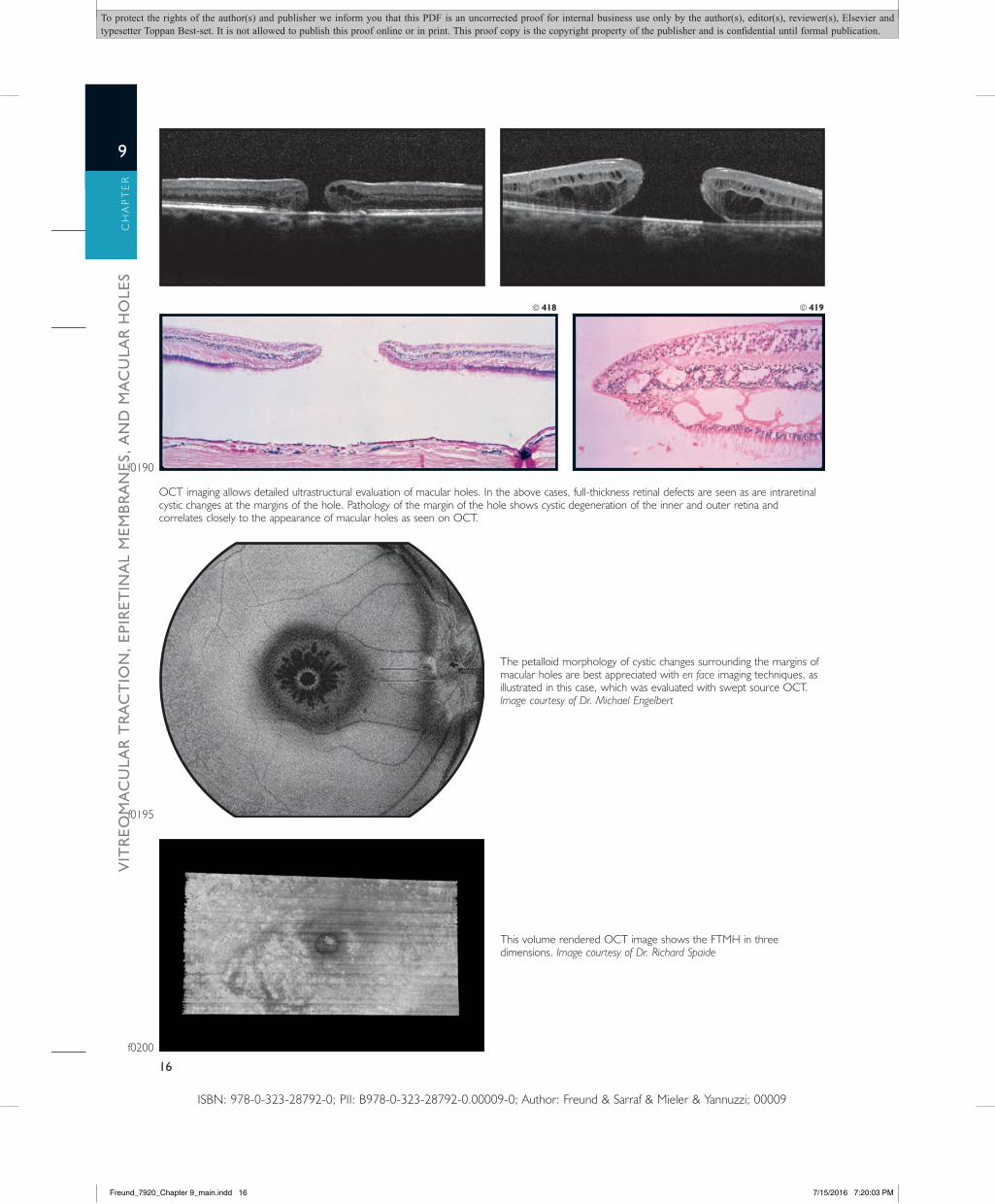
OCT imaging allows detailed ultrastructural evaluation of macular holes In the above cases, full-thickness retinal defects are seen as are intraretinal cystic changes at the margins of the hole Pathology of the margin of the hole shows cystic degeneration of the inner and outer retina and correlates closely to the appearance of macular holes as seen on OCT
418 419
The petalloid morphology of cystic changes surrounding the margins of macular holes are best appreciated with en face imaging techniques, as illustrated in this case, which was evaluated with swept source OCT Image courtesy of Dr. Michael Engelbert
This volume rendered OCT image shows the FTMH in three dimensions Image courtesy of Dr. Richard Spaide
ISBN: 978-0-323-28792-0; PII: B978-0-323-28792-0 00009-0; Author: Freund & Sarraf & Mieler & Yannuzzi; 00009
f0190
f0195
f0200
Freund_7920_Chapter 9_main.indd 16 7/15/2016 7:20:03 PM
To protect the rights of the author(s) and publisher we inform you that this PDF is an uncorrected proof for internal business use only by the author(s), editor(s), reviewer(s), Elsevier and typesetter Toppan Best-set. It is not allowed to publish this proof online or in print. This proof copy is the copyright property of the publisher and is confidential until formal publication.

FuLL
-TH
ICkN
ESS
MA
Cu
LAR
HO
LE
9
CH
AP
TE
R
17
Fluorescein angiography of FTMHs demonstrates hyperfluorescence due to loss of overlying retinal tissue Attenuation of the RPE at the site of the hole, as seen on the histological specimen (right image), is another reason for this hyperfluorescence
These patients with long-standing macular holes have a demarcation ring that is atrophic in nature This appears as a hyperfluorescent window defect on fluorescein angiography
spontaneous closure
The rate of spontaneous closure of primary FTMHs has been reported to range from 3% to 6% The exact mechanism of spontaneous macular hole closure is still unclear, but four differ-ent hypotheses have been proposed: (1) complete vitreous
detachment over the fovea releasing the tractional forces, (2) for-mation of an epiretinal membrane resulting in hole shrinkage, (3) glial cell proliferation at the base of the hole, and (4) growth of retinal tissue bridging the hole
This patient was noted to have VMT with adhesions of the posterior hyaloid to the roof of the macular hole (left image) One week later, there was spontaneous and complete posterior vitreous separation, with bridging of the retinal tissue at the level of the inner retina Often, there are outer retinal layer defects, which may either persist or recover with time (middle image) After 1 year, foveal architecture appears normal (right image)
This patient had a FTMH of approximately 200 µm at the narrowest diameter with an epiretinal membrane (left image) Five months later, there was spontaneous apposition of the outer layers of the retina with transformation of the hole into a lamellar configuration (middle image) At 6 months, the foveal architecture appears almost normal (right image) Images courtesy of Dr. Andrea Scopulo
ISBN: 978-0-323-28792-0; PII: B978-0-323-28792-0 00009-0; Author: Freund & Sarraf & Mieler & Yannuzzi; 00009
f0205
f0210
s0070
p0115
f0215
f0220
Freund_7920_Chapter 9_main.indd 17 7/15/2016 7:20:09 PM
To protect the rights of the author(s) and publisher we inform you that this PDF is an uncorrected proof for internal business use only by the author(s), editor(s), reviewer(s), Elsevier and typesetter Toppan Best-set. It is not allowed to publish this proof online or in print. This proof copy is the copyright property of the publisher and is confidential until formal publication.

ViT
rEo
Ma
cu
lar
Tr
ac
Tio
n,
Epir
ETin
al
MEM
br
an
Es,
an
d M
ac
ula
r H
olE
s
9
CH
AP
TE
R
18
Vitreolytic Treatment
Intravitreal ocriplasmin therapy has been shown to have a success rate between 10-40% for small stage 2 FTMHs (≤250 µm) Ocri-plasmin therapy is used by some surgeons as first-line therapy for
carefully selected cases of FTMH Eyes that fail vitreolytic therapy are managed surgically
This patient with a stage 2 FTMH was treated with intravitreal ocriplasmin (left image) One week later, there was separation of the posterior hyaloid and closure of the inner retina (middle image) Six months later, there were persistent outer retinal defects and ellipsoid zone disruptions (right image) Images courtesy of Dr. John Miller
surgical Treatment
Patients with FTMHs may be treated with pars plana vitrectomy, internal limiting membrane peeling, and gas tamponade Post-vit-rectomy, inner retinal dimpling is seen on the inner retinal surface in approximately a third of cases and typically appears 3 months
after surgery The exact mechanism is unknown, although it has been proposed that the dimples may represent defects in Müller cell regrowth
This patient with FTMH underwent pars plana vitrectomy and membrane peeling Four months after surgery, there is inner retinal dimpling and some disruption at the level of the ellipsoid zone
Three-dimensional volume rendered OCT imaging provides precise spatial visualization of inner retinal dimpling following macular hole surgery Image courtesy of Dr. Richard Spaide
ISBN: 978-0-323-28792-0; PII: B978-0-323-28792-0 00009-0; Author: Freund & Sarraf & Mieler & Yannuzzi; 00009
s0075
p0120
f0225
s0080
p0125
f0230
f0235
Freund_7920_Chapter 9_main.indd 18 7/15/2016 7:20:09 PM
To protect the rights of the author(s) and publisher we inform you that this PDF is an uncorrected proof for internal business use only by the author(s), editor(s), reviewer(s), Elsevier and typesetter Toppan Best-set. It is not allowed to publish this proof online or in print. This proof copy is the copyright property of the publisher and is confidential until formal publication.

FuLL
-TH
ICkN
ESS
MA
Cu
LAR
HO
LE
9
CH
AP
TE
R
19
secondary Macular Holes
The pathogenesis of secondary macular holes is different from primary macular holes Surgical hole closure rates are lower
in eyes with secondary macular holes due to the varied pathophysiology
This patient had a central retinal vein occlusion (left image) and cystoid macular edema (top right image) that was treated with intravitreal steroids and anti-vascular endothelial growth factor therapy A FTMH appeared after resolution of the central macular edema (bottom right image). Images courtesy of Dr. Jay Klancnik
This multicolor image highlights the perifoveal lesion that is characteristic of macular telangiectasia type 2 (left image). OCT imaging reveals characteristic atrophic and cavitary retinal defects at the fovea (top right image). With time, a FTMH developed as the area of cavitation and atrophy enlarged (bottom right image).
This patient with a macular hole overlying a serous pigment epithelial detachment was treated with multiple intravitreal anti-vascular endothelial growth factor injections and photodynamic therapy over a period of 1 year to induce flattening of the pigment epithelial detachment A pars plana vitrectomy with peeling of the internal limiting membrane was performed subsequently with successful closure of the macular hole
ISBN: 978-0-323-28792-0; PII: B978-0-323-28792-0 00009-0; Author: Freund & Sarraf & Mieler & Yannuzzi; 00009
s0085
p0130
f0240
f0245
f0250
Freund_7920_Chapter 9_main.indd 19 7/15/2016 7:20:13 PM
To protect the rights of the author(s) and publisher we inform you that this PDF is an uncorrected proof for internal business use only by the author(s), editor(s), reviewer(s), Elsevier and typesetter Toppan Best-set. It is not allowed to publish this proof online or in print. This proof copy is the copyright property of the publisher and is confidential until formal publication.

ViT
rEo
Ma
cu
lar
Tr
ac
Tio
n,
Epir
ETin
al
MEM
br
an
Es,
an
d M
ac
ula
r H
olE
s
9
CH
AP
TE
R
20
Macular holes may uncommonly develop during the natural course of acquired vitelliform lesions In this case, a macular hole developed during the process of spontaneous vitelliform reabsorption (arrow) Anatomical closure was achieved following vitrectomy and membrane peeling (bottom image). Images courtesy of Dr. Michael Engelbert
Months later, the patient in the above case developed a pigmented, hyperautofluorescent scar at the site of injury OCT imaging shows a FTMH at the site of RPE proliferation and scarring
Bilateral macular holes may be induced by lightning strike injury as seen in this case Images courtesy of Dr. Vikram Jain
This patient developed a FTMH following self-inflicted injury with a handheld laser pointer The curvilinear streaks of retinal pigment epithelial damage appear hyperfluorescent on fluorescein angiography Hyper-reflective vertical steaks in the outer retina, as seen on OCT, characterize the acute stages of laser-induced maculopathy (bottom right image).
Blunt trauma may also induce an acute FTMH Blunt injury in this case resulted in a large, subretinal hemorrhage and a FTMH that was clearly seen on OCT Images courtesy of Dr. Carmen Puliafito
ISBN: 978-0-323-28792-0; PII: B978-0-323-28792-0 00009-0; Author: Freund & Sarraf & Mieler & Yannuzzi; 00009
f0255
f0260
f0265
f0270
f0275
Freund_7920_Chapter 9_main.indd 20 7/15/2016 7:20:17 PM
To protect the rights of the author(s) and publisher we inform you that this PDF is an uncorrected proof for internal business use only by the author(s), editor(s), reviewer(s), Elsevier and typesetter Toppan Best-set. It is not allowed to publish this proof online or in print. This proof copy is the copyright property of the publisher and is confidential until formal publication.

LAM
ELLA
R M
AC
uLA
R H
OLE
9
CH
AP
TE
R
21
lamellar Macular HoleThe diagnosis of lamellar macular hole requires the following three criteria to be satisfied: (1) an irregular foveal contour or defect of the inner retina, (2) thinning at the base of the fovea, and (3) absence of a full-thickness defect Lamellar macular holes may develop following abrupt termination of pathophysiological pro-cesses that would otherwise have resulted in a FTMH Lamellar macular holes may also be due to contraction of an existing
perifoveal epiretinal membrane–internal limiting membrane com-plex Studies have shown that 80% to 100% of lamellar macular holes are associated with epiretinal membranes Although lamellar macular holes may progress to FTMHs, approximately 80% were found to be stable, both functionally and morphologically, over time As such, lamellar macular holes are usually observed and surgical management is considered only if there is visual decline
Spectral-domain OCT shows the variable appearance of lamellar macular holes In these patients, there is an associated epiretinal membrane, an irregularity in the foveal contour, defects in the inner retina, and thinning of the retina at the base of the fovea without a full-thickness defect There may also be intraretinal schisis due to traction (right image)
Macular pseudohole
Macular pseudohole is not a true FTMH and is due to contraction of an epiretinal membrane With spectral-domain OCT, a macular pseudohole has a very similar appearance to a lamellar macular hole, especially when the foveal tissue loss is subtle Some clinicians believe that lamellar macular hole and macular pseudohole are two distinct entities, best distinguished with fundus autofluores-cence by demonstrating foveal tissue loss in true lamellar macular
holes However, other clinicians believe that the two are very similar in that they both possess a perifoveal epiretinal membrane–internal limiting membrane complex, with lamellar macular holes demonstrating centrifugal contraction and macular pseudoholes demonstrating a centripetal contraction Management of macular pseudoholes is observation unless there is progressive visual decline that warrants surgery
The color fundus photograph shows a macular pseudohole due to an epiretinal membrane (left image) OCT demonstrates an epiretinal membrane with associated tenting of inner retinal tissue due to centripetal contraction (right image)
Fundus autofluorescence of a lamellar hole may reveal hyperautofluorescence due to loss of foveal tissue (left image) compared to the normal appearance of a macular pseudohole (right image)
ISBN: 978-0-323-28792-0; PII: B978-0-323-28792-0 00009-0; Author: Freund & Sarraf & Mieler & Yannuzzi; 00009
s0090
p0135
f0280
s0095
p0140
f0285
f0290
Freund_7920_Chapter 9_main.indd 21 7/15/2016 7:20:19 PM
To protect the rights of the author(s) and publisher we inform you that this PDF is an uncorrected proof for internal business use only by the author(s), editor(s), reviewer(s), Elsevier and typesetter Toppan Best-set. It is not allowed to publish this proof online or in print. This proof copy is the copyright property of the publisher and is confidential until formal publication.
ViT
rEo
Ma
cu
lar
Tr
ac
Tio
n,
Epir
ETin
al
MEM
br
an
Es,
an
d M
ac
ula
r H
olE
s
9
CH
AP
TE
R
22
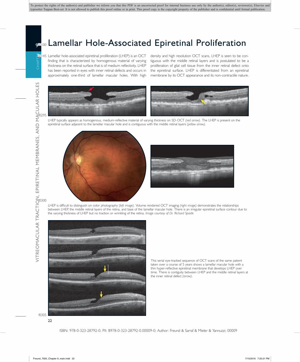
lamellar Hole-associated Epiretinal proliferationLamellar hole-associated epiretinal proliferation (LHEP) is an OCT finding that is characterized by homogenous material of varying thickness on the retinal surface that is of medium reflectivity LHEP has been reported in eyes with inner retinal defects and occurs in approximately one-third of lamellar macular holes With high
density and high resolution OCT scans, LHEP is seen to be con-tiguous with the middle retinal layers and is postulated to be a proliferation of glial cell tissue from the inner retinal defect onto the epiretinal surface LHEP is differentiated from an epiretinal membrane by its OCT appearance and its non-contractile nature
LHEP typically appears as homogenous, medium-reflective material of varying thickness on SD-OCT (red arrow). The LHEP is present on the epiretinal surface adjacent to the lamellar macular hole and is contiguous with the middle retinal layers (yellow arrow).
LHEP is difficult to distinguish on color photography (left image) Volume rendered OCT imaging (right image) demonstrates the relationships between LHEP, the middle retinal layers of the retina, and base of the lamellar macular hole There is an irregular epiretinal surface contour due to the varying thickness of LHEP but no traction or wrinkling of the retina Image courtesy of Dr. Richard Spaide
This serial eye-tracked sequence of OCT scans of the same patient taken over a course of 5 years shows a lamellar macular hole with a thin hyper-reflective epiretinal membrane that develops LHEP over time There is contiguity between LHEP and the middle retinal layers at the inner retinal defect (arrow)
ISBN: 978-0-323-28792-0; PII: B978-0-323-28792-0 00009-0; Author: Freund & Sarraf & Mieler & Yannuzzi; 00009
s0100
p0145
f0295
f0300
f0305
Freund_7920_Chapter 9_main.indd 22 7/15/2016 7:20:21 PM
To protect the rights of the author(s) and publisher we inform you that this PDF is an uncorrected proof for internal business use only by the author(s), editor(s), reviewer(s), Elsevier and typesetter Toppan Best-set. It is not allowed to publish this proof online or in print. This proof copy is the copyright property of the publisher and is confidential until formal publication.

SuG
GES
TED
REA
DIN
G
9
CH
AP
TE
R
23
suggested readingEpiretinal MembraneAppiah, A P , 1989 Secondary causes of premacular
fibrosis Ophthalmology 96, 389–392 koizumi, H , Spaide, R F , Fisher, Y L , et al , 2008
Three-dimensional evaluation of vitreomacular traction and epiretinal membrane using spectral-domain optical coherence tomography Am J Ophthalmol 145 (3), 509–517
Pang, C E , Spaide, R F , Freund, k B , 2014 Epiretinal proliferation seen in association with lamellar macular holes: a distinct clinical entity Retina 34 (8), 1513–1523
Pang, C E , Spaide, R F , Freund, k B , 2015 Comparing functional and morphologic characteristics of lamellar macular holes with and without lamellar hole-associated epiretinal proliferation Retina 35 (4), 720–726
Pesin, S R , Olk, R G , Grand, M G , et al , 1991 Vitrectomy for premacular fibroplasia Prognostic factors, long-term follow-up, and time course of visual improvement Ophthalmology 98, 1109–1114
Scheerlinck, L M , van der Valk, R , van Leeuwen, R , 2015 Predictive factors for postoperative visual acuity in idiopathic epiretinal membrane: a systematic review Acta Ophthalmol 93 (3), 203–212
Smiddy, W E , Maguire, A M , Green, W R , et al , 1989 Idiopathic epiretinal membranes: ultrastructural characteristics and clinicopathologic correlation Ophthalmology 96, 811–821
Tari, S R , Vidne-Hay, O , Greenstein, V C , et al , 2007 Functional and structural measurements for the assessment of internal limiting membrane peeling in idiopathic macular pucker Retina 27, 567–572
Vitreomacular Traction and Macular HoleBalaratnasingam, C , Dansingani, k , Dhrami-Gavazi,
E , et al , 2015 Documentation of spontaneous macular hole closure in macular telangiectasia type 2 using multimodal imaging Ophthalmic Surg Lasers Imaging Retina 46 (8), 883–886
Bhavsar, k V , Wilson, D , Margolis, R , et al , 2015 Multimodal imaging in handheld laser-induced maculopathy Am J Ophthalmol 159 (2), 227–231
Campo, R V , Lewis, R S , 1984 Lightning-induced macular hole Am J Ophthalmol 97, 792–794
Chang, L k , Fine, H F , Spaide, R F , et al , 2008 ultrastructural correlation of spectral-domain optical coherence tomographic findings in vitreomacular traction syndrome Am J Ophthalmol 146 (1), 121–127
Chew, E Y , Sperduto, R D , Hiller, R , et al , 1999 Clinical course of macular holes: the eye disease case-control study Arch Ophthalmol 117, 248–249
Duker, J S , kaiser, P k , Binder, S , et al , 2013 The International Vitreomacular Traction Study Group classification of vitreomacular adhesion, traction,
and macular hole Ophthalmology 120 (12), 2611–2619
Fisher, Y L , Slakter, J S , Yannuzzi, L A , et al , 1994 A prospective natural history study and kinetic ultrasound evaluation of idiopathic macular holes Ophthalmology 101, 5–11
Goldberg, N , Freund, k B , 2012 Progression of an acquired vitelliform lesion to a full-thickness macular hole documented by eye-tracked spectral-domain optical coherence tomography Arch Ophthalmol 130 (9), 1221–1223
Seider, M I , Lujan, B J , Gregori, G , et al , 2009 ultra-high resolution spectral domain optical coherence tomography of traumatic maculopathy Ophthalmic Surg Lasers Imaging 40 (5), 516–521
Stalmans, P , Benz, M S , Gandorfer, A , et al , MIVI-TRuST Study Group 2012 Enzymatic vitreolysis with ocriplasmin for vitreomacular traction and macular holes N Engl J Med 367 (7), 606–615
Steel, D H , Lotery, A J , 2013 Idiopathic vitreomacular traction and macular hole: a comprehensive review of pathophysiology, diagnosis, and treatment Eye (Lond) 27 (Suppl 1), S1–S21
Tzu, J H , John, V J , Flynn, H W Jr , et al , 2015 Clinical course of vitreomacular traction managed initially by observation Ophthalmic Surg Lasers Imaging Retina 46 (5), 571–576
ISBN: 978-0-323-28792-0; PII: B978-0-323-28792-0 00009-0; Author: Freund & Sarraf & Mieler & Yannuzzi; 00009
Freund_7920_Chapter 9_main.indd 23 7/15/2016 7:20:21 PM
To protect the rights of the author(s) and publisher we inform you that this PDF is an uncorrected proof for internal business use only by the author(s), editor(s), reviewer(s), Elsevier and typesetter Toppan Best-set. It is not allowed to publish this proof online or in print. This proof copy is the copyright property of the publisher and is confidential until formal publication.


