Chapter 6 Bone Tissue - Faribault Public Schools -...
23
Chapter 6 Bone Tissue
Transcript of Chapter 6 Bone Tissue - Faribault Public Schools -...

Chapter 6 Bone Tissue

A. Functions/Extracellular Matrix
1. Bone
Rigid
Support/protect/storage/lever/blood
Collagen/Ca & P crystals: hydroxyapatite = concrete
Inorganic matrix:
Hydroxyapatite
Scaffolding for
the skeleton
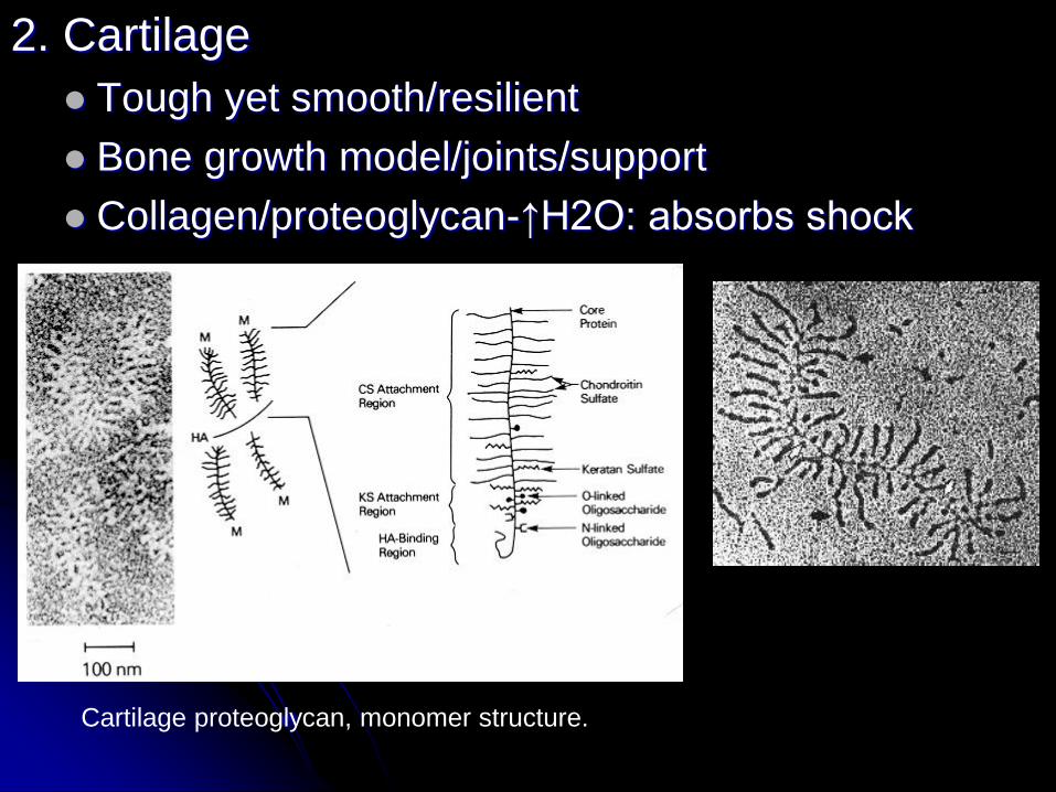
2. Cartilage
Tough yet smooth/resilient
Bone growth model/joints/support
Collagen/proteoglycan-↑H2O: absorbs shock
Cartilage proteoglycan, monomer structure.

3. Tendon (MuscleTBone); ligaments (BoneLBone)
Attachment sites
Collagen: tough/ropelike
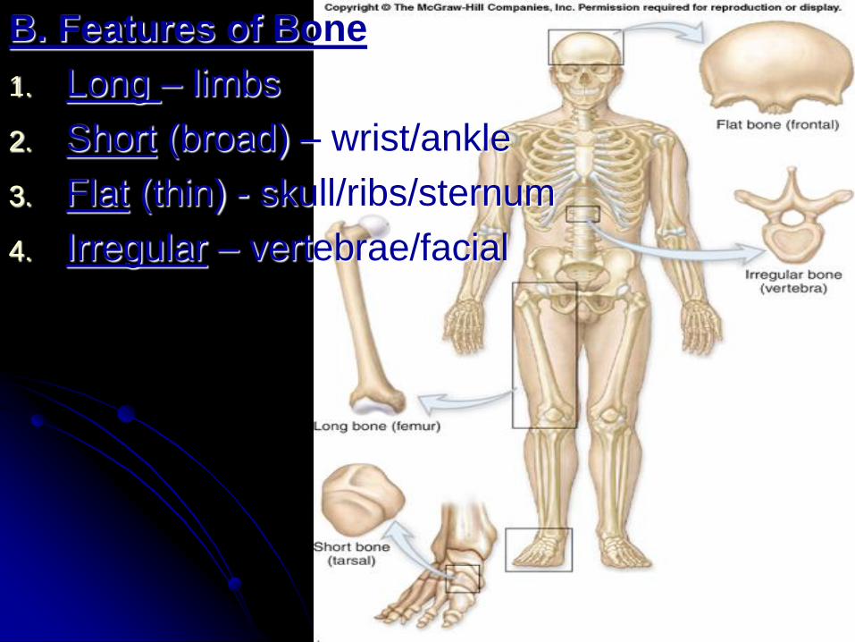
B. Features of Bone
1. Long – limbs
2. Short (broad) – wrist/ankle
3. Flat (thin) - skull/ribs/sternum
4. Irregular – vertebrae/facial
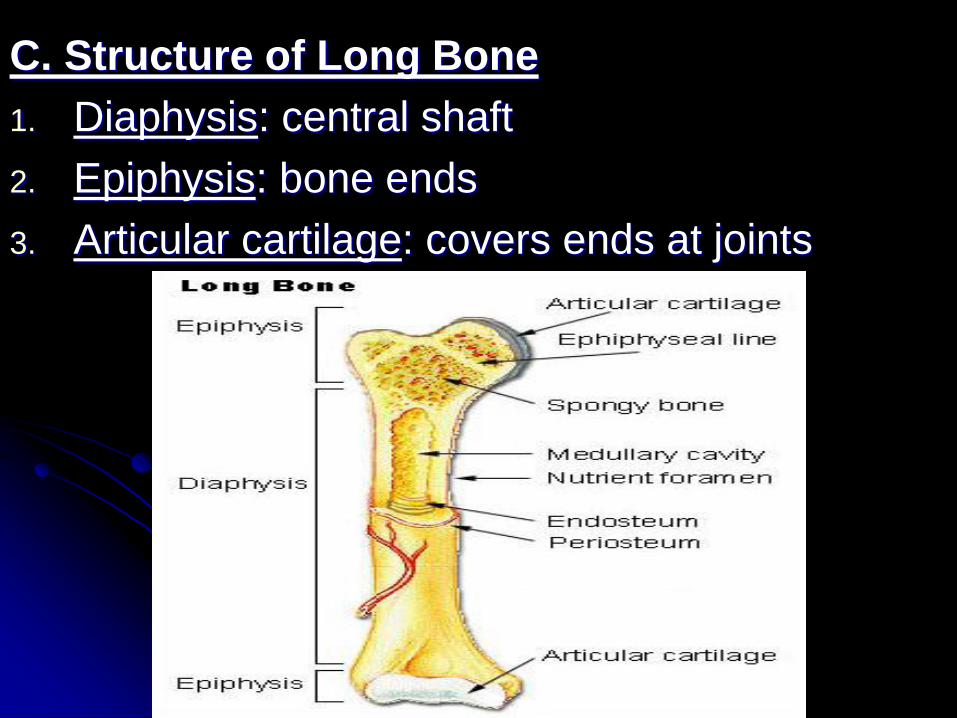
C. Structure of Long Bone
1. Diaphysis: central shaft
2. Epiphysis: bone ends
3. Articular cartilage: covers ends at joints

4. Epiphyseal plate
Growth plate
Bone model cartilage between end & shaft
Epiphyseal line when bone growth stops
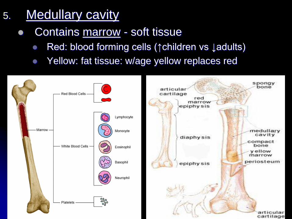
5. Medullary cavity
Contains marrow - soft tissue
Red: blood forming cells (↑children vs ↓adults)
Yellow: fat tissue: w/age yellow replaces red

6. Periosteum (outer)/Endosteum (thin inner):
CT w/bld vessels, nerve cells & osteoblasts-bone-
forming cells (growth, repair)

D. Two Types

1. Compact Bone: Long bone diaphysis & surfaces (all)

• Haversian Systems: (Osteons)
A. Lamellae: extracellular matrix
B. Osteocytes: bone cells
C. Lacunae: spaces containing bone cells
D. Canaliculi: canals connecting osteocytes
E. Central canal contains BVs
lamella

2. Cancellous Bone (spongy):
Long bone epiphysis & centers (all)
Trabeculae: plates of bone w/spaces

E. Bone Ossification (formation)
1. Cell types
a. Osteoblast: bone forming cell
b. Osteocyte: mature bone cell
c. Osteoclast: bone eating (remodeling cell)

Intramembranous (membrane bones) Endochondral (cartilage bones)
no cartilage starts with cartilage
numerous small blood vessels avascular
many flat bones (skull) long bones and flat bones
Two Methods of Ossification in Fetal Bone Formation

2. Intramembranous Ossification (upper fetal skull):
Osteoblasts in CT form spongy, then compact bone

3. Endochondral Ossification (fetal skull base & rest):
a. Fetal cartilage model
b. Primary ossification/
diaphysis
c. Secondary ossification/
epiphysis
d. Bone matures
(epiphysial line forms)
The hyaline cartilage model

F. Bone Growth:
Periosteum ↑diameter; Epiphyseal plate
↑length

G. Remodel: (osteoclast/osteoblast)
Hormones, diet

H. Repair:
1. Clot
2. Callus network (repair area)
3. Osteoblasts form cancellous first
(cast needed for support)
4. Remodel w/both

Incomplete Multiple Right Open/bone
fragments Angle protrudes (Review p. 120
for Chp test)

I. Calcium Homeostasis
• Osteoblast (Ca to bone) = osteoclast (to Bld)
• Parathyroid gland regulates ↓ Bld Ca levels
• Parathyroid Hormone:
• ↑ Osteoclast activity (Ca from bone to bld)
• Kidney reabsorb Ca from urine
• Kidney = ↑ Vit. D = ↑ Ca from SI
• Thyroid gland regulates ↑ Bld Ca levels
• Calcitonin Hormone: ↓ Osteoclast activity

J. Effects of Aging:
Osteoblast less active than osteoclast
Bone loss after age 35 (esp. post-menopause) =
osteoporosis: bone reabsorption ↑ than formation
Production of articular cartilage/synovial fluid slows
Prevent w/physical activity, Ca & Vit. D supplements
Normal
cancellous bone
Osteoporotic
cancellous bone