Chapter 20 Reproduction. Copyright © The McGraw-Hill Companies, Inc. Permission required for...
74
Chapter 20 Reproducti on
-
Upload
adela-barton -
Category
Documents
-
view
252 -
download
1
Transcript of Chapter 20 Reproduction. Copyright © The McGraw-Hill Companies, Inc. Permission required for...
Chapter 20
Reproduction
Copyright © The McGraw-Hill Companies, Inc. Permission required for reproduction or display.
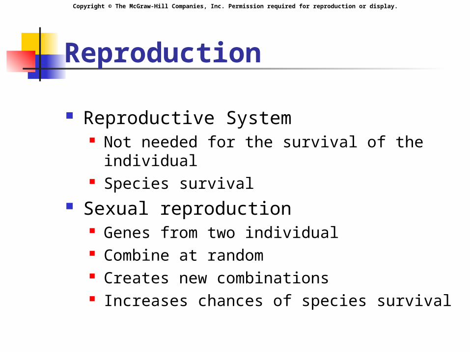
Reproduction
Reproductive System Not needed for the survival of the individual Species survival
Sexual reproduction Genes from two individual Combine at random Creates new combinations Increases chances of species survival
Copyright © The McGraw-Hill Companies, Inc. Permission required for reproduction or display.
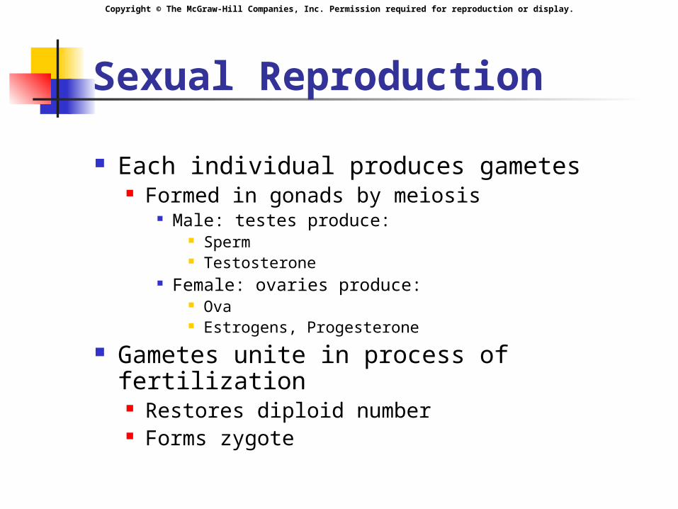
Sexual Reproduction
Each individual produces gametes Formed in gonads by meiosis
Male: testes produce: Sperm Testosterone
Female: ovaries produce: Ova Estrogens, Progesterone
Gametes unite in process of fertilization Restores diploid number Forms zygote

Copyright © The McGraw-Hill Companies, Inc. Permission required for reproduction or display.
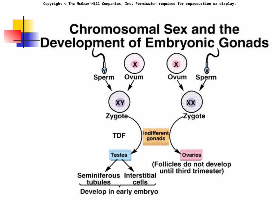
Sexual Determination Each zygote inherits
23 chromosomes from mother 23 chromosomes from father. 23 pairs of homologous chromosomes.
alleles Kinds of chromosomes
1-22 pairs of chromosomes: autosomal 23rd pair are sex chromosomes.
Male: XY Female: XX
Chromosomal gender of zygote determined by fertilizing sperm.
Copyright © The McGraw-Hill Companies, Inc. Permission required for reproduction or display.
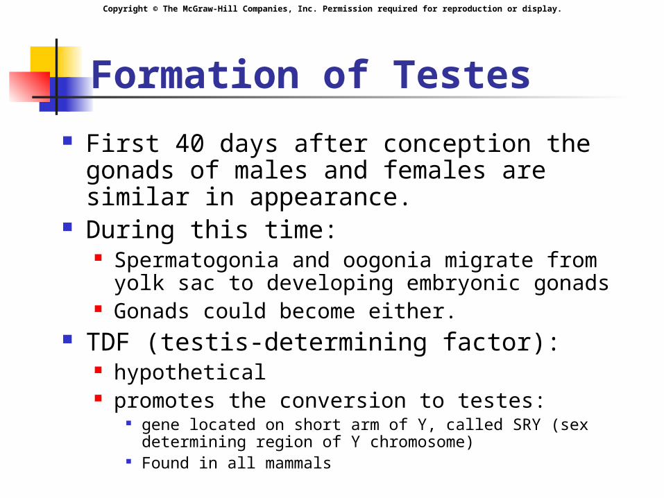
Formation of Testes
First 40 days after conception the gonads of males and females are similar in appearance.
During this time: Spermatogonia and oogonia migrate from yolk
sac to developing embryonic gonads Gonads could become either.
TDF (testis-determining factor): hypothetical promotes the conversion to testes:
gene located on short arm of Y, called SRY (sex determining region of Y chromosome)
Found in all mammals
Copyright © The McGraw-Hill Companies, Inc. Permission required for reproduction or display.
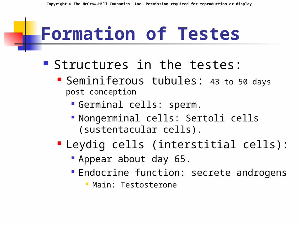
Formation of Testes
Structures in the testes: Seminiferous tubules: 43 to 50 days post
conception Germinal cells: sperm. Nongerminal cells: Sertoli cells
(sustentacular cells). Leydig cells (interstitial cells):
Appear about day 65. Endocrine function: secrete androgens
Main: Testosterone

Copyright © The McGraw-Hill Companies, Inc. Permission required for reproduction or display.
Formation of Testes
Leydig cells secrete testosterone. Begins 8th week and peaks at 12-14th week. Masculinizes embryonic structures.
Testosterone then declines to very low levels until puberty. Decline occurs by end of second trimester
Testes descend into scrotum shortly before birth. Temp about 3 degrees below internal temp 35 degrees C
Copyright © The McGraw-Hill Companies, Inc. Permission required for reproduction or display.
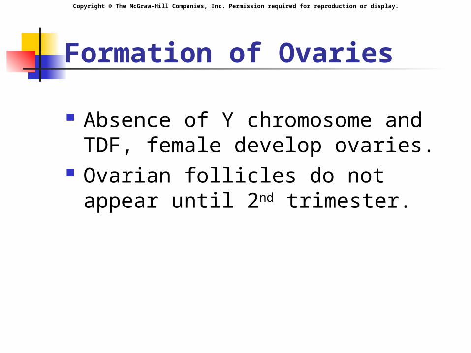
Formation of Ovaries
Absence of Y chromosome and TDF, female develop ovaries.
Ovarian follicles do not appear until 2nd trimester.
Copyright © The McGraw-Hill Companies, Inc. Permission required for reproduction or display.
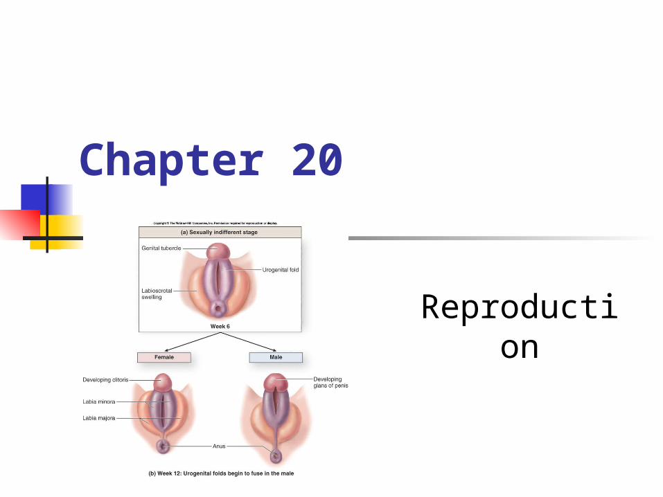
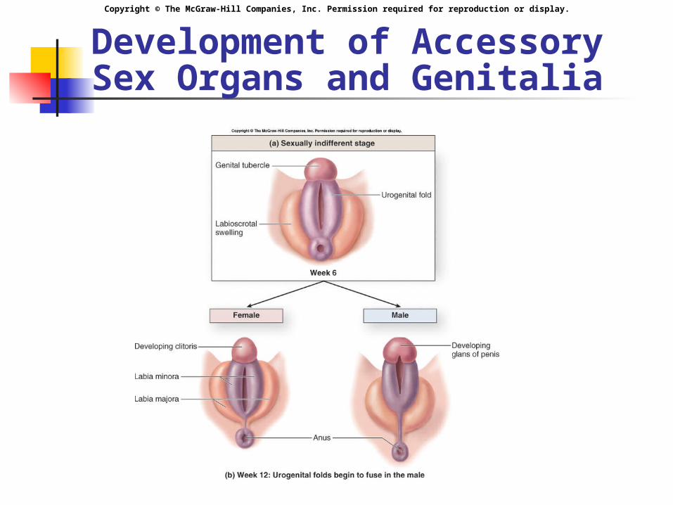
Copyright © The McGraw-Hill Companies, Inc. Permission required for reproduction or display.Development of Accessory Sex Organs and Genitalia
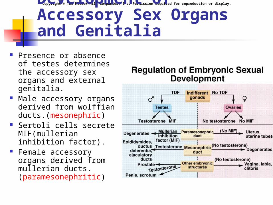
Presence or absence of testes determines the accessory sex organs and external genitalia.
Male accessory organs derived from wolffian ducts.(mesonephric)
Sertoli cells secrete MIF(mullerian inhibition factor).
Female accessory organs derived from mullerian ducts. (paramesonephritic)
Copyright © The McGraw-Hill Companies, Inc. Permission required for reproduction or display.

Copyright © The McGraw-Hill Companies, Inc. Permission required for reproduction or display.
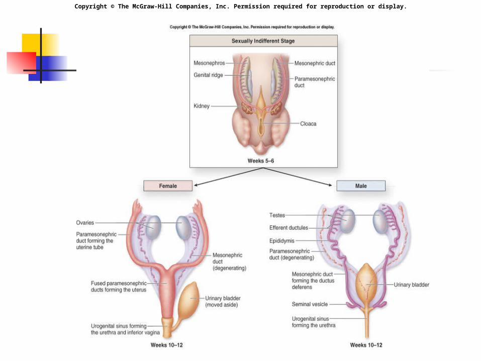
Development of Accessory Sex Organs and Genitalia Both duct systems in both sexes between
days 25 and 50 Regression of mullarian ducts begins about
day 60 Testosterone
responsible for development of male accessory sex organs
External genitalia identical first 6 weeks, then testosterone stimulates development of penis
Not the active agent in all cells converted to dihydrotestosterone (DHT) in some
target cells Needed for penis, spongy urethra, scrotum,
prostrate Testosterone directly needed for wolfian
derivatives: Epididymis, ductus deferens, ejaculatory duct, SV
Copyright © The McGraw-Hill Companies, Inc. Permission required for reproduction or display.
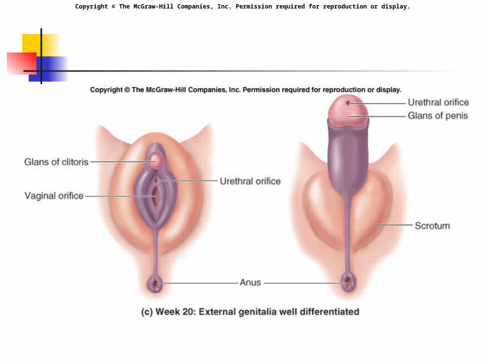
Development of Accessory Sex Organs and Genitalia
Copyright © The McGraw-Hill Companies, Inc. Permission required for reproduction or display.

Copyright © The McGraw-Hill Companies, Inc. Permission required for reproduction or display.
Endocrine Regulation of Reproduction First trimester
Embryonic testes are active endocrine glands Secrete large amounts of testosterone
Embryonic ovaries not mature until third trimester
Time of birth: Gonads in both sexes relatively inactive
Before puberty: Low levels of sex steroids in both Due to lack of stimulation
Puberty: Increased stimulation from gonadotropic
hormones Induce increase in sex steroids

Copyright © The McGraw-Hill Companies, Inc. Permission required for reproduction or display.
Endocrine Regulation of Reproduction
Hypothalamus releases LHRH (GnRH) into hypothalamo-hypophyseal portal vessels.
Anterior pituitary secretes: LH: luteinizing hormone.
In male: interstitial-cell stimulating hormone (ICSH) FSH: follicle-stimulating hormone.
Secreted in pulsatile fashion to prevent desensitization and down regulation of receptors.
Copyright © The McGraw-Hill Companies, Inc. Permission required for reproduction or display.
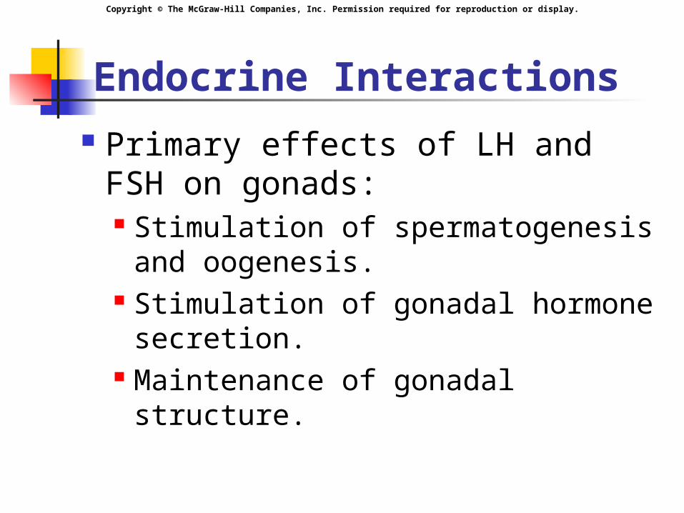
Endocrine Interactions Primary effects of LH and FSH
on gonads: Stimulation of spermatogenesis
and oogenesis. Stimulation of gonadal hormone
secretion. Maintenance of gonadal
structure.

Copyright © The McGraw-Hill Companies, Inc. Permission required for reproduction or display.
Endocrine Regulation
Negative Feedback: Inhibit GnRH from
hypothalamus. Inhibit anterior
pituitary response to GnRH.
Inhibin secretion inhibit anterior pituitary release of FSH.
By sertoli cells
Female: estrogen and progesterone.
Male: testosterone.
Copyright © The McGraw-Hill Companies, Inc. Permission required for reproduction or display.

Copyright © The McGraw-Hill Companies, Inc. Permission required for reproduction or display.
Onset of Puberty
FSH and LH high in newborn, falls to low levels in few weeks.
Puberty: driven by increased secretion of FSH and LH

Copyright © The McGraw-Hill Companies, Inc. Permission required for reproduction or display.
Onset of Puberty
FSH and LH Brain maturation increases GnRH secretion. Decreased sensitivity of GnRH to negative
feedback. LH:
Increased secretion triggers puberty Late puberty, pulsatile secretion of LH and
FSH increase during sleep. Stimulate a rise in sex steroid secretion.
Copyright © The McGraw-Hill Companies, Inc. Permission required for reproduction or display.
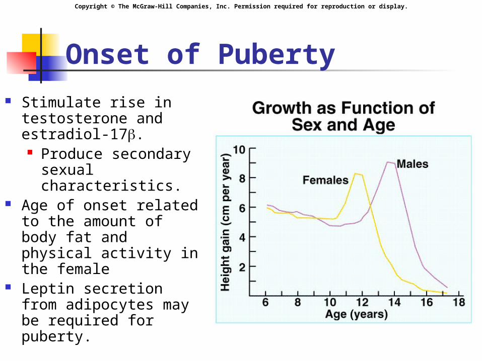
Onset of Puberty Stimulate rise in
testosterone and estradiol-17. Produce secondary
sexual characteristics.
Age of onset related to the amount of body fat and physical activity in the female
Leptin secretion from adipocytes may be required for puberty.

Copyright © The McGraw-Hill Companies, Inc. Permission required for reproduction or display.
Pineal Gland
Secretes melatonin. Secretion influenced by light-dark
cycles. Inhibit gonadotropin secretion. Role in humans not established.
Copyright © The McGraw-Hill Companies, Inc. Permission required for reproduction or display.
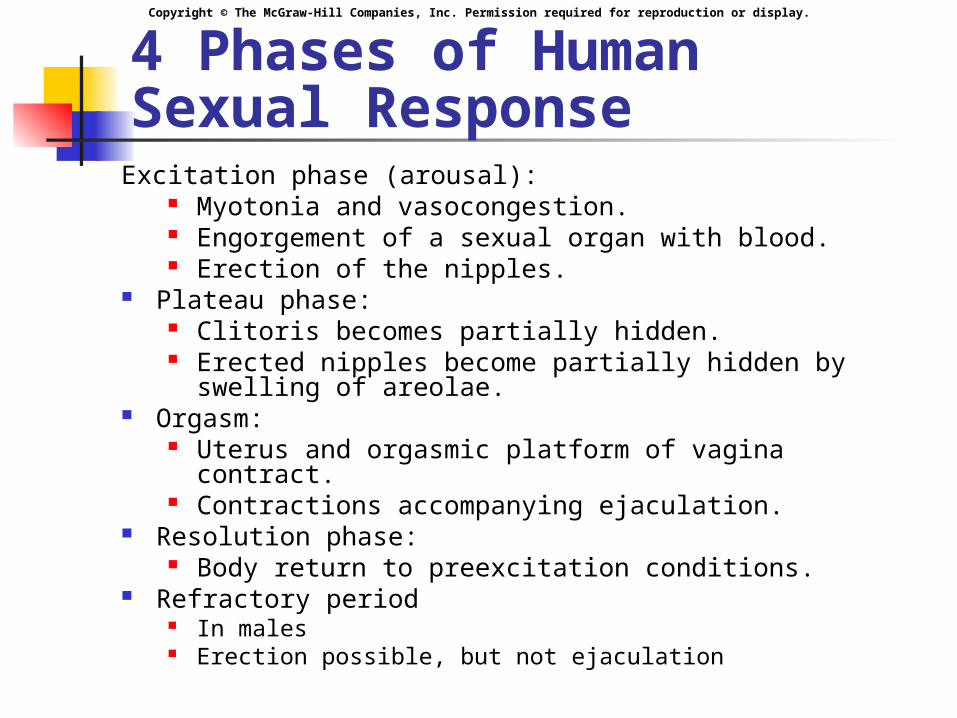
4 Phases of Human Sexual Response
Excitation phase (arousal): Myotonia and vasocongestion. Engorgement of a sexual organ with blood. Erection of the nipples.
Plateau phase: Clitoris becomes partially hidden. Erected nipples become partially hidden by swelling
of areolae. Orgasm:
Uterus and orgasmic platform of vagina contract. Contractions accompanying ejaculation.
Resolution phase: Body return to preexcitation conditions.
Refractory period In males Erection possible, but not ejaculation
Copyright © The McGraw-Hill Companies, Inc. Permission required for reproduction or display.
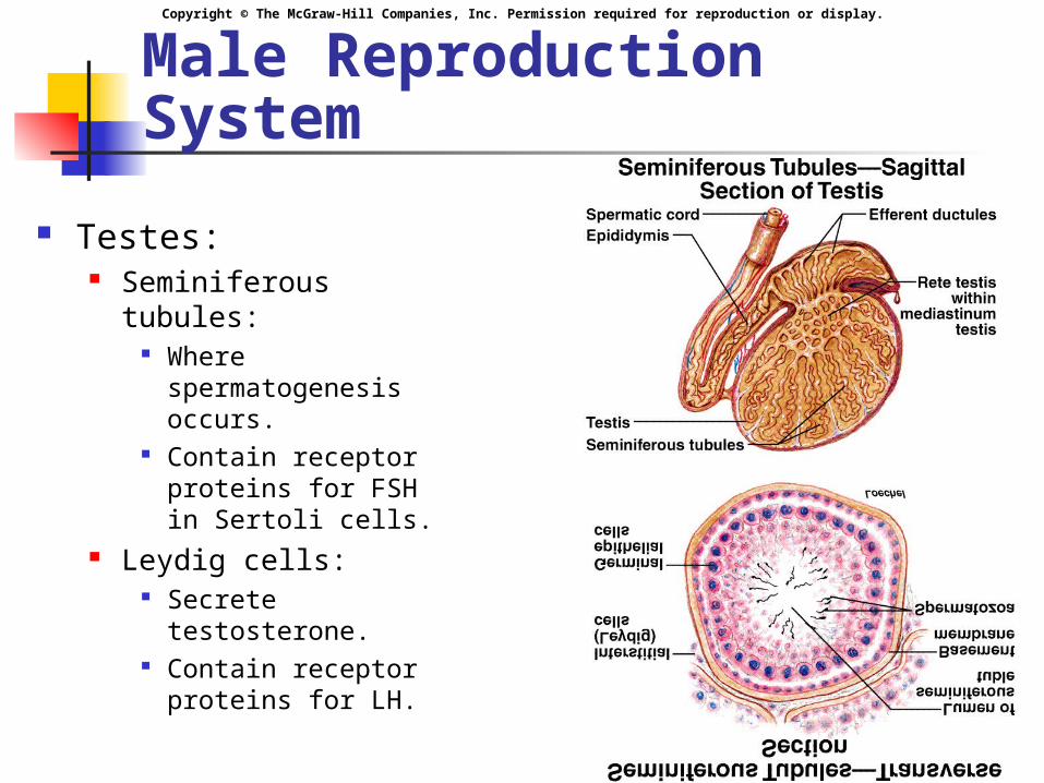
Male Reproduction System
Testes: Seminiferous tubules:
Where spermatogenesis occurs.
Contain receptor proteins for FSH in Sertoli cells.
Leydig cells: Secrete testosterone. Contain receptor
proteins for LH.

Copyright © The McGraw-Hill Companies, Inc. Permission required for reproduction or display.
Control of LH and FSH Secretion
Negative feedback: Testosterone
inhibits LH and GnRH production.
Inhibin inhibits FSH secretion.
Aromatization reaction producing estadiol in the brain is required for the negative feedback effects of testosterone on LH.
Brain is a target organ for testosterone
Converted to derivatives
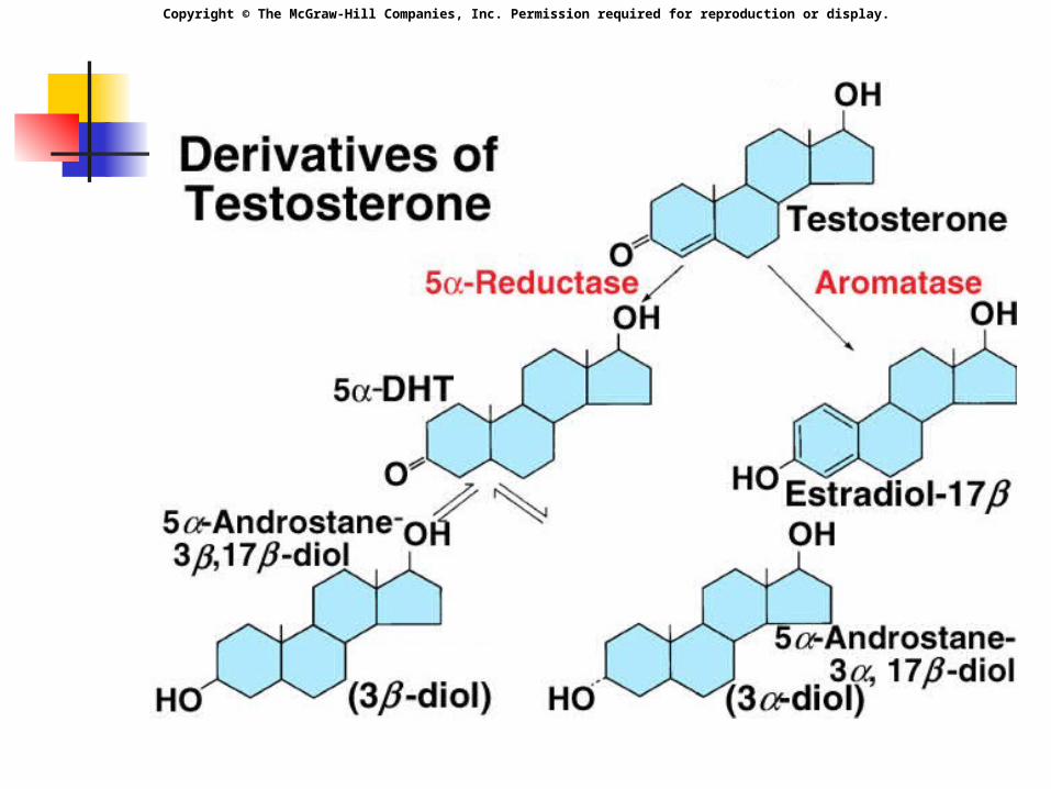
Copyright © The McGraw-Hill Companies, Inc. Permission required for reproduction or display.

Copyright © The McGraw-Hill Companies, Inc. Permission required for reproduction or display.
Testosterone Secretion Responsible for
initiation and maintenance of body changes in puberty.
Stimulate growth of muscles, larynx, and bone growth until sealing of the epiphyseal discs.
Promote hemoglobin synthesis.
Acts in paracrine fashion and is responsible for spermatogenesis.
Copyright © The McGraw-Hill Companies, Inc. Permission required for reproduction or display.
Testosterone Secretion
Negative feedback of testosterone and inhibin Keep relatively constant levels of
gonadotropins Results in relatively constant levels Different in female
At menopause: no more sex steroids In males, gradual decrease
Copyright © The McGraw-Hill Companies, Inc. Permission required for reproduction or display.
Endocrine function: testes
Testosterone: main androgen Sertoli and Leydig cells secrete small
amounts of estradiol. Have receptors for estradiol (as do other
male structures) May be needed for spermatogenesis
Estradiol may be responsible for: Negative feedback in brain. Sealing of epiphyseal plates. Regulatory function in fertility.

Copyright © The McGraw-Hill Companies, Inc. Permission required for reproduction or display.
Spermatogenesis
Spermatogonia: Replicate initially by mitosis. Produce two cells
One becomes a primary spermatocytes undergoes meiosis: 2 nuclear divisions. 2nd meiotic division produce 4
spermatids.
Copyright © The McGraw-Hill Companies, Inc. Permission required for reproduction or display.
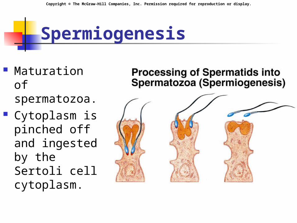
Spermiogenesis
Maturation of spermatozoa.
Cytoplasm is pinched off and ingested by the Sertoli cell cytoplasm.

Copyright © The McGraw-Hill Companies, Inc. Permission required for reproduction or display.
Sertoli Cells Blood-testes barrier:
Prevents autoimmune destruction of sperm. Produce FAS ligand which binds to the FAS
receptor on surface to T lymphocytes, triggering apoptosis.
Secretes inhibin. Phagocytize residual bodies:
Transmit information molecules from germ cells to Sertoli cells.
Secrete ABP (androgen-binding protein): Binds to testosterone and concentrates
testosterone in the tubules.
Copyright © The McGraw-Hill Companies, Inc. Permission required for reproduction or display.
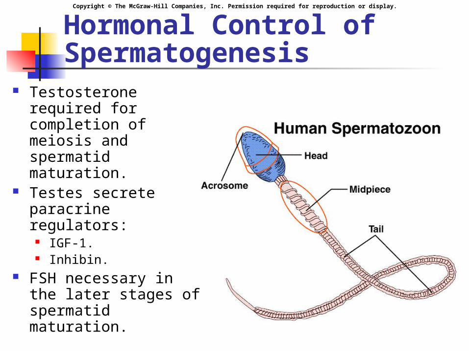
Hormonal Control of Spermatogenesis
Testosterone required for completion of meiosis and spermatid maturation.
Testes secrete paracrine regulators:
IGF-1. Inhibin.
FSH necessary in the later stages of spermatid maturation.
Copyright © The McGraw-Hill Companies, Inc. Permission required for reproduction or display.
Male Accessory Organs Epididymis:
Maturational changes. Resistance to pH changes and temperature. Storage.
Prostate secretes: Alkaline fluid. Citric acid. Ca++. Coagulation proteins.
Seminal vesicles secrete: Fructose.
Copyright © The McGraw-Hill Companies, Inc. Permission required for reproduction or display.
Erection, Emission, and Ejaculation
Erection: Increased vasodilation of arterioles. NO is the NT. Blood flow into the erectile tissues of the penis. Parasympathetic
Emission: Movement of semen into the urethra. Sympathetic
Ejaculation: Forcible expulsion of semen from the urethra
out of the penis. Sympathetic

Copyright © The McGraw-Hill Companies, Inc. Permission required for reproduction or display.
Female Reproductive System
Ovaries: Contain large number of follicles
which enclose ova. Extensions called fimbriae
partially cover each ovary. At ovulation, secondary oocyte is
extruded.
Copyright © The McGraw-Hill Companies, Inc. Permission required for reproduction or display.
Female Reproductive System
Fallopian (uterine) tubes: Ova drawn into the tube by cilia.
Uterus: Womb. Endometrium shed during
menstruation. Vagina:
Cervical mucus plug.
Copyright © The McGraw-Hill Companies, Inc. Permission required for reproduction or display.
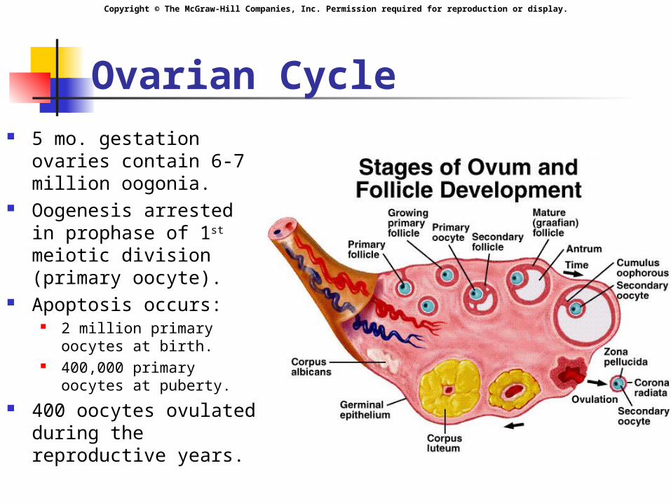
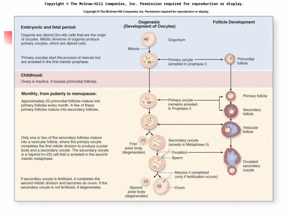
Ovarian Cycle 5 mo. gestation
ovaries contain 6-7 million oogonia.
Oogenesis arrested in prophase of 1st meiotic division (primary oocyte).
Apoptosis occurs: 2 million primary oocytes
at birth. 400,000 primary oocytes
at puberty. 400 oocytes ovulated
during the reproductive years.
Copyright © The McGraw-Hill Companies, Inc. Permission required for reproduction or display.
Ovarian Cycle
Primary oocytes contained in primary follicles. FSH stimulates cell growth.
Develop into secondary follicles. Fusion of its vesicles into the antrum. Mature graafian follicle: 1st meiotic division completed
(secondary oocyte).
Copyright © The McGraw-Hill Companies, Inc. Permission required for reproduction or display.
Copyright © The McGraw-Hill Companies, Inc. Permission required for reproduction or display.
Ovarian Cycle
Secondary oocyte: Under FSH stimulation:
Theca cells secrete testosterone. Granulosa cells: contain the enzyme aromatase to convert testosterone into estrogen.
Copyright © The McGraw-Hill Companies, Inc. Permission required for reproduction or display.
Ovulation Graafian follicle forms bulge on
surface of ovary. Extrudes secondary oocyte into
the uterine tube. Empty follicle becomes corpus
luteum and secretes: Progesterone. Estrogen. If not fertilized becomes corpus
albicans.

Copyright © The McGraw-Hill Companies, Inc. Permission required for reproduction or display.
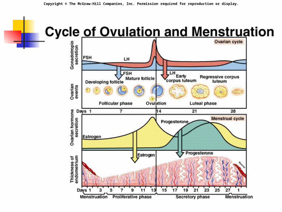
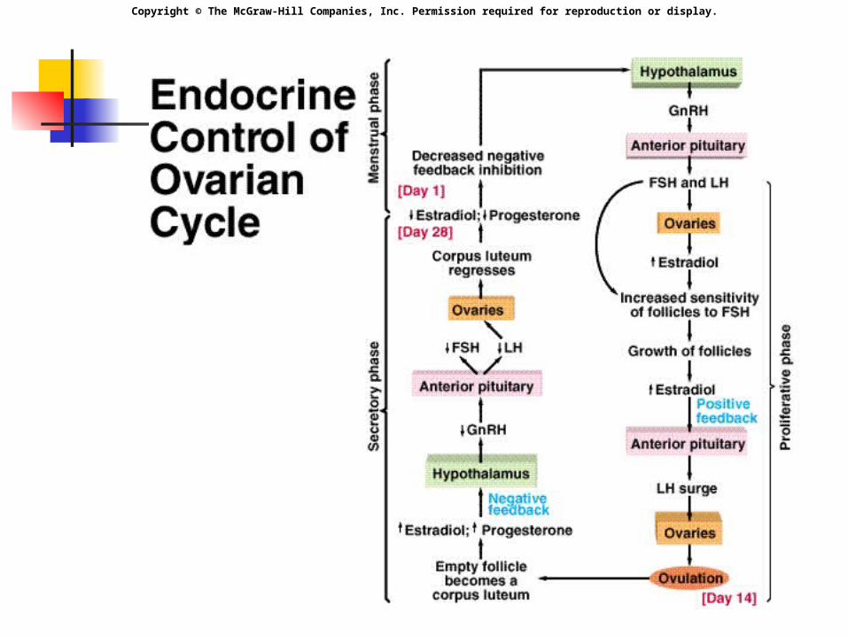
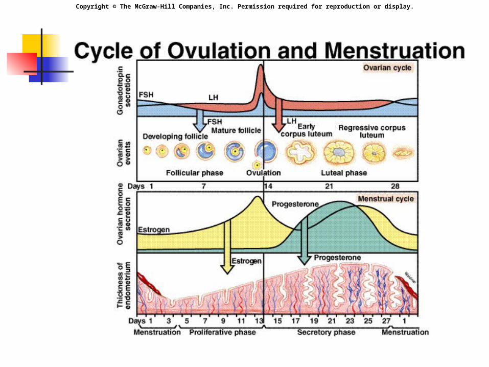
Menstrual Cycle
3 phases: Ovarian Follicular Phase Ovulation Luteal Phase
Duration approximately 28 days. Day 1 is the first day of
menstruation.
Copyright © The McGraw-Hill Companies, Inc. Permission required for reproduction or display.
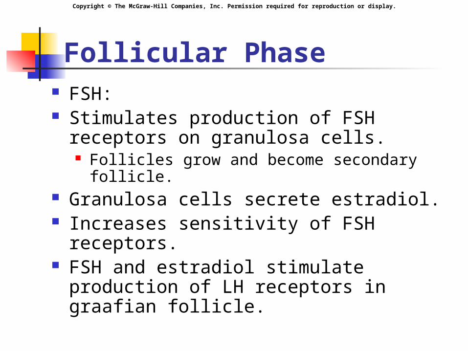
Follicular Phase FSH: Stimulates production of FSH receptors
on granulosa cells. Follicles grow and become secondary follicle.
Granulosa cells secrete estradiol. Increases sensitivity of FSH receptors. FSH and estradiol stimulate production
of LH receptors in graafian follicle.
Copyright © The McGraw-Hill Companies, Inc. Permission required for reproduction or display.
Follicular Phase
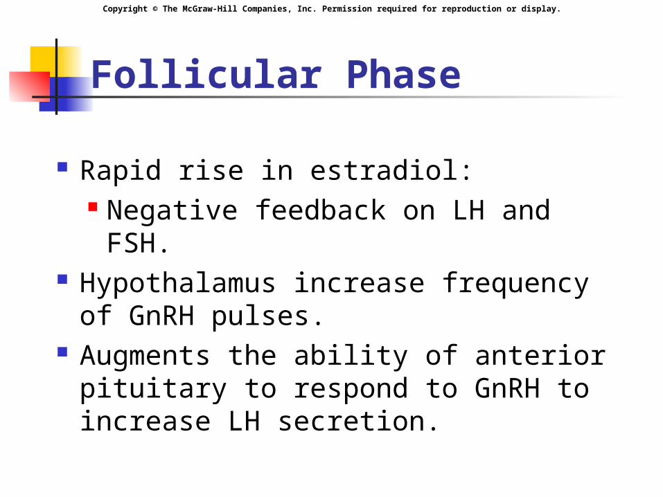
Rapid rise in estradiol: Negative feedback on LH and
FSH. Hypothalamus increase frequency
of GnRH pulses. Augments the ability of anterior
pituitary to respond to GnRH to increase LH secretion.
Copyright © The McGraw-Hill Companies, Inc. Permission required for reproduction or display.
Follicular Phase
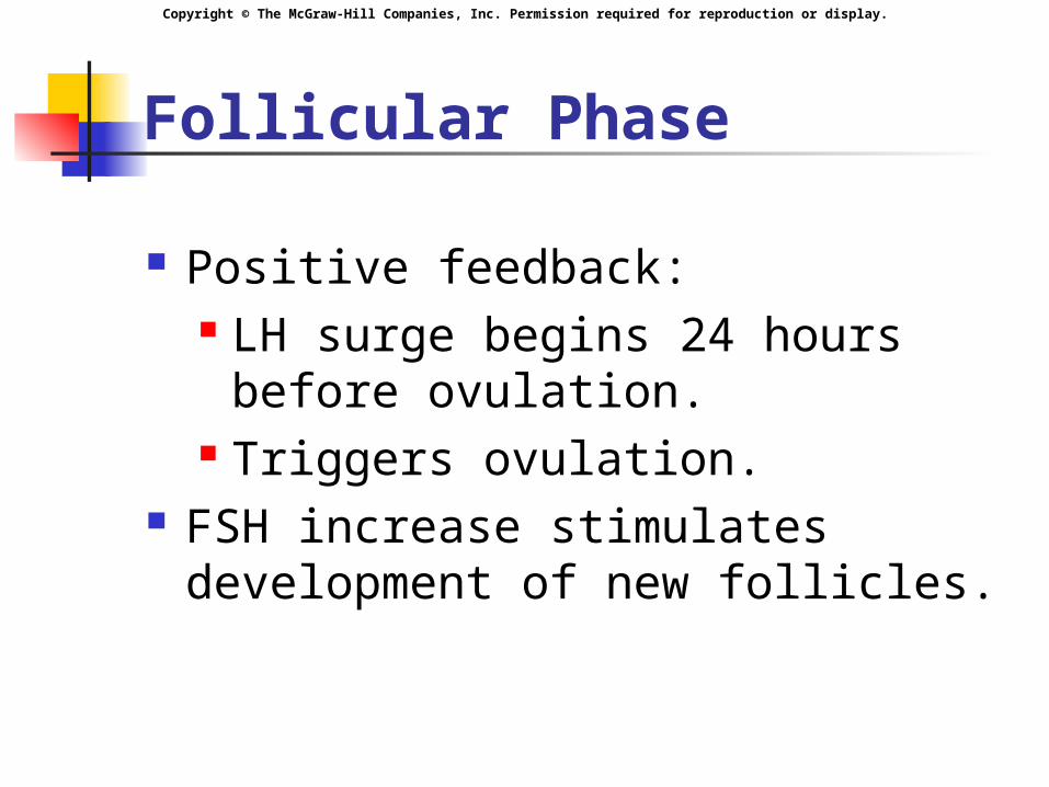
Positive feedback: LH surge begins 24 hours before
ovulation. Triggers ovulation.
FSH increase stimulates development of new follicles.
Copyright © The McGraw-Hill Companies, Inc. Permission required for reproduction or display.
Ovulation
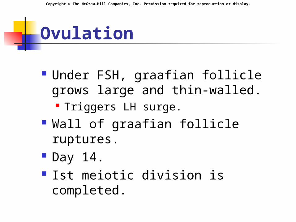
Under FSH, graafian follicle grows large and thin-walled. Triggers LH surge.
Wall of graafian follicle ruptures. Day 14. Ist meiotic division is completed.

Copyright © The McGraw-Hill Companies, Inc. Permission required for reproduction or display.
Luteal Phase
LH stimulates formation of the empty follicle into corpus luteum.
Corpus luteum secretes: Progesterone: Plasma concentration rapidly rises. Estradiol.
Negative feedback on LH and FSH. Inhibin: suppress FSH.

Copyright © The McGraw-Hill Companies, Inc. Permission required for reproduction or display.
Luteal Phase
Corpus luteum regresses unless fertilization occurs: Estradiol decreases. Progesterone decreases.
Withdrawal of estradiol and progesterone cause menstruation to occur.
Copyright © The McGraw-Hill Companies, Inc. Permission required for reproduction or display.
Copyright © The McGraw-Hill Companies, Inc. Permission required for reproduction or display.
Copyright © The McGraw-Hill Companies, Inc. Permission required for reproduction or display.
Endometrial Changes
3 phases of endometrium changes: Proliferative phase. Secretory phase. Menstrual phase.
Copyright © The McGraw-Hill Companies, Inc. Permission required for reproduction or display.
Proliferative Phase
Ovary is in follicular phase. Estradiol stimulate growth of
endometrium. Spiral arteries develop. Stimulate production of receptor
proteins for progesterone. Cornification of vaginal epithelium
occurs.
Copyright © The McGraw-Hill Companies, Inc. Permission required for reproduction or display.
Secretory Phase
Ovary is in luteal phase. Progesterone stimulates
development of uterine glands, which become engorged with glycogen.
Endometrium becomes thick, vascular and spongy.
Cervical mucus thickens and becomes sticky.

Copyright © The McGraw-Hill Companies, Inc. Permission required for reproduction or display.
Menstrual Phase
Progesterone cause constriction of spiral arteries.
Necrosis and sloughing of endometrium occurs.
Lasts 1-5 days.
Copyright © The McGraw-Hill Companies, Inc. Permission required for reproduction or display.
Copyright © The McGraw-Hill Companies, Inc. Permission required for reproduction or display.
Menopause
Cessation of ovarian activity. Age ~ 50 years. Ovaries depleted of follicles. Estradiol and inhibin withdrawl
causes hot flashes, and atrophy of the vaginal wall.
LH and FSH increase.
Copyright © The McGraw-Hill Companies, Inc. Permission required for reproduction or display.
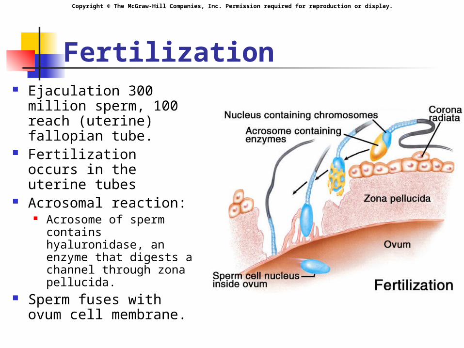
Fertilization Ejaculation 300
million sperm, 100 reach (uterine) fallopian tube.
Fertilization occurs in the uterine tubes
Acrosomal reaction: Acrosome of sperm
contains hyaluronidase, an enzyme that digests a channel through zona pellucida.
Sperm fuses with ovum cell membrane.
Copyright © The McGraw-Hill Companies, Inc. Permission required for reproduction or display.
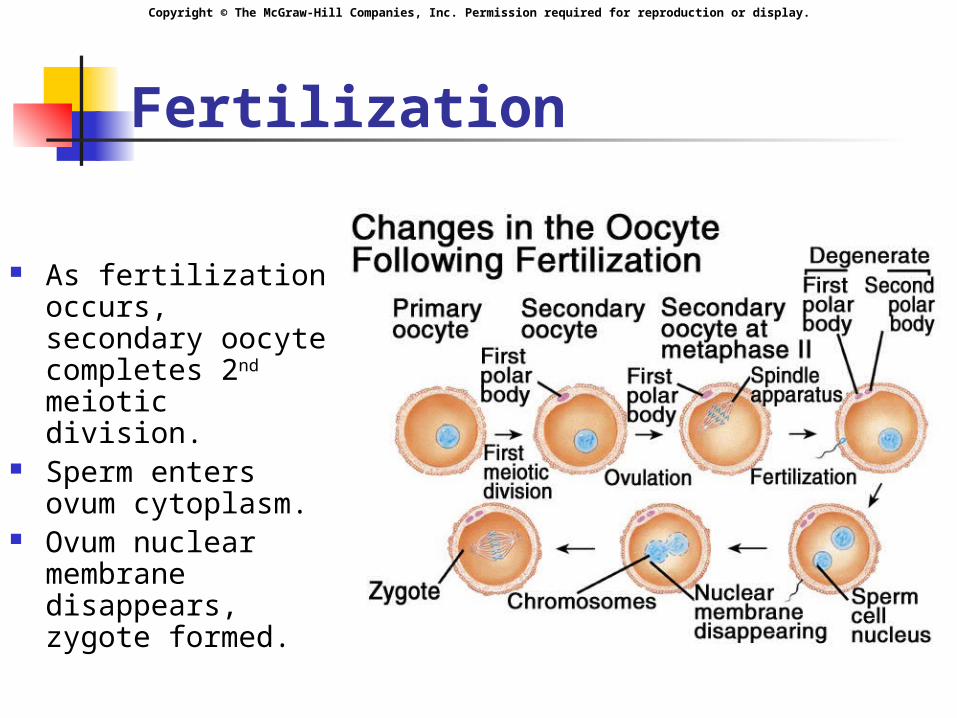
Fertilization
As fertilization occurs, secondary oocyte completes 2nd meiotic division.
Sperm enters ovum cytoplasm.
Ovum nuclear membrane disappears, zygote formed.
Copyright © The McGraw-Hill Companies, Inc. Permission required for reproduction or display.
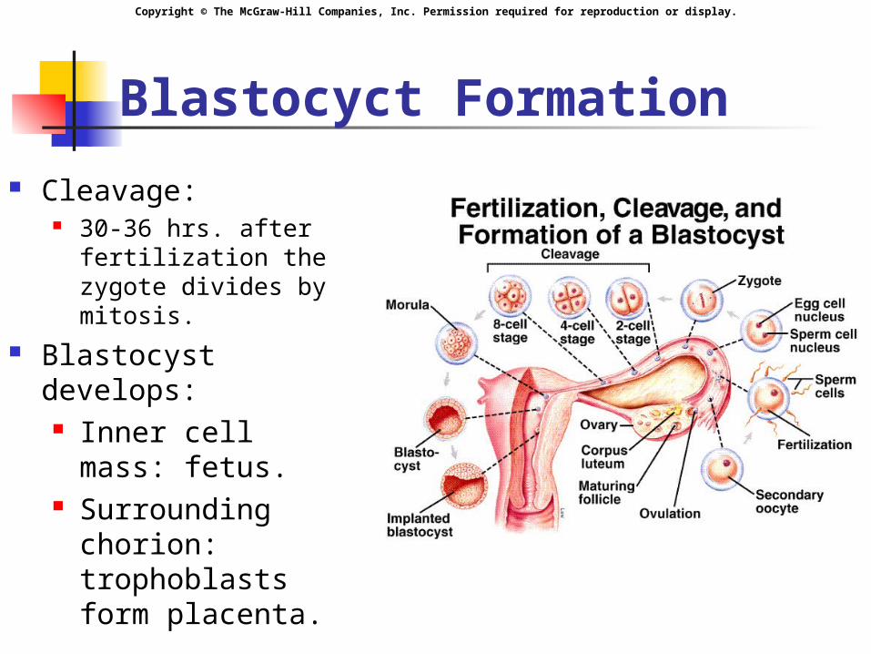
Blastocyct Formation
Cleavage: 30-36 hrs. after
fertilization the zygote divides by mitosis.
Blastocyst develops: Inner cell mass:
fetus. Surrounding
chorion: trophoblasts form placenta.

Copyright © The McGraw-Hill Companies, Inc. Permission required for reproduction or display.
Implantation 6th day after
fertilization, blastocyst attaches to uterine wall.
Blastocyst secretes enzymes that allow blastocyst to burrow into endometrium.
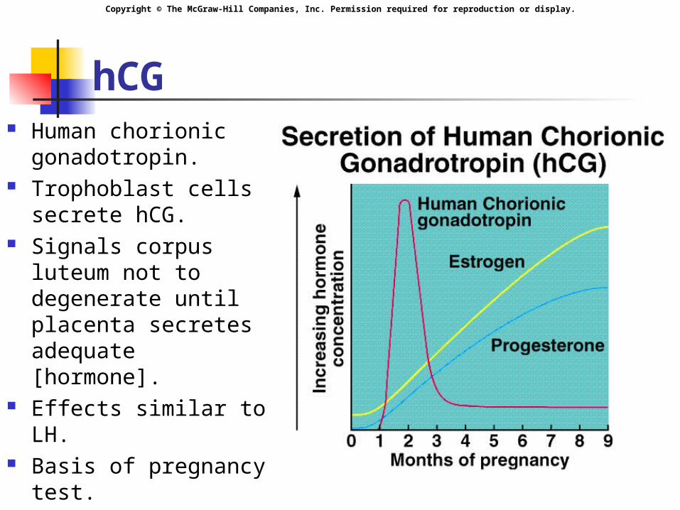
Trophoblast cells secrete hCG.
Copyright © The McGraw-Hill Companies, Inc. Permission required for reproduction or display.
hCG Human chorionic
gonadotropin. Trophoblast cells
secrete hCG. Signals corpus
luteum not to degenerate until placenta secretes adequate [hormone].
Effects similar to LH. Basis of pregnancy
test.
Copyright © The McGraw-Hill Companies, Inc. Permission required for reproduction or display.
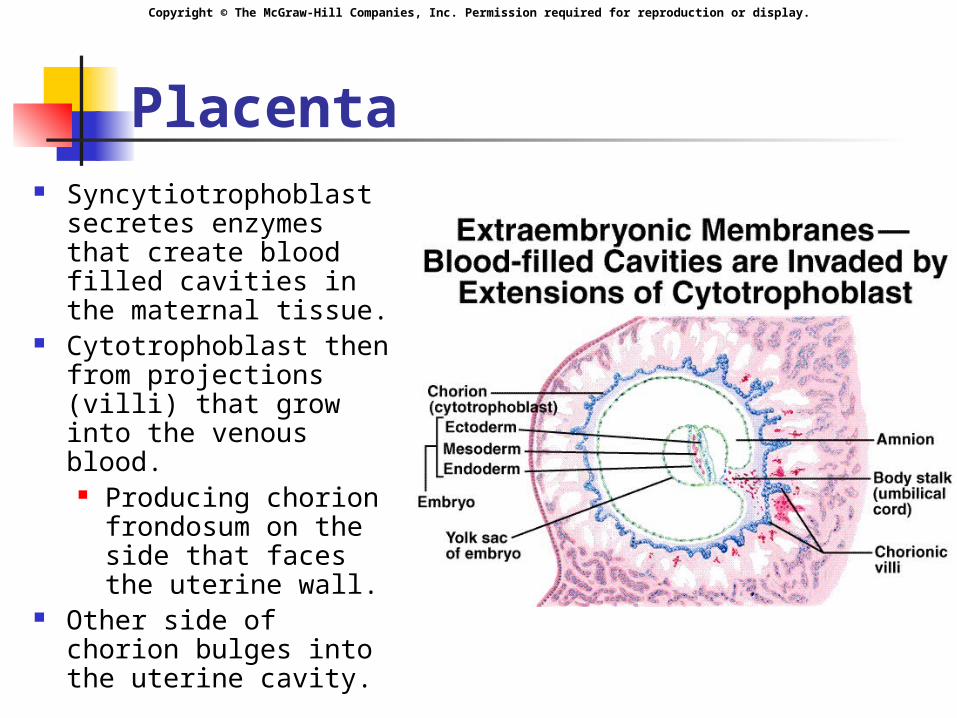
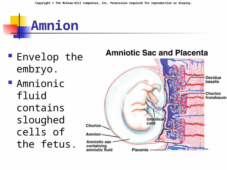
Placenta Syncytiotrophoblast
secretes enzymes that create blood filled cavities in the maternal tissue.
Cytotrophoblast then from projections (villi) that grow into the venous blood.
Producing chorion frondosum on the side that faces the uterine wall.
Other side of chorion bulges into the uterine cavity.
Copyright © The McGraw-Hill Companies, Inc. Permission required for reproduction or display.
Placental Changes
Decidual reaction: Endometrial growth. Accumulation of glycogen.
Decidua basalis: maternal tissue in contact with the chorion frondosum.
Maternal and fetal blood do not mix.
Copyright © The McGraw-Hill Companies, Inc. Permission required for reproduction or display.
Amnion
Envelop the embryo.
Amnionic fluid contains sloughed cells of the fetus.

Copyright © The McGraw-Hill Companies, Inc. Permission required for reproduction or display.
Placenta Function
Gas exchange: 02 and C02.
Nutrient exchange. Waste exchange. Synthesis of proteins and
enzymes.
Copyright © The McGraw-Hill Companies, Inc. Permission required for reproduction or display.
Placental Hormones
hCS: Chorionic somatomammotropin. GH effects. Diabetic-like effect:
Glucose sparing effect. Polyuria. Lipolysis.
Copyright © The McGraw-Hill Companies, Inc. Permission required for reproduction or display.
Placental Hormones
Fetal-placental unit: Placenta must cooperate with the
adrenal cortex in the fetus to produce estrogen.
Estrogen stimulates: Endometrial growth. Inhibit prolactin secretion. Growth of mammary ducts. Enlargement of mother’s uterus.
Copyright © The McGraw-Hill Companies, Inc. Permission required for reproduction or display.
Placental Hormones
Progesterone: Suppresses uterine contractions. Stimulates uterine growth . Suppresses LH and FSH. Stimulate development of
alveolar tissue of the mammary gland.
Copyright © The McGraw-Hill Companies, Inc. Permission required for reproduction or display.
Parturition
Estrogen in late pregnancy: Increases amount of oxytocin stored. Stimulate production of oxytocin
receptors in myometrium. Stimulate prostaglandin production.
Uterine contractions: Oxytocin. Prostaglandins.
Copyright © The McGraw-Hill Companies, Inc. Permission required for reproduction or display.
Lactation
Hypothalamus releases PRH. Anterior pituitary releases
prolactin: Stimulate milk production.
Oxytocin needed for “milk letdown”.
Copyright © The McGraw-Hill Companies, Inc. Permission required for reproduction or display.

Copyright © The McGraw-Hill Companies, Inc. Permission required for reproduction or display.