Chapter 2: Spine & Upper Extremity - DaVinci AcademyInferior ligamentum nuchae Cervical and thoracic...
17
Spine & Upper Extremity Chapter 2: Spine & Upper Extremity
Transcript of Chapter 2: Spine & Upper Extremity - DaVinci AcademyInferior ligamentum nuchae Cervical and thoracic...

Spine & Upper Extremity
Chapter 2: Spine & Upper Extremity

Spine & Upper Extremity
Spine
I. Osteology
A. Overview
▪ Consists of stacks of vertebrae forming the vertebral column
▪ Five sections:
‒ Cervical: 7 vertebrae
‒ Thoracic: 12 vertebrae
‒ Lumbar: 5 vertebrae
‒ Sacral: 5 fused vertebrae
‒ Coccygeal: 5 fused vertebrae
▪ Forms an S-shape as a result of two types of curvature
‒ Primary curvature: develops intrauterine and is concave anterior
○ Shape of thoracic and sacral sections
‒ Secondary curvature: develops after birth and is concave posterior
○ Shape of cervical and lumbar sections
○ Cervical section develops when baby lifts head up at six months old
○ Lumbar section develops when infant assumes upright position at one year old
‒ Functions to maintain our upright posture by evenly displacing the weight of the body throughout
the S-shaped vertebral column
Figure 2.1: Lateral View of Vertebral Column Figure 2.2: Axial View of Lumbar Vertebra -
B. Vertebrae
▪ Vertebral body is the thick oval segment of a vertebra
‒ Intervertebral discs separate the vertebrae and provide cushion for weight-bearing and movement
of the vertebral column
‒ One of the sites of adult hematopoiesis

Spine & Upper Extremity
▪ Pedicles project posterolateral from the vertebral body
▪ Laminae begin at the pedicles and join posteriorly to form the base of the spinous process, which
projects posteriorly
▪ Transverse processes project laterally from the junction of the pedicles and lamina
▪ Vertebral foramen is a space that is formed by the posterior aspect of the vertebral body, pedicles and
lamina
‒ Contents: spinal cord, meninges, vertebral vessels, adipose tissue and spinal roots
▪ Vertebral canal is formed by the stacking of the vertebral foramen
▪ Vertebral arch is formed by the pedicles and lamina
▪ Superior and inferior articular processes are bony prominences that extend out from the junction of the
pedicle and lamina
Figure 2.3: Lateral View of Superior and Inferior Articulating Processes Figure 2.4: Lateral View of Intervertebral Facet Joints -
C. Intervertebral Facet Joints
▪ Formed by the articulation of a superior articular facet of the inferior vertebra with the inferior articular
facet of the superior vertebra
D. Pars Interarticularis
▪ Bony part of the vertebra at the junction of the pedicle and lamina that lies between the superior and
inferior articular processes of the facet joint
▪ On a radiograph of the lumbar spine the pars is referred to as the neck of a Scottie dog image formed
by an outline of surrounding vertebral structures
E. Cervical Spine
▪ C1 vertebra (Atlas)
‒ Articulates with the occipital condyles of the occipital bone to form the junction between the spine
and the skull called the atlanto-occipital joint
○ Allows the head to nod up and down
‒ Has no vertebral body
‒ Ring like structure formed by anterior and posterior arches and two lateral masses
‒ Anterior arch
○ Anterior surface is convex shaped and at its center is the anterior tubercle, which serves as the
attachment of the longus coli muscles and the anterior longitudinal ligament
○ Posterior surface is concave shaped and contains at its center an oval facet for articulation
with the dens (odontoid process) of the axis (C2 vertebra)
‒ Posterior arch contains the posterior tubercle, which gives origin to the nuchal ligament
‒ Lateral masses
○ Superior facets receive the occipital condyles
○ Inferior facets articulate with the axis and allow rotational movements of the head
‒ Large transverse processes that project laterally from the lateral masses
○ Transverse foramen contains the vertebral arteries before they enter the skull

Spine & Upper Extremity
Figure 2.5: Axial CT of a C1 Vertebra with a Jefferson Fracture (White Arrows) Published with permission from LearningRadiology.com
-
‒ Jefferson Fracture
○ Fracture of the anterior and posterior arches in one or more places
○ Mechanisms of injury: axial loading of the head onto the spine or direct impact to the
posterior aspect of the cervical spine
○ Presentation: neck pain, often with no neurological symptoms
○ Fracture can result in damage to the arteries in the neck potentially causing Horner’s
syndrome, ataxia and inability to sense pain or temperature
○ Diagnosis: X-ray and CT
○ Treatment: if stable put patient in collar immobilization, if unstable surgical fixation
i. Stability is partially determined by the integrity of the transverse ligament connecting the
lateral masses of the atlas
ii. If on an axial CT, the atlantodental interval (ADI) is measured to be larger than 3mm,
then the transverse ligament is considered torn and the fracture is unstable
▪ C2 vertebra (Axis)
‒ Forms the rotational base for the atlas and skull
‒ Unlike C1, C2 has a vertebral body with a prominent projection called the dens
○ Oval circular facet on its anterior surface for articulating with the anterior arch of C1
○ Allows C1-C2 rotation
○ Primary horizontal stabilizer of the atlanto-axial joint
‒ Hangman’s Fracture
○ Fracture of both the pedicles or pars interarticularis of the C2 vertebra
○ Causes: falls, especially in the elderly; motor vehicle accidents due to hyperextension of the
neck; axial loading onto the C2 vertebra
○ Mechanism of injury: sudden forceful hyperextension of the head just under the chin that
occurs mainly with deceleration injuries
○ Treatment: neck collar or surgery
Figure 2.6: Posterior View of C2 Figure 2.7: Lateral View of C2 Figure 2.8: Axial View of C3-C7 -
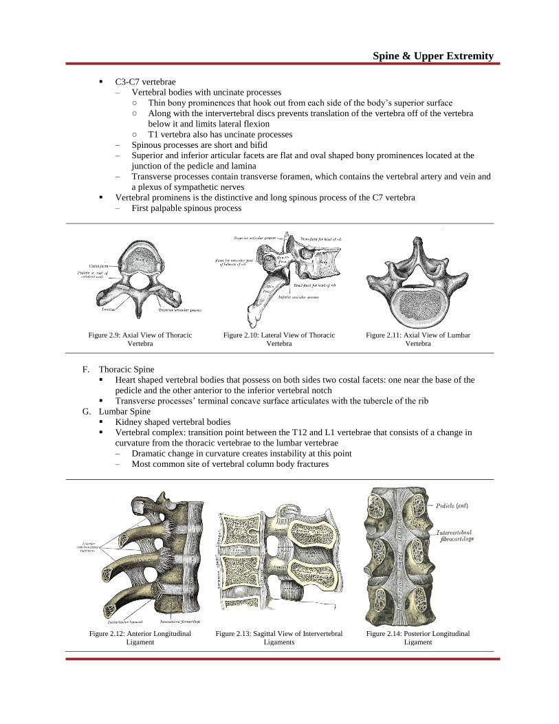
Spine & Upper Extremity
▪ C3-C7 vertebrae
‒ Vertebral bodies with uncinate processes
○ Thin bony prominences that hook out from each side of the body’s superior surface
○ Along with the intervertebral discs prevents translation of the vertebra off of the vertebra
below it and limits lateral flexion
○ T1 vertebra also has uncinate processes
‒ Spinous processes are short and bifid
‒ Superior and inferior articular facets are flat and oval shaped bony prominences located at the
junction of the pedicle and lamina
‒ Transverse processes contain transverse foramen, which contains the vertebral artery and vein and
a plexus of sympathetic nerves
▪ Vertebral prominens is the distinctive and long spinous process of the C7 vertebra
‒ First palpable spinous process
Figure 2.9: Axial View of Thoracic
Vertebra
Figure 2.10: Lateral View of Thoracic
Vertebra
Figure 2.11: Axial View of Lumbar
Vertebra -
F. Thoracic Spine
▪ Heart shaped vertebral bodies that possess on both sides two costal facets: one near the base of the
pedicle and the other anterior to the inferior vertebral notch
▪ Transverse processes’ terminal concave surface articulates with the tubercle of the rib
G. Lumbar Spine
▪ Kidney shaped vertebral bodies
▪ Vertebral complex: transition point between the T12 and L1 vertebrae that consists of a change in
curvature from the thoracic vertebrae to the lumbar vertebrae
‒ Dramatic change in curvature creates instability at this point
‒ Most common site of vertebral column body fractures
Figure 2.12: Anterior Longitudinal
Ligament
Figure 2.13: Sagittal View of Intervertebral
Ligaments
Figure 2.14: Posterior Longitudinal
Ligament -

Spine & Upper Extremity
H. Ligaments
▪ Anterior Longitudinal Ligament (ALL): thick sheet like ligament that runs down the anterior surfaces
of all the vertebral bodies
‒ Prevents anterior displacement of intervertebral discs and hyperextension of the spine
▪ Posterior Longitudinal Ligament (PLL): runs within the spinal canal and extends from the C2 vertebra
along the posterior surfaces of the vertebral bodies
‒ Prevents posterior displacement of intervertebral discs and hyperflexion of the spine
▪ Ligamentum flavum: attaches adjacent vertebral lamina
‒ High elastic tissue content that provides elastic recoil to vertebral column after flexion
‒ Protects spinal cord posteriorly
▪ Supraspinous ligament: spans between the tips of adjacent spinous processes beginning at the C7-T1
vertebral level
▪ Interspinous ligament: attaches adjacent spinous processes
▪ Intertransverse ligament: attaches between adjacent transverse processes
▪ ALL is much stronger than PLL
Figure 2.15: Transverse Ligament of C1 Vertebra Figure 2.16: Posterior View of Cervical Ligaments -
▪ Cervical ligaments
‒ Cruciform ligament is composed of:
○ Transverse ligament: attaches the lateral masses of the atlas (C1) and arches over the dens of
the axis (C2)
○ Longitudinal ligament: connects the apex of the dens to the anterior region of the foramen
magnum and the body of the axis (C1)
‒ Tectorial membrane: extends superiorly from the PLL at the body of C2 vertebra to the base of the
occipital bone
‒ Nuchal ligament: extends from the external occipital protuberance on the posterior skull and
across the tips of cervical spinous processes
○ Continuous with the supraspinous ligament at C7 vertebra
II. Intervertebral Discs
A. Disc Structure
▪ Fibrocartilaginous ovoid shaped structures that lie between adjacent vertebral bodies forming a
symphysis joint
▪ Functions to allow movement between adjacent vertebral bodies and act as shock absorbers
▪ Two components
‒ Nucleus Pulposus: gel-like core that consists of proteoglycans, glycosaminoglycans and
collagenous fibers immersed in mucoid material
‒ Annulus Fibrosus: fibrocartilage ring made up of collagen types I and II
B. Disc Herniations
▪ Results from breakdown of the annulus fibrosus allowing a portion of the nucleus pulposus to herniate
out, which can potentially compress the spinal nerves and/or roots
▪ Intervertebral discs usually herniate adjacent to the lateral edge of the PLL

Spine & Upper Extremity
▪ Most commonly occurs in the cervical and lumbar regions of the spine
‒ Rare in the thoracic region because those vertebrae undergo less movement due to being fixated
by the ribs
Figure 2.17: Axial View of Intervertebral Disc Figure 2.18: Deep Back Muscles -
III. Back Muscles
A. Deep Muscles
▪ Extend from the pelvic bones along the length of the spine to the base of the skull
▪ Involved in movements of the spine
▪ All are innervated by the dorsal rami of the spinal nerves
▪ Divided into three layers: deep, intermediate and superficial
▪ Deep layer
‒ Group of three muscles that attach between the transverse and spinous processes of the vertebral
column
Table 2.1: Deep Layer of the Deep Muscles
Muscle Origin Insertion Movement Innervation
Semispinalis ▪ Transverse processes
of C4-T10
▪ Spinous processes
of C2-T4
▪ Extension of the
head and spine
▪ Contralateral rotation
of the head
▪ Dorsal primary
rami of spinal
nerves
Multifidus ▪ Sacrum
▪ Posterior iliac spine
▪ Transverse processes
of T1-T3
▪ Articular processes of
C4-C7
▪ Spinous processes
of vertebrae
▪ Stabilization of the
spine
▪ Dorsal primary
rami of spinal
nerves
Rotatores ▪ Vertebral transverse
processes
▪ Lamina and spinous
processes of the
immediately
superior vertebra
▪ Stabilization of the
spine
▪ Proprioception
▪ Dorsal primary
ramus of spinal
nerves
.
▪ Intermediate Layer
‒ Group of three muscles collectively called the erector spinae muscles
‒ All three arise from a common tendinous origin that attaches to: sacroiliac and supraspinous
ligaments, iliac crest, sacrum, lumbar and thoracic vertebrae

Spine & Upper Extremity
Table 2.2: Intermediate Layer of Deep Muscles
Muscle Origin Insertion Movement Innervation
Iliocostalis ▪ Sacrum
▪ Iliac crest
▪ Spinous processes
of lumbar/thoracic
vertebrae
▪ Ribs ▪ Unilaterally: flex
the spine
▪ Bilaterally: extend
the spine and head
▪ Dorsal primary rami
of spinal nerves
Longissimus ▪ Sacrum
▪ Iliac crest
▪ Spinous processes
of lumbar/thoracic
vertebrae
▪ Ribs
▪ Transverse
processes of
C2-T12
▪ Mastoid process
▪ Unilaterally: flex
the spine
▪ Bilaterally: extend
the spine and head
▪ Dorsal primary rami
of spinal nerves
Spinalis ▪ Sacrum
▪ Iliac crest
▪ Spinous processes
of lumbar/thoracic
vertebrae
▪ Spinous processes
of C2 and T1-T8
▪ Occipital bone
▪ Unilaterally: flex
the spine
▪ Bilaterally: extend
the spine and head
▪ Dorsal primary rami
of spinal nerves
.
▪ Superficial Layer
‒ Two muscles on the posterior aspect of the cervical spine that move the head and neck
Table 2.3: Superficial Layer of Deep Muscles
Muscle Origin Insertion Movement Innervation
Splenius Capitis ▪ Inferior aspect of
ligamentum nuchae
▪ Spinous processes of
C7-T3
▪ Mastoid process
▪ Occipital bone
▪ Rotate the head to
the ipsilateral side
▪ Dorsal primary
rami of spinal
nerves C3 and C4
Splenius
Cervicis
▪ Spinous processes of
T3-T6
▪ Transverse
processes of
C1-C4
▪ Rotates the head to
the ipsilateral side
▪ Dorsal primary
rami of lower
cervical spinal
nerves .
Figure 2.19: Superficial Layer of Deep Back Muscles Figure 2.20: Intermediate and Superficial Back Muscles -
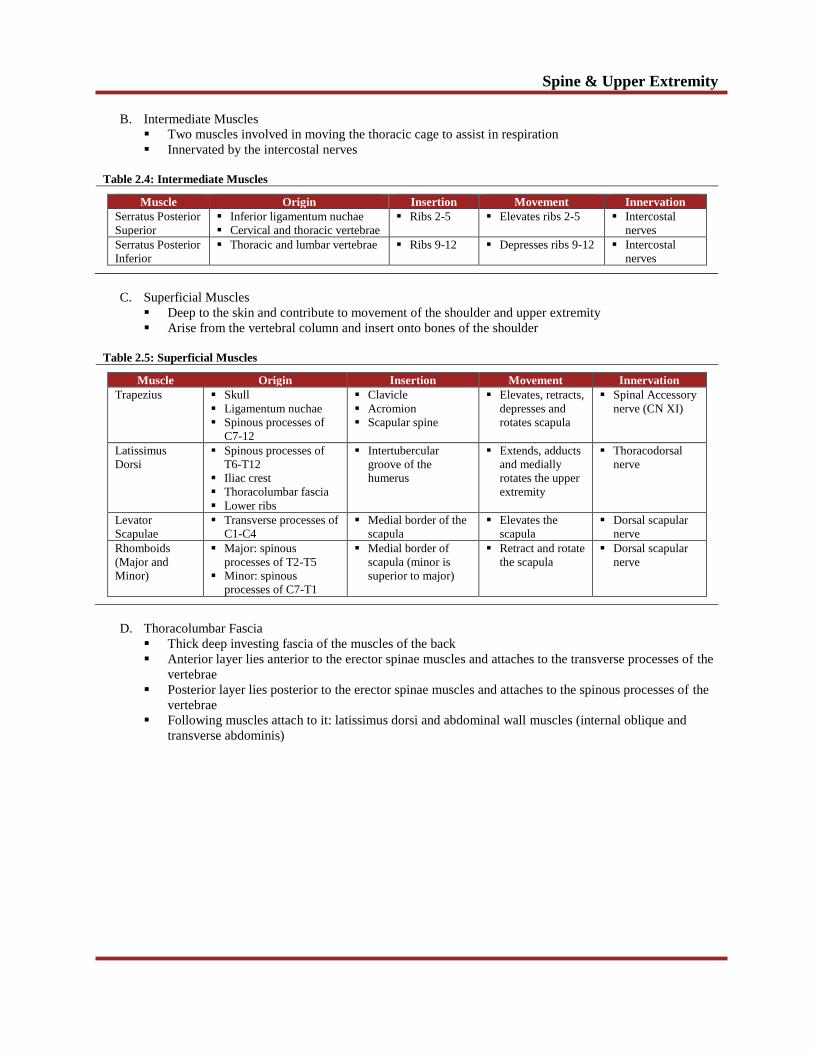
Spine & Upper Extremity
B. Intermediate Muscles
▪ Two muscles involved in moving the thoracic cage to assist in respiration
▪ Innervated by the intercostal nerves
Table 2.4: Intermediate Muscles
Muscle Origin Insertion Movement Innervation
Serratus Posterior
Superior
▪ Inferior ligamentum nuchae
▪ Cervical and thoracic vertebrae
▪ Ribs 2-5 ▪ Elevates ribs 2-5 ▪ Intercostal
nerves
Serratus Posterior
Inferior
▪ Thoracic and lumbar vertebrae ▪ Ribs 9-12 ▪ Depresses ribs 9-12 ▪ Intercostal
nerves .
C. Superficial Muscles
▪ Deep to the skin and contribute to movement of the shoulder and upper extremity
▪ Arise from the vertebral column and insert onto bones of the shoulder
Table 2.5: Superficial Muscles
Muscle Origin Insertion Movement Innervation
Trapezius ▪ Skull
▪ Ligamentum nuchae
▪ Spinous processes of
C7-12
▪ Clavicle
▪ Acromion
▪ Scapular spine
▪ Elevates, retracts,
depresses and
rotates scapula
▪ Spinal Accessory
nerve (CN XI)
Latissimus
Dorsi
▪ Spinous processes of
T6-T12
▪ Iliac crest
▪ Thoracolumbar fascia
▪ Lower ribs
▪ Intertubercular
groove of the
humerus
▪ Extends, adducts
and medially
rotates the upper
extremity
▪ Thoracodorsal
nerve
Levator
Scapulae
▪ Transverse processes of
C1-C4
▪ Medial border of the
scapula
▪ Elevates the
scapula
▪ Dorsal scapular
nerve
Rhomboids
(Major and
Minor)
▪ Major: spinous
processes of T2-T5
▪ Minor: spinous
processes of C7-T1
▪ Medial border of
scapula (minor is
superior to major)
▪ Retract and rotate
the scapula
▪ Dorsal scapular
nerve
.
D. Thoracolumbar Fascia
▪ Thick deep investing fascia of the muscles of the back
▪ Anterior layer lies anterior to the erector spinae muscles and attaches to the transverse processes of the
vertebrae
▪ Posterior layer lies posterior to the erector spinae muscles and attaches to the spinous processes of the
vertebrae
▪ Following muscles attach to it: latissimus dorsi and abdominal wall muscles (internal oblique and
transverse abdominis)

Spine & Upper Extremity
IV. Neural Structures
Figure 2.21: Axial View of Spinal Cord Figure 2.22: Lateral View of Terminal Spinal Cord Figure 2.23: Cauda Equina -
A. Spinal Cord
▪ Occupies ⅔ of the vertebral canal and is enveloped by the meninges along its entire length
▪ Grey matter is in the central region of the cord and contains neuron cell bodies
‒ Motor neuron cell bodies are located in the ventral horn
‒ Autonomic neuron cell bodies are located in the lateral horn
‒ Sensory neuron cell bodies are located in the dorsal horn and in the dorsal root ganglion
▪ White matter is located peripherally and contains neural tracts traveling to and from the brain
▪ Conus medullaris: terminal end of the spinal cord located at the T12/L1 vertebral level
▪ Dural sac: continuation of the dura beyond the conus medullaris that houses the cauda equina and is
the terminal fixation point via the filum terminale
▪ Cauda equina: collection of the final lumbar and sacral spinal nerves (L2-S5) that continue on from the
conus medullaris within the dural sac and are bathed in CSF
‒ Provide visceral innervation to the pelvic organs (e.g. bladder) and motor and sensory innervation
to the perineum and lower extremity
▪ Filum terminale is a long fibrous tissue cord that spans from the apex of the conus medullaris to
coccygeal vertebrae and is composed of two parts:
‒ Filum terminale internum: extension from the pia mater that travels within the dural sac and
inserts into the tip of the dural sac around the second sacral vertebra
‒ Filum terminale externum: extends from the apex of the dural sac and inserts into the first segment
of the coccyx (AKA coccygeal ligament)
▪ Lumbar puncture is a diagnostic procedure used to collect sample CSF
‒ Insert the needle below the L2 vertebral level to avoid spinal cord injury
‒ Needle is advanced into the dural sac between the L3-L5 vertebral levels to collect CSF around the
cauda equina
○ Iliac crest can be used as a landmark because it is at the same level as the intervertebral disc
between L4/L5
‒ Layers pierced: skin, superficial fascia, supraspinous ligament, interspinous ligament, ligamentum
flavum, epidural space, dura mater and arachnoid mater
○ Epidural anesthesia block stops at the epidural space and is inserted in the same region to
avoid injury to the spinal cord

Spine & Upper Extremity
B. Spinal Nerves
▪ Exit through the intervertebral (neural) foramen, which are formed by the following structures:
‒ Anterior: posterolateral aspect of superior vertebra and intervertebral disc
‒ Posterior: facet joints
‒ Superior: inferior vertebral notch of the superior pedicle
‒ Inferior: superior vertebral notch of the inferior pedicle
Figure 2.24: Lateral View of Cervical Vertebra Figure 2.25: Cervical Spinal Nerves -
▪ Cervical spinal nerves
‒ Travel above the corresponding vertebra’s pedicle, then exit through the intervertebral foramen
○ Example: C5 spinal nerve travels above the C5 vertebra’s pedicle, then exits through the
intervertebral foramen between the C4 and C5 vertebrae
‒ Cervical disc herniations compress the spinal nerve corresponding to the lower vertebra
○ Example: disc herniation at C5/C6 vertebral level will compress the C6 spinal nerve
○ Most common cervical levels for disc herniation: C4/C5, C5/C6 and C6/C7
○ Symptoms manifest in the upper extremity
○ Can involve sensory and motor deficits and/or changes in reflexes
○ Cervical radiculopathy: impingement of cervical roots and/or spinal nerves, which results in
pain/numbness radiating down the upper extremity
‒ Special cervical spinal nerves
○ C1 spinal nerve only carries motor fibers
○ C2 spinal nerve directly gives rise to the greater occipital nerve, which pierces the trapezius
muscle and provides cutaneous innervation to the posterior part of the scalp
○ C8 spinal nerve exits below the pedicle of C7 vertebra and all subsequent spinal nerves exit
below the corresponding vertebra’s pedicle
‒ Cervical spinal nerve reflexes
○ C5 – biceps brachii
○ C6 – brachioradialis
○ C7 – triceps brachii
C7
T1
C5
C6
C4

Spine & Upper Extremity
Figure 2.26: Lateral View of Lumbar Vertebrae Figure 2.27: Lumbar Spinal Nerves -
▪ Lumbar spinal nerves
‒ Travels below the corresponding vertebra’s pedicle, then exits through the intervertebral foramen
○ Example: L4 spinal nerve travels below the L4 vertebra’s pedicle, then exits through the
intervertebral foramen between the L4 and L5 vertebrae
‒ Lumbar disc herniations compress the spinal nerve corresponding to the lower vertebrae
○ Example: disc herniation at L4/L5 vertebral level will compress the L5 spinal nerve
○ Symptoms manifest in the buttocks and lower extremity
○ Can involve sensory and motor deficits and/or changes in reflexes
○ Lumbar radiculopathy: impingement of lumbar roots and/or spinal nerves that results in
pain/numbness radiating down the lower extremity (e.g. sciatica)
‒ Lumbar spinal nerve reflexes
○ L4 – patellar tendon
○ S1 – Achilles tendon
C. Sympathetic Chain
▪ Pair of trunk structures that travel along the lateral surface of the vertebral bodies
▪ Begins in the thorax region at T1 vertebra and descends the vertebral column
▪ Both trunks course medially in the sacral region and converge on the ganglion impar →
anterior to the coccyx
▪ Passageway and synapse point for sympathetic fibers
▪ Left sympathetic trunk travels just lateral to the thoracic and abdominal aorta
V. Blood Vessels
A. Aorta
▪ Descends along anterior surface of the vertebral bodies in the thorax and abdomen
‒ At risk during discectomy spine surgery procedures
▪ Bifurcates into the common iliac arteries at L4 vertebral level in the abdomen
B. Vertebral Artery
▪ Arises from the subclavian artery, then enters the transverse processes of the cervical vertebrae at the
C6 vertebral level
▪ Ascends through the transverse foramen of each cervical vertebra and exits at the C1 vertebral level
▪ Travels across the posterior arch of C1 and through the suboccipital triangle before entering the skull
through the foramen magnum
▪ Within the skull, the two vertebral arteries join to form the basilar artery, which is the main supply to
the brainstem
▪ Excessive manipulation or trauma to the cervical vertebrae can create an embolism in the vertebral
arteries that can travel to the cerebral circulation and result in a stroke
L5
S1
L3
L4
L2

Spine & Upper Extremity
C. Vertebral Venous Plexuses
▪ Internal vertebral venous plexus is located in the epidural space forming anterior and posterior venous
networks that travel the length of the vertebral column
‒ Communicates superiorly with the cranial dural sinuses
‒ Inferiorly communicates with the prostatic venous plexus, which is believed to be a conduit for
prostate carcinoma to metastasize to the spine
▪ External vertebral venous plexus forms an anterior part that travels anterior to the vertebral column and
a posterior part that travels on the vertebral arch externally
‒ Forms anastomosis with the internal vertebral venous plexus
Figure 2.28: Sympathetic Trunk Figure 2.29: Vertebral Artery Figure 2.30: Vertebral Venous System -
VI. Clinical Pearls
A. Lumbosacral Strain
▪ Strain of the lower back muscles
▪ Causes: physical exertion, poor form when lifting, overuse or trauma
▪ Presentation: low back pain that does not radiate, usually following a clear causative incident
▪ Physical exam: tenderness to palpation over the paraspinal muscles, (-) Straight Leg Test and normal
neurological exam
▪ Treatment: NSAIDs and early mobilization, exercises or physical therapy
B. Disc Herniation
▪ Cervical
‒ Presentation: neck, shoulder and/or arm pain, weakness and numbness
‒ Rule out: torticollis, shoulder pathology, carpal tunnel, neurological disease and peripheral
vascular disease (brachial and radial pulses)
Table 2.6: Physical Exam Findings for Cervical Disc Herniations
Physical Exam C5 C6 C7 C8 T1
Sensory Lateral shoulder Thumb Middle finger Ring and small
fingers
Medial forearm
and hand
Motor Weak shoulder
abduction
Weak elbow
flexion
Weak elbow
extension
Weak finger
abduction
Weak finger
abduction
Reflex Hypoactive
biceps brachii
Hypoactive
brachioradialis
Hypoactive
triceps
No abnormal
reflex
No abnormal
reflex -

Spine & Upper Extremity
Figure 2.31: Cervical Disc Herniations Figure 2.32: Lumbar Disc Herniations Figure 2.33: Lateral MRI of Lumbar Disc Herniation Published with permission from LearningRadiology.com
-
▪ Lumbar
‒ Presentation: low back, buttocks and/or leg pain, weakness and numbness
‒ Special tests: (+) Straight Leg Test, abnormal gait (walking on heels/toes)
‒ Rule out: abdominal/pelvis pathology, hip pathology, knee pathology, neurological disease and
peripheral vascular disease (posterior tibial and dorsalis pedis pulses)
Table 2.7: Physical Exam Findings for Lumbar Disc Herniations
Physical Exam L3 L4 L5 S1 S2-S4
Sensory Anterior and
medial thigh
Medial leg and
ankle
Dorsal foot and
1st webspace
Lateral and
plantar foot
Perianal sensation
Motor Knee extension Ankle
dorsiflexion
Big toe
dorsiflexion
Ankle plantar
flexion
Anal squeeze
Reflex No abnormal
reflex
Hypoactive
patellar tendon
No abnormal
reflex
Hypoactive
Achilles tendon
Hypoactive
bulbocavernosus
Gait N/A N/A Walking on
heels, foot drop
Walking on toes N/A
-
▪ Diagnosis: MRI confirms diagnosis and can show impingement of spinal nerves
▪ Treatment: physical therapy, corticosteroid injections and/or surgery
C. Spinal Stenosis
▪ Narrowing of the spinal canal and/or the transforaminal foramina
▪ Usually due to degenerative joint disease resulting in bone spurs, ligament hypertrophy, facet joint
breakdown and cyst formation
▪ Other causes: degenerative disc disease, retropulsion of bone fragments during vertebral fractures,
spondylolisthesis, spine trauma and tumors
▪ Presentation: low back pain that radiates bilaterally into the buttocks and lower extremities; numbness;
weakness; bladder/bowel incontinence
‒ Pain is worse with standing and extension
‒ Pain is better with flexion and leaning forward (Shopping Cart Sign)
▪ Neurogenic claudication: patient is unable to walk distances due to significant leg pain, similar to
patients with peripheral vascular disease of the lower extremities
C7
T1
C5
C6
C4
L5
S1
L3
L4
L2

Spine & Upper Extremity
▪ Physical exam: bilateral numbness, weakness, hypoactive reflexes, abnormal gait and/or absent anal
reflex
▪ Diagnosis: X-ray, CT and MRI
▪ Treatment: physical therapy, corticosteroid injections and/or surgery
D. Spondylolysis
▪ Fracture or defect of the pars interarticularis without slippage of the vertebral bodies
▪ Common in injuries that involve hyperextension of the spine (gymnasts, linemen in football) and often
seen in children
▪ Most common site is L5 vertebra
▪ Presentation: low back pain that is worse with activity
▪ Diagnosis: X-ray, CT and MRI
▪ Treatment: rest, physical therapy – lumbar flexion exercises, lumbar brace, surgery is uncommon
unless the patient also develops vertebral instability (e.g. spondylolisthesis)
E. Spondylolisthesis
▪ Slippage of one vertebra on another (usually refers to anterior translation)
▪ Causes: degenerative arthritis, traumatic, isthmic and dysplastic
▪ Bilateral spondylolysis can lead to a spondylolisthesis
▪ Presentation: low back pain worse with activity, pain that radiates down the lower extremity,
numbness, weakness and difficulty ambulating
▪ Physical exam: decreased range of motion, semi-kyphotic posture, abnormal gait and/or motor and
sensory deficits
▪ Diagnosis: X-ray, CT and MRI for visualizing damage to neurologic structures
▪ Treatment: rest, physical therapy, bracing and/or surgery
Figure 2.34: Oblique X-ray of a Lumbar
Spondylolysis
Figure 2.35: Lateral X-ray of Lumbar
Spondylolisthesis
Figure 2.36: AP X-ray of Sacroiliitis in
Ankylosing Spondylitis
Published with permission from LearningRadiology.com -
F. Abnormal Curvatures
▪ Excessive Kyphosis (hyperkyphosis)
‒ Excessive thoracic (primary) curvature
‒ Causes: osteoporosis eroding anterior thoracic vertebrae, disuse or atrophy of deep back muscles
and whiplash injury (cervical spine)
‒ Presentation: difficulty getting up from a chair, feeling off-balance, difficulty ambulating, fatigue
and respiratory problems in severe disease
‒ Diagnosis: X-ray
‒ Treatment: treat underlying cause and physical therapy

Spine & Upper Extremity
▪ Excessive Lordosis (hyperlordosis)
‒ Excessive lumbar (secondary) curvature
‒ Causes: muscle strength and length imbalances, weak anterior abdominal wall muscles,
osteoporosis, achondroplasia, spondylolisthesis and osteoporosis
‒ Pregnant women temporarily acquire during late gestation – adjust center of gravity due to fetal
growth
‒ Diagnosis: X-ray
‒ Treatment: muscle stretching, bracing and treating an underlying cause if present
▪ Scoliosis
‒ Abnormal lateral curvature and long axis rotation of the spine
‒ Types: congenital, idiopathic and neuromuscular
‒ Presentation: asymptomatic; pain in the back, shoulders and neck; respiratory problems in severe
disease
‒ Physical exam: spinal deformity on Forward Bending Test, asymmetric weakness of intrinsic back
muscles and asymmetric lower extremity lengths
○ Neurological deficits usually only seen in neuromuscular type
‒ Diagnosis: full length spinal X-rays (AP and lateral views)
‒ Treatment: observation, bracing, activity restriction and/or surgery
G. Ankylosing Spondylitis
▪ Systemic autoimmune disease that primarily affects the spine, especially where it articulates with the
pelvis
▪ Leads to fusion of the spine in an ascending manner from lumbar to the cervical region
▪ Presentation: low back pain and stiffness, neck pain later in the disease, inflammation at tendinous
insertions into bone (Achilles and supraspinatus tendons), chest pain and respiratory problems with
thoracic spine involvement
▪ Labs: ESR and HLA-B27
▪ Complications: eventually the spine can become brittle and prone to fractures with minimal trauma
‒ With advanced disease, the lumbar spine can lose lordosis resulting in an inability to stand upright
▪ Diagnosis: X-ray showing sacroiliitis – sclerotic changes in the sacroiliac joints
▪ Treatment: NSAIDs, immunotherapies, physical therapy and surgery for severe cases
H. Conus Medullaris Syndrome
▪ Injury or compression of the conus medullaris
▪ Causes: trauma, tumor, spinal stenosis and pathology in the epidural space compressing the conus
medullaris (e.g. abscess, hematoma)
▪ Presentation: acute onset severe low back pain; bilateral motor and sensory deficits in the lower
extremities; perianal numbness; early onset bowel and bladder dysfunction
▪ Physical exam: hyperreflexia and absent anal reflex
▪ Diagnosis: MRI
▪ Treatment: emergent surgical decompression
I. Cauda Equina Syndrome
▪ Injury or compression of the cauda equina
▪ Causes: trauma, space-occupying lesions within the lumbosacral canal – disc herniation (most
common), spinal stenosis, tumor, epidural hematoma and epidural abscess
▪ Presentation: acute onset severe low back pain; severe radicular pain that is often unilateral, perianal
numbness, asymmetric motor and sensory deficits in the lower extremities; later onset bowel and
bladder dysfunction
▪ Physical exam: diminished or absent reflexes and absent anal reflex
▪ Diagnosis: MRI
▪ Treatment: emergent surgical decompression
Spine & Upper Extremity
J. Epidural Abscess
▪ Collection of pus that has formed in the epidural space of the spinal canal
▪ Most common organism: Staphylococcus aureus
▪ Risk factors: IV drug abuse, diabetes and immunodeficiency
▪ Presentation: low back pain, fever and neurological deterioration
▪ Labs: CBC, ESR and CRP
▪ Diagnosis: MRI
▪ Treatment: antibiotics, emergent surgical decompression and evacuation
K. Spinal Tumors
▪ Common site for metastasis: lymphoma, melanoma, breast, lung, prostate, colorectal and renal
carcinoma
‒ Presentation: back pain, muscle weakness/paralysis, abnormal reflexes and/or acute neurological
deterioration
‒ Diagnosis: X-ray, CT and MRI
‒ Treatment: surgical resection, radiation therapy and chemotherapy
▪ Multiple myeloma is a cancer of plasma cells that results in abnormally large production of antibodies
that affects several organ systems, often including the spine
‒ Presentation: back pain, bone pain, renal failure, infection, hypercalcemia and anemia
○ Can have spinal cord compression leading to neurological deficits
‒ Spine complications: lytic lesions, pathological fractures, structural deformity and anemia from
infiltration of the vertebral bone marrow
▪ Tumors of the spinal cord and meninges are very rare, but can cause significant neurological deficits
and disability
‒ Examples: schwannomas, neurofibromas, meningiomas, astrocytomas and ependymomas