The association of hypertriglyceridemia with cardiovascular events ...

Chapter 1 Introduction

Chapter 1
10
Introduction
11
INTRoDUCTIoN
The clinical challenge of acute pancreatitis
The clinical course of acute pancreatitis is difficult to predict, complications are
potentially lethal and timing and modality of surgical intervention are of great
influence to outcome. Clinical decision making and surgical management are
challenged by the complex pathophysiology of local and systemic complications.
Prevention of infectious complications during the course of acute pancreatitis
would reduce the need for surgical intervention and mortality. However, despite
extensive efforts to reduce the occurrence of infectious complications, currently
applied prophylactic treatment strategies (e.g. prophylactic antibiotics) still do not
yield significant improvement.
These challenges are a strong incentive to further experimental and clinical research
with the aim to improve insight into the pathophysiology of acute pancreatitis
and its complications. This will facilitate development of new prophylactic and
(surgical) treatment strategies, aid clinical decision making, and ultimately improve
outcome.
Aims of this thesis are to assess current surgical challenges of acute pancreatitis,
to gain further insight into pathophysiology and to explore the potential of
prophylactic probiotics. Part I describes the clinical course and surgical treatment
of patients with severe acute pancreatitis. In Part II pathophysiology, the use of
animal models and mechanisms of infectious complications are further explored.
Part III is focussed on the use of prophylactic multispecies probiotics to reduce
the incidence of infectious complications and to change the course of the disease.
Ten major questions addressed in this thesis are shown in Box 1 (Page 16).
PART I: SURGERY
Clinical course
In the majority of cases (85%), acute pancreatitis is limited to pancreatic edema and
mild pancreatic inflammation.1,2 This mild form is usually self-limited, with excellent
outcome (< 2% mortality).1-3 However, in 15% of the cases pancreatic inflammation
is severe, leading to extensive tissue damage and necrosis of pancreatic tissue. In
these cases, local pancreatic inflammation triggers a severe systemic inflammatory
response, potentially leading to pulmonary injury or multiple organ failure.4-6 overall
mortality in patients with severe acute pancreatitis is reported to be 10 to 40%.2,7-9
Morbidity and mortality of severe acute pancreatitis increase significantly if infection
of pancreatic necrosis occurs. Mortality increases from 10-40% for patients with
sterile pancreatic necrosis to 20-70% for patients with infected pancreatic necrosis
(Chapter 2).2,10-12
Management of acute pancreatitis
Mild acute pancreatitis requires only minimal medical support measures.2,7
Management of severe acute pancreatitis however, is much more challenging.
Surgical intervention is indicated when infection of (peri-)pancreatic necrosis
is suspected or proven, necrosis has been sealed off and can be completely
removed.2,8 Surgery should be avoided during the early (pro-inflammatory)
phase.2, 7-9 During this early phase, the immunological response to surgical trauma
in addition to the present pro-inflammatory state results in an overwhelming pro-
inflammatory immune response that could add to fatal outcome.
In general, three surgical treatment strategies for infected pancreatic necrosis are
at hand: 1) laparotomy and necrosectomy followed by open management of the
abdomen, 2) laparotomy and necrosectomy followed by continuous postoperative
lavage or 3) minimally invasive procedures. No randomized clinical trials comparing
these surgical strategies have been published yet. Surgical management of acute
pancreatitis is addressed in Chapter 2.
Due to the anatomical relationship between the ascending, transverse or
descending colon and the pancreas, activated pancreatic enzymes can spread
to these segments of the large intestine. Activated pancreatic digestive enzymes
cause local tissue damage potentially leading to necrosis or perforation of the
affected colonic segment. This requires immediate surgical intervention (Chapter 3).
Perforation of the colon causes severe bacterial peritonitis, infection of pancreatic
necrosis and sepsis. This complication however, is relatively rare and as described
in Parts II and III, the colon is not the only intestinal segment to play a role in the
pathophysiology of infectious complications during severe acute pancreatitis.
PART II: PATHoPHYSIoLoGY
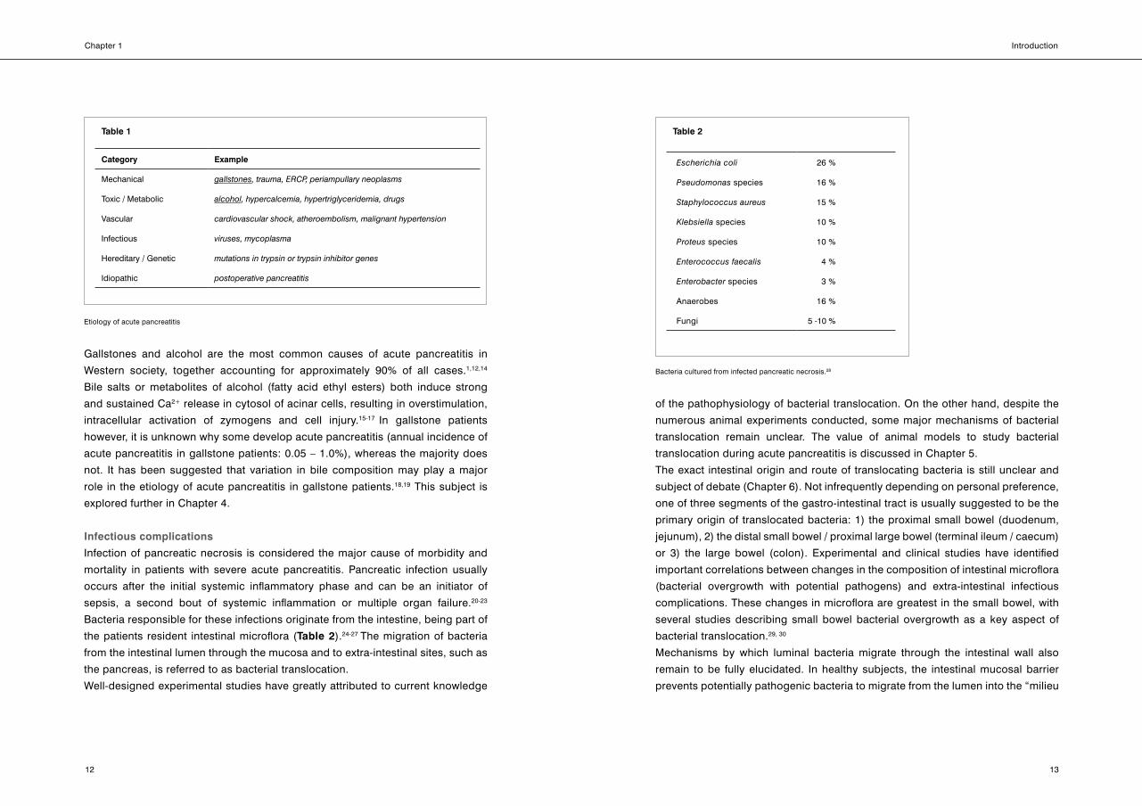
Etiology of acute pancreatitis
There are several causes for injury or dysfunction of pancreatic acinar cells or
ducts, leading to acute pancreatitis*.13 As shown in Table 1, etiology of acute
pancreatitis can be divided into six major categories.
* BackgroundinformationonhistoryandfunctionofthepancreasisprovidedinBox 2.(Pages17-20). Box 3containsbackgroundinformationonthepathogenesisofacutepancreatitis.(Page21)
Chapter 1
12
Introduction
13
Gallstones and alcohol are the most common causes of acute pancreatitis in
Western society, together accounting for approximately 90% of all cases.1,12,14
Bile salts or metabolites of alcohol (fatty acid ethyl esters) both induce strong
and sustained Ca2+ release in cytosol of acinar cells, resulting in overstimulation,
intracellular activation of zymogens and cell injury.15-17 In gallstone patients
however, it is unknown why some develop acute pancreatitis (annual incidence of
acute pancreatitis in gallstone patients: 0.05 – 1.0%), whereas the majority does
not. It has been suggested that variation in bile composition may play a major
role in the etiology of acute pancreatitis in gallstone patients.18,19 This subject is
explored further in Chapter 4.
Infectious complications
Infection of pancreatic necrosis is considered the major cause of morbidity and
mortality in patients with severe acute pancreatitis. Pancreatic infection usually
occurs after the initial systemic inflammatory phase and can be an initiator of
sepsis, a second bout of systemic inflammation or multiple organ failure.20-23
Bacteria responsible for these infections originate from the intestine, being part of
the patients resident intestinal microflora (Table 2).24-27 The migration of bacteria
from the intestinal lumen through the mucosa and to extra-intestinal sites, such as
the pancreas, is referred to as bacterial translocation.
Well-designed experimental studies have greatly attributed to current knowledge
of the pathophysiology of bacterial translocation. on the other hand, despite the
numerous animal experiments conducted, some major mechanisms of bacterial
translocation remain unclear. The value of animal models to study bacterial
translocation during acute pancreatitis is discussed in Chapter 5.
The exact intestinal origin and route of translocating bacteria is still unclear and
subject of debate (Chapter 6). Not infrequently depending on personal preference,
one of three segments of the gastro-intestinal tract is usually suggested to be the
primary origin of translocated bacteria: 1) the proximal small bowel (duodenum,
jejunum), 2) the distal small bowel / proximal large bowel (terminal ileum / caecum)
or 3) the large bowel (colon). Experimental and clinical studies have identified
important correlations between changes in the composition of intestinal microflora
(bacterial overgrowth with potential pathogens) and extra-intestinal infectious
complications. These changes in microflora are greatest in the small bowel, with
several studies describing small bowel bacterial overgrowth as a key aspect of
bacterial translocation.29, 30
Mechanisms by which luminal bacteria migrate through the intestinal wall also
remain to be fully elucidated. In healthy subjects, the intestinal mucosal barrier
prevents potentially pathogenic bacteria to migrate from the lumen into the “milieu
Table 1
Category Example
Mechanical gallstones,trauma,ERCP,periampullaryneoplasms
Toxic / Metabolic alcohol,hypercalcemia,hypertriglyceridemia,drugs
Vascular cardiovascularshock,atheroembolism,malignanthypertension
Infectious viruses,mycoplasma
Hereditary / Genetic mutationsintrypsinortrypsininhibitorgenes
Idiopathic postoperativepancreatitis
Etiology of acute pancreatitis
Table 2
Escherichiacoli 26 %
Pseudomonas species 16 %
Staphylococcusaureus 15 %
Klebsiella species 10 %
Proteus species 10 %
Enterococcusfaecalis 4 %
Enterobacter species 3 %
Anaerobes 16 %
Fungi 5 -10 %
Bacteria cultured from infected pancreatic necrosis.28

Chapter 1
14
Introduction
15
interieur” of the host. The intestinal mucosal barrier roughly consists of three
main components: the luminal mucus layer, the enterocyte lining and the mucosal
immune system. During acute pancreatitis, mucosal barrier function is impaired
and bacterial translocation frequently occurs.26,31
In patients with acute pancreatitis, the reaction of the immune system plays a key
role in the course of the disease. In the initial (early) phase the immune system
is “primed” into a pro-inflammatory state. In the second (late) phase, negative
immunological feedback renders the immune system in a state of immune
depression. In this second phase, bacteria translocating from the intestinal lumen
have “free reign” when entering extra-intestinal sites of the vulnerable host. It is for
this reason that infectious complications most frequently occur in the late phase of
the disease. Throughout this thesis, the working model of bacterial translocation
is based on the three aspects described above: 1) intestinal bacterial overgrowth,
2) mucosal barrier failure and 3) inadequate immune response.
PART III: PRoBIoTIC PRoPHYLAxIS
Prevention of bacterial translocation
Since the intestine was suggested to be the origin of bacteria found in pancreatic
necrosis of patients with severe acute pancreatitis, numerous experimental and
clinical studies have been performed to reduce or prevent bacterial translocation.32-
34 Many treatment strategies have been suggested.
Prevention of intestinal bacterial overgrowth with repeated administration of oral
and rectal antibiotics in patients admitted to intensive care units reduced the
occurrence of infectious complications.35,36 This treatment modality however,
has never been adopted worldwide due to the burden of the elaborate treatment
scheme and the fear for microbial resistance (Chapter 7).
Prophylactic systemic antibiotics have been advocated by many as the preferred
treatment strategy for patients with severe acute pancreatitis. However, reluctance
remained to adopt prophylactic systemic antibiotics as the treatment strategy of
choice. This reluctance was based on the lack of proof in favor for antibiotics,
and on concerns for the occurrence of multiple resistant microbes and their
complications.37,38 Many international meetings and discussions in world literature
have addressed the topic without production of irrefutable arguments for or against
antibiotics. Recently in 2004 and in 2006, two independent, large, double blinded
placebo controlled multicenter clinical trials both demonstrated that prophylactic
antibiotics are not beneficial to patients with severe acute pancreatitis.24,39
Alternatives to antibiotic prophylaxis therefore seem to be needed. Prophylactic
multispecies probiotics may offer a promising concept.
Multispecies probiotics
Probiotics are defined as “specific live or inactivated microbial cultures that have
documented targets in reducing the risk of human disease or in their nutritional
management”.40 Many bacterial strains have been documented to be non-pathogenic
to humans, and to have favorable effects to the host.41 These favorable effects of
probiotic bacterial strains can be used to reduce bacterial translocation during severe
acute pancreatitis. In this context, the “targets” of probiotics to prevent bacterial
translocation should include the three major aspects of its pathogenesis described
above: small bowel bacterial overgrowth, mucosal barrier failure and immune
response. With this knowledge in mind, six bacterial strains have been identified
in vitro to positively influence these targets: Lactobacillus acidophilus (W70),
Lactobacillus casei (W56), Lactobacillus salivarius (W24), Lactococcus lactis
(W58),Bifidobacteriumbifidum (W23)and Bifidobacterium infantis (W52). These
strains were also selected for their capabilities to thrive in each other’s presence,
without competition between or suppression of individual strains. The result is a
mixture of six probiotic strains, especially designed to target the pathophysiology
of bacterial translocation during severe acute pancreatitis: Ecologic®641(Winclove
Bio Industies BV, Amsterdam, The Netherlands). In Chapters 8-10, Ecologic®641
was used to assess the functionality of probiotic prophylaxis during experimental
acute pancreatitis.

Chapter 1
16
Introduction
17
Box 1
Major questions addressed in this thesis
1. What are the current views on surgical treatment of necrotizing acute
pancreatitis?
2. How does the colon get involved in acute pancreatitis and how should
this be treated?
3. What is the role of bile composition in the pathogenesis of biliary
pancreatitis?
4. What is the role of intestinal flora in the course of acute pancreatitis?
5. How does translocation of intestinal bacteria to extra-intestinal sites take
place?
6. Are animal models suitable tools to study bacterial translocation during
acute pancreatitis?
7. Do prophylactic antibiotics improve outcome in acute pancreatitis?
8. Does modification of intestinal flora with multispecies probiotics improve
outcome in experimental acute pancreatitis?
9. At which levels do probiotics exert their effects and what are their
mechanisms of action?
10. Do prophylactic multispecies probiotics offer an alternative to prophylactic
antibiotics in predicted severe acute pancreatitis?
Answers to these questions are summarized in Box 1 of the General discussion
(Pages 247-249)
Box 2
The pancreas
The Greek surgeon Herophilus of Chalcedon (335-280 BC) is considered to be
the first to describe the pancreas.42 Albeit there is debate on the subject, it is
thought that it was not until four hundred years later that the Greek anatomist
Ruphos of Ephesus named the pancreas after its appearance and consistency
(pan-creas, meaning “all flesh”, or “al-vlees-klier” in Dutch).42,43
The pancreas is situated transversely across the posterior abdominal wall and
lies in close anatomical relationship to the duodenum and the base of the
transverse mesocolon (Figure 1).
The pancreas serves as a gland which drains its products into the digestive tract
(exocrine function) or into the circulation (endocrine function). Recognition of
the exocrine and endocrine function of the pancreas came with the discovery
Anatomical topography of the pancreas. (with permission from McGraw-Hill: K.W. Warren,
Atlas of Surgery of the Liver, Pancreas and Biliary Tract, 1991, Appleton & Lange)
Figure 1

Chapter 1
18
Introduction
19
Pancreatic enzymes are essential for digestion of proteins (proteases:
including trypsin, chymotrypsin, elastase and carboxypeptidase), lipids
(lipases: including phospholipase A2) and carbohydrates (e.g. amylase). The
enzymatic properties of proteases and lipases are harmful to pancreatic tissue
itself. Therefore, these harmful proteases and lipases in the pancreas are
stored and secreted in their inactive form (zymogens), not to be activated until
they reach the duodenum. Zymogens include trypsinogen, chymotrypsinogen,
proelastase, procarboxypeptidase and prophospholipase A2, respectively.
Pancreatic secretion is regulated neurohormonally.47,48 Activation of acinar
cells leads to increased cytosolic levels of Ca2+ (stored in endoplasmatic
reticulum) or increased cytosolic cyclic adenosine monophosphate (cAMP).
Both triggers lead to increased transport of zymogen granules to the cell apex.
There, the granules fuse with the apical (luminal) membrane of the acinar cell,
releasing zymogens into the acinar lumen. Secretions are then transported
through the pancreatic ductal system to the duodenum.
Acinar cells are roughly pyramidal in shape, radially surrounding the acinar lumen (L) with the apex pointing inwards. The base of the cells contains the nucleus (N) and endoplasmatic reticulum (ER). The cytosol (C) is rich in zymogen granules, which drain their contents into the central acinar lumen. (H&E staining, 600x)
Figure 3of pancreatic digestive enzymes in the mid 19th century and insulin in the early
20th century, respectively.44 The dual role of the pancreas clearly reflects in its
histological “design”; pancreatic acini form the functional units of the exocrine
pancreas, whereas the islets of Langerhans are responsible for its endocrine
function (Figure 2).
Exocrine function of the pancreas
The part of the pancreas with exocrine function comprises about 85% of the
organ.45-47 Pancreatic acini can be considered individual small glands, with
acinar cells encircling a central lumen (Figure 3). Acinar cell secretions drain
into the central lumen, which empties via the intercalated duct, intralobular
ducts and larger extralobular ducts into the main or accessory pancreatic duct,
leading pancreatic secretions into the duodenum. Acinar cell secretions are
the enzymatic component of pancreatic juice, whereas epithelial cells lining
the pancreatic ducts produce the aqueous component. In total, the pancreas
secretes up to 2.5 liters of fluid per day.46
Figure 2
The exocrine and endocrine function of the pancreas are clearly represented in pancreatic histology. Pancreatic acini form the functional units of the exocrine pancreas, whereas islets of Langerhans (one shown, upper left) are responsible for its endocrine function. (H&E staining, 100x)

Chapter 1
20
Introduction
21
In the duodenum, trypsinogen is converted into active trypsin by duodenal
brush border enzyme enteropeptidase (enterokinase). Trypsin on its turn,
also actives trypsinogen (autoactivation), but also, very importantly, the other
zymogens (positive feedback).
Acinar cells contain intricate protective mechanisms to prevent premature
intracellular activation of zymogens, which would result in digestion of the
pancreatic tissue itself (autodigestion).49 An important protective mechanism
is the storage of enzymes in their inactive form in zymogen granules. The
granules have a membrane preventing leakage of zymogens/enzymes into
the cytoplasmatic compartment of the cell. Additional protection is provided
by the presence of trypsin inhibitor in zymogen granules. In case, for any
reason, intracellular trypsinogen activation occurs, the activity of trypsin can
be suppressed by trypsin inhibitor, preventing further activation of trypsinogen
and other zymogens.
Box 3
Pathogenesis of acute pancreatitis
Physiological acinar cell stimulation (e.g. cholecystokinin) results in a short
and self-resolving peak in cytosolar Ca2+ concentration, triggering zymogen
production and secretion.47 If the stimulus persists or cytosolar Ca2+ can not be
cleared, the sustained and toxic Ca2+ signal results in activation of inflammatory
signals (e.g. NF-κB activation) and local trypsinogen activation.49
Premature activation of trypsinogen in acinar cells or pancreatic ductuli
is considered the key event in the development of acute pancreatitis.1,50-53
Disruption of enzyme production or secretion (e.g. sustained intracellular
Ca2+, traumatic disruption of pancreatic ductuli, genetic causes) can cause the
protective mechanisms discussed in Box 2 to fail and zymogens to become
activated enzymes before they reach the duodenum. The positive feedback
mechanisms triggered by trypsin cause additional zymogen activation within
the pancreas. Consequently, the amount of available trypsin inhibitor becomes
insufficient to quench the overwhelming release of activated enzymes. A
vicious cycle of enzyme induced acinar cell death and enzyme release results
in tissue autodigestion, (micro)vascular damage, edema, inflammation and
necrosis, typifying severe acute pancreatitis.

Chapter 1
22
Introduction
23
12. Baron TH, Morgan DE. Acute necrotizing pancreatitis. N Engl J Med 1999;340:1412-
1417.
13. Buchler MW, Uhl W, Malfertheiner P, Sarr MG. Acute pancreatitis: Etiology. In: Buchler
MW, Uhl W, Malfertheiner P, and Sarr MG, eds. Diseases of the Pancreas. Basel,
Switzerland: S. Karger AG, 2004:16-21.
14. Steinberg W, Tenner S. Acute pancreatitis. N Engl J Med 1994;330:1198-1210.
15. Voronina S, Longbottom R, Sutton R, Petersen oH, Tepikin A. Bile acids induce calcium
signals in mouse pancreatic acinar cells: implications for bile-induced pancreatic
pathology. J Physiol 2002;540:49-55.
16. Kim JY, Kim KH, Lee JA, Namkung W, Sun AQ, Ananthanarayanan M, Suchy FJ, Shin
DM, Muallem S, Lee MG. Transporter-mediated bile acid uptake causes Ca2+-
dependent cell death in rat pancreatic acinar cells. Gastroenterology 2002;122:1941-
1953.
17. Criddle DN, Murphy J, Fistetto G, Barrow S, Tepikin AV, Neoptolemos JP, Sutton R,
Petersen oH. Fatty acid ethyl esters cause pancreatic calcium toxicity via inositol
trisphosphate receptors and loss of ATP synthesis. Gastroenterology 2006;130:781-793.
18. Ros E, Navarro S, Bru C, Garcia-Puges A, Valderrama R. occult microlithiasis in
'idiopathic' acute pancreatitis: prevention of relapses by cholecystectomy or
ursodeoxycholic acid therapy. Gastroenterology 1991;101:1701-1709.
19. Lee SP, Nicholls JF, Park HZ. Biliary sludge as a cause of acute pancreatitis. N Engl J
Med 1992;326:589-593.
20. Zhao x, Andersson R, Wang x, Dib M, Wang x. Acute pancreatitis-associated lung
injury: pathophysiological mechanisms and potential future therapies. Scand J
Gastroenterol 2002;37:1351-1358.
21. Gloor B, Muller CA, Worni M, Martignoni ME, Uhl W, Buchler MW. Late mortality in
patients with severe acute pancreatitis. Br J Surg 2001;88:975-979.
References
1. Mitchell RM, Byrne MF, Baillie J. Pancreatitis. Lancet 2003;361:1447-1455.
2. Werner J, Feuerbach S, Uhl W, Buchler MW. Management of acute pancreatitis: from
surgery to interventional intensive care. Gut 2005;54:426-436.
3. Isenmann R, Beger HG. Natural history of acute pancreatitis and the role of infection.
Baillieres Best Pract Res Clin Gastroenterol 1999;13:291-301.
4. Wilson PG, Manji M, Neoptolemos JP. Acute pancreatitis as a model of sepsis. J
Antimicrob Chemother 1998;41 Suppl A:51-63.
5. Brady M, Christmas S, Sutton R, Neoptolemos J, Slavin J. Cytokines and acute
pancreatitis. Baillieres Best Pract Res Clin Gastroenterol 1999;13:265-289.
6. Norman J. The role of cytokines in the pathogenesis of acute pancreatitis. Am J Surg
1998;175:76-83.
7. Nieuwenhuijs VB, Besselink MG, van Minnen LP, Gooszen HG. Surgical management of
acute necrotizing pancreatitis: a 13-year experience and a systematic review. Scand J
Gastroenterol Suppl 2003;111-116.
8. Buchler MW, Gloor B, Muller CA, Friess H, Seiler CA, Uhl W. Acute necrotizing
pancreatitis: treatment strategy according to the status of infection. Ann Surg
2000;232:619-626.
9. Hartwig W, Maksan SM, Foitzik T, Schmidt J, Herfarth C, Klar E. Reduction in mortality
with delayed surgical therapy of severe pancreatitis. J Gastrointest Surg 2002;6:481-
487.
10. Swaroop VS, Chari ST, Clain JE. Severe acute pancreatitis. JAMA 2004;291:2865-2868.
11. Beger HG, Bittner R, Block S, Buchler M. Bacterial contamination of pancreatic
necrosis. A prospective clinical study. Gastroenterology 1986;91:433-438.

Chapter 1
24
Introduction
25
22. Renner IG, Savage WT, III, Pantoja JL, Renner VJ. Death due to acute pancreatitis. A
retrospective analysis of 405 autopsy cases. Dig Dis Sci 1985;30:1005-1018.
23. Beger HG, Rau B, Mayer J, Pralle U. Natural course of acute pancreatitis. World J Surg
1997;21:130-135.
24. Isenmann R, Runzi M, Kron M, Kahl S, Kraus D, Jung N, Maier L, Malfertheiner P,
Goebell H, Beger HG. Prophylactic antibiotic treatment in patients with predicted severe
acute pancreatitis: a placebo-controlled, double-blind trial. Gastroenterology
2004;126:997-1004.
25. Runkel NS, Moody FG, Smith GS, Rodriguez LF, LaRocco MT, Miller TA. The role of the
gut in the development of sepsis in acute pancreatitis. J Surg Res 1991;51:18-23.
26. Ammori BJ. Role of the gut in the course of severe acute pancreatitis. Pancreas
2003;26:122-129.
27. Samel S, Lanig S, Lux A, Keese M, Gretz N, Nichterlein T, Sturm J, Lohr M, Post S. The
gut origin of bacterial pancreatic infection during acute experimental pancreatitis in rats.
Pancreatology 2002;2:449-455.
28. Buchler M, Malfertheiner P, Friess H, Isenmann R, Vanek E, Grimm H, Schlegel P, Friess
T, Beger HG. Human pancreatic tissue concentration of bactericidal antibiotics.
Gastroenterology 1992;103:1902-1908.
29. Nieuwenhuijs VB, Verheem A, Duijvenbode-Beumer H, Visser MR, Verhoef J, Gooszen
HG, Akkermans LM. The role of interdigestive small bowel motility in the regulation of
gut microflora, bacterial overgrowth, and bacterial translocation in rats. Ann Surg
1998;228:188-193.
30. Van Felius ID, Akkermans LM, Bosscha K, Verheem A, Harmsen W, Visser MR, Gooszen
HG. Interdigestive small bowel motility and duodenal bacterial overgrowth in
experimental acute pancreatitis. Neurogastroenterol Motil 2003;15:267-276.
31. Rahman SH, Ammori BJ, Holmfield J, Larvin M, McMahon MJ. Intestinal hypoperfusion
contributes to gut barrier failure in severe acute pancreatitis. J Gastrointest Surg
2003;7:26-35.
32. Foitzik T, Stufler M, Hotz HG, Klinnert J, Wagner J, Warshaw AL, Schulzke JD, Fromm
M, Buhr HJ. Glutamine Stabilizes Intestinal Permeability and Reduces Pancreatic
Infection in Acute Experimental Pancreatitis. J Gastrointest Surg 1997;1:40-47.
33. Widdison AL, Karanjia ND, Reber HA. Routes of spread of pathogens into the pancreas
in a feline model of acute pancreatitis. Gut 1994;35:1306-1310.
34. Marotta F, Geng TC, Wu CC, Barbi G. Bacterial translocation in the course of acute
pancreatitis: beneficial role of nonabsorbable antibiotics and lactitol enemas. Digestion
1996;57:446-452.
35. Luiten EJ, Hop WC, Lange JF, Bruining HA. Differential prognosis of gram-negative
versus gram-positive infected and sterile pancreatic necrosis: results of a randomized
trial in patients with severe acute pancreatitis treated with adjuvant selective
decontamination. Clin Infect Dis 1997;25:811-816.
36. Luiten EJ, Hop WC, Lange JF, Bruining HA. Controlled clinical trial of selective
decontamination for the treatment of severe acute pancreatitis. Ann Surg 1995;222:57-
65.
37. Webb CH. Antibiotic resistance associated with selective decontamination of the
digestive tract. J Hosp Infect 1992;22:1-5.
38. de Jonge E, Schultz MJ, Spanjaard L, Bossuyt PM, Vroom MB, Dankert J, Kesecioglu J.
Effects of selective decontamination of digestive tract on mortality and acquisition of
resistant bacteria in intensive care: a randomised controlled trial. Lancet
2003;362:1011-1016.
39. Dellinger EP, Telado JM, Soto N. Prophylactic antibiotic treatment in patients with
severe acute necrotizing pancreatitis: a double-blind placebo-controlled study. Ann.
Surg. In press. 2006.
Chapter 1
26
Introduction
27
40. Isolauri E, Rautava S, Kalliomaki M, Kirjavainen P, Salminen S. Role of probiotics in food
hypersensitivity. Curr opin Allergy Clin Immunol 2002;2:263-271.
41. Timmerman HM, Koning CJ, Mulder L, Rombouts FM, Beynen AC. Monostrain,
multistrain and multispecies probiotics--A comparison of functionality and efficacy. Int J
Food Microbiol 2004;96:219-233.
42. Busnardo AC, DiDio LJ, Tidrick RT, Thomford NR. History of the pancreas. Am J Surg
1983;146:539-550.
43. Tsuchiya R, Fujisawa N. on the etymology of "pancreas". Int J Pancreatol 1997;21:269-
272.
44. Banting FG, Best CH. Pancreatic extracts. 1922. J Lab Clin Med 1990;115:254-272.
45. Bolender RP. Stereological analysis of the guinea pig pancreas. I. Analytical model and
quantitative description of nonstimulated pancreatic exocrine cells. J Cell Biol
1974;61:269-287.
46. Crawford JM, Cotran RS. The Pancreas. In: Robbins RS, Kumar V, and Collins T, eds.
Pathologic Basis of Disease. Sixth ed. Philadelphia, Pennsylvania, USA: W.B. Saunders
Company, 1999:903-929.
47. Kutchai HC. Gastrointestinal secretions. In: Berne RM and Levy MN, eds. Physiology.
Third ed. St. Louis, Missouri, USA: Mosby Year Book, 1993:652-687.
48. van Acker GJ, Perides G, Steer ML. Co-localization hypothesis: a mechanism for the
intrapancreatic activation of digestive enzymes during the early phases of acute
pancreatitis. World J Gastroenterol 2006;12:1985-1990.
49. Bhatia M, Wong FL, Cao Y, Lau HY, Huang J, Puneet P, Chevali L. Pathophysiology of
acute pancreatitis. Pancreatology 2005;5:132-144.
50. Gorelick FS, otani T. Mechanisms of intracellular zymogen activation. Baillieres Best
Pract Res Clin Gastroenterol 1999;13:227-240.
51. Raraty MG, Connor S, Criddle DN, Sutton R, Neoptolemos JP. Acute pancreatitis and
organ failure: pathophysiology, natural history, and management strategies. Curr
Gastroenterol Rep 2004;6:99-103.
52. Frossard JL, Pastor CM. Experimental acute pancreatitis: new insights into the
pathophysiology. Front Biosci 2002;7:d275-d287.
53. Leung PS, Ip SP. Pancreatic acinar cell: Its role in acute pancreatitis. Int J Biochem Cell
Biol 2006;38:1024-1030.