Chalcone Attenuates Staphylococcus aureus Virulence by ...€¦ · could down-regulate the...
12
ORIGINAL RESEARCH published: 06 September 2017 doi: 10.3389/fmicb.2017.01715 Frontiers in Microbiology | www.frontiersin.org 1 September 2017 | Volume 8 | Article 1715 Edited by: Hui Wu, University of Alabama at Birmingham, United States Reviewed by: Shashank Gupta, Brown University, United States Devendra Hiraman Dusane, The Ohio State University, United States *Correspondence: Xiaodi Niu [email protected] Jianfeng Wang [email protected] † These authors have contributed equally to this work. Specialty section: This article was submitted to Infectious Diseases, a section of the journal Frontiers in Microbiology Received: 27 April 2017 Accepted: 24 August 2017 Published: 06 September 2017 Citation: Zhang B, Teng Z, Li X, Lu G, Deng X, Niu X and Wang J (2017) Chalcone Attenuates Staphylococcus aureus Virulence by Targeting Sortase A and Alpha-Hemolysin. Front. Microbiol. 8:1715. doi: 10.3389/fmicb.2017.01715 Chalcone Attenuates Staphylococcus aureus Virulence by Targeting Sortase A and Alpha-Hemolysin Bing Zhang 1, 2† , Zihao Teng 1† , Xianhe Li 1 , Gejin Lu 1 , Xuming Deng 1, 2 , Xiaodi Niu 1 * and Jianfeng Wang 1, 2 * 1 Key Laboratory of Zoonosis, Ministry of Education, College of Veterinary Medicine, Jilin University, Changchun, China, 2 Center of Infection and Immunity, The First Hospital, Jilin University, Changchun, China Staphylococcus aureus (S.aureus) resistance, considered a dilemma for the clinical treatment of this bacterial infection, is becoming increasingly intractable. Novel anti-virulence strategies will undoubtedly provide a path forward in combating these resistant bacterial infections. Sortase A (SrtA), an enzyme responsible for anchoring virulence-related surface proteins, and alpha-hemolysin (Hla), a pore-forming cytotoxin, have aroused great scientific interest, as they have been regarded as targets for promising agents against S. aureus infection. In this study, we discovered that chalcone, a natural small compound with little anti-S. aureus activity, could significantly inhibit SrtA activity with an IC 50 of 53.15 μM and Hla hemolysis activity with an IC 50 of 17.63 μM using a fluorescence resonance energy transfer (FRET) assay and a hemolysis assay, respectively. In addition, chalcone was proven to reduce protein A (SpA) display in intact bacteria, binding to fibronectin, formation of biofilm and S. aureus invasion. Chalcone could down-regulate the transcriptional levels of the hla gene and the agrA gene, thus leading to a reduction in the expression of Hla and significant protection against Hla-mediated A549 cell injury; more importantly, chalcone could also reduce mortality in infected mice. Additionally, molecular dynamics simulations and mutagenesis assays were used to identify the mechanism of chalcone against SrtA, which implied that the inhibitory activity lies in the bond between chalcone and SrtA residues Val168, Ile182, and Arg197. Taken together, the in vivo and in vitro experiments suggest that chalcone is a potential novel therapeutic compound for S. aureus infection via targeting SrtA and Hla. Keywords: Staphylococcus aureus, sortase A, alpha-hemolysin, chalcone, inhibitor INTRODUCTION Staphylococcus aureus (S. aureus) is the etiologic agent of a wide range of clinical infections, including bacteremia, infective endocarditis, and osteoarticular infections, as well as skin and soft tissue infections and metastatic abscess formation (Lowy, 1998; Coates et al., 2014). The continuous emergence of S. aureus strains developing a resistance to antibiotics, such as methicillin-resistant S. aureus (MRSA) and vancomycin-resistant S. aureus (VASA), has been largely responsible for the severe clinical complications and increase in the incidence rate of unfavorable prognoses (Ippolito et al., 2010; Gould, 2013). Simultaneously, there are few high-efficiency antibiotics in the drug discovery pipeline. Thus, treatment options are severely limited. The pressing challenge is the identification of new drug targets and the discovery of new agents against S. aureus infection.
Transcript of Chalcone Attenuates Staphylococcus aureus Virulence by ...€¦ · could down-regulate the...

ORIGINAL RESEARCHpublished: 06 September 2017doi: 10.3389/fmicb.2017.01715
Frontiers in Microbiology | www.frontiersin.org 1 September 2017 | Volume 8 | Article 1715
Edited by:
Hui Wu,
University of Alabama at Birmingham,
United States
Reviewed by:
Shashank Gupta,
Brown University, United States
Devendra Hiraman Dusane,
The Ohio State University,
United States
*Correspondence:
Xiaodi Niu
Jianfeng Wang
†These authors have contributed
equally to this work.
Specialty section:
This article was submitted to
Infectious Diseases,
a section of the journal
Frontiers in Microbiology
Received: 27 April 2017
Accepted: 24 August 2017
Published: 06 September 2017
Citation:
Zhang B, Teng Z, Li X, Lu G, Deng X,
Niu X and Wang J (2017) Chalcone
Attenuates Staphylococcus aureus
Virulence by Targeting Sortase A and
Alpha-Hemolysin.
Front. Microbiol. 8:1715.
doi: 10.3389/fmicb.2017.01715
Chalcone Attenuates Staphylococcusaureus Virulence by TargetingSortase A and Alpha-HemolysinBing Zhang 1, 2†, Zihao Teng 1†, Xianhe Li 1, Gejin Lu 1, Xuming Deng 1, 2, Xiaodi Niu 1* and
Jianfeng Wang 1, 2*
1 Key Laboratory of Zoonosis, Ministry of Education, College of Veterinary Medicine, Jilin University, Changchun, China,2Center of Infection and Immunity, The First Hospital, Jilin University, Changchun, China
Staphylococcus aureus (S.aureus) resistance, considered a dilemma for the clinical
treatment of this bacterial infection, is becoming increasingly intractable. Novel
anti-virulence strategies will undoubtedly provide a path forward in combating these
resistant bacterial infections. Sortase A (SrtA), an enzyme responsible for anchoring
virulence-related surface proteins, and alpha-hemolysin (Hla), a pore-forming cytotoxin,
have aroused great scientific interest, as they have been regarded as targets for promising
agents against S. aureus infection. In this study, we discovered that chalcone, a natural
small compound with little anti-S. aureus activity, could significantly inhibit SrtA activity
with an IC50 of 53.15 µM and Hla hemolysis activity with an IC50 of 17.63 µM
using a fluorescence resonance energy transfer (FRET) assay and a hemolysis assay,
respectively. In addition, chalcone was proven to reduce protein A (SpA) display in intact
bacteria, binding to fibronectin, formation of biofilm and S. aureus invasion. Chalcone
could down-regulate the transcriptional levels of the hla gene and the agrA gene,
thus leading to a reduction in the expression of Hla and significant protection against
Hla-mediated A549 cell injury; more importantly, chalcone could also reduce mortality
in infected mice. Additionally, molecular dynamics simulations and mutagenesis assays
were used to identify the mechanism of chalcone against SrtA, which implied that the
inhibitory activity lies in the bond between chalcone and SrtA residues Val168, Ile182, and
Arg197. Taken together, the in vivo and in vitro experiments suggest that chalcone is a
potential novel therapeutic compound for S. aureus infection via targeting SrtA and Hla.
Keywords: Staphylococcus aureus, sortase A, alpha-hemolysin, chalcone, inhibitor
INTRODUCTION
Staphylococcus aureus (S. aureus) is the etiologic agent of a wide range of clinical infections,including bacteremia, infective endocarditis, and osteoarticular infections, as well as skin and softtissue infections andmetastatic abscess formation (Lowy, 1998; Coates et al., 2014). The continuousemergence of S. aureus strains developing a resistance to antibiotics, such as methicillin-resistantS. aureus (MRSA) and vancomycin-resistant S. aureus (VASA), has been largely responsible for thesevere clinical complications and increase in the incidence rate of unfavorable prognoses (Ippolitoet al., 2010; Gould, 2013). Simultaneously, there are few high-efficiency antibiotics in the drugdiscovery pipeline. Thus, treatment options are severely limited. The pressing challenge is theidentification of new drug targets and the discovery of new agents against S. aureus infection.

Zhang et al. Attenuation of Staphylococcus aureus Virulence
A formidable array of secreted exotoxins and surface proteinsanchored in the cell wall plays a crucial role in the pathogenicprocess of S. aureus (Dinges et al., 2000;Wardenburg et al., 2007).Among all of the virulence factors, sortase and alpha-hemolysin(Hla) generate the most interest. Interest in sortase as a targetfor the establishment of anti-virulence strategies primarily stemsfrom studies in which loss of the gene encoding sortase led tothe decreased virulence of S. aureus in a mouse model of S.aureus infection (Albus et al., 1991; Mazmanian et al., 2000). Asthe so-called “house-keeping” sortase, sortase A (SrtA) plays acritical role in the anchoring of surface proteins of S. aureus tothe cell wall envelope. Surface proteins of Gram-positive bacteria,as one of the virulence factors, play a significant part in theprocess of invading the host. One of the shared features ofthese proteins that mediate bacterial adhesion and evade hostimmune defenses is that they all contain LPXTG (Leu-Pro-X-Thr-Gly) sorting signals, which SrtA can identify and use tocatalyze the anchor further (Fischetti et al., 1990; Scott andBarnett, 2006). Soon after SrtA was discovered and cloned, manystudies reported that the in vivo inhibition of SrtA could bemeasured. Subsequently, a number of SrtA inhibitors, includingnatural products and synthetic small molecules, among others,were identified (Clancy et al., 2010; Zhang et al., 2014, 2016; Linet al., 2015).
Another important target, Hla, which is encoded by the hlagene and is generally secreted late in the exponential phase ofgrowth, is a water-soluble pore-forming cytotoxin leading to thedamage and death of cells, such as erythrocytes and epithelialcells, owing to its lytic property (Berube and Bubeck, 2013).Previous studies reported that Hla damaged the air-blood barrierof the lung in a rat model, and similar to sortase, S. aureuslacking Hla exhibited an obvious attenuated pathogenicity ina mouse model of S. aureus infection (Mcelroy et al., 1999;Wardenburg et al., 2007). A number of previous studies haveshown that inhibitors targeting Hla could significantly preventMASA infection, indicating a novel and effective strategy forcombating S. aureus (Ragle et al., 2010).
Notably, an anti-virulence strategy targeting sortase or Hlawas able to disrupt the pathogenesis of bacterial infectionswithout a direct interference with bacterial growth (Levy et al.,1976) and, thus, may be less likely to induce selective pressuresand may slow down the development of drug resistance. Inaddition, we reasoned that a strategy simultaneously targetingboth sortase and Hla could cause a double blow to S. aureusinfection and thereby represent a more effective anti-infectiontreatment. In this study, we first found that one class of dietarycompound, called chalcone (Figure 1A), could be a promisinginhibitor for targeting both S. aureus SrtA and Hla and thensystematically evaluated the inhibitory activity of chalcone witha fluorescence resonance energy transfer (FRET) assay and ahemolysis assay, respectively, where chalcone was found tosignificantly neutralize SrtA activity and inhibit Hla production.The therapeutic effect in a S. aureus infection mouse modelwas found to be obvious as well. With these approaches, weprovide powerful evidence that chalcone is a potential agentfor the treatment of S. aureus infection via targeting SrtAand Hla.
METHODS
Bacterial Strains, Growth Conditions, andReagentsStaphylococcus aureus strain USA 300 and S. aureus 1SrtAstrain USA 300 1SrtA, obtained from our lab, were used inthe present study and cultured in brain-heart infusion (BHI)broth (Sigma) at 37◦C. Chalcone was purchased from the TianjinYifang S&T Co., Ltd. (Tianjin, China). The fluorescent peptideDabcyl-QALPETGEE-Edans was purchased from GL Biochem(Shanghai, China).
Construction of Plasmids EncodingWild-Type (WT)-SrtA, V168A-SrtA,I182A-SrtA, and R197A-SrtAThe DNA sequence encoding theWT-SrtA protein was amplifiedfrom S. aureus USA 300 genomic DNA and used as a templatealong with the corresponding primers (Table 1) to amplify theS. aureus SrtA sequence through PCR. The PCR products weredigested with Nde1 and BamH1 and then cloned into the pGEX-6P-1 expression vector. The pGEX-6P-1-srtA plasmid encodingWT-SrtA was generated after the sequence was confirmedby DNA sequencing. Site-directed mutagenesis for V168A-SrtA, I182A-SrtA, and R197A-SrtA was carried out using theQuickChange site-directed mutagenesis kit (Stratagene, La Jolla,CA, USA). The mutagenic primer pairs employed to produce thethree mutants are listed in Table 1.
Expression and Purification of WT-SrtA,V168A-SrtA, I182A-SrtA, and R197A-SrtAWT-SrtA and all mutant constructs were transformedinto Escherichia coli BL21 (DE3) cells. Subsequently, thetransformants were grown and selected in Luria-Bertani (LB)media with ampicillin (100 mg/L) at 37◦C. To induce proteinexpression, 1 mM IPTG (Sigma) was added into the bacteriasuspension when the OD (600 nm) reached 0.6–0.8, followed byovernight growth at 16◦C. The bacteria were harvested throughcentrifugation at 3,000 × g for 30 min at 4◦C and resuspendedin the reaction buffer (50 mM Tris-HCl, 5 mM CaCl2 and150 mM NaCl, pH 7.5). The cell debris was removed throughcentrifugation at 10,000 × g for 1 h at 4◦C after sonication. Thesupernatant was applied to a self-packaged GST-affinity column(2 ml glutathione Sepharose 4B) (GE Amersham Biosciences,Piscataway, NJ, USA). First, the reaction buffer was used toremove the unbound contaminating proteins, then PrecisionProtease was used to digest the GST-tagged protein at 4◦Covernight. Finally, the reaction buffer was used again to washoff the target protein. The point mutations V168A-SrtA, I182A-SrtA, and R197A-SrtA were expressed and purified similarly toWT-SrtA.
WT and Mutant SrtA Activity MeasurementTo measure the activity of WT and mutant SrtA, a FRET assaywas used. As detailed earlier (Tonthat et al., 1999), 100 µl ofa mixture consisting of the reaction buffer, purified proteinsand different concentrations of chalcone were added in theappropriate order to 96-well plates and incubated for 30 min at
Frontiers in Microbiology | www.frontiersin.org 2 September 2017 | Volume 8 | Article 1715

Zhang et al. Attenuation of Staphylococcus aureus Virulence
FIGURE 1 | Chalcone inhibits S. aureus SrtA activity. (A) The inhibitory effect of chalcone on S. aureus SrtA activity. After pre-incubation with various concentrations of
chalcone, followed by adding the model substrate peptide Dabcyl-QALPETGEE-Edans, a microplate reader was used to determine the catalytic activity of each
sample. (B) The growth curve of S. aureus treated with the indicated concentrations of chalcone.
TABLE 1 | Oligonucleotide primers used in this study.
Primer name Oligonucleotide(5–3)a
WT-SrtA-F GCGGGATCCCAAGCTAAACCTCAAATTCC
WT-SrtA-R CCGCTCGAGTTATTTGACTTCTGTAGCTACAA
V168A-SrtA-F CCTACAGATGTAGGAGCGCTAGATGAACAAAAAGG
V168A-SrtA-R CCTTTTTGTTCATCTAGCGCTCCTACATCTGTAGG
I182A-SrtA-F GATAAACAATTAACATTAGCGACTTGTGATGATTACAATG
I182A-SrtA-R CATTGTAATCATCACAAGTCGCTAATGTTAATTGTTTATC
R197A-SrtA-F GACAGGCGTTTGGGAAAAAGCGAAAATCTTTGTAGCTACAG
R197A-SrtA-R CTGTAGCTACAAAGATTTTCGCTTTTTCCCAAACGCCTGTC
aRestriction endonuclease recognition sites or mutated codons are underlined.
37◦C. Subsequently, upon the addition of the fluorescent peptidesubstrate Dabcyl-QALPETGEE-Edans, the reaction was initiated,and after incubation for 1 h at 37◦C, the fluorescence was readusing emission and excitation wavelengths of 350 and 520 nm,respectively.
Anti-S. aureus Activity of ChalconeThe minimum inhibitory concentration (MIC) of chalconeagainst S. aureus was determined by broth microdilutionaccording to the NCCLS guideline M31-A2. For growth curveplotting, 1 ml of the overnight bacterial cultures was transferred(1: 50) to BHI broth containing different concentrations ofchalcone. The absorbance reading was taken at OD (600 nm).
Protein A (SpA)-Related FluorescenceAnalysisOvernight cultures of S. aureuswild-type strain (WT strain) wereinoculated 1: 1000 into fresh BHI broth and grown to an OD (600nm) of 1.0 with chalcone or dimethyl sulfoxide (DMSO, as thesolvent control) at 37◦C. Meanwhile, the USA300 1SrtA strain(WT1SrtA strain) was used as the positive control. The bacteriawere fixed with a 4% formaldehyde solution for 20 min aftercentrifugal collection (3,000 × g for 5 min) and washed twicewith PBS. The bacteria were then resuspended in PBS containing
a 1: 25 dilution of FITC-labeled goat anti-rabbit IgG (eBioscience)and incubated for 2.5 h at room temperature. Subsequently, thecells were washed three times with PBS and added to poly-L-lysine-coated glass slides. The SpA-related fluorescence wasobserved using a confocal laser-scanning microscope (Olympus,Shanghai, China).
Fibronectin-Binding AssayThe WT strain was grown in BHI broth to an OD (600 nm) of0.5 with chalcone or DMSO in a shaking incubator with a shakerrate of 190 rpm at 37◦C; the WT1SrtA strain was used as apositive control. The bacteria were collected centrifugally (3,000× g for 5 min), and after washing with PBS three times, the cellswere resuspended using PBS and adjusted to an OD (600 nm) of1.0. Fibronectin from bovine plasma with a concentration of 2µg/ml was added into 96-well flat-bottom polystyrene microtiterplates, 100µl in every well, and incubated overnight at 4◦C. Afterwashing three times with PBS, 100 µl of bovine serum albumin(BSA) at a concentration of 5% was added into the plate andincubated for 2 h at 37◦C. The plates were washed three timesafterwards, then 100 µl of the bacterial suspension was addedand incubated for 2 h at 37◦C. Subsequently, after removing thesuspension and washing three times with PBS, 100 µl of crystalviolet with a concentration of 0.4% was added and incubated for30 min at 37◦C. The absorbance of the plates was then read at570 nm with a microplate reader (Tecan, Austria) after the plateswere washed again and dried.
Biofilm Formation AssayThe WT strain was grown in BHI broth to an OD (600 nm)of 0.6 with chalcone or DMSO with a shaker rate of 190rpm at 37◦C (the WT1SrtA strain was used as the positivecontrol), and then, 10 µl of the bacterial solution was added intothe 96-well flat-bottom polystyrene microtiter plates containing290 ml BHI of broth and 3% (w/v) sucrose with or withoutchalcone. The mixture was incubated anaerobically under stillculture conditions for 18 h at 37◦C. After incubation, the liquidcontaining the bacteria and medium was removed, followed by
Frontiers in Microbiology | www.frontiersin.org 3 September 2017 | Volume 8 | Article 1715
Zhang et al. Attenuation of Staphylococcus aureus Virulence
the addition of 100 µl of 10% formaldehyde solution, whichwas then left overnight at room temperature to fix the biofilm.Subsequently, the formaldehyde was removed, and each well wasstained with 100 µl of 0.1% v/v crystal violet for 30 min at roomtemperature. After rinsing with double distilled water and drying,200 µl of 33% acetic acid was added to each well and all of thecontents were mixed manually. The absorbance of the plates wassubsequently read at 490 nm.
Cell Invasion AssaysJ774 cells were suspended in DMEM / HIGH GLUCOSEsupplemented with 10% heat-inactivated fetal bovine serum (HI-FBS; Invitrogen), and approximately 3 × 10⁵ cells were seededinto each well of 24-well flat-bottom polystyrenemicrotiter platescontaining 12 mm diameter coverslips overnight (37◦C, 5%CO2). The WT strain was grown in BHI broth to an OD (600nm) of 1.0 with chalcone or DMSO in a shaking incubator with ashaker rate of 190 rpm at 37◦C; the WT1SrtA strain was used asthe positive control. The bacteria were then adjusted to an OD(600 nm) of 1.0, and 1 ml of the bacterial solution was addedinto each well and incubated for 60 min at 37◦C. After eachwell was washed three times with PBS, 1 ml of DMEM / HIGHGLUCOSE supplemented with 300µg /ml gentamicin was addedand incubated for 30min at 37◦C. The J774 cells on the coverslipswere then lysed in the sterile distilled water and plated onto BHIbroth agar plates for CFU after washing three times with PBS. Theplates were placed overnight at 37◦C.
Molecular ModelingIn this work, the initial structure of SrtA was obtained from theX-ray crystallography 3D structure (PDB code: 3CI5). To obtainthe starting structure of the ligand/SrtA complex for a moleculardynamics (MD) simulation, a standard docking procedure for arigid protein and a flexible ligand was performed with AutoDock4 (Morris et al., 2009; Hu et al., 2010). Subsequently, themolecular dynamics simulation of the complex’s systems wasperformed, and the details of the processes of the computationalbiology method were described in a previous report (Dong et al.,2013; Lv et al., 2013; Niu et al., 2013).
Binding Affinity Determination of Ligandswith ProteinsIn our paper, the fluorescence-quenching method was used tomeasure the binding constants (KA) of ligands with proteins. A280-nm excitation wavelength with a 5-nm bandpass and a 345-nm emission wavelength with a 10-nm bandpass were used forthe measurements. Details of the measurements were describedpreviously (Bandyopadhyay et al., 2002; Jurasekova et al., 2009).
Hemolysis AssayOvernight cultures of the WT strain were inoculated 1: 100into fresh BHI broth and grown to an OD (600 nm) of 0.3 at37◦C. DMSO or different concentrations of chalcone were thenadded for further culture. To determine the effect of chalconeon the hemolysis activity of S. aureus, 1 ml of supernatant washarvested (3,000 × g, 5 min), and 100 µl of the bacterial culturesupernatants were incubated with rabbit erythrocytes whose final
concentration was 2.5% in PBS at 37◦C for 20min, after which thesamples were centrifuged (10,000× g, 1 min). Finally, the releaseof hemoglobin was measured at OD (543 nm).
To determine the effect of chalcone on hemolysis inducedby the bacterial culture supernatants, the bacterial supernatantswere pre-cultured with different concentrations of chalcone asdescribed above.
Western Blotting AssayAn equal volume of the bacterial culture supernatants thatwere harvested in the hemolysis assay was resolved using SDS-PAGE, and the proteins were transferred onto PVDFmembranes.After blocking in 5% non-fat milk for 2 h, the membraneswere incubated with a primary rabbit anti-Hla antibody (Sigma-Aldrich) diluted 1: 8,000 for 2 h and a horseradish peroxidase-conjugated secondary antibody (Proteintech) diluted 1: 4,000for 2 h. The signals were visualized on a Tanon-4200 imagerusing Amersham ECL Western blotting detection reagents (GEHealthcare, Buckinghamshire, UK).
Real-Time RT-PCR AssayTheWT strain with DMSO or various concentrations of chalconewas cultured and grown to an OD (600 nm) of 2.5 at 37◦C. Asdescribed previously (Qiu et al., 2011), the total RNA from thecultured bacteria was isolated and then reverse transcribed intocDNA using the Takara RNA PCR kit (AMV), ver. 3.0 (Takara,Kyoto, Japan). According to the manufacturer’s instructions, thePCR reactions were performed in 25-µl volumes using SYBRPremix Ex Taq TM (Takara). The 7000 Sequence DetectionSystem (Applied Biosystems, Courtaboeuf, France) was used toassess the PCR amplification. All primer pairs used for this assayconformed with a previous study (Qiu et al., 2011). It is worthnoting that the housekeeping gene, gyrBRNA, was used as anendogenous control to normalize the expressional levels betweenthe samples.
Live/Dead and Cytotoxicity AssaysHla, as a vital factor, has been shown to participate in mediatingA549 cell injury and death (Bubeck and Olaf, 2008). For thisreason, A549 cells were cultured and transferred into 96-wellflat-bottom polystyrene microtiter plates at a density of 2 × 10⁴
cells per well in 200 µl of culture medium and incubated withthe bacterial suspensions harvested above. After a 5-h incubationat 37◦C, the Live/Dead (green/red) reagent (Roche) and theCytotoxicity Detection kit (LDH, Roche) were used to assessthe therapeutic effect of chalcone on A549 cells. The imagesof the Live/Dead cells were acquired using a confocal laser-scanning microscope, and the release of LDH was determined ona microplate reader (Tecan, Austria) at 490 nm.
Animal ExperimentsThe mice (6- to 8-week-old-female C57BL/6J) used for theanimal experiments were obtained from the ExperimentalAnimal Center of Jilin University. The animal experiments wereapproved by and conducted in accordance with the guidelinesof the Animal Care and Use Committee of Jilin University.Overnight cultures of S. aureus were inoculated 1: 100 into fresh
Frontiers in Microbiology | www.frontiersin.org 4 September 2017 | Volume 8 | Article 1715

Zhang et al. Attenuation of Staphylococcus aureus Virulence
BHI broth and grown to an OD (600 nm) of 0.6 at 37◦C. Aftercentrifugal collection (3,000 × g for 5 min) and washing threetimes with PBS, the bacteria were resuspended in PBS to therequired concentration for survival studies. Bacteria (4 × 10⁸
CFUs) were dropped into the left nare of the mice. To investigatethe therapeutic effect of chalcone, the mice were treated with asubcutaneous injection of chalcone of 150 mg/kg after infectionwith S. aureus and then at 12-h intervals thereafter. The controlmice were treated with equal volumes of DMSO simultaneously.The mortality analysis was monitored after 96 h, with ten micecontained in each experimental group.
Statistical AnalysisThe statistical significance of the treated and control group wereassessed using the log-rank tests for the survival curves, and forother assays, a Student’s t-test was used. The differences wereanalyzed using SPSS 13.0 (SPSS Inc., Chicago, IL, USA) statisticalsoftware, and the differences were considered statisticallysignificant when P < 0.05. The data are presented as the mean± SD.
RESULTS
Chalcone Inhibits the Activity of SrtA andthe Expression of HlaChalcone (Figure 1A), one of the effective components of herbalplants, has been identified to be a remarkable inhibitor againstbacteria virulence factors (Wallockrichards et al., 2015; Li et al.,2016). In this study, chalcone was shown to be a potentialinhibitor against S. aureus SrtA and Hla. A FRET assay was usedto determine the inhibitory activity of chalcone against SrtA,and the result indicated that the inhibition of SrtA activity wassignificant in the presence of various concentrations of chalcone(Figure 1A). We determined that the half maximal inhibitoryconcentration (IC50) of chalcone was 53.15 µM. It is worthnoting that the minimum inhibitory concentration (MIC) ofchalcone against the tested S. aureus strain was >4,864 µM.Meanwhile, the growth of S. aureus USA 300 was not visiblyaffected by chalcone at concentrations sufficient to inhibit SrtA(Figure 1B).
Furthermore, a hemolysis assay was used to determine theeffect of chalcone on the hemolysis activity of the bacterialcultural supernatants. The results showed that chalcone at theconcentrations tested in this study could significantly decreasethe hemolysis activity of S. aureus in a dose-dependent mannerwhen co-cultured with USA 300 strain (Figures 2A,B). Notably,minimal hemolysis was detected when the strain was culturedwith 38 µM chalcone, and the IC50 value for chalcone-inducedinhibition of Hla was 17.63 µM. In addition, pre-incubationwith chalcone had almost no effect on the hemolytic activitiesof the bacterial culture supernatants (Figures 2A,B), implyingthat chalcone-induced inhibition of Hla might act by meansof blocking its expression. To verify this, a Western blottingassay with the bacterial culture supernatants harvested aboveand a real-time RT-PCR assay were performed. In line withour conjecture, the Hla levels in the supernatants were reducedin a dose-independent manner (Figure 2C), and under our
experimental conditions, the transcription levels of the hla geneand the agrA gene in USA 300 were both down-regulated bychalcone in a dose-independent manner (Figure 2D). Takentogether, our results established that chalcone was able toeffectively inhibit SrtA activity and the expression of Hla whileexerting little antimicrobial activity, implying that chalconecould represent an effective anti-infective agent for S. aureusinfection.
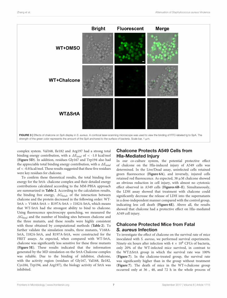
Chalcone Influences the SpA Display inIntact CellsStaphylococcus aureus SrtA anchors many different surfaceproteins in the cell wall, including SpA, a multifunctionalmolecule responsible for binding the Fcγ portion of hostimmunoglobulins (Falugi et al., 2013). Therefore, SpA is vital forimmune evasion, and because of its function, a change in theamount of SpA in the cell wall after the addition of chalconerequires investigation. In this assay, S. aureus cultured with orwithout chalcone was stained with FITC-labeled goat anti-rabbitIgG, which made the cells display a green color whose strengthwas determined by the amount of SpA in the cell wall. With theaid of confocal laser-scanning microscope, the content variationof SpA in the cell wall was investigated. The result showed thatthe WT1SrtA strain, used as a control, displayed much less SpAon its surface compared with the WT strain, and as expected,76 µM chalcone present in the cell culture caused significantreductions in the SpA display (Figure 3). These consequencesindicated that chalcone was capable of disturbing the assemblyof sortase-mediated SpA in the cell wall.
Chalcone Reduces the Adherence ofS. aureus to FibronectinIn the previous study, we have drawn the conclusion thatchalcone can visibly inhibit the SrtA-catalyzed transpeptidationat relatively low concentrations. The fibronectin-bindingproteins FnBPA and FnBPB are LPXTG proteins expressedby S. aureus. Fibronectin (Fn) is regarded as a bridge betweenthe bacterial adhesion FnBP and the mammalian cell integrin,accelerating the process of phagocytosis, thereby stimulatingthe internalization of bacteria; consequently, Fn and FnBPs playa vital role in the process of invading host cells (Foster andHöök, 1998; Roche et al., 2003). Because FnBPs anchored by S.aureus SrtA can make bacteria invasive, if their display in thecell wall is blocked by weakening SrtA activity, then bacterialinvasion would be decreased. For this reason, we employedFn-binding assays to determine if chalcone could reduce theadherence of S. aureus to Fn. As expected, the WT1SrtA strainhad a minimum adhesion rate to Fn owing to the damage toits Fn-binding function and no inhibition was observed in thesamples treated with chalcone (Figure 4A). However, when theWT strain was treated with chalcone at different concentrations,the adhesion rates decreased in turn, respectively, indicating thatthe effect occurred in a dose-dependent manner. The variancebetween the 38 µM chalcone group and the WT group was avery significant difference, as was the 76 µM chalcone group(Figure 4A).
Frontiers in Microbiology | www.frontiersin.org 5 September 2017 | Volume 8 | Article 1715

Zhang et al. Attenuation of Staphylococcus aureus Virulence
FIGURE 2 | Chalcone inhibits S. aureus Hla expression. (A) The hemolysis activity of culture supernatants by S. aureus co-cultured with chalcone. (B) Bacterial
supernatants pre-incubated with chalcone and then the hemolysis activity was determined. (C) Western blotting assay to detect the Hla expression in the bacterial
culture supernatants by S. aureus co-cultured with or without chalcone. (D) The relative gene expression of the hla gene and the agrA gene in S. aureus exposed to
chalcone at different concentrations. Three independent experiments were performed to obtain stable results. *P < 0.05 vs. the WT group and **P < 0.01 vs. the WT
group.
Chalcone Reduced Biofilm FormationBacteria enhance their ability to resist antibiotics and otherantimicrobial agents by embedding in biofilms (Pugliese andFavero, 2002; Cerca et al., 2005; Høiby et al., 2010). Biofilmphenotypes are promoted by the FnBPs, the major cell wallautolysin or the icaADBC-encoded polysaccharide intercellularadhesin/poly-N-acetylglucosamine (PIA/PNAG) (O’Neill et al.,2007, 2008; Clarissa Pozzi et al., 2012). To investigate whetherchalcone hindered biofilm formation, S. aureus biofilm formationwas determined with or without chalcone. After dye treatment,theWT1SrtA group was muchmore lightly stained than theWTgroup, and the visible difference between the biofilms formedunder the conditions of untreated and treated with 76 µMchalcone was quite significant (Figure 4B). These results were inaccordance with those of the quantitative analysis (Figure 4C).In summary, chalcone reduced biofilm formation, suggestingthat the inhibitory effect of chalcone on S. aureus SrtA hadoccurred.
Chalcone Weakened the S. aureus InvasionSrtA function is normally important for the display of sortase-mediated surface proteins, thus enabling bacteria to invadethe host cells. To examine how chalcone affects S. aureusinvasion, J774 cells were cultured with bacteria and chalconeat different concentrations. Not surprisingly, the numbers ofbacteria entering the cells in the WT1SrtA group showed a
significant decrease compared with the WT group (Figure 4D).Consistent with the hypothesis, the addition of a dose of chalconeto the experimental system significantly decreased the quantityof bacteria, indicating that chalcone weakened the S. aureusinvasion by inhibiting SrtA activity (Figure 4D).
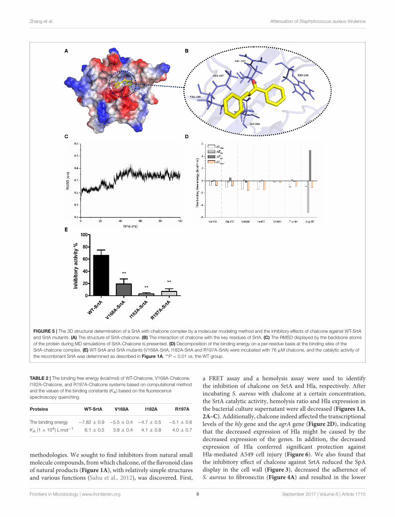
Molecular Dynamics Simulation forSrtA-ChalconeThrough the computational biology method, the potentialbinding mode of chalcone with SrtA in the active site wasexplored in this study. The chalcone was bound to SrtA, andaccording to the binding mode given (Figures 5A,B), it wasclear that chalcone could bind to SrtA via Van der Waalsand electrostatic interactions. During the time course of thesimulation, chalcone could localize to the catalytic pocket of SrtA(residue 160–200). In detail, the binding model of chalcone withSrtA revealed that chalcone could form strong interactions withVal166, Gly167, Val168, Ile182, Val193, and Arg197, respectively.The complex was found to reach equilibrium at 100 ns basedon the analysis of the root-mean-square deviations (RMSD) ofthe backbone Cα atoms (Figure 5C), which indicated that thecomplex system has reached equilibrium.
To explore the energy contributions from the residuesof the binding sites in the SrtA- chalcone complex, theenergy decomposition was calculated for the SrtA-chalcone
Frontiers in Microbiology | www.frontiersin.org 6 September 2017 | Volume 8 | Article 1715
Zhang et al. Attenuation of Staphylococcus aureus Virulence
FIGURE 3 | Effects of chalcone on SpA display in S. aureus. A confocal laser-scanning microscope was used to view the binding of FITC-labeled Ig to SpA. The
strength of the green color represents the amount of the SpA anchored to the surface of bacteria. Scale bar, 1 µm.
complex system. Val168, Ile182 and Arg197 had a strong totalbinding energy contribution, with a ∆Etotal of < -1.0 kcal/mol(Figure 5D). In addition, residues Gly167 and Trp194 also hadthe appreciable total binding energy contribution, with a ∆Etotalof< -0.8 kcal/mol. These results suggested that these five residueswere key residues for chalcone.
To confirm these theoretical results, the total binding freeenergy for the SrtA- chalcone complex and their detailed energycontributions calculated according to the MM-PBSA approachare summarized in Table 2. According to the calculation results,the binding free energy, ∆Gbind, of the interaction betweenchalcone and the protein decreased in the following order: WT-SrtA > V168A-SrtA > R197A-SrtA > I182A-SrtA, which meansthat WT-SrtA had the strongest ability to bind to chalcone.Using fluorescence spectroscopy quenching, we measured the∆Gbind and the number of binding sites between chalcone andthe three mutants, and these results were highly consistentwith those obtained by computational methods (Table 2). Tofurther validate the simulation results, three mutants, V168A-SrtA, I182A-SrtA, and R197A-SrtA, were constructed for theFRET assays. As expected, when compared with WT-SrtA,chalcone was significantly less sensitive for these three mutants(Figure 5E). These results indicated that the informationgenerated by the MD simulation on the SrtA-Chalcone complexwas reliable. Due to the binding of inhibitor, chalcone,with the activity region (residues of Gly167, Val168, Ile182,Cys184, Trp194, and Arg197), the biology activity of SrtA wasinhibited.
Chalcone Protects A549 Cells fromHla-Mediated InjuryIn our co-culture system, the potential protective effectof chalcone on the Hla-induced injury of A549 cells wasdetermined. In the Live/Dead assay, uninfected cells retainedgreen fluorescence (Figure 6A), and inversely, injured cellsretained red fluorescence. As expected, 38 µM chalcone showedan obvious reduction in cell injury, with almost no cytotoxiceffect observed in A549 cells (Figures 6B–E). Simultaneously,the LDH assay showed that treatment with chalcone couldsignificantly decrease the release of LDH into the supernatantsin a dose-independent manner compared with the control group,indicating less cell death (Figure 6E). Above all, the resultsshowed that chalcone had a protective effect on Hla–mediatedA549 cell injury.
Chalcone Protected Mice from FatalS. aureus InfectionTo investigate the effect of chalcone on the survival rate of miceinoculated with S. aureus, we performed survival experiments.Ninety-six hours after infection with 4 × 10⁸ CFUs of bacteria,only 20% of the WT-infected mice survived, in contrast tothe WT1SrtA group in which the survival rate was 100%(Figure 7). In the chalcone-treated group, the survival ratewas significantly higher than in the group without treatment(Figure 7). The death of mice in the WT+chalcone groupoccurred only at 36 , 48, and 72 h in the whole process of
Frontiers in Microbiology | www.frontiersin.org 7 September 2017 | Volume 8 | Article 1715

Zhang et al. Attenuation of Staphylococcus aureus Virulence
FIGURE 4 | Chalcone reduces the adhesion of S. aureus to Fn, biofilm formation and S. aureus invasion. (A) Adhesion rate of S. aureus to Fn in the presence of
different concentrations of chalcone. The WT1SrtA strain was incubated with 76 µM chalcone. (B) Photographs of biofilms grown in 96-well flat-bottom polystyrene
microtiter plates. (C) Quantification of the biofilm mass. (D) Chalcone weakened S. aureus invasion. Bacteria and J774 cells were cultured with chalcone at different
concentrations at 37◦C for 1 h, followed by gentamicin addition and incubation at 37◦C for 30 min, after which number of intracellular bacteria was determined. Three
independent experiments were performed to obtain stable results. *P < 0.05 vs. the WT group, **P < 0.01 vs. the WT group and NS represents no significance.
infection (Figure 7). These findings suggested that chalconecould prolong survival of the mice in S. aureus–inducedinfection.
DISCUSSION
Staphylococcus aureus is a major human pathogen and is also aleading cause of sepsis and infective endocarditis. The continuousspread of antibiotic-resistant strains, such as MRSA and VRSAmakes treatment very difficult (Lowy, 1998; Petti and Fowler,2003; Menichetti, 2005), and new valid strategies are greatlyneeded to address this severe situation. The virulence factors ofbacteria, such as adhesins, invasins and toxins, among others,facilitate infection by evading the immune response and leadingto colonization, spread and tissue damage (Lowy, 1998; Foster,2005; Rooijakkers et al., 2005). In addition, previous studies havedemonstrated that the vast majority of these various virulencefactors are nonessential for bacterial survival and are crucial in
the process of targeting host cells and causing disease (Lowy,1998; Berube and Bubeck, 2013). Some previous studies have alsodemonstrated that S. aureus lacking the genes encoding SrtA orHla will show a weakened bacterial virulence (Albus et al., 1991;Mcelroy et al., 1999; Mazmanian et al., 2000; Wardenburg et al.,2007). For this reason, therapeutic agents targeting virulencefactors, such as SrtA and Hla, which do not threaten survival,may not lead to the development of resistance as quickly asconventional antibiotics typically do, and this will have importantimplications for S. aureus infection. Some agents targeting S.aureus SrtA or Hla have been found previously (Liu et al.,2015; Zhou et al., 2015; Zhang et al., 2016). However, as thesecompounds are targeted only at one kind of virulence factor, thelimitations for clinical treatment will certainly exist. A hypothesiswas then proposed as to whether we could find an agent targetingSrtA and Hla simultaneously to produce a better therapeuticeffect.
The scope of screening for SrtA and Hla inhibitors waswide, involving natural, synthetic and high-throughput screening
Frontiers in Microbiology | www.frontiersin.org 8 September 2017 | Volume 8 | Article 1715
Zhang et al. Attenuation of Staphylococcus aureus Virulence
FIGURE 5 | The 3D structural determination of a SrtA with chalcone complex by a molecular modeling method and the inhibitory effects of chalcone against WT-SrtA
and SrtA mutants. (A) The structure of SrtA-chalcone. (B) The interaction of chalcone with the key residues of SrtA. (C) The RMSD displayed by the backbone atoms
of the protein during MD simulations of SrtA-Chalcone is presented. (D) Decomposition of the binding energy on a per-residue basis at the binding sites of the
SrtA-chalcone complex. (E) WT-SrtA and SrtA mutants (V168A-SrtA, I182A-SrtA and R197A-SrtA) were incubated with 76 µM chalcone, and the catalytic activity of
the recombinant SrtA was determined as described in Figure 1A. **P < 0.01 vs. the WT group.
TABLE 2 | The binding free energy (kcal/mol) of WT-Chalcone, V168A-Chalcone,
I182A-Chalcone, and R197A-Chalcone systems based on computational method
and the values of the binding constants (KA) based on the fluorescence
spectroscopy quenching.
Proteins WT-SrtA V168A I182A R197A
The binding energy −7.82 ± 0.9 −5.5 ± 0.4 −4.7 ± 0.5 −5.1 ± 0.6
KA (1 × 10⁴) L·mol−1 6.1 ± 0.5 3.8 ± 0.4 4.1 ± 0.8 4.0 ± 0.7
methodologies. We sought to find inhibitors from natural smallmolecule compounds, fromwhich chalcone, of the flavonoid classof natural products (Figure 1A), with relatively simple structuresand various functions (Sahu et al., 2012), was discovered. First,
a FRET assay and a hemolysis assay were used to identifythe inhibition of chalcone on SrtA and Hla, respectively. Afterincubating S. aureus with chalcone at a certain concentration,the SrtA catalytic activity, hemolysis ratio and Hla expression inthe bacterial culture supernatant were all decreased (Figures 1A,2A–C). Additionally, chalcone indeed affected the transcriptionallevels of the hly gene and the agrA gene (Figure 2D), indicatingthat the decreased expression of Hla might be caused by thedecreased expression of the genes. In addition, the decreasedexpression of Hla conferred significant protection againstHla-mediated A549 cell injury (Figure 6). We also found thatthe inhibitory effect of chalcone against SrtA reduced the SpAdisplay in the cell wall (Figure 3), decreased the adherence ofS. aureus to fibronectin (Figure 4A) and resulted in the lower
Frontiers in Microbiology | www.frontiersin.org 9 September 2017 | Volume 8 | Article 1715

Zhang et al. Attenuation of Staphylococcus aureus Virulence
FIGURE 6 | Chalcone protects A549 cells from injury mediated by S. aureus
Hla. Live/Dead reagent-A549 cells were observed with fluorescent imaging.
(A) The untreated A549 cells. (B) A549 cells treated with 38 µM chalcone only.
(C) A549 cells infected with S. aureus culture supernatants harvested in the
absence of chalcone. (D) A549 cells treated with S. aureus culture
supernatants harvested in the presence of 38 µM chalcone. (E) The LDH
release by A549 cells treated with S. aureus culture supernatants harvested
previously and 38 µM chalcone only, respectively. Scale bar, 10 µm. **P <
0.01 vs. the WT group.
biofilm formation (Figures 4B,C). In addition, as far as we know,our study is the first to use a cell invasion assay to evaluate theinhibitory activity of a natural compound on SrtA, which showedthat the quantity of bacterial entry into cells was significantlydecreased by chalcone (Figure 4D). To explore the interactionmechanism between chalcone and SrtA, a molecular dynamicssimulation for a SrtA-chalcone complex system was carried out.By means of molecular dynamics simulation, it was found thatchalcone could localize to the catalytic pocket of SrtA (residues160–200), which is very close to the binding site of substrate.
FIGURE 7 | Effects of chalcone on survival rates after 96 hours in C57BL/6J
mice. Mice were infected with 4 × 10⁸ CFUs of S. aureus and the WT1SrtA
strain. Treatment with chalcone (150 mg/kg, twice a day) was initiated after
infection. **P < 0.01 vs. the WT group.
Due to the binding of chalcone to SrtA, the binding of substrateto SrtA was blocked, leading to the loss of biological activityof SrtA (Figure 5). It is worth mentioning that just at a muchlower concentration than the MIC, chalcone could significantlystop S. aureus SrtA-mediated transpeptidation and the hemolysisactivity of Hla in vitro by inhibiting SrtA activity and theexpression of Hla, respectively, instead of killing bacteria directly(Figure 1B), which means a less selective pressure for bacteriaand a lower risk of drug resistance.More importantly, a C57BL/6Jmouse model was established to determine the therapeutic effectof chalcone in vivo, and treatment with chalcone significantlyattenuated the virulence of S. aureus and protected mice frominfection caused by the bacteria (Figure 7). To the best of ourknowledge, this is the first report of the discovery and proof ofan inhibitor targeting S. aureus SrtA and Hla simultaneously, andthus, this kind of inhibitor can be considered as a novel promisingcandidate against S. aureus infection.
In conclusion, our study discovered that the natural smallcompound chalcone could effectively inhibit S. aureus SrtAactivity and the expression of Hla by occupying the sites of theenzyme to prevent anchoring of surface proteins and decreasingtranscription levels of the hla gene and the agrA gene, therebyattenuating S. aureus virulence both in vitro and in vivo. Thesefindings could be the foundation for further design of novel anti-infection agents, and a SrtA-Hla-centered strategy has emergedaccordingly, thus making a contribution for opening a newhorizon for the treatment of S. aureus infection.
AUTHOR CONTRIBUTIONS
JW and XN conceived and designed the experiments. BZ,ZT, XL, and GL performed the experiments. XD contributedreagents/materials/analysis tools. JW, BZ, and XD wrote thepaper.
FUNDING
This work was supported by the National Key Technology R&DProgram (No. 2016YFD05013), the National Basic ResearchProgram of China (grant 2013CB127205) and the ProjectFunded by China Postdoctoral Science Foundation (Project No.2016M591486).
Frontiers in Microbiology | www.frontiersin.org 10 September 2017 | Volume 8 | Article 1715
Zhang et al. Attenuation of Staphylococcus aureus Virulence
REFERENCES
Albus, A., Arbeit, R. D., and Lee, J. C. (1991). Virulence of Staphylococcus aureus
mutants altered in type 5 capsule production. Infect. Immun. 59, 1008–1014.
Bandyopadhyay, S., Valder, C. R., Huynh, H. G., Ren, H., and Allison, W. S. (2002).
The beta G156C substitution in the F1-ATPase from the thermophilic Bacillus
PS3 affects catalytic site cooperativity by destabilizing the closed conformation
of the catalytic site. Biochemistry 41, 14421–14429. doi: 10.1021/bi026243g
Berube, B. J., and Bubeck, W. J. (2013). Staphylococcus aureus α-Toxin: nearly a
century of intrigue. Toxins (Basel) 5, 1140–1166. doi: 10.3390/toxins5061140
Bubeck,W. J., andOlaf, S. (2008). Vaccine protection against Staphylococcus aureus
pneumonia. J. Exp. Med. 205, 287–294. doi: 10.1084/jem.20072208
Cerca, N., Martins, S., Cerca, F., Jefferson, K. K., Pier, G. B., Oliveira, R.,
et al. (2005). Cerca, N. Comparative assessment of antibiotic susceptibility
of coagulase negative staphylococci in biofilm versus planktonic culture as
assessed by bacterial enumeration or rapid XTT colorimetry. J. Antimicrob.
Chemother. 56, 331–336. doi: 10.1093/jac/dki217
Clancy, K. W., Melvin, J. A., and Mccafferty, D. G. (2010). Sortase transpeptidases:
insights into mechanism, substrate specificity, and inhibition. Biopolymers 94,
385–396. doi: 10.1002/bip.21472
Clarissa Pozzi, E. M. W., Justine Rudkin, K., Carolyn Schaeffer, R.,
Amanda Lohan, J., Pin, T., Brendan Loftus, J., et al. (2012). Methicillin
resistance alters the biofilm phenotype and attenuates virulence in
Staphylococcus aureus device-associated infections. PLoS Pathog. 8:e1002626.
doi: 10.1371/journal.ppat.1002626
Coates, R., Moran, J., and Horsburgh, M. J. (2014). Staphylococci: colonizers and
pathogens of human skin. Future Microbiol. 9, 75–91. doi: 10.2217/fmb.13.145
Dinges, M. M., Orwin, P. M., and Schlievert, P. M. (2000). Exotoxins of
Staphylococcus aureus. Clin. Microbiol. Rev. 13, 16–34, table of contents.
doi: 10.1128/CMR.13.1.16-34.2000
Dong, J., Qiu, J., Zhang, Y., Lu, C., Dai, X., Wang, J., et al. (2013).
Oroxylin A inhibits hemolysis via hindering the self-assembly of α-
hemolysin heptameric transmembrane pore. PLoS Comput. Biol. 9:e1002869.
doi: 10.1371/journal.pcbi.1002869
Falugi, F., Kim, H. K., Missiakas, D.M., and Schneewind, O. (2013). Role of protein
a in the evasion of host adaptive immune responses by Staphylococcus aureus.
MBio 4:e00575. doi: 10.1128/mBio.00575-13
Fischetti, V. A., Pancholi, V., and Schneewind, O. (1990). Conservation
of a hexapeptide sequence in the anchor region of surface
proteins from Gram-positive cocci. Mol. Microbiol. 4, 1603–1605.
doi: 10.1111/j.1365-2958.1990.tb02072.x
Foster, T. J. (2005). Immune evasion by staphylococci. Nat. Rev. Microbiol. 3,
948–958. doi: 10.1038/nrmicro1289
Foster, T. J., and Höök, M. (1998). Surface protein adhesins of Staphylococcus
aureus. Trends Microbiol. 6:484. doi: 10.1016/S0966-842X(98)01400-0
Gould, I. M. (2013). Treatment of bacteraemia: meticillin-resistant Staphylococcus
aureus (MRSA) to vancomycin-resistant S. aureus (VRSA). Int. J. Antimicrob.
Agents 42, 23–30. doi: 10.1016/j.ijantimicag.2013.04.006
Høiby, N., Bjarnsholt, T., Givskov, M., Molin, S., and Ciofu, O. (2010). Antibiotic
resistance of bacterial biofilms. Int. J. Antimicrob. Agents 35, 322–332.
doi: 10.1016/j.ijantimicag.2009.12.011
Hu, R., Barbault, F., Maurel, F., Delamar, M., and Zhang, R. (2010). Molecular
dynamics simulations of 2-Amino-6-arylsulphonylbenzonitriles analogues as
HIV inhibitors: interaction modes and binding free energies. Chem. Biol. Drug
Des. 76, 518–526. doi: 10.1111/j.1747-0285.2010.01028.x
Ippolito, G., Leone, S., Lauria, F. N., Nicastri, E., Wenzel, R. P., and Ippolito, G.
(2010). Methicillin-resistant Staphylococcus aureus: the superbug. Int. J. Infect.
Dis. 14(Suppl. 4), S7–S11. doi: 10.1016/j.ijid.2010.05.003
Jurasekova, Z., Marconi, G., Sanchez-Cortes, S., and Torreggiani, A. (2009).
Spectroscopic and molecular modeling studies on the binding of the
flavonoid luteolin and human serum albumin. Biopolymers 91, 917–927.
doi: 10.1002/bip.21278
Levy, S. B., Fitzgerald, G. B., and Macone, A. B. (1976). Spread of antibiotic-
resistant plasmids from chicken to chicken and from chicken to man. Nature
260, 40–42. doi: 10.1038/260040a0
Li, H., Chen, Y., Zhang, B., Niu, X., Song, M., Luo, Z., et al. (2016). Inhibition
of sortase A by chalcone prevents Listeria monocytogenes infection. Biochem.
Pharmacol. 106, 19–29. doi: 10.1016/j.bcp.2016.01.018
Lin, W., Bi, C., Cai, H., Liu, B., Zhong, X., Deng, X., et al. (2015). The therapeutic
effect of chlorogenic acid against Staphylococcus aureus infection through
sortase A inhibition. Front. Microbiol. 6:1031. doi: 10.3389/fmicb.2015.01031
Liu, S., Zhou, X., Li, W., Zhang, H., Zhang, B., Li, G., et al. (2015). Diosmetin
inhibits the expression of alpha-hemolysin in Staphylococcus aureus. Antonie
Van Leeuwenhoek 108:383. doi: 10.1007/s10482-015-0491-6
Lowy, F. D. (1998). Staphylococcus aureus infections. N. Eng. J. Med. 339,
2026–2027.
Lv, Z., Wang, H. S., and Niu, X. D. (2013). Molecular dynamics simulations
reveal insight into key structural elements of aaptamines as sortase
inhibitors with free energy calculations. Chem. Phys. Lett. 585, 171–177.
doi: 10.1016/j.cplett.2013.08.097
Mazmanian, S. K., Liu, G., Jensen, E. R., Lenoy, E., and Schneewind, O. (2000).
Staphylococcus aureus sortase mutants defective in the display of surface
proteins and in the pathogenesis of animal infections. Proc. Natl. Acad. Sci.
U.S.A. 97, 5510–5515. doi: 10.1073/pnas.080520697
Mcelroy, M. C., Harty, H. R., Hosford, G. E., Boylan, G. M., Pittet, J. F., and Foster,
T. J. (1999). Alpha-toxin damages the air-blood barrier of the lung in a rat
model of Staphylococcus aureus-induced pneumonia. Infect. Immun. 67:5541.
Menichetti, F. (2005). Current and emerging serious Gram-
positive infections. Clin. Microbiol. Infect. 11(Suppl. S3), 22–28.
doi: 10.1111/j.1469-0691.2005.01138.x
Morris, G. M., Huey, R., Lindstrom, W., Sanner, M. F., Belew, R. K.,
Goodsell, D. S., et al. (2009). AutoDock4 and AutoDockTools4: automated
docking with selective receptor flexibility. J. Comput. Chem. 30, 2785–2791.
doi: 10.1002/jcc.21256
Niu, X., Qiu, J., Wang, X., Gao, X., Dong, J., Wang, J., et al. (2013).
Molecular insight into the inhibition mechanism of cyrtominetin to α-
hemolysin by molecular dynamics simulation. Eur. J. Med. Chem. 62, 320–328.
doi: 10.1016/j.ejmech.2013.01.008
O’Neill, E., Pozzi, C., Houston, P., Humphreys, H., Robinson, D. A., Loughman, A.,
et al. (2008). A novel Staphylococcus aureus biofilm phenotype mediated by the
fibronectin-binding proteins, FnBPA and FnBPB. J. Bacteriol. 190, 3835–3850.
doi: 10.1128/JB.00167-08
O’Neill, E., Pozzi, C., Houston, P., Smyth, D., Humphreys, H., Robinson, D.
A., et al. (2007). Association between methicillin susceptibility and biofilm
regulation in Staphylococcus aureus isolates from device-related infections. J.
Clin. Microbiol. 45:1379. doi: 10.1128/JCM.02280-06
Petti, C. A., and Fowler, V. G. Jr. (2003). Staphylococcus aureus bacteremia and
endocarditis. Cardiol. Clin. 21:219. doi: 10.1016/S0733-8651(03)00030-4
Pugliese, G., and Favero, M. S. (2002). biofilms: survival mechanisms
of clinically relevant microorganisms. Clin. Microbiol. Rev. 15:167.
doi: 10.1128/CMR.15.2.167-193.2002
Qiu, J., Li, H., Meng, H., Hu, C., Li, J., Luo, M., et al. (2011). Impact of luteolin on
the production of alpha-toxin by Staphylococcus aureus. Lett. Appl. Microbiol.
53:238. doi: 10.1111/j.1472-765X.2011.03098.x
Ragle, B. E., Karginov, V. A., and Bubeck,W. J. (2010). Prevention and treatment of
Staphylococcus aureus pneumonia with a -cyclodextrin derivative. Antimicrob.
Agents Chemother. 54, 298–304. doi: 10.1128/AAC.00973-09
Roche, F. M., Massey, R., Peacock, S. J., Day, N. P., Visai, L., Speziale,
P., et al. (2003). Characterization of novel LPXTG-containing proteins of
Staphylococcus aureus identified from genome sequences. Microbiology 149(Pt
3), 643. doi: 10.1099/mic.0.25996-0
Rooijakkers, S. H. M., Kessel, K. P. M. V., and Strijp, J. A. G. V.
(2005). Staphylococcal innate immune evasion. Trends Microbiol. 13:596.
doi: 10.1016/j.tim.2005.10.002
Sahu, N. K., Balbhadra, S. S., Choudhary, J., and Kohli, D. V. (2012). Exploring
pharmacological significance of chalcone scaffold: a review. Curr. Med. Chem.
19, 209–225. doi: 10.2174/092986712803414132
Scott, J. R., and Barnett, T. C. (2006). Surface proteins of gram-positive
bacteria and how they get there. Annu. Rev. Microbiol. 60, 397–423.
doi: 10.1146/annurev.micro.60.080805.142256
Tonthat, H., Liu, G., Mazmanian, S. K., Faull, K. F., and Schneewind, O. (1999).
Purification and characterization of sortase, the transpeptidase that cleaves
surface proteins of Staphylococcus aureus at the LPXTGmotif. Proc. Natl. Acad.
Sci. U.S.A. 96:12424. doi: 10.1073/pnas.96.22.12424
Wallockrichards, D. J., Marleswright, J., Clarke, D. J., Maitra, A., Dodds, M.,
Hanley, B., et al. (2015). Molecular basis of Streptococcus mutans sortase A
Frontiers in Microbiology | www.frontiersin.org 11 September 2017 | Volume 8 | Article 1715

Zhang et al. Attenuation of Staphylococcus aureus Virulence
inhibition by the flavonoid natural product trans-chalcone. Chem. Commun.
51:10483. doi: 10.1039/C5CC01816A
Wardenburg, J. B., Patel, R. J., and Schneewind, O. (2007). Surface proteins
and exotoxins are required for the pathogenesis of Staphylococcus aureus
pneumonia. Infect. Immun. 75, 1040–1044. doi: 10.1128/IAI.01313-06
Zhang, B., Wang, X., Wang, L., Chen, S., Shi, D., and Wang, H. (2016).
Molecular mechanism of the flavonoid natural product dryocrassin
ABBA against Staphylococcus aureus sortase A. Molecules 21:1428.
doi: 10.3390/molecules21111428
Zhang, J., Liu, H., Zhu, K., Gong, S., Dramsi, S., Wang, Y. T., et al.
(2014). Antiinfective therapy with a small molecule inhibitor of
Staphylococcus aureus sortase. Proc. Natl. Acad. Sci. U.S.A. 111, 13517–13522.
doi: 10.1073/pnas.1408601111
Zhou, X., Liu, S., Li, W., Zhang, B., Liu, B., Liu, Y., et al. (2015).
Phloretin derived from apple can reduce alpha-hemolysin expression
in methicillin-resistant Staphylococcus aureus USA300. World J.
Microbiol. Biotechnol. 31, 1259–1265. doi: 10.1007/s11274-015-
1879-1
Conflict of Interest Statement: The authors declare that the research was
conducted in the absence of any commercial or financial relationships that could
be construed as a potential conflict of interest.
Copyright © 2017 Zhang, Teng, Li, Lu, Deng, Niu and Wang. This is an open-
access article distributed under the terms of the Creative Commons Attribution
License (CC BY). The use, distribution or reproduction in other forums is permitted,
provided the original author(s) or licensor are credited and that the original
publication in this journal is cited, in accordance with accepted academic practice.
No use, distribution or reproduction is permitted which does not comply with these
terms.
Frontiers in Microbiology | www.frontiersin.org 12 September 2017 | Volume 8 | Article 1715