Cervical Spine
30
UNC Radiology Residency Educational Scholarship University of North Carolina School of Medicine Department of Radiology 2020 Jeremy Kim MD Sheryl Jordan MD Cervical Spine
Transcript of Cervical Spine

UNC Radiology Residency Educational Scholarship
University of North Carolina School of Medicine Department of Radiology 2020
Jeremy Kim MD Sheryl Jordan MD
Cervical Spine

Learning objectives
By the end of this activity, participants will be able to:
1. Understand cervical spine anatomy
2. Know what test to expect and order in the setting of trauma
3. Relay mechanisms of cervical spine injury

Module Outline
I. Background
II. Anatomy
III. Cases
III. Wrap up/Questions

Background
• 7-10k cervical spine injuries in US annually
• ~50% have associated spinal cord trauma
• Alert stable patients without distracting injuriesClinical decision rules to guide imaging
NEXUS: National Emergency X-Radiograph Utilization Study
CCR: Canadian C-Spine Rule
• Symptomatic, disoriented, obtunded patients
IMAGING NECESSARY

Canadian C Spine Rule Note: Complete exam of the cervical spine MUST include all of C7 and at least superior endplate of T1

If cervical spine trauma known -> CT

If ligamentous or spine cord injury -> CT + MR

Anatomy

Anatomy

Anatomy: Frontal radiograph & CT

Anatomy: Lateral radiograph

Anatomy: Sagittal CT

Anatomy: Open mouth odontoid & CT

Anatomy
Atlanto-occipital alignment:Anterior margin of foramen magnum should line up with densBasion to dens interval <10 mm (BDI)
Normal Anatomy:Axial CT image of C1 ring

Three Column ConceptTHREE COLUMN CONCEPTAnterior, Middle, and Posterior columns as indicated
UNSTABLE: fracture of middle column and either anterior or posterior column

Module Outline
I. Background
II. Anatomy
III. Cases
III. Wrap up/Questions

34yoM riding an ATV, lost control and crashed
Cervical spine CT ordered as part of trauma CT series

Findings: On 3 sagittal CT images, there is Basion-dens interval excessive, >10 mm, in this case almost 5 cm = Atlanto-occipital dislocation
Atlanto-occipital dislocation:
More common in childrenNearly always fatalHyperextension with distraction injuryUNSTABLE
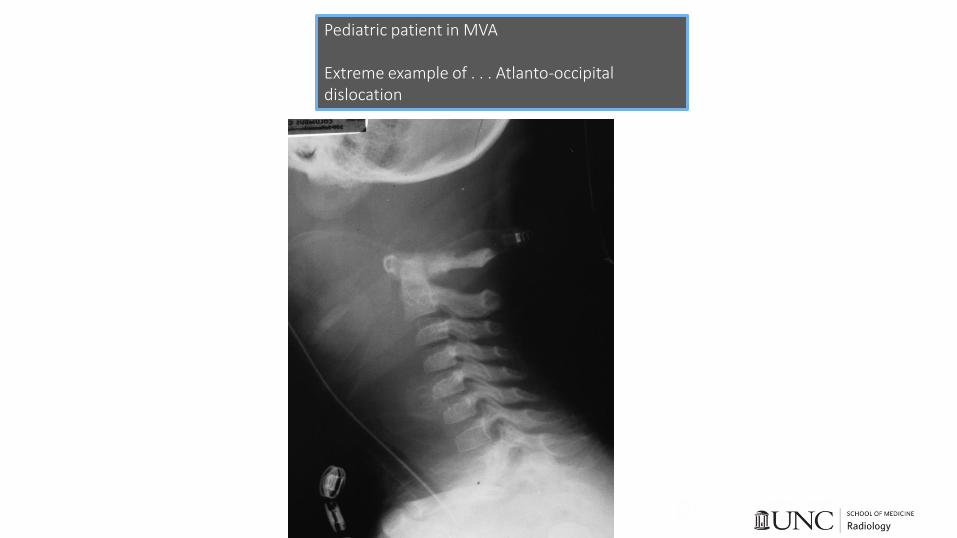
Pediatric patient in MVA
Extreme example of . . . Atlanto-occipital dislocation
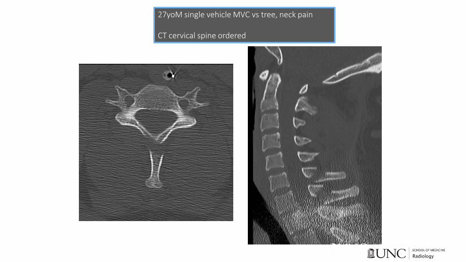
27yoM single vehicle MVC vs tree, neck pain
CT cervical spine ordered

Findings: On axial and sagittal cervical spine CT, postero-inferiorly displaced fractures of C6 and C7 spinous processes = Clay Shoveler fractures
Clay Shoveler fracture:
Hyperflexion injury
Most commonly C6, C7, or T1
Usually with contraction of paraspinous muscles pulling on spinous processes
STABLE

31yoM head on MVC, neck pain
Cervical spine CT ordered

Case #5:On 3 sagittal and 1 axial cervical spine CT, grade II-III anterolisthesis of C6 on C7, posterior elements fractures (C6), bilateral jumped and locked facets with fractures = Traumatic C6-7 spondylolisthesis with bilateral locked facets
Bilateral Locked Facets:
Anterior dislocation of vertebral body
Jumping of inferior articular process over the superior articular process of vertebral body below – locked in this position
Can be uni- or bilateral
Extreme flexion type injury of head/neck
High risk of cord damageUNSTABLE

45yoF brought in by EMS following high speed motorcycle collision
Cervical spine CT ordered as part of trauma protocol
Case #6:

On 1 axial and 2 sagittal cervical spine CT images, fractures of the bilateral pars interarticularis of C2 = Hangman fracture
Hangman fracture:
Also known as traumatic spondylolisthesis of the axis
Result of hyperextension and distraction, classically after high speed MVC with chin hitting dashboardSTABLE
(Despite name, not commonly seen in hangings - more likely cause of death in that case = asphyxiation)

19yoF boating at a lake with friends, dove head first into shallow water and now with neck pain
Radiographs were ordered (before you saw the patient)

Findings: On open mouth odontoid and lateral radiographs, lateral masses of C1 do not line up appropriately and there is a posterior C1 arch fracture

Findings: On CT there are C1 anterior and posterior arch fractures (normal is intact C1 ring) = Jefferson fracture
Jefferson fracture:
Compression fracture of bony C1 ring involving both anterior and posterior C1 archesAxial loading injuryTransverse ligament may also be injuredTreatment is conservative with hard collarSTABLE
Exception: transverse ligament disrupted (unstable)
Wrap Up
• If C-spine trauma -> think CT!
• If ligamentous or spinal cord injury -> think MR!
• 4 spinal lines: anterior & posterior vertebral, spinolaminar, posterior spinous
• UNSTABLE: middle column + either anterior or posterior
• Named fractures: Jefferson, Clay Shoveler, Hangman

More at www.rads.web.unc.edu www.msrads.web.unc.eduand @UNCRadRes
Thank you!