


Cernunnos deficiency reduces thymocyte lifespan and alters the T ...
36
Cernunnos deficiency reduces thymocyte lifespan and 1 alters the T cell repertoire in mice and humans 2 3 Gabriella Vera, MSc 1,2,* , Paola Rivera-Munoz, PhD, 1,2,* , Vincent Abramowski, MSc 1,* , 4 Laurent Malivert, PhD 1,2 , Annick Lim, PhD 4 , Christine Bole-Feysot, PhD 2 , Christelle 5 Martin, PhD 5 , Benoit Florkin, MD 6 , Sylvain Latour, PhD 1,2 , Patrick Revy, PhD 1,2 , and Jean- 6 Pierre de Villartay,PhD 1,2,3# 7 8 1 Laboratory “Genome Dynamics in the Immune System”, INSERM U768, 75015 Paris, 9 France; 10 2 Université Paris Descartes-Sorbonne Paris Cité, Institut Imagine, Site Necker, IFR94, 11 75015 Paris , France; 12 3 Assistance Publique-Hôpitaux de Paris, Service d’Immunologie et d’Hématologie 13 Pédiatrique, Hôpital Necker Enfants Malades, 75015 Paris, France; 14 4 Département d'Immunologie, Institut Pasteur, 75724 Paris, France; 15 5 SEAT, CNRS UPS44, 94800 Villejuif, France; 16 6 Service de Pédiatrie, Centre Hospitalier Universitaire (CHU) de la Citadelle, Liège, 17 Belgium. 18 19 *: These authors contributed equally to this work 20 #: Correspondence to: Dr. Jean-Pierre de Villartay 21 INSERM U768, Hôpital Necker 22 149 rue de Sèvres, 75015 Paris 23 tel: +33 1 44 49 50 81 24 fax: +33 1 42 73 06 40 25 [email protected] 26 27 28 Running Title: Cernunnos and T cell repertoire 29 30 31 Word count M&M: 644 words 32 Character count Intro/Res/Disc/Fig leg: 26153 char 33 34 Copyright © 2012, American Society for Microbiology. All Rights Reserved. Mol. Cell. Biol. doi:10.1128/MCB.01057-12 MCB Accepts, published online ahead of print on 3 December 2012 on April 9, 2018 by guest http://mcb.asm.org/ Downloaded from
-
Upload
truongnguyet -
Category
Documents
-
view
220 -
download
1
Transcript of Cernunnos deficiency reduces thymocyte lifespan and alters the T ...

Cernunnos deficiency reduces thymocyte lifespan and 1
alters the T cell repertoire in mice and humans 2
3
Gabriella Vera, MSc1,2,*, Paola Rivera-Munoz, PhD,1,2,*, Vincent Abramowski, MSc1,*, 4
Laurent Malivert, PhD1,2, Annick Lim, PhD4, Christine Bole-Feysot, PhD2, Christelle 5
Martin, PhD5, Benoit Florkin, MD6, Sylvain Latour, PhD1,2, Patrick Revy, PhD1,2 , and Jean-6
Pierre de Villartay,PhD1,2,3# 7
8
1 Laboratory “Genome Dynamics in the Immune System”, INSERM U768, 75015 Paris, 9
France; 10
2 Université Paris Descartes-Sorbonne Paris Cité, Institut Imagine, Site Necker, IFR94, 11
75015 Paris , France; 12
3 Assistance Publique-Hôpitaux de Paris, Service d’Immunologie et d’Hématologie 13
Pédiatrique, Hôpital Necker Enfants Malades, 75015 Paris, France; 14
4 Département d'Immunologie, Institut Pasteur, 75724 Paris, France; 15
5 SEAT, CNRS UPS44, 94800 Villejuif, France; 16
6 Service de Pédiatrie, Centre Hospitalier Universitaire (CHU) de la Citadelle, Liège, 17
Belgium. 18
19
*: These authors contributed equally to this work 20
#: Correspondence to: Dr. Jean-Pierre de Villartay 21
INSERM U768, Hôpital Necker 22
149 rue de Sèvres, 75015 Paris 23
tel: +33 1 44 49 50 81 24
fax: +33 1 42 73 06 40 25
27
28
Running Title: Cernunnos and T cell repertoire 29
30
31
Word count M&M: 644 words 32
Character count Intro/Res/Disc/Fig leg: 26153 char 33
34
Copyright © 2012, American Society for Microbiology. All Rights Reserved.Mol. Cell. Biol. doi:10.1128/MCB.01057-12 MCB Accepts, published online ahead of print on 3 December 2012
on April 9, 2018 by guest
http://mcb.asm
.org/D
ownloaded from

Vera et al.
2
Abstract 35
Cernunnos is a DNA repair factor of the Non Homologous End Joining machinery. Its 36
deficiency in humans causes radiosensitive SCID with microcephaly, characterized in part 37
by a profound lymphopenia. In contrast to the human condition, the immune system of 38
Cernunnos KO mice is not overwhelmingly affected. In particular, Cernunnos is dispensable 39
during V(D)J recombination in lymphoid cells. Nevertheless, the viability of thymocytes is 40
reduced in Cernu KO mice, owing to the chronic activation of a P53-dependent DNA 41
damage response. This translates into the qualitative alteration of the T cell repertoire, in 42
which the most distal Vα and Jα segments are missing. This results in the contraction of 43
discrete T cell populations such as iNKT and MAIT in both humans and mice. 44
45
on April 9, 2018 by guest
http://mcb.asm
.org/D
ownloaded from

Vera et al.
3
Introduction 46
The immune system is the site of intense genome dynamics, in particular during the 47
development and maturation of B and T lymphocytes in bone marrow and thymus when 48
antigen receptor genes are rearranged through V(D)J recombination prior to their 49
expression. DNA damages are also likely to occur during the several phases of intense 50
proliferation, which accompany the development of B and T cells . 51
V(D)J recombination is the prototypical example for the generation of programmed DNA 52
double strand break (DNA-dsb) during lymphoid development through the activity of 53
Recombination activating genes 1 and 2 (Rag1/2) on immunoglobulin (Ig) and T cell 54
receptor (TCR) genes of (see Helmink et al.(15) for a recent review). The resulting DNA-55
dsb is resolved by the Non Homologous End Joining (NHEJ) DNA repair pathway, 56
composed of seven core components (see Lieber et al.(24) for a recent review). The 57
Cernunnos/Xrcc4/DNA-Ligase IV complex ultimately reseals the DNA-dsb. Cernunnos, 58
also known as Xrcc4 like factor (XLF), is the last NHEJ factor that was independently 59
identified through the survey of RS-SCID patients(7) and a yeast two-hybrid screen with 60
Xrcc4 as a bait(1). Cernunnos and Xrcc4 adopt the same overall three-dimensional crystal 61
structure(2, 22), and together with DNA-Ligase IV are parts of the same complex(1, 8). 62
Cernunnos stimulates the DNA joining activity of the Xrcc4/DNA-ligaseIV complex(25, 63
30). 64
V(D)J recombination constitutes a central checkpoint in the development of the immune 65
system, as its defect leads to abortive B and T cell maturation in vivo, resulting in severe 66
combined immune deficiency (SCID) (9), but its first recognized function is the generation 67
of a diverse antigenic repertoire through the combinatorial association of Variable, 68
Diversity, and Joining segments that encode the Variable domains of both Ig and TCRs(5). 69
Numerous examples show that a reduced V(D)J recombinase activity affects the extent of 70
on April 9, 2018 by guest
http://mcb.asm
.org/D
ownloaded from

Vera et al.
4
antigenic diversity of immune receptors in mice and humans. The resulting immune 71
deregulation may then lead to autoimmunity, increased susceptibility to infections, or to the 72
development of various forms of cancer(35). 73
A Cernunnos KO mouse in which deletion of exons 4-5 caused in-frame alternative splicing 74
of exon 3 to 6 and residual (less than 1% of wt.) protein expression was previously 75
reported(21). In the present study we used a different gene targeting approach to develop a 76
complete Cernunnos null mouse model to explore the role of Cernunnos in the development 77
and maturation of the murine immune system in comparison to what is known in Cernunnos 78
deficient human patients. 79
80
Materials and Methods 81
Generation of Cernu-/- mice 82
Cernu-/- mice were developed at the Institut Clinique de la Souris (ICS, Illkirch, France) by 83
flanking exon 4 with loxP sites (Figure 1). Mice were genotyped by standard PCR on tail 84
DNA using the following primers: Cernu4R (5’-GTCCCCAGCTGTTAAGAGTTTC-3’) for 85
KO and Flox ; CernuExo4F (5’-GGATGAAGGACCTTGAGATCC-3’) for flox ; Cernu3F 86
(5’-CTATGGAAGCCAGGAGAGAATG-3’) for KO. All animals were maintained in a 87
specific pathogen-free environment. Analyses were performed on Cernu KO and littermate 88
control animals with a mix B6/129 background. P53 KO mice were on a mix B6/129 89
background as well. All experiments and procedures were performed in compliance with the 90
French Ministry of Agriculture’s Regulations for Animal Experiments (Act no. 87847, 19 91
October 1987, as modified in May 2001). 92
93 Analysis of lymphocyte populations 94
Cell phenotyping was performed on blood, thymus, bone marrow, lymph nodes, and splenic 95
lymphoid populations by four-color fluorescence analysis as previously described(33). 96
on April 9, 2018 by guest
http://mcb.asm
.org/D
ownloaded from
Vera et al.
5
iNKT cell determination was performed on splenocytes from 4-8 weeks old mice by staining 97
with APC mouse-CD1d tetramer loaded, or not, with αGal-cer (kindly provided NIH 98
Tetramer Core Facility), and 2.4G2 anti-Fc antibody and FITC anti-mouse TCRβ (BD 99
bioscience) and PE anti-mouse CD3ε or anti-mouse NK1.1 (Becton Dickson). Human 100
iNKT(29) and MAIT(26) cells were determined using anti-Va24 and anti-Va7.2 antibodies 101
on PBLs. 102
103
V(D)J recombination assay in thymocytes 104
CD4-CD8- thymocytes were negatively purified by magnetic sorting and infected with the 105
MX-RSS12/23 supernatant(23). The level of V(D)J recombination was determined 48h after 106
transduction by scoring the GFP+ (rearranged) cells among the huCD4+ transduced cells. 107
Thymocytes from Artemis -/- mice were used as negative control. 108
109
Thymocyte proliferation assay in vivo 110
Cernu KO mice were bred into the Rag1-/- background and 6-8 wks. old double KO mice 111
were injected i.p. with 100µg of anti-CD3 antibody to mimic the TCR-β selection induced 112
thymocyte proliferative burst as previously described(38). 113
114
Thymocyte survival assay in vitro 115
Single cell suspensions were obtained from thymus and cultured at 1x10⁶ cells/ml in 116
DMEM supplemented with glutamine and 10% decomplemented fetal bovine serum. Viable 117
cells were determined after 24h by trypan blue staining and apoptosis was analyzed by 118
FACS after labeling with Annexin-V and 7AAD (Apoptosis detection kit, BD Pharmingen). 119
120
on April 9, 2018 by guest
http://mcb.asm
.org/D
ownloaded from

Vera et al.
6
RT-PCR analysis of TCR-Jα usage 121
For mTRAV3.1, mTRAV5.1, and mTRAV3.4 combined determination, thymocyte and 122
splenocyte derived cDNAs were PCR-amplified using mTRAV3 (5’-123
TCATCTGCACCTACACAGACAGTGC-3’) and mTCR-EX4R (5’-124
GATGGAGCTTGGGAGTCAG-3’) primers, cloned into TOPO-TA vector (Invitrogen), 125
and sequenced with T7 or M13 primers. Immunoscope was determined as previously 126
described(28). 127
128
Analysis of global TCR-α usage by deep sequencing 129
RNA from human PBLs was reverse transcribed using SMARTER 5’RACE cDNA kit 130
(Clontech laboratories) according to manufacturer’s recommendations. 5’RACE PCR was 131
achieved using a mix of long: 5'-132
CTAATACGACTCACTATAGGGCAAGCAGTGGTATCAACGCAGAGT–3' and short: 5'–133
CTAATACGACTCACTATAGGGC –3' Universal Primer Mix (UPM) and the Cα reverse primer 134
CAR3: 5’-GTCTCTCAGCTGGTACACG-3’. 500bp PCR products were gel purified and 135
processed for single molecule sequencing using the Ion Personal Genome Machine (PGM, 136
Ion Torrent, Life Technologies) according to manufacturer’s recommendations. hTRAV and 137
hTRAJ gene segments were identified using IMGT database and software 138
(http://www.imgt.org/HighV-QUEST)(6). The genomic location of each hTRAV and 139
hTRAJ element was obtained from UCSC Genome Browser (http://genome.ucsc.edu/). 140
141
Quantitative real-time RT-PCR analysis 142
TaqMan PCR were performed in triplicates on 20 ng of reverse transcribed RNA using 143
Predesigned primers and probe sets from Applied Biosystems (mouse cernunnos exons1-2: 144
Mm 01259071_m1; mouse Bid exons5-6: m00432073_m1 ; mouse Cdkn1a or P21 exons1-145
on April 9, 2018 by guest
http://mcb.asm
.org/D
ownloaded from

Vera et al.
7
2: Mm 01303209 _m1 ; mouse Bax exons4-5: Mm 00432050 _m1; mouse Bbc3 or PUMA 146
exons3-4: Mm 00519268 _m1; and GAPDH: Mm99999915_g1). mRNA content was 147
calculated with the sequence detector SDS2.1 software (Applied Biosystems). GAPDH was 148
used for normalization of expression and RNA from Cernu+/- mice as the calibrator. The 149
relative amount of mRNA in samples was determined using the 2-ΔΔCT methods where ΔΔCT 150
is the difference between ΔCT(Cttarget-CTGAPDH)sample and ΔCT(Cttarget 151
CTGAPDH)calibrator. Final results were expressed as the n-fold differences in target gene 152
expression in tested samples when compared with the mean expression value of a control. 153
154
Results 155
Generation of Cernunnos KO mice. 156
Based on previous structure/function studies, we designed our Cernunnos KO model by 157
deleting exon 4 (Figure 1A). Cernu KO was bred to homozygosity by crossing Cernu+/- 158
mice. All genotype combinations were born at the expected Mendelian frequency (Figure 1B 159
and 1C). Quantitative RT-PCR analysis covering exons 1-2 demonstrated the absence of 160
detectable Cernunnos transcript in both thymus and spleen from Cernu-/- mice as a probable 161
consequence of non-sense mediated RNA decay resulting from the out of frame splicing of 162
exon 3 to 5 (Figure 1D). Accordingly, Cernunnos protein was not detected in Cernu-/- MEFs 163
by western blot (Figure 1E). MEFs from Cernu-/- demonstrated an increased sensitivity to 164
the radiomimetic drug phleomycin in vitro (Figure 1F) as expected for a NHEJ defect. 165
166
The immune system is not overwhelmingly affected in Cernunnos KO mice 167
The lymphocyte counts of Cernu-/- mice were slightly, yet significantly, diminished (Figure 168
2A) compared to those of their wt. littermates in thymus (31.6x10⁶ vs. 108.7x10⁶, 169
p=0.0009), spleen (27.4x10⁶ vs. 72.8x10⁶, p<0.0001), and lymph nodes (6.7x10⁶ vs. 170
on April 9, 2018 by guest
http://mcb.asm
.org/D
ownloaded from

Vera et al.
8
4.7x10⁶, p=0.25), which contrasted with the known severe lymphopenia of human 171
Cernunnos patients(7). This was independent of the age of the animals (data not shown) and 172
homogeneous between all the T and B cell populations analyzed in thymus, spleen, and 173
lymph nodes (data not shown). The lark-shaped image of CD4/CD8 dual staining of 174
thymocytes attests for the normal development of thymocytes through all maturation stages 175
and thus indicates a proficient V(D)J recombination process. To confirm this, we analyzed 176
the V(D)J recombination activity ex vivo in purified DN thymocytes using the retroviral 177
V(D)J recombination substrate MX-RSS12/23(23) (Figure 2B). About 26% of thymocytes 178
from wt. mice transduced with the reporter (identified by the expression of HuCD4) have 179
rearranged the substrate and express GFP. No recombination could be identified in Artemis 180
deficient thymocytes as expected. DN thymocytes from Cernu-/- mice showed a two fold 181
reduction in the V(D)J recombination activity in this setting. Moreover, sequence of coding 182
joints recovered from Cernu-/- thymocytes were only marginally affected with a slight, yet 183
not statistically significant, increase in coding ends erosion as compared with wt. 184
thymocytes (Figure 3). This is in accord with the relatively normal V(D)J recombination 185
activity noted in A-MuLV transformed pro-B cell lines derived from a distinct Cernu-/- 186
mouse model(21). Cernunnos is also dispensable for V(D)J recombination in humans as 187
shown by the normal B cell differentiation in the bone marrow of Cernunnos patients as 188
opposed to Rag1/2, Artemis, or DNA LigaseIV patients (36). 189
We then asked if the reduction in thymocyte counts could be a consequence of an impaired 190
proliferative capacity during TCR-β selection. We introduced the Cernu KO mutation on the 191
Rag1-/- background and mimicked the TCR-β selection driven thymocyte proliferative burst 192
by treating mice i.p. with 100µg anti-CD3 as previously described(38). There was no 193
difference in anti-CD3 driven thymocytes expansion after 9 days between Rag1-/-xCernu-/- 194
on April 9, 2018 by guest
http://mcb.asm
.org/D
ownloaded from

Vera et al.
9
and Rag1-/- mice, with about 100x106 recovered cells, a 2-log increase as compared to the 195
thymocyte cellularity of untreated mice in both cases (Figure 2C). 196
197
The T cell repertoire is biased in Cernunnos KO mice 198
We analyze T cell repertoire diversity in Cernu-/- mice by Immunoscope (28). Both TCR-β 199
(Figure 4A) and TCR-α (Figure 4B) repertoires were diversified with the representation of 200
all Vβ and Vα families in accord with the proficient V(D)J recombination activity. 201
Nevertheless, a close examination of most 5’ (“distal”) and 3’ (“proximal”) Vα segments 202
revealed a striking difference in their usage among Cernu-/- and Cernu+/- mice (Figure 4B, 203
C). Whereas the distal mTRAV1 and mTRAV2 account for 2.2% of all Vα in Cernu+/- T 204
cells, they only represent 0.4% of Vα in Cernu-/- mice (Figure 4C). Conversely, the most 205
proximal mTRAV17 to mTRAV21 segments represent 2.7% and 8.8% of Vα segments in 206
Cernu+/- and Cernu-/- T lymphocytes respectively. In other words, while the frequency of 207
distal Vα is reduced by 80% in Cernu-/- T cells, the usage of proximal Vα segments is 208
increased 3.2 fold in the same mice. To consolidate this observation we designed a PCR 209
assay whereby TCR-α transcripts expressing either distal mTRAV3.1 and mTRAV5.1 or 210
proximal mTRAV3.4 genes (Figure 5A) are co-amplified with a single pair of primers and 211
the frequency of the three Vαs is then determined by cloning and sequencing. As shown in 212
Figure 5B, the use of distal Vα segments is significantly reduced in Cernu-/- mice as 213
compared to Cernu+/- mice, both in thymus (32% vs. 81%, p<0.0001) and spleen (61% vs. 214
81%, p<0.0001). We next determined Jα usage in each set of Vα dist. and Vα prox. 215
containing TCR-α transcripts (Figure 5C). In accord with the theory of sequential waves of 216
TCR-α rearrangements in the thymus (see discussion)(27), the Vα prox. (Vα3’) containing 217
transcripts tend to use the most 5’ (proximal to Vαs) Jα segments with a median at 218
on April 9, 2018 by guest
http://mcb.asm
.org/D
ownloaded from

Vera et al.
10
mTRAJ51 in thymus and mTRAJ46 in spleen for both Cernu+/- and Cernu-/- T cells. The 219
Vα dist. (Vα5’) containing transcripts from Cernu+/- T cells follow the same rule by using 220
more 3’ (distal to Vαs) Jα segments, centered around mTRAJ16 and mTRAJ25 for thymus 221
and spleen respectively. In sharp contrast, Vα dist. containing transcripts from Cernu-/- T 222
cells contain Jα segments centered on mTRAJ40 and mTRAJ46 in thymus and spleen 223
respectively, close to the beginning of the TCR-Jα cluster. In summary, the molecular 224
analysis of TCR-α rearrangements in Cernu-/- mice revealed two main differences when 225
compared to Cernu+/- mice: 1) a strong tendency to use the most 3’ (proximal to Jαs) Vα 226
segments and 2) a considerable shift in Jα usage toward the most 5’ (proximal to Vαs) 227
segments even when TCR-α rearrangements involved distal Vαs. 228
229
The TCRα repertoire is skewed in human Cernunnos patients 230
We then wished to appreciate to what extent the variation of Vα and Jα utilization identified 231
in Cernu-/- mice could be generalized to human Cernunnos deficiency condition. We 232
evaluated TCR-α repertoire in PBLs by means of 5’ RACE PCR and Next Generation 233
Sequencing. Vα and Jα were plotted according to their relative positions on the genome 234
(Figure 6A, B). Whereas the median positions of Vα from the control was located around 235
hTRAV9.2 (pos. 3.2 105 bp), the Vα median position in TCR-α transcripts from the 236
Cernunnos patient was significantly displaced downstream of the Vα locus around 237
hTRAV26-1 (pos. 5.2 105 bp), in proximity to the TCR-Jα cluster. Conversely, the median 238
Jαs were centered around hTRAJ28 and hTRAJ39 (pos. 4.0 and 2.6 104 bp) for the control 239
and the Cernunnos patient respectively, a statistically significant shift toward the 5’ side of 240
the TCR-Jα cluster, more proximal to the Vα segments, in TCR-α from the patient. The 241
contour plot representation of the TCR-α transcripts according to the position of Vα and Jα 242
on April 9, 2018 by guest
http://mcb.asm
.org/D
ownloaded from

Vera et al.
11
segments (Figure 6B) illustrates the shift of the TCR-α point cloud towards the most 243
downstream Vα and most upstream Jα in PBLs from Cernunnos patient as compared to 244
control. Indeed, the barycenter (Vα; Jα) moves from (3.2 105 bp; 4.0 104 bp) in control to 245
(5.2 105 bp; 2.6 104 bp) in Cernunnos patient. 246
Mucosal-associated invariant T cells (MAIT) and invariant Natural killer T cells (iNKT) are 247
two innate-like populations of T lymphocytes that express highly conserved and semi 248
invariant T cell receptors (4, 18). iNKT cells are bona fide T lymphocytes that also express 249
NK lineage receptors. They recognize microbial ligands, suggesting their probable innate-250
like antimicrobial functions. MAIT cells also react to bacterially infected cells in MHC class 251
I-like molecule (MR1)-dependent manner. Vitamin B metabolites were recently identified as 252
ligands for MAIT cells (16). In humans, iNKTs use hTRAV10 (pos. 2.0 105 bp)) and 253
hTRAJ18 (pos. 5.0 104 bp) and MAITs express hTRAV1.2 (pos. 0.2 105 bp) and hTRAJ33 254
(pos. 3.3 104 bp). Figure 6C represents the position of invariant TCR-α transcripts from 255
iNKT and MAIT T cells together with the 75% cloud point of TCR-α transcripts from a 256
Cernunnos patient and a control individual. It appears that MAIT and iNKT specific TCR-α 257
transcripts are well outside the TCR-α 75% cloud point in Cernunnos patient arguing that 258
these two minor T cell populations should be largely underrepresented in the context of 259
Cernunnos deficiency. In mice, the invariant TCRα expressed by iNKT cells is composed of 260
Vα14 (mTRAV11) rearranged to mTRAJ18. The frequency of iNKT cells identified by dual 261
staining using anti-TCRβ and CD1-d tetramer significantly decreased from 1.06% to 0.28% 262
in the spleen of Cernu-/- mice (Figure 7B) as expected given the reduced usage of distal 263
TCR-Jα segments. Determination of iNKT and MAIT cells in PBLs from one Cernunnos 264
patient showed the same tendency with a 10-fold decrease in both populations in comparison 265
with the control of the day (data not shown). Nevertheless, frequency of iNKT and MAIT 266
cells being highly variable among individuals, this sole observation in humans awaits other 267
on April 9, 2018 by guest
http://mcb.asm
.org/D
ownloaded from

Vera et al.
12
cases for statistical support. 268
Collectively, the shift in TCRα usage towards the most proximal Vα and Jα segments and 269
the resulting disappearance of iNKT cells represent the signature of reduced thymocyte 270
viability as detailed in the discussion. This series of analyses thus suggested that the lack of 271
Cernunnos decreases thymocyte lifespan both in humans and mice. 272
273
Decreased viability of Cernu-/- thymocytes 274
To confirm this hypothesis we analyzed thymocyte lifespan upon Cernunnos inactivation by 275
surveying thymocyte viability after 24h in culture in vitro (Figure 8A). The cell recovery 276
decreased from 50.22% to 24.84% in Cernu+/- and Cernu-/- cultures respectively. This 277
statistically significant 50% reduction was imputable to an increase in thymocytes apoptosis 278
as measured by dual staining with 7AAD and Annexin-V (Figure 8B). 279
To evaluate the possible implication of the DNA repair/tumor suppressor factor P53 in this 280
phenomenon, we determined the level of expression of several P53 target genes by real time 281
quantitative PCR (Figure 8C). The expression of four P53-dependent genes involved in 282
apoptosis, PUMA, P21, Bid, and BAX was significantly increased in thymocytes from 283
Cernu-/- mice (Figure 8C). The almost complete return of expression levels to baseline for 284
these four genes in the Cernu-/-xP53-/- double KO (DKO) animals further attested for the 285
implication of P53 in thymocyte apoptosis caused by the loss of Cernunnos. 286
287
P53 KO partly complements the reduced Cernunnos KO thymocyte viability 288
To further ascertain the role of P53 in the decreased thymocyte fitness observed in Cernu-/- 289
mice, we generated Cernu-/-xP53-/- DKO animals and surveyed the various parameters 290
associated with thymocytes viability. The first striking consequence of introducing the P53 291
KO background was a complete normalization of the thymocyte counts (243.2x10⁶ in DKO 292
on April 9, 2018 by guest
http://mcb.asm
.org/D
ownloaded from

Vera et al.
13
vs. 92.78x10⁶ in Cernu-/- mice, p=0.0074) in DKO mice (Figure 9A), together with the 293
statistically significant increase in thymocytes recovery in 24h cultures in vitro (Figure 9B). 294
Introduction of the P53 KO background had also a significant impact on the frequency of 295
TCR-Vα and Jα usage (Figure 9C). Indeed, the proportion of upstream Vα usage increased 296
from 32% to 58% in splenic T cells from Cernu-/- and DKO mice respectively, a statistically 297
significant shift towards the values obtained in T cells from wt. (93%) and P53-/- (82%) 298
mice. This partial normalization of TCR-Vα usage was accompanied by a concomitant 299
correction of Jα usage in 5’Vα containing transcripts with median Jα at mTRAJ38 and 300
mTRAJ29 in Cernu-/- and DKO T cells respectively, a mTRAJ position in the later 301
approaching the median values of mTRAJ23 seen in wt. and P53-/- mice (Figure 9C). Lastly, 302
the increased thymocytes viability in DKO as revealed by TCR Vα and Jα usage translated 303
into the partial recovery of the iNKT T cell population in DKO spleens (Figure 9D). 304
305
We conclude that Cernunnos deficiency results in the chronic activation of the DNA damage 306
response (DDR), and the subsequent P53 driven upregulation of pro-apoptotic factors 307
leading to thymocytes decreased viability and the qualitative alteration of the T cell 308
repertoire both in humans and mice. 309
310
Discussion 311
The Cernunnos loss of function is not embryonic lethal in mice. likewise, we identified a 312
human Cernunnos patient harboring a homozygous genomic deletion spanning exons 2 to 5, 313
resulting in a null allele (unpublished observation). Cernunnos is therefore not essential both 314
in humans and mice in contrast to Xrcc4 and DNA-Ligase IV. 315
Surprisingly, the immune system is not overwhelmingly affected in Cernu-/- mice in contrast 316
to the human Cernunnos deficiency condition(7). Indeed, while Cernunnos patients harbor a 317
on April 9, 2018 by guest
http://mcb.asm
.org/D
ownloaded from

Vera et al.
14
remarkable lymphopenia, the number of T and B lymphocytes is only slightly reduced in 318
Cernu-/- mice when compared to heterozygous or wt. littermates. Indeed, Cernunnos is 319
largely dispensable during V(D)J recombination as first noted by Li et al.(21). Accordingly, 320
Cernu-/- mice crossed onto the P53 KO background did not develop B cell lymphoma as 321
opposed to other NHEJ deficiency situations(10). We would like to propose a speculative 322
yet plausible explanation for this intriguing observation based on our recent report on the 323
Cernunnos/XRCC4 complex structure(31). Crystal of Cernunnos-Xrcc4 complexes revealed 324
that both homodimers associate with each other in long filaments through their head 325
domains, helping tether the broken DNA ends by creating a “DNA ligation synapse” (3, 14, 326
31, 39). The absence of Cernunnos would then result in this DNA ligation synapse 327
destabilization rather than a catalytic malfunction of the X4/Cernu/L4 complex per se. 328
During V(D)J recombination, the Rag1 and Rag2 proteins persist on DNA ends in the so 329
called Post Cleavage Complex (PCC) (see Schatz et al.(32) for a recent review), to shepherd 330
the Rag1/2 induced DNA breaks toward the NHEJ pathway (20). The DNA-ends tethering 331
function of Xrcc4-Cernunnos filaments would then become partially redundant to the 332
Rag1/2 PCC, arguing for the almost normal V(D)J recombination in the absence of 333
Cernunnos. Interestingly, V(D)J recombination is severely altered in mice carrying both 334
ATM and Cernunnos KO alleles(40), which is in accord with the known role of ATM in 335
stabilizing the PCC complex. 336
We noted a significant alteration in TCR Vα and Jα usage in both murine and human 337
Cernunnos deficient T cells. The TCR-α locus is unique compared to the other TCR and Ig 338
loci in the sense that multiple waves of TCR-α rearrangements can occur until the resulting 339
TCR expressing thymocytes are positively selected (see Krangel et al.(17) for review). The 340
initial waves of TCR-α rearrangement involve the most 5’ Jα segments(37). Subsequent 341
waves of VαJα rearrangements involve upstream Vα to downstream Jα segments (Figure 342
on April 9, 2018 by guest
http://mcb.asm
.org/D
ownloaded from

Vera et al.
15
10). Thymocyte viability directly impacts on the numbers of successive VαJα 343
rearrangements(13, 34). The shift in TCRα usage toward the most 3’Vα and the most 5’Jα 344
in Cernunnos deficient T cells in both humans and mice is therefore an indication of reduced 345
thymocyte viability in these settings. 346
This is a consequence of a chronic P53 activation likely caused by a lasting-DNA damage 347
response. Indeed, introduction of the P53 KO background partially reverted the 348
characteristics associated with thymocyte fragility. This is reminiscent of the situation of 349
DNA-Ligase IV and Xrcc4 KO mice(11, 12), in which, the backcrossing onto the P53 KO 350
background circumvent the embryonic lethality caused by the apoptosis of post-mitotic 351
neurons. 352
In conclusion, we found that although Cernunnos is dispensable for V(D)J recombination, 353
its absence results in a skewing of TCR Vα and Jα usage resulting in the quantitative and 354
qualitative alteration of the T cell repertoire. This is in particular highlighted by the deficit in 355
iNKT and MAIT cells, two populations of lymphocytes expressing invariant TCRs 356
composed of upstream Vα rearranged to downstream Jα and therefore relying on extended 357
thymocyte lifespan. Given the possible anti-microbial activity of these two discrete T cell 358
population (4,19), their deficit could impact the immunocompetence of Cernunnos defective 359
hosts. Likewise, one cannot exclude that some other yet undefined T cell populations 360
bearing particular TCR specificities could be compromised (or over-represented) in the 361
absence of Cernunnos. Analysis of TCR-α rearrangement in peripheral mature T cells as an 362
indirect mean to evaluate thymocyte viability could help understand some immune 363
deregulation situations leading to autoimmunity or susceptibility to develop cancer. 364
365
Acknowledgments 366
We thank P. Ferrier and M. Krangel for critical reading of the manuscript, Kamel Abdoun 367
on April 9, 2018 by guest
http://mcb.asm
.org/D
ownloaded from
Vera et al.
16
for animal care and anti-CD3 injections, and Christelle Lenoir for assistance in iNKT 368
determination in humans. We thank The Institut Clinique de la Souris (ICS, Illkirch, France) 369
for development of the Cernu KO mouse, the NIH Tetramer Facility for CD1d tetramers, 370
and O. Lantz for the anti-hVα7.2 antibody. This work was supported by institutional grants 371
from INSERM, ERC (249816 PIDIMMUN), Ligue Nationale contre le Cancer (Equipe 372
Labellisée LA LIGUE), and GIS-Maladies Rares. GV was supported by the Ministère de la 373
Recherche et de la Technologie (MRT), P R-M by Institut National du Cancer (INCa) and 374
Association de Recherche sur le Cancer (ARC), and L M by MRT and ARC. P R and S L 375
are scientists from CNRS. 376
377
References 378
1. Ahnesorg, P., P. Smith, and S. P. Jackson. 2006. XLF interacts with the XRCC4-379
DNA ligase IV complex to promote DNA nonhomologous end-joining. Cell 380
124:301-313. 381
2. Andres, S. N., M. Modesti, C. J. Tsai, G. Chu, and M. S. Junop. 2007. Crystal 382
structure of human XLF: a twist in nonhomologous DNA end-joining. Molecular cell 383
28:1093-1101. 384
3. Andres, S. N., A. Vergnes, D. Ristic, C. Wyman, M. Modesti, and M. Junop. 385
2012. A human XRCC4-XLF complex bridges DNA. Nucleic acids research 386
40:1868-1878. 387
4. Bendelac, A., P. B. Savage, and L. Teyton. 2007. The biology of NKT cells. 388
Annual review of immunology 25:297-336. 389
5. Brack, C., M. Hirama, R. Lenhard-Schuller, and S. Tonegawa. 1978. A complete 390
immunoglobulin gene is created by somatic recombination. Cell 15:1-14. 391
on April 9, 2018 by guest
http://mcb.asm
.org/D
ownloaded from

Vera et al.
17
6. Brochet, X., M. P. Lefranc, and V. Giudicelli. 2008. IMGT/V-QUEST: the highly 392
customized and integrated system for IG and TR standardized V-J and V-D-J 393
sequence analysis. Nucleic acids research 36:W503-508. 394
7. Buck, D., L. Malivert, R. de Chasseval, A. Barraud, M. C. Fondaneche, O. 395
Sanal, A. Plebani, J. L. Stephan, M. Hufnagel, F. le Deist, A. Fischer, A. 396
Durandy, J. P. de Villartay, and P. Revy. 2006. Cernunnos, a novel 397
nonhomologous end-joining factor, is mutated in human immunodeficiency with 398
microcephaly. Cell 124:287-299. 399
8. Callebaut, I., L. Malivert, A. Fischer, J. P. Mornon, P. Revy, and J. P. de 400
Villartay. 2006. Cernunnos interacts with the XRCC4 x DNA-ligase IV complex 401
and is homologous to the yeast nonhomologous end-joining factor Nej1. The Journal 402
of biological chemistry 281:13857-13860. 403
9. de Villartay, J. P. 2009. V(D)J recombination deficiencies. Advances in 404
experimental medicine and biology 650:46-58. 405
10. Ferguson, D. O., and F. W. Alt. 2001. DNA double strand break repair and 406
chromosomal translocation: lessons from animal models. Oncogene 20:5572-5579. 407
11. Frank, K. M., N. E. Sharpless, Y. Gao, J. M. Sekiguchi, D. O. Ferguson, C. Zhu, 408
J. P. Manis, J. Horner, R. A. DePinho, and F. W. Alt. 2000. DNA ligase IV 409
deficiency in mice leads to defective neurogenesis and embryonic lethality via the 410
p53 pathway. Molecular cell 5:993-1002. 411
12. Gao, Y., D. O. Ferguson, W. Xie, J. P. Manis, J. Sekiguchi, K. M. Frank, J. 412
Chaudhuri, J. Horner, R. A. DePinho, and F. W. Alt. 2000. Interplay of p53 and 413
DNA-repair protein XRCC4 in tumorigenesis, genomic stability and development. 414
Nature 404:897-900. 415
on April 9, 2018 by guest
http://mcb.asm
.org/D
ownloaded from

Vera et al.
18
13. Guo, J., A. Hawwari, H. Li, Z. Sun, S. K. Mahanta, D. R. Littman, M. S. 416
Krangel, and Y. W. He. 2002. Regulation of the TCRalpha repertoire by the 417
survival window of CD4(+)CD8(+) thymocytes. Nature immunology 3:469-476. 418
14. Hammel, M., M. Rey, Y. Yu, R. S. Mani, S. Classen, M. Liu, M. E. Pique, S. 419
Fang, B. L. Mahaney, M. Weinfeld, D. C. Schriemer, S. P. Lees-Miller, and J. A. 420
Tainer. 2011. XRCC4 protein interactions with XRCC4-like factor (XLF) create an 421
extended grooved scaffold for DNA ligation and double strand break repair. The 422
Journal of biological chemistry 286:32638-32650. 423
15. Helmink, B. A., and B. P. Sleckman. 2011. The Response to and Repair of RAG-424
Mediated DNA Double-Strand Breaks. Annual review of immunology. 425
16. Kjer-Nielsen, L., O. Patel, A. J. Corbett, J. Le Nours, B. Meehan, L. Liu, M. 426
Bhati, Z. Chen, L. Kostenko, R. Reantragoon, N. A. Williamson, A. W. Purcell, 427
N. L. Dudek, M. J. McConville, R. A. O'Hair, G. N. Khairallah, D. I. Godfrey, 428
D. P. Fairlie, J. Rossjohn, and J. McCluskey. 2012. MR1 presents microbial 429
vitamin B metabolites to MAIT cells. Nature. 430
17. Krangel, M. S., J. Carabana, I. Abbarategui, R. Schlimgen, and A. Hawwari. 431
2004. Enforcing order within a complex locus: current perspectives on the control of 432
V(D)J recombination at the murine T-cell receptor alpha/delta locus. Immunological 433
reviews 200:224-232. 434
18. Le Bourhis, L., L. Guerri, M. Dusseaux, E. Martin, C. Soudais, and O. Lantz. 435
2011. Mucosal-associated invariant T cells: unconventional development and 436
function. Trends in immunology 32:212-218. 437
19. Le Bourhis, L., E. Martin, I. Peguillet, A. Guihot, N. Froux, M. Core, E. Levy, 438
M. Dusseaux, V. Meyssonnier, V. Premel, C. Ngo, B. Riteau, L. Duban, D. 439
on April 9, 2018 by guest
http://mcb.asm
.org/D
ownloaded from

Vera et al.
19
Robert, S. Huang, M. Rottman, C. Soudais, and O. Lantz. 2010. Antimicrobial 440
activity of mucosal-associated invariant T cells. Nature immunology 11:701-708. 441
20. Lee, G. S., M. B. Neiditch, S. S. Salus, and D. B. Roth. 2004. RAG proteins 442
shepherd double-strand breaks to a specific pathway, suppressing error-prone repair, 443
but RAG nicking initiates homologous recombination. Cell 117:171-184. 444
21. Li, G., F. W. Alt, H. L. Cheng, J. W. Brush, P. H. Goff, M. M. Murphy, S. 445
Franco, Y. Zhang, and S. Zha. 2008. Lymphocyte-specific compensation for 446
XLF/cernunnos end-joining functions in V(D)J recombination. Molecular cell 447
31:631-640. 448
22. Li, Y., D. Y. Chirgadze, V. M. Bolanos-Garcia, B. L. Sibanda, O. R. Davies, P. 449
Ahnesorg, S. P. Jackson, and T. L. Blundell. 2008. Crystal structure of human 450
XLF/Cernunnos reveals unexpected differences from XRCC4 with implications for 451
NHEJ. The EMBO journal 27:290-300. 452
23. Liang, H. E., L. Y. Hsu, D. Cado, L. G. Cowell, G. Kelsoe, and M. S. Schlissel. 453
2002. The "dispensable" portion of RAG2 is necessary for efficient V-to-DJ 454
rearrangement during B and T cell development. Immunity 17:639-651. 455
24. Lieber, M. R. 2010. The mechanism of double-strand DNA break repair by the 456
nonhomologous DNA end-joining pathway. Annual review of biochemistry 79:181-457
211. 458
25. Lu, H., U. Pannicke, K. Schwarz, and M. R. Lieber. 2007. Length-dependent 459
binding of human XLF to DNA and stimulation of XRCC4.DNA ligase IV activity. 460
The Journal of biological chemistry 282:11155-11162. 461
26. Martin, E., E. Treiner, L. Duban, L. Guerri, H. Laude, C. Toly, V. Premel, A. 462
Devys, I. C. Moura, F. Tilloy, S. Cherif, G. Vera, S. Latour, C. Soudais, and O. 463
on April 9, 2018 by guest
http://mcb.asm
.org/D
ownloaded from
Vera et al.
20
Lantz. 2009. Stepwise development of MAIT cells in mouse and human. PLoS 464
biology 7:e54. 465
27. Mauvieux, L., I. Villey, and J. P. de Villartay. 2001. T early alpha (TEA) regulates 466
initial TCRVAJA rearrangements and leads to TCRJA coincidence. European 467
journal of immunology 31:2080-2086. 468
28. Pannetier, C., M. Cochet, S. Darche, A. Casrouge, M. Zoller, and P. Kourilsky. 469
1993. The sizes of the CDR3 hypervariable regions of the murine T-cell receptor 470
beta chains vary as a function of the recombined germ-line segments. Proceedings of 471
the National Academy of Sciences of the United States of America 90:4319-4323. 472
29. Pasquier, B., L. Yin, M. C. Fondaneche, F. Relouzat, C. Bloch-Queyrat, N. 473
Lambert, A. Fischer, G. de Saint-Basile, and S. Latour. 2005. Defective NKT cell 474
development in mice and humans lacking the adapter SAP, the X-linked 475
lymphoproliferative syndrome gene product. The Journal of experimental medicine 476
201:695-701. 477
30. Riballo, E., L. Woodbine, T. Stiff, S. A. Walker, A. A. Goodarzi, and P. A. 478
Jeggo. 2009. XLF-Cernunnos promotes DNA ligase IV-XRCC4 re-adenylation 479
following ligation. Nucleic acids research 37:482-492. 480
31. Ropars, V., P. Drevet, P. Legrand, S. Baconnais, J. Amram, G. Faure, J. A. 481
Marquez, O. Pietrement, R. Guerois, I. Callebaut, E. Le Cam, P. Revy, J. P. de 482
Villartay, and J. B. Charbonnier. 2011. Structural characterization of filaments 483
formed by human Xrcc4-Cernunnos/XLF complex involved in nonhomologous 484
DNA end-joining. Proceedings of the National Academy of Sciences of the United 485
States of America 108:12663-12668. 486
32. Schatz, D. G., and P. C. Swanson. 2011. V(D)J recombination: mechanisms of 487
initiation. Annual review of genetics 45:167-202. 488
on April 9, 2018 by guest
http://mcb.asm
.org/D
ownloaded from

Vera et al.
21
33. Soulas-Sprauel, P., G. Le Guyader, P. Rivera-Munoz, V. Abramowski, C. 489
Olivier-Martin, C. Goujet-Zalc, P. Charneau, and J. P. de Villartay. 2007. Role 490
for DNA repair factor XRCC4 in immunoglobulin class switch recombination. The 491
Journal of experimental medicine 204:1717-1727. 492
34. Sun, Z., D. Unutmaz, Y. R. Zou, M. J. Sunshine, A. Pierani, S. Brenner-Morton, 493
R. E. Mebius, and D. R. Littman. 2000. Requirement for RORgamma in thymocyte 494
survival and lymphoid organ development. Science 288:2369-2373. 495
35. Vajdic, C. M., L. Mao, M. T. van Leeuwen, P. Kirkpatrick, A. E. Grulich, and 496
S. Riminton. 2010. Are antibody deficiency disorders associated with a narrower 497
range of cancers than other forms of immunodeficiency? Blood 116:1228-1234. 498
36. van der Burg, M., and A. R. Gennery. 2011. Educational paper. The expanding 499
clinical and immunological spectrum of severe combined immunodeficiency. 500
European journal of pediatrics 170:561-571. 501
37. Villey, I., D. Caillol, F. Selz, P. Ferrier, and J. P. de Villartay. 1996. Defect in 502
rearrangement of the most 5' TCR-J alpha following targeted deletion of T early 503
alpha (TEA): implications for TCR alpha locus accessibility. Immunity 5:331-342. 504
38. Villey, I., P. Quartier, F. Selz, and J. P. de Villartay. 1997. Germ-line 505
transcription and methylation status of the TCR-J alpha locus in its accessible 506
configuration. European journal of immunology 27:1619-1625. 507
39. Wu, Q., T. Ochi, D. Matak-Vinkovic, C. V. Robinson, D. Y. Chirgadze, and T. 508
L. Blundell. 2011. Non-homologous end-joining partners in a helical dance: 509
structural studies of XLF-XRCC4 interactions. Biochemical Society transactions 510
39:1387-1392, suppl 1382 p following 1392. 511
40. Zha, S., C. Guo, C. Boboila, V. Oksenych, H. L. Cheng, Y. Zhang, D. R. 512
Wesemann, G. Yuen, H. Patel, P. H. Goff, R. L. Dubois, and F. W. Alt. 2011. 513
on April 9, 2018 by guest
http://mcb.asm
.org/D
ownloaded from
Vera et al.
22
ATM damage response and XLF repair factor are functionally redundant in joining 514
DNA breaks. Nature 469:250-254. 515
516
on April 9, 2018 by guest
http://mcb.asm
.org/D
ownloaded from

Vera et al.
23
Figure legends 517
Figure 1. Generation of Cernu-/- mice. (A) Schematic representation of the KO strategy and 518
location of the external 5’probe. Exon 4 was flanked by LoxP sites. (B) Southern blot 519
analysis of tail DNA showing the 6.3Kb wt. band and 4.9Kb KO allele on BclI digest using 520
the 5’probe. (C) Mendelian distribution in offspring of Cernu+/-xCernu+/- and Cernu+/-521
xCernu-/- intercrosses. (D) Quantitative RT-PCR evaluation of Cernunnos transcript levels in 522
thymus and spleen of +/+, +/-, and -/- littermates. (E) WB analysis showing the absence of 523
35kDa Cernunnos protein in MEFs. Anti-Ku-70 ab was used as loading control. **: non-524
specific band (F) Phleomycin sensitivity of Cernu-/- (green circles), Xrcc4-/- (grey circles), 525
DNA-Lig4-/- (black circles), and wt. (orange circles) MEFs. Represented is one of two 526
separate experiments, with values representing the mean of two independent determinations 527
for each point. 528
529
Figure 2. (A) lymphocyte cellularity in thymus, spleen, and lymph nodes (LN) of Cernu+/- 530
and Cernu-/- mice. (B) V(D)J recombination activity in purified CD4-CD8- thymocytes. 531
Transduced cells are identified through the expression of the huCD4 gene from the reporter. 532
The % of recombination represents the fraction of GFP positive cells among the huCD4 533
positive cells. The relative V(D)J activity is calculated par comparison to the level attained 534
in thymocytes from Cernu+/- mice used as 100% activity control. (C) Proliferative capacity 535
of thymocytes. Cernu-/-Rag1-/- and Rag1-/- mice were injected i.p. with anti-CD3. 536
537
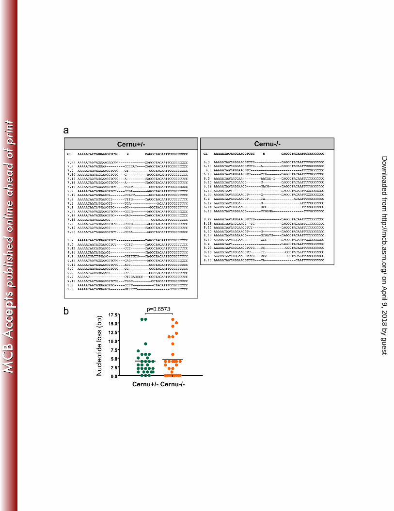
Figure 3. (A) Nucleotide sequence analysis of Coding Joins recovered from pMX-RSS-538
12/23 transduced thymocytes from 2 Cernu+/- and 2 Cernu-/- mice. (B) Similar frequency of 539
nucleotide loss at Coding Joins recovered from pMX-RSS-12/23 transduced thymocytes 540
from Cernu+/- and Cernu-/- mice. 541
on April 9, 2018 by guest
http://mcb.asm
.org/D
ownloaded from
Vera et al.
24
542
Figure 4. Immunoscope of TCRβ (A) and TCRα (B) from 2 Cernu-/- (R6 and R8) and 2 543
Cernu+/- (R7 and R9) mice. (C) Differential representation of distal (5’) and proximal (3’) 544
Vα in Cernu-/- and Cernu+/- T cells. 545
546
Figure 5. (A) Schematic representation of the murine TCR Vα and Jα clusters and position 547
of the three Vα genes co-amplified in PCR conditions using a unique set of primers. (B) 548
Differential representation of distal Vαs (mTRAV3.1 and mTRAV5.1) versus proximal Vαs 549
(mTRAV3.4) after cloning and sequencing of RT-PCR products from thymus and spleen. 550
(C) TCR-Jα representation in dist. and prox. Vαs containing transcripts from thymus and 551
spleen of Cernu-/- and Cernu+/- mice. Vertical red lines correspond to the median for Jα 552
usage. 553
554
Figure 6. TCRα repertoire in human Cernunnos patient. (A) Box and whiskers 555
representation of Vα and Jα usage in TCRα transcripts from one Cernunnos patient and one 556
control individual as determined by 5’RACE PCR and Next Generation Sequencing (NGS). 557
Each VαJα transcript (black and grey dots for Cernunnos patients and control respectively) 558
is positioned according to the relative location (in bp) of its Vα and Jα element in the 559
genome. (B) Contour plots representation of TCRα transcripts from Cernunnos patient (431 560
sequences) and control (1423 sequences). Blue and red bullets represent the VαJα 561
barycenter in each case. (C) Boxes representing 75% of TCRα transcript sequences for a 562
Cernunnos patient (orange square) and a control (blue square). The two green bullets 563
represent the theoretical position of TCRα chains from iNKT (hTRAV10, pos. 2.0 105 bp; 564
on April 9, 2018 by guest
http://mcb.asm
.org/D
ownloaded from

Vera et al.
25
hTRAJ18 pos. 5.0 104) and MAIT (hTRAV1.2, pos. 0.2 105 bp; hTRAJ33, pos. 3.3 104 bp) 565
cells respectively. 566
567
Figure 7. (A) Determination of iNKT cells in spleen by dual staining with anti-TCRβ and 568
CD1-d tetramer. (B) Schematic representation of the human and murine TCR Vα and Jα 569
clusters with the location of TRAV and TRAJ used by iNKT cells. Two Jα can be used by 570
murine NKT cells, TRAV11D or the most downstream TRAV11 because of the duplication 571
of the central TCR-Jα segments in mice. 572
573
Figure 8. Thymocyte survival (A) and apoptosis (B) in 24h culture in vitro. (C) Quantitative 574
RT-PCR analysis of P53 target genes in Cernu-/- thymocytes. 575
576
Figure 9. Partial normalization of Cernu-/- thymocyte viability upon P53 inactivation. (A) 577
Total thymocyte counts. (B) Thymocyte relative survival in 24h culture. (C) Differential 578
TCR-Vα usage in splenic T cells using PCR co-amplification of two 5’Vα (Va3.1 and 579
Va5.1) and one 3’Vα(Va3.4). The vertical red lines represent the medians for Jα usage. (D) 580
iNKT cell recovery in spleen. 581
582
Figure 10. Multiple waves of TCRα rearrangements during thymocyte development. DNA 583
accessibility in the TCRJα cluster is regulated such that the first VαJα rearrangements are 584
targeted to the 5’ side of the TCR-Jα cluster. If the thymocytes expressing the resulting TCR 585
are not positively selected, Rag1 and Rag2 expression continues and a second wave of 586
rearrangement involving upstream Vα and downstream Jα occurs. In the absence of 587
positive selection of this newly expressed TCR, a third wave of VαJα recombination occurs 588
and so on. The possibility of thymocytes to undergo several waves of TCRα rearrangement 589
on April 9, 2018 by guest
http://mcb.asm
.org/D
ownloaded from

Vera et al.
26
depends on their lifespan. In RORγT-/- mice the thymocyte viability is decreased resulting in 590
a bias of Jα usage towards the most 5’ elements (first wave). In contrary, the thymocyte 591
lifespan is increased in BclXL Tg mice allowing several waves of recombination and the 592
resulting skewing of Jα usage toward the most downstream elements. 593
on April 9, 2018 by guest
http://mcb.asm
.org/D
ownloaded from


![Thymoglobulin (anti-thymocyte globulin [rabbit]) · 2020. 12. 14. · DESCRIPTION . Thymoglobulin® (Anti-thymocyte globulin [rabbit]) is a purified, pasteurized, gamma immune globulin](https://static.fdocuments.net/doc/165x107/60c2dece3812e518472963b9/thymoglobulin-anti-thymocyte-globulin-rabbit-2020-12-14-description-thymoglobulin.jpg)
![Thymoglobulin (anti-thymocyte globulin [rabbit]) - Sanofiproducts.sanofi.ca/en/thymoglobulin.pdf · Thymoglobulin® (Anti-thymocyte Globulin [Rabbit]) ... (ATG) products, as protein](https://static.fdocuments.net/doc/165x107/5aa587a17f8b9ab4788d4753/thymoglobulin-anti-thymocyte-globulin-rabbit-anti-thymocyte-globulin-rabbit.jpg)


