
Thymocyte-Specific Truncation of the Deubiquitinating ...
13
of March 9, 2020. This information is current as Modulator-Dependent Manner B Essential κ Positive Selection in a NF- Deubiquitinating Domain of CYLD Impairs Thymocyte-Specific Truncation of the Dimitris L. Kontoyiannis and George Mosialos Ageliki Tsagaratou, Eirini Trompouki, Sofia Grammenoudi, http://www.jimmunol.org/content/185/4/2032 doi: 10.4049/jimmunol.0903919 July 2010; 2010; 185:2032-2043; Prepublished online 19 J Immunol Material Supplementary 9.DC1 http://www.jimmunol.org/content/suppl/2010/07/20/jimmunol.090391 References http://www.jimmunol.org/content/185/4/2032.full#ref-list-1 , 16 of which you can access for free at: cites 43 articles This article average * 4 weeks from acceptance to publication Fast Publication! • Every submission reviewed by practicing scientists No Triage! • from submission to initial decision Rapid Reviews! 30 days* • Submit online. ? The JI Why Subscription http://jimmunol.org/subscription is online at: The Journal of Immunology Information about subscribing to Permissions http://www.aai.org/About/Publications/JI/copyright.html Submit copyright permission requests at: Email Alerts http://jimmunol.org/alerts Receive free email-alerts when new articles cite this article. Sign up at: Print ISSN: 0022-1767 Online ISSN: 1550-6606. Immunologists, Inc. All rights reserved. Copyright © 2010 by The American Association of 1451 Rockville Pike, Suite 650, Rockville, MD 20852 The American Association of Immunologists, Inc., is published twice each month by The Journal of Immunology at Max-Planck-Institut f. Immunbiologie on March 9, 2020 http://www.jimmunol.org/ Downloaded from at Max-Planck-Institut f. Immunbiologie on March 9, 2020 http://www.jimmunol.org/ Downloaded from
Transcript of Thymocyte-Specific Truncation of the Deubiquitinating ...
of March 9, 2020.This information is current as
Modulator-Dependent MannerB EssentialκPositive Selection in a NF-
Deubiquitinating Domain of CYLD Impairs Thymocyte-Specific Truncation of the
Dimitris L. Kontoyiannis and George MosialosAgeliki Tsagaratou, Eirini Trompouki, Sofia Grammenoudi,
http://www.jimmunol.org/content/185/4/2032doi: 10.4049/jimmunol.0903919July 2010;
2010; 185:2032-2043; Prepublished online 19J Immunol
MaterialSupplementary
9.DC1http://www.jimmunol.org/content/suppl/2010/07/20/jimmunol.090391
Referenceshttp://www.jimmunol.org/content/185/4/2032.full#ref-list-1
, 16 of which you can access for free at: cites 43 articlesThis article
average*
4 weeks from acceptance to publicationFast Publication! •
Every submission reviewed by practicing scientistsNo Triage! •
from submission to initial decisionRapid Reviews! 30 days* •
Submit online. ?The JIWhy
Subscriptionhttp://jimmunol.org/subscription
is online at: The Journal of ImmunologyInformation about subscribing to
Permissionshttp://www.aai.org/About/Publications/JI/copyright.htmlSubmit copyright permission requests at:
Email Alertshttp://jimmunol.org/alertsReceive free email-alerts when new articles cite this article. Sign up at:
Print ISSN: 0022-1767 Online ISSN: 1550-6606. Immunologists, Inc. All rights reserved.Copyright © 2010 by The American Association of1451 Rockville Pike, Suite 650, Rockville, MD 20852The American Association of Immunologists, Inc.,
is published twice each month byThe Journal of Immunology
at Max-Planck-Institut f. Im
munbiologie on M
arch 9, 2020http://w
ww
.jimm
unol.org/D
ownloaded from
at M
ax-Planck-Institut f. Imm
unbiologie on March 9, 2020
http://ww
w.jim
munol.org/
Dow
nloaded from

The Journal of Immunology
Thymocyte-Specific Truncation of the DeubiquitinatingDomain of CYLD Impairs Positive Selection in a NF-kBEssential Modulator-Dependent Manner
Ageliki Tsagaratou,*,† Eirini Trompouki,†,1 Sofia Grammenoudi,† Dimitris L. Kontoyiannis,†
and George Mosialos*,†
The cylindromatosis tumor suppressor gene (Cyld) encodes a deubiquitinating enzyme (CYLD) with immunoregulatory function.
In this study, we evaluated the role of Cyld in T cell ontogeny by generating a mouse (CyldD9) with a thymocyte-restricted Cyld
mutation that causes a C-terminal truncation of the protein and reciprocates catalytically inactive human mutations. Mutant mice
had dramatically reduced single positive thymocytes and a substantial loss of peripheral T cells. The analyses of polyclonal and
TCR-restricted thymocyte populations possessing the mutation revealed a significant block in positive selection and an increased
occurrence of apoptosis at the double-positive stage. Interestingly, in the context of MHC class I and II restricted TCR transgenes,
lack of functional CYLD caused massive deletion of thymocytes that would have been positively selected, which is consistent with
an impairment of positive selection. Biochemical analysis revealed that CyldD9 thymocytes exhibit abnormally elevated basal
activity of NF-kB and JNK. Most importantly, inactivation of NF-kB essential modulator fully restored the NF-kB activity of
CyldD9 thymocytes to physiologic levels and rescued their developmental and survival defect. This study identifies a fundamental
role for functional CYLD in establishing the proper threshold of activation for thymocyte selection by a mechanism dependent on
NF-kB essential modulator. The Journal of Immunology, 2010, 185: 2032–2043.
Ubiquitination is currently emerging as a dominant reg-ulatory mechanism in cancer and immunity by meansof posttranslational modification of intracellular signal-
ing mediators of activation, proliferation, stress, and death. Thecylindromatosius tumor suppressor gene (Cyld) encodes a 956 aapolypeptide (CYLD) with deubiquitinating activity (1–3). Thedeubiquitinating domain of CYLD is located in the carboxyl ter-minal 365 aa and constitutes the most highly conserved region ofthe protein (2–4). Cyld originally emerged as a tumor suppressorgene through the identification of human mutations predisposingto tumors of skin appendages (4). Subsequently, insufficient CYLDfunctions correlated with a range of tumors including hepatocel-lular, colon, and lung carcinomas and multiple myelomas (5–9).Interestingly, all the mutations identified to date in human Cyldare predicted to cause carboxyl terminal truncations or frameshiftalterations of the protein that inactivate its deubiquitinating acti-vity (2, 3). Numerous in vitro and in vivo experimental approaches
have established an inhibitory role for CYLD on the basal andTNFR-family–induced activity of the transcription factor NF-kB,which was associated with the ability of CYLD to induce thedeubiquitination of TNFR-associated factor 2, TNFR-associatedfactor 6, NF-kB essential modulator (NEMO), TGFb-activatedkinase 1, B cell CLL-lymphoma 3 and receptor-interacting protein 1(10–12). Notably, the combination of genetic and biochemical stud-ies associated the deubiquitinating activity of CYLD with its tu-mor-suppressing and NF-kB–inhibitory properties (2, 3).Interestingly, the armamentarium of CYLD functions extends
beyond cellular transformation and includes immune modulation.For example, impaired CYLD expression is associated with im-mune diseases like inflammatory bowel disease (13). However,a number of recent studies in different mice bearing obligatorynull alleles demonstrated putative, essential, yet contrasting rolesfor CYLD in the regulation of adaptive and innate immuneresponses (14–18). A first report aimed to eliminate CYLDfunctions in the mouse through the germ-line removal of the ATG-containing exon 2 from the murine Cyld locus (15). Mutant micewere viable but showed reduced numbers of mature CD4+ andCD8+ single positive (SP) T cells in their thymic and peripheralcompartments. The study did not reveal the stage of T cell mat-uration that failed in the context of the mutation. Instead, theauthors proposed that defects in the TCR-induced interaction ofthe lymphocyte-specific protein tyrosine kinase (Lck) adaptor ki-nase with the ZAP70 kinase obscured the processes of T cellselection in immature Cyld-deficient CD4+CD8+ double positive(DP) thymocytes. Paradoxically, peripheral Cyld-deficient T cellsdid not share the same defect in TCR signaling, but instead werehyperresponsive to TCR stimulation (14). Subsequent studiesrevealed that these mutant mice exhibited additional abnormalitiesin B cell development, peripheral B cell subset composition anda puzzling B cell hyperactivity (17). Thus, and although indicativefor the involvement of CYLD in the modulation of lymphocytedevelopment, these studies raised a number of puzzling issues and
*School of Biology, Aristotle University of Thessaloniki, Thessaloniki; and †Insti-tute of Immunology, Biomedical Sciences Research Center Alexander Fleming, Vari,Greece
1Current address: Harvard Medical School, Children’s Hospital, 1 Blackfan Circle,Boston, MA.
Received for publication December 8, 2009. Accepted for publication June 3, 2010.
This work was supported by the Sixth Research Framework Program of the EuropeanUnion, Project INCA (LSHC-CT-2005-018704), and a Leukemia and LymphomaSociety Scholarship (to G.M.).
Address correspondence and reprint requests to George Mosialos, School of Biology,Aristotle University of Thessaloniki, 54124, Thessaloniki, Greece. E-mail address:[email protected]
The online version of this article contains supplemental material.
Abbreviations used in this paper: BCLX L/S, B cell lymphoma X L/S; DD, doubledull; DN, double negative; DP, double positive; IKK, I-kB kinase; LAT, linker foractivation of T cells; Lck, lymphocyte-specific protein tyrosine kinase; NEMO,NF-kB essential modulator; PI, propidium iodide; SP, single positive; tg, transgenic.
Copyright� 2010 by TheAmericanAssociation of Immunologists, Inc. 0022-1767/10/$16.00
www.jimmunol.org/cgi/doi/10.4049/jimmunol.0903919
at Max-Planck-Institut f. Im
munbiologie on M
arch 9, 2020http://w
ww
.jimm
unol.org/D
ownloaded from

could not definitively conclude on the roles of CYLD in governingthese processes. The involvement of CYLD in lymphocyte devel-opment was questioned further by another line of mouse mutantsin which the expression of CYLD was disrupted by the germ-lineremoval of exons 2 and 3 of the Cyld gene (16). Surprisingly, thesemice did not appear to have any defects in the development ofmature lymphocytes, although these cells showed enhanced acti-vation of NF-kB and JNK in response to stimulators of innateand adaptive immunity. The apparent discrepancy between the dif-ferent mutants cannot be resolved easily because of the differencein targeting strategies, but they indicate a potential multicellulareffect of CYLD in lymphocyte development in addition to potentialredundancies in CYLD functions that cannot be resolved in a Cyld-deficient setting.To circumvent these problems and to address the specific role of
CYLD in T cell ontogeny, we generated conditionally targetedmice possessing a T cell-restricted mutation in the Cyld locusengineered to encode a functionally impaired C-terminal truncatedCYLD protein. Through this mutation, we establish a cell-intrinsicrole of Cyld in T cell development and demonstrate its involve-ment in thymocyte selection in a manner that depends on itsfunctional interaction with NEMO.
Materials and MethodsMice
The generation of mice with loxP-targeted Cyld locus has been describedpreviously (19). The transgenic Lck-Cre (20) mice were provided by J.D.Marth (University of California, San Diego, CA). The transgenic H-Y TCRmice were provided by Dr. B. Malissen (Centre d’Immunologie de Mar-seille Luminy, Marseille, France). The BL/6 Tg (TCRaTCRb)425 Cbn/j(OT2) mice were purchased from the Jackson Laboratory (Bar Harbor,ME). All mice were maintained in mixed C57BL/6, 129Ola configuration.The mice were bred and maintained in the animal facilities of the Bio-medical Sciences Research Centre Alexander Fleming under specific-pathogen free conditions. Experiments on live animals were approved bythe Hellenic Ministry of Rural Development (Directorate of VeterinaryServices) and by Biomedical Sciences Research Center Alexander Flem-ing’s Animal Research and Ethics Committee for compliance to Federa-tion of Laboratory Animal Science Associations regulations. Screening oftail DNA for inheritance of the floxed Cyld gene was performed by PCRusing the following primers: F6: 59-CGTGAACAGATGTGAAGGC-39;R6: 59-CTACCATCCCTGCTAACCAC-39; F5: 59-GCAGGCTGTACAG-ATGGAAC-39; R1: 59-CTGCAAATTTCAGGTTGCTGTTG-39. Inheritanceof the LckCre transgene was determined by PCR using the following primers:forward, 59-ATTACCGGTCGATGCAACGAGT-39; and reverse, 59-CAGG-TATCTCTGACCAGAGTCA-39. Inheritance of the H-Y TCR transgene wasdetermined by PCR using the following primers: Vb8.2HY: 59-GGC-TGCAGTCACCCAAAGCCCAAG-39 and 59-CAGTCAGTCTGGTTCCT-GAGCC-39. Inheritance of the OT2 TCR transgene was determined by PCRusing the following primers:
oIMR1825: 59-GCT GCT GCA CAG ACC TAC T-39 andoIMR1826: 59-CAG CTC ACC TAA CAC GAG GA-39.
DNA analysis for Nemo was performed using the following three pri-mers in one reaction, as described elsewhere (21):
27: 59-GCC TTG GTG CTC CCT AAC TCT-39,40: 59-TCA CAT CAC ATC GTT ATC CTT-39, and61: 59-ATG AAC AAG CAC CCC TGG AAG-39.
Abs
Abs against the following molecules coupled to the indicated fluorochromeswere purchased from BD Pharmingen (San Diego, CA): CD4-FITC, CD4-PE, CD8-PE, CD3–biotin, B220-Cychrome, TCR-Cychrome, CD11b-PE,Gr1-biotin, CD25-biotin, CD44-FITC, CD62L-biotin, CD69 PECy7, andTCRVb 5.1-PE. The following purified Abs were purchased from BDPharmingen: anti-NEMO (C73-764), anti-CD53 (OX-79), and anti-CD4(GK1.5). Biotin-conjugated anti-CD24, allophycocyanin–Cy7-conjugatedanti-CD8, anti-CD3ε, and anti-CD28 were purchased from Biolegend (SanDiego, CA). A700-conjugated anti-CD4, PercP-conjugated anti-CD8,
FITC-conjugated anti–H-Y TCR (clone T3.70), FITC-conjugated anti-TCRVa2 were purchased from eBioscience (San Diego, CA). Determina-tion of cell survival in fresh or cultured thymocytes was conducted bystaining with annexin V (BD Biosciences, San Jose, CA) and propidiumiodide ([PI] Sigma-Aldrich, St. Louis, MO) after surface staining for CD4and CD8 (when evaluating survival of total thymocytes) or after CD53-dependent isolation of DP thymocytes. The anti-CYLD Ab that was usedin Fig. 1 was a gift from Dr. Shao Cong Sun (MDAnderson Cancer Center,Houston, TX), and it has been described elsewhere (22). The anti-CYLDAb (E-4) that was used in Supplemental Fig. 6 was purchased from SantaCruz Biotechnology (Santa Cruz, CA). The anti-Lck (3A5), anti–ZAP-70(1E7.2), anti-p65 (A), anti–p-JNK (G-7), anti-JNK2 (D-2), anti-linker foractivation of T cells (LAT; M-19), and anti-tubulin (B-7) Abs wereobtained from Santa Cruz Biotechnology. Abs to Src phosphorylated attyrosine 416 (which recognizes Lck phosphorylated at tyrosine 394),Zap70 phosphorylated at tyrosine 319, and LAT phosphorylated at tyrosine191 were obtained from Cell Signaling Technology (Beverly, MA). Theanti-actin mouse mAb was purchased from MP Biomedicals (Solon, OH)and the anti-FLAG (M5) mouse mAb was purchased from Sigma-Aldrich.
Flow cytometric analysis
Single-cell suspensions were obtained from the thymus, spleen, and lymphnodes by dissociation of isolated tissues through a 60-mm mesh. Red bloodcells were excluded by Gey’s lysis solution, and debris was removed bya cell strainer. Cells were processed for the detection of a panel of cellmarkers by incubation in PBS, 0.1% NaN2, and 2% FBS for 20 min on iceby titrated concentrations of reagents. Cell-associated fluorescence wasanalyzed by a FACSCantoII flow cytometer and the DIVA V6 software(BD Biosciences). Flow cytometry figures were prepared using the FlowJosoftware (Tree Star, Ashland, OR). Differences in lymphocyte populationswere evaluated with the unpaired t test using the Sigmaplot 9 statisticalsoftware.
DP thymocyte purification
Total thymocytes were incubated with anti–CD53-biotin (BD Pharmingen)and then with anti-rat IgG-biotin (Jackson ImmunoResearch Laboratories,West Grove, PA). CD53+ cells were subsequently removed using anti-biotin microbeads (MACS; Miltenyi Biotec, Auburn, CA). DP thymocyteswere incubated on ice before stimulation to evaluate TCR signaling.
Immunoblotting and immunoprecipitation assays
Immunoblotting and immunoprecipitation assays were performed as pre-viously described (2).
EMSA
Nuclear extracts were prepared and EMSA was performed as previouslydescribed (19). The sequences of the oligonucleotides that were used forthe determination of Oct-1 binding were the following:
Oct-1 F: 59-TGT CGA ATG CAA ATC ACT AG-39 andOct-1 R: 59-TTC TAG TGA TTT GCA TTC G-39.
The sequences of the oligonucleotides that were used for the de-termination of NF-kB binding contained two tandem repeated NF-kBbinding sites (underlined), and they were as follows:
NF-kBf: 59-ATC AGG GAC TTT CCG CTG GGG ACT TT-39 andNF-kBr: 59-CGG AAA GTC CCC AGC GGA AAG TCC CT-39.
For supershift analysis, nuclear extracts were preincubated with anti-p65Abs.
T cell stimulation assays
To evaluate TCR signaling, 5 3 106 to 10 3 106 T cells resuspended inserum-free Iscove’s medium were incubated for 15 min on ice with anti-mouse CD3ε mAb (1 mg/ml, 145-2c11; Biolegend) and anti-CD4 (1 mg/ml, L3T4; BD Pharmingen), followed by crosslinking with goat anti-hamster IgG (6060-01; SouthernBiotech, Birmingham, AL) for the indi-cated times at 37˚C with gentle shaking. Stimulated T cells were imme-diately lysed in RIPA buffer containing protease and phosphataseinhibitors. DP thymocyte survival was calculated as the percentage of alivecells (negative for staining with annexin V and PI) after overnight incuba-tion with 20 mg/ml of immobilized anti-mouse CD28 (37.51) and the in-dicated concentrations of anti-mouse CD3 (145-2c11; Biolegend).
The Journal of Immunology 2033
at Max-Planck-Institut f. Im
munbiologie on M
arch 9, 2020http://w
ww
.jimm
unol.org/D
ownloaded from

RT-PCR
Total RNA was isolated from total thymocytes or DP cells with Trizol(Invitrogen, Carlsbad, CA) and oligo-dT primed cDNAwas prepared usingImprom Reverse Transcriptase (Promega, Madison, WI) according to themanufacturer’s instructions.
ResultsMutation of Cyld prohibits the generation of maturethymocytes
To examine the functional role of Cyld in specific tissues, wedevised a conditional strategy to render murine CYLD catalyti-cally inactive in tissues of interest. Furthermore, this approach wasadopted specifically to elucidate the biologic role of the catalyticdomain of CYLD. Previous approaches analyzed the phenotype ofCyld null mice, which reflects the combined lack of biologicfunctions that depend on the multiple functional domains ofCYLD. Using gene targeting, we previously flanked the ninthexon of the murine Cyld gene with loxP sequences (Cyldflx9 allele;Fig. 1A) to permit its excision by the Cre recombinase (CyldD9;Fig. 1A) (19). Exon 9 encodes an essential part of the deubiqui-tinating domain of CYLD; therefore, its removal should inhibit itscatalytic activity. Furthermore, the removal of exon 9 should alsoinduce a frameshift mutation because of the aberrant splicing ofexons 8 to 10, thus mimicking the naturally occurring carboxylterminal mutations of Cyld in patients with familial cylindro-matosis. Previously, we used Cyldflx9/flx9 to introduce the CyldD9
mutation in the mouse germline (19).To generate mice with a thymocyte-specific CyldD9 mutation,
Cyldflx9/flx9 mice were crossed to LckCre-transgenic mice (20). TheLck promoter restricts the expression of Cre in the early stage ofimmature double negative (DN) thymocytes. In accordance withthe thymocyte-specific expression pattern of the Cre recombinasein LckCre-Cyldflx9/flx9 mice, the excision of Cyld exon 9 wasreadily evident in thymocytes (Fig. 1B), but not in other non-lymphoid tissues from these mice (Fig. 1C). In addition, RT-PCR analysis verified the presence of Cyld mRNA encoding exons1–13, but were devoid of the sequence encoding exon 9 (Fig. 1Dand data not shown). Interestingly, the mutant mRNA was pri-marily present in LckCre-Cyldflx9/flx9, whereas it was clearlyunderrepresented in heterozygous LckCre-Cyldflx9/+, suggestinga predominance of wild type CYLD expression in the lattersetting (Fig. 1D). Finally, immunoblotting with an anti-CYLDAb indicated a nearly complete absence of full-length CYLD pro-tein in LckCre-Cyldflx9/flx9 thymic or peripheral T cell extracts,respectively (Fig. 1E, 1F). A carboxyl terminally truncated formof CYLD could not be detected in LckCre-Cyldflx9/flx9 thymocytes,most likely because the highest level of CYLD expression is ob-served in SP thymocytes (15), which are practically eliminated inLckCre-Cyldflx9/flx9 mice (see below).Both thymic and peripheral T cells isolated from Cyldflx9/+ and
LckCre mice maintained a physiologic representation of T cellsubtypes, as assessed by their enumeration after the evaluation ofsurface expression of CD4, CD8, CD25, CD44, CD62L, CD69,CD5, and TCR (Supplemental Fig. 1 and data not shown). Simi-larly, the LckCre-Cyldflx9/+ and LckCre mice showed similar rep-resentation of T cell subtypes based on the expression of the afore-mentioned markers (Supplemental Fig. 1B and data not shown).Thus, we present LckCre-Cyldflx9/+ mice (termed T-Cyld+ in allthe figures) as the control animals for mutant LckCre-Cyldflx9/flx9
mice (termed T-CyldD9 in all the figures).The introduction of CyldD9 in mouse thymocytes in LckCre-
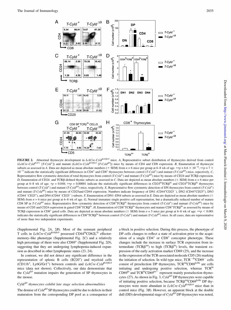
Cyldflx9/flx9 did not affect significantly the representation of DNand DP thymocytes; strikingly however, both CD4+ and CD8+ SPcompartments were reduced to almost nominal values (Fig. 2A,
2B, 2E, 2F). Furthermore, the reduction in CD4- or CD8-expressing thymocytes reflected the loss of mature single positivesubsets, as confirmed by the enumeration of CD24lo cells express-ing high levels of surface TCRb (Fig. 2C, 2D). However, theTCRbloCD8+CD52CD24hi thymocytes that constitute immaturesingle positive cells were present, and generally the levels ofCD8+TCRblo thymocytes in CyldD9 and control mice were com-parable, whereas CD8+TCRbhi thymocytes that represent matureCD8 SP were significantly underrepresented in CyldD9 mice (Fig2G, 2H). Similarly, the reduction of CyldD9 SP thymocyte popu-lations was also reflected in the dramatic reduction of CD4+ andCD8+ T cells in the periphery of LckCre-Cyldflx9/flx9, as assessedby their enumeration in mesenteric lymph nodes and the spleen
FIGURE 1. Generation and characterization of LckCre-Cyldflx9/flx9 mice.
A, Gene-targeting strategy. Schematic representation of exons 7–13 (E7–
E13) of the murine Cyld locus (Cyld+). The structures of the floxed Cyld
locus in the absence (Cyldflx9) and presence (CyldD9) of the Cre recombi-
nase are shown. The loxP sites are shown as solid triangles, and the neo-
mycin resistance gene is shown as a gray rectangle. The positions of
primers F5, F6, R1, and R6 are shown by arrows. B, PCR of genomic
DNA from thymocytes of Cyldflx9/flx9 (Cyldflx9) and LckCre-Cyldflx9/flx9 (T-
CyldD9) mice with the indicated primer pairs for the Cyld locus or primers
for the Gapdh locus. The positions of the specific PCR products are
indicated. More than four animals per genotype were analyzed. At least
three independent experiments were performed. C, PCR of genomic DNA
from perfused thymus, lung, liver, heart, and kidney of LckCre-Cyldflx9/flx9
mice with the indicated primer pairs for the Cyld locus or primers for the
Gapdh locus. The positions of the specific PCR products corresponding to
the recombined CyldD9 locus or the Gapdh locus are indicated. Organs
isolated from two animals were used. Two independent experiments were
performed. D, RT-PCR of RNA from thymocytes of wild type (Cyld+),
LckCre-Cyldflx9/+ (T-Cyld+) and LckCre-Cyldflx9/flx9 (T-CyldD9) mice with
the primers F5 and R1 for the Cyld mRNA or primers for the Gapdh
mRNA. The position of the specific PCR products corresponding to full-
length Cyld (T-Cyld+) and CyldD9 (T-CyldD9) are indicated. At least three
mice per genotype were used. Data are representative of three independent
experiments. E, Immunoblot of thymocyte lysates from LckCre (T-Cyld+)
and LckCre-Cyldflx9/flx9 (T-Cyld D9) mice. The positions of full length
CYLD and actin are shown. At least three animals per genotype were used.
Data are representative of three independent experiments. F, Immunoblot
of splenic T cells from LckCre (T-Cyld+) and LckCre-Cyldflx9/flx9 (T-CyldD9)
mice. The positions of full-length CYLD and tubulin are shown. Two ani-
mals per genotype were used. Data are representative of two independent
experiments.
2034 CYLD IS REQUIRED FOR THYMOCYTE POSITIVE SELECTION
at Max-Planck-Institut f. Im
munbiologie on M
arch 9, 2020http://w
ww
.jimm
unol.org/D
ownloaded from
(Supplemental Fig. 2A, 2B). Most of the remnant peripheralT cells in LckCre-Cyldflx9/flx9 possessed CD44hiCD62Llo effector-memory–like phenotype (Supplemental Fig. 2C) and a relativelyhigh percentage of them were also CD69+ (Supplemental Fig. 2D),suggesting that they are undergoing lymphopenia-induced expan-sion as described in other lymphopenic states (23, 24).In contrast, we did not detect any significant difference in the
representation of splenic B cells (B220+) and myeloid cells(CD11b+, Ly6G/Gr1+) between controls and LckCre-Cyldflx9/flx9
mice (data not shown). Collectively, our data demonstrate thatthe CyldD9 mutation impairs the generation of SP thymocytes inthe mouse.
CyldD9 thymocytes exhibit late stage selection abnormalities
The demise ofCyldD9 SP thymocytes could be due to defects in theirmaturation from the corresponding DP pool as a consequence of
a block in positive selection. During this process, the phenotype ofDP cells changes to reflect a state of activation prior to the acqui-sition of a single CD4+ or CD8+ coreceptor phenotype. Thesechanges include the increase in surface TCR expression from in-termediate (TCRbint) to high (TCRbhi) levels, the transient ex-pression of the early activation marker CD69 (25), and the increasein the expression of the TCR-associatedmolecule CD5 (26) markingthe initiation of selection. In wild type mice, TCR2/loCD692 cellsconsist of preselection DP thymocytes, TCRintCD69lo/hi are cellsinitiating and undergoing positive selection, whereas TCRhi
CD69hi and TCRhiCD69lo/2 represent mainly postselection thymo-cytes (27). As shown in Fig. 3, CyldD9 DP thymocytes were capableof initiating positive selection, because TCRbintCD69lo/hi DP thy-mocytes were more abundant in LckCre-Cyldflx9/flx9 mice than incontrol mice (Fig. 3B). However, an apparent block at the doubledull (DD) developmental stage ofCyldD9DP thymocytes was noted,
FIGURE 2. Abnormal thymocyte development in LckCre-Cyldflx9/flx9 mice. A, Representative subset distribution of thymocytes derived from control
(LckCre-Cyldflx9/+ [T-Cyld+]) and mutant (LckCre-Cyldflx9/flx9 [T-CyldD9]) mice by means of CD4 and CD8 expression. B, Enumeration of thymocyte
subsets as assessed in A. Data are depicted as mean absolute numbers (6 SEM) from n = 6 mice per group at 6–8 wk of age. ppp = 6.6 3 1026; ppp = 7 31025 indicate the statistically significant differences in CD4+ and CD8+ thymocytes between control (T-Cyld+) and mutant (T-CyldD9) mice, repectively. C,
Representative flow cytometric detection of total thymocytes from control (T-Cyld+) and mutant (T-CyldD9) mice by means of CD24 and TCRb expression.
D, Enumeration of CD24- and TCRb-defined thymic subsets as assessed in C. Data are depicted as mean absolute numbers (6 SEM) from n = 6 mice per
group at 6–8 wk of age. pp = 0.048; ppp = 0.00001 indicate the statistically significant differences in CD24hiTCRbhi and CD24loTCRbhi thymocytes
between control (T-Cyld+) and mutant (T-CyldD9) mice, respectively. E, Representative flow cytometric detection of DN thymocytes from control (T-Cyld+)
and mutant (T-CyldD9) mice by means of CD25and CD44 expression. Numbers indicate frequency of DN1 (CD44+CD252), DN2 (CD44+CD25+), DN3
(CD442CD25+), and DN4 (CD442CD252) subsets. F, Enumeration of DN1–DN4 subsets as assessed in E. Data are depicted as mean absolute numbers (6SEM) from n = 6 mice per group at 6–8 wk of age. G, Normal immature single positive cell representation, but a dramatically reduced number of mature
CD8 SP in T-CyldD9 mice. Representative flow cytometric detection of CD8+TCRblo thymocytes from control (T-Cyld+) and mutant (T-CyldD9) mice by
means of CD5 and CD24 expression in gated CD8+TCRblo. H, Enumeration of CD8+TCRblo thymocytes and mature CD8+TCRbhi as assessed by means of
TCRb expression in CD8+ gated cells. Data are depicted as mean absolute numbers (6 SEM) from n = 5 mice per group at 6–8 wk of age. ppp , 0.001
indicates the statistically significant differences in CD8+TCRbhi between control (T-Cyld+) and mutant (T-CyldD9) mice. In all cases, data are representative
of more than two independent experiments.
The Journal of Immunology 2035
at Max-Planck-Institut f. Im
munbiologie on M
arch 9, 2020http://w
ww
.jimm
unol.org/D
ownloaded from

because CD4+CD8lo cells were dramatically reduced inCyldD9 mice(Fig. 3C). Furthermore, immature TCRhiCD69hi SP thymocytes andmature TCRhiCD69lo/2 SP thymocytes were dramatically reduced,whereas remnant cells that express these markers exhibited a latentDD identity and showed similar percentages of CD4 and CD8 SPthymocytes instead of the typical 3:1 ratio of CD4 to CD8 SPthymocytes (Fig. 3C). Similar conclusions were reached whenevaluating the upregulation of CD5 in combination with TCR.More specifically, the following four different developmentalstages of thymocytes can be discerned based on the expression ofCD5 and TCR: TCRloCD5lo cells that consist of preselection DPcells; TCRloCD5int cells that consist of DP undergoing positiveselection; TCRintCD5hi cells that are mainly DD as well as CD4+
CD8lo cells that are in the process of positive selection; and, finally,TCRhiCD5hi cells that consist of postselection CD4 and CD8thymocytes (27). As shown in Supplemental Fig. 3A, CyldD9 DPcells initiated the process of positive selection as indicated by theoverrepresentation of TCRloCD5int cells compared with controlthymocytes. However, CyldD9 cells were blocked in the DD
developmental stage and failed to upregulate TCR, a hallmark ofthe successful completion of positive selection and the generation ofmature naive SP cells ready to migrate to the periphery (Sup-plemental Fig. 3A, 3B) (28). As expected, CD4+CD8lo cells wereseverely reduced and SP cells were practically absent in mutantmice (Supplemental Fig. 3A–C). Collectively, our data indicate thatCyldD9DP cells initiate the process of positive selection and proceeduntil the DD stage where they are apparently blocked, resulting ina nearly complete absence of CD4+CD8lo cells and CD5hiCD69lo/2
TCRhi mature SPs. This deviation of thymocytes from the normaldevelopmental process could be attributed either to their deletion orto their inability to complete positive selection, potentially becauseof impaired TCR signaling. However, the normal initiation of posi-tive selection and the presence of cells undergoing positive selectionuntil theDD stage—that is, until the upregulation of TCR (Fig. 3 andSupplemental Fig. 3)—suggests that there is nomajor defect in TCRsignaling in CyldD9 mice (27). Indeed, proximal TCR signaling inresponse to CD3/CD4 costimulation was similar in CyldD9 and con-trol DP thymocytes (Supplemental Fig. 4A). To explore further the
FIGURE 3. Cyld D9 DP thymocytes initiate positive selection but are blocked in the double dull developmental stage. A, Monitoring of thymocyte positive
selection based onCD69 upregulation. Representative flow cytometric detection of CD69 and TCRb expression in total thymocytes from control (T-Cyld+) and
mutant (T-CyldD9) mice (left panels). Defined subsets representing distinct developmental stages (TCRb2/loCD692, TCRintCD69lo/hi, TCRhiCD69hi, and
TCRhiCD69lo/2)were further analyzed forCD4andCD8expression (right panels indicated by arrows).B, Absolute numbers of thymocyte subsets in control (T-
Cyld+) and mutant (T-CyldD9) mice as assessed by CD69 and TCR expression in A. Data are depicted as mean absolute numbers (6 SEM) from n = 6 mice per
group at 5–7wkof age.ppp,0.001 indicates the statistically significant differences in the different developmental stages between control (T-Cyld+) andmutant
(T-CyldD9) mice. C, Enumeration of thymic subsets defined by CD4 and CD8 expression within the CD69/TCR defined developmental stages that showed
significant statistical difference in B. Data are depicted as mean absolute numbers (6 SEM) from n = 6 mice per group at 5–7 wk of age. The statistically
significant differences between control (T-Cyld+) and mutant (T-CyldD9) mice: ppp, 0.02 for DD cells; ppp, 0.001 for the other subsets.
2036 CYLD IS REQUIRED FOR THYMOCYTE POSITIVE SELECTION
at Max-Planck-Institut f. Im
munbiologie on M
arch 9, 2020http://w
ww
.jimm
unol.org/D
ownloaded from

reason for the loss of CyldD9 DP thymocytes, we evaluated theirsurvival rate in ex vivo cultures. As assessed by annexin V and PIstaining,more than 70%of control LckCre-Cyldflx9/+DP thymocytessurvived after a 20-hr ex vivo culturing period. In sharp contrast,less than 30% of LckCre-Cyldflx9/flx9DP thymocytes survived underthe same conditions (Fig. 4A, 4B). More precisely, both the earlyand late apoptotic CyldD9 DP were 2-fold higher when comparedwith control DP thymocytes, whereas necrotic DP cells could bedetected in similar percentages (Fig. 4A). Intriguingly, the expres-sion of the prosurvival factor B cell lymphoma X L/S (BCLXL/S),which is a critical player in the survival of DP thymocytes (29, 30),was significantly reduced in CyldD9 compared with control DP thy-mocytes, providing a molecular basis for the impaired survival ofCyldD9 DP thymocytes (Fig. 4C, 4D).
Transgenic TCRs that favor positive selection failed to rescuethe development of CyldD9 DP thymocytes and revealed theirincreased propensity for deletion
The complex process of thymocyte selection is difficult to unravelin mice with a polyclonal T cell repertoire. To circumvent thisobstacle, we examined the selection of MHC class I restricted
thymocytes expressing the transgenic HY TCR. This transgenicTCR reacts with an Ag derived from the male-specific Smcy pro-tein in the context of H-2Db MHC class I. Hence, thymocytesexpressing HY-specific TCR are negatively selected in male H-2Db and undergo massive deletion. This phenomenon leads toa dramatic decrease in total cellularity, which is accompaniedby severe underrepresentation of DP and CD8 SP T cells.However, HY-specific TCR promotes positive selection of theCD8 lineage in female mice (31).In transgenic (tg)HY+LckCre-Cyldflx9/flx9 female mice, total
thymic cellularity was severely reduced (Fig. 5A). DP thymocytesbehaved similarly (data not shown). Both total thymocytes and DPthymocytes isolated from tgHY+LckCre-Cyldflx9/flx9 mice showedan even greater tendency to become apoptotic in culture relativeto control HY-TCR-expressing thymocytes (Fig. 5B and data notshown, respectively). Thus, after 20 h of in vitro culture, the rem-nant CyldD9 thymocytes were reduced to almost nominal values(Fig. 5B, 5C).Flow cytometric analysis revealed uniform expression of the
transgenic HY TCR in tgHY+LckCre-Cyldflx9/flx9 thymocytes (Fig.5D). Moreover, the number of DP and CD8 SP thymocytes bearingthe HY TCR was reduced in tgHY+LckCre-Cyldflx9/flx9mice (Fig. 5,E and F). However, CyldD9 CD8 SP thymocytes were mature asassessed by flow cytometric analysis of the markers CD69, CD5and CD24 (Fig. 5G). More precisely, CD5 expression wasincreased both in DP and CD8 SP HY-expressing thymocytes,whereas both CD69 and CD24 were markedly downregulated inCD8 SP HY-expressing thymocytes. Thus, the maturation of theproduced CyldD9 CD8 SP thymocytes readily takes place in thiscontext. This finding suggests that HY TCR recognizes the Agand that the process of positive selection is initiated. However,a large number of thymocytes that would normally be positivelyselected under these conditions are eliminated in the absence offunctional CYLD. Collectively, our data indicate that the CyldD9
mutation leads to a change in the threshold of activation, whichapparently diverts CD8 SP thymocytes from a destiny of positiveselection to enhanced deletion.Negative selection in HY male mice is characterized by severely
reduced thymocyte numbers as well as nearly complete absence ofDP and SP thymocytes. This profile probably reflects negative se-lection that occurs early in the DP stage. In HY tgLckCre Cyldflx9/flx9
male mice, negative selection owing to high-affinity TCR ligandengagement takes place to the same extent as in control male mice,because the number of DP and CD8 cells were comparable (Fig.5H). This finding is in accordance with the similar effect of TCRengagement on the survival of control and LckCre Cyldflx9/flx9 DPthymocytes in vitro, although the interpretation of this result iscompromised by the much greater tendency of LckCre Cyldflx9/flx9
DP thymocytes to undergo apoptosis (Supplemental Fig. 5).To further evaluate the implication of CYLD in positive selection
of CD4 lineage, we crossed LckCre-Cyldflx9/flx9 mice with theOT-2 TCR transgenic mice that express a TCR composed of Va2/V b5 chains, recognizing the OVA peptide 323–339 in the con-text of MHC class II I-Ab (32). The analysis indicated a severereduction in total thymic cellularity of tgOT2+ LckCre-Cyldflx9/flx9
mice compared with control mice (Fig. 6A) that was accompaniedby a decrease in the ex vivo survival of thymocytes (Fig. 6B).Moreover, there was an apparent reduction in TCR expression(Fig. 6C) that, combined with the absence of CD4 T cells in theVa2/Vb5 TCR high thymocytes, reveals that positive selection iscompletely abolished in the absence of functional CYLD protein(Fig. 6D, 6E). Furthermore, tgOT2+ LckCre-Cyldflx9/flx9 mice hada tendency to lose CD4+CD8lo thymocytes, which represent anintermediate population that has initiated selection in response to
FIGURE 4. Reduced exvivo survival ofCyldD9DP thymocytes.A, CD53-
depleted DP thymocytes from LckCre-Cyldflx9/+ (T-Cyld+) and LckCre-
Cyldflx9/flx9 (T-CyldD9) mice were stained for annexin/PI and assessed by
flow cytometry following their isolation and 16 h of culture. The
percentages of live cells (annexin–PI–), early apoptotic (annexin+PI–), late
apoptotic (annexin+PI+), and necrotic (annexin–PI+) after 16 h of culture are
indicated. One representative experiment of two is depicted. At least three
mice per genotype were evaluated. B, Enumeration of live DP thymocytes
from control (T-Cyld+) and mutant (T-CyldD9) mice after 16 h in culture as
assessed in A. Survival of DP cells in vitro was estimated as the frequency of
live cells (annexin2PI2) after 16 h in culture relative to the input population
and represented as mean absolute numbers (6 SEM) from three control (T-
Cyld+) and four mutant (T-CyldD9) mice per group at 5–6 wk of age. ppp,0.001 indicates the statistically significant difference between the survival of
control (T-Cyld+) and mutant (T-CyldD9) mice.C, Reduced expression of the
anti-apoptotic BCLXL/S in CyldD9 DP thymocytes. RT-PCR analysis using
RNA isolated from CD53 depleted control (T-Cyld+) and mutant (T-CyldD9)
DP, revealed reduced expression of BCLXL/S in CyldD9 DP thymocytes. At
least four mice per genotype were evaluated. D, Quantitation of BCLXL/S
expression inLckCre-Cyldflx9/flx9 (T-CyldD9) andLckCre-Cyldflx9/+ (T-Cyld+)
mice. Densitometric analysis was performed, and the ratio of the BCLX L/S
to GAPDH (expression index) was calculated. The expression index of
BCLX L/S from purified DP thymocytes of control mice was divided by the
expression index from mutant DP thymocytes, to derive the corresponding
folds of upregulation of BCLXL/S expression.
The Journal of Immunology 2037
at Max-Planck-Institut f. Im
munbiologie on M
arch 9, 2020http://w
ww
.jimm
unol.org/D
ownloaded from

TCR signaling and are midway in the transition from DP to SP cells(Fig. 6D, 6E) (27). However, CyldD9 DP thymocytes with Va2/Vb5TCR high expression upregulated CD5 and CD69 similarly tocontrol DP thymocytes, whereas they downregulated CD24 moreprominently than control DP thymocytes, a sign of prematurematuration (Fig. 6F) (33). Moreover, flow cytometric analysis ofthe spleen (Fig. 6G, 6H) and mesenteric lymph nodes (data notshown) of TCR Vb5 and CD4 expression revealed a nearlycomplete absence of peripheral T cells. These data establishCYLD as a key player in the positive selection of the CD4 lineage,most likely by maintaining a proper threshold of activation and thussafeguarding the outcome of positive selection.
Aberrant activation of NF-kB and JNK in CyldD9 thymocytes
It has been shown previously that CYLD is a negative regulator ofNF-kB and JNK. This finding prompted an investigation of theactivity of the two pathways in CyldD9 thymocytes. Importantly,basal levels of NF-kB DNA-binding activity were dramatically
increased in CyldD9 thymocytes compared with control thymo-cytes (Fig. 7A, upper panel, compare lanes 3 and 4). The activityof NF-kB was mainly due to the binding of p65 and p50 subunits(Fig. 7A, upper panel, lane 6 and data not shown). Similar re-sults were obtained with nuclear extracts from DP thymocytes(Supplemental Fig. 4B, compare lanes 3 and 4). In addition, theactivity of JNK was also elevated in CyldD9 thymocytes comparedwith control thymocytes (Fig. 7B, compare lanes 2 and 3 and Fig.7C). These findings implicate the deregulated activation of NF-kBand JNK in defective CyldD9 thymocyte selection.
Inactivation of NEMO rescues the developmental defects ofCyldD9 thymocytes
CYLD has been shown to interact with NEMO, which plays a fun-damental role in the activation of NF-kB as an integral componentof the I-kB kinase (IKK) complex (2, 3). More recently, NEMOhas been implicated in the activation of JNK (34, 35). To de-termine whether NEMO is involved in the defective selection of
FIGURE 5. Impaired positive selection in LckCreCyldflx9/flx9 MHC class I restricted transgenic mice. A, Total thymocyte number in female control
(tgHY+ LckCre-Cyldflx9/+ [HY+ T-Cyld+]) and mutant (tgHY+ LckCre-Cyldflx9/flx9 [HY+ T-CyldD9]) mice. Data are depicted as mean absolute numbers
(6 SEM) from n = 7–9 mice per group. ppp = 0.001 indicates the statistically significant difference in the total number of thymocytes between control (HY+
T-Cyld+) and mutant (HY+ T-CyldD9) mice. B, Histograms depicting annexin V staining in PI2 fresh (0 h) or cultured (for 20 h) thymocytes from control
(HY+ T-Cyld+) and mutant (HY+ T-CyldD9) mice . C, In vitro survival of fresh and cultured thymocytes isolated from two control (HY+ T-Cyld+) and three
mutant (HY+ T-CyldD9) mice. Average values 6 SE are shown. ppp , 0.00024 indicates statistically significant differences. D, Representative histogram
depicting the difference in the expression of the HY-TCR (T3.70) between total thymocytes from control ([HY+ T-Cyld+], open histogram) and mutant
([HY+ T-CyldD9], shaded histogram) mice. E, Representative subset distribution of gated HY-TCR–expressing thymocytes by means of CD4 and CD8
expression in control (HY+ T-Cyld+) and mutant (HY+ T-CyldD9) mice. F, Enumeration of HY-TCR–expressing DP and CD8+ thymocytes in control (HY+
T-Cyld+) and mutant (HY+ T-CyldD9) mice. Data are depicted as mean absolute numbers (6 SEM) from n = 7–9 mice per group. ppp = 0.0005; ppp =
0.0006 indicate the statistically significant differences in DP and CD8 thymocytes between control and mutant mice, respectively. G, Representative
histograms that depict the expression of CD69, CD5, and CD24 in both DP and CD8 SP that express HY-TCR in control ([HY+ T-Cyld+], open histogram)
and mutant ([HY+ T-CyldD9], shaded histogram) mice. If not otherwise stated, data are representative of at least three independent experiments. H, Analysis
of CD4 and CD8 thymic subsets in male control (HY+ T-Cyld+) and mutant (HY+ T-CyldD9) mice by flow cytometry. Thymocytes expressing high levels of
T3.70-reactive TCR were evaluated for CD4 and CD8 expression. Data from one representative experiment of three are depicted. Three 6-wk-old mice per
genotype were evaluated.
2038 CYLD IS REQUIRED FOR THYMOCYTE POSITIVE SELECTION
at Max-Planck-Institut f. Im
munbiologie on M
arch 9, 2020http://w
ww
.jimm
unol.org/D
ownloaded from

thymocytes in LckCre-Cyldflx9/flx9 mice, these mice were crossedwith mice (Nemoflx) carrying a conditionally-targeted Nemo allele.More specifically, Nemoflx mice bear a premature stop codon thatcan be conditionally introduced in a Cre-dependent manner andhave been used already to evaluate the function of NEMO in T celldevelopment (21). The double mutant mice (LckCre-Cyldflx9/flx9-Nemoflx) were viable and fertile and showed no obviousabnormalities. The loxP-targeted Nemo and Cyld alleles were effi-ciently recombined in the thymocytes of LckCre-Cyldflx9/flx9-Nemoflx
mice, as determined by competitive genomic PCR (SupplementalFig. 6A, 6B). It should be noted that the recombination efficiencyof the Cyldflx9 allele in LckCre-Cyldflx9/flx9-Nemoflx mice was com-parable to its recombination in LckCre-Cyldflx9/flx9 mice (Sup-plemental Fig. 6B). Furthermore, the efficient recombination ofthe Cyldflx9 allele was confirmed by the absence of a full-lengthCyld transcript and the expression of a CyldD9 transcript in DPthymocytes from mice (Supplemental Fig. 6C). Finally, full-length CYLD protein was undetectable (Supplemental Fig. 6D),
and NEMO was significantly reduced, in accordance with previ-ous reports (36), in thymocytes from LckCre-Cyldflx9/flx9-Nemoflx
double mutant mice as determined by immunoblotting (Supplemen-tal Fig. 6E). Concomitant inactivation of NEMO in thymocytes withmutated Cyld fully restored the basal activity of NF-kB to phys-iologic levels (Fig. 7A, compare lanes 2, 3, and 4) and attenuatedJNK activation, which remained above physiologic levels (Fig. 7B,compare lanes 1, 2, 3, and Fig. 7C). Most importantly, both CD4and CD8 SP populations in mice with mutated Cyld and Nemowererestored to levels that were comparable to those seen in controlmice (Fig. 8A, 8B). Nevertheless, the absolute number of CD8 SPin the double mutants was lower than the corresponding populationof control mice, a finding that can be attributed to Nemo deficiencyas previously described (Fig. 8B) (36). Furthermore, and in accor-dance with the previously described phenotype of mice with T cell-specific Nemo deficiency, CD8 SP thymocytes from double mutantmice showed an immature phenotype, as reflected by their inabilityto downregulate CD24 (data not shown), whereas in the periphery
FIGURE 6. Positive selection of MHC class II-restricted TCR transgenic mice was practically abolished in CyldD9 T cells. A, Total thymocyte number in
female control (tgOT2 LckCre-Cyldflx9/+ [OT2 T-Cyld+]) and mutant (tgOT2 LckCre-Cyldflx9/flx9 [OT2 T-CyldD9]) mice. Data are depicted as mean absolute
numbers (6 SEM) from n = 5 mice per group. ppp = 0.001 indicates the statistically significant difference in the total number of thymocytes between
control and mutant mice. B, In vitro survival of fresh and cultured thymocytes isolated from seven control (OT2 T-Cyld+) and five mutant (OT2 T-CyldD9)
mice. Average values 6 SE are shown. ppp , 0.008 indicates statistically significant differences. C, Impaired positive selection in OT2 T-CyldD9 mice as
assessed by their inability to upregulate TCRVb. Representative histograms are shown for the surface expression of TCRVa and TCRVb. The open
histogram represents control mice, whereas the shaded histogram represents mutant mice. At least seven mice per genotype were evaluated. Data are
representative of three separate experiments. D, Representative subset distribution of gated TCRVb2/Va5-expressing thymocytes by means of CD4 and
CD8 expression in control (OT2 T-Cyld+) and mutant (OT2 T-CyldD9) mice. E, Absolute numbers of DP, CD4+CD8lo, and CD4 SP in TCRVa2/Vb5 gated
thymocytes from control (OT2 T-Cyld+) and mutant (OT2 T-CyldD9) mice. Data are depicted as mean absolute numbers (6 SEM) from n = 5 mice per
group. At least two independent experiments were performed. pp = 0.031; ppp = 0.003; ppp = 0.001 indicate the statistically significant differences in DP,
CD4+CD8lo, and CD4 thymocytes between control and mutant mice, respectively. F, Representative histograms that depict the expression of CD69, CD5,
and CD24 in gated TCRVa2/Vb5-expressing DP in control (OT2 T-Cyld+, open histogram) and mutant (OT2 T-CyldD9, shaded histogram) mice. G,
Representative histograms that depict the surface expression of TCR Vb2 in splenocytes isolated from control (OT2 T-Cyld+, open histogram) and mutant
(OT2 T-CyldD9, shaded histogram) mice. At least five mice per genotype were evaluated. H, Surface expression of CD4 and CD8 in splenocytes from control
(OT2 T-Cyld+) and mutant (OT2 T-CyldD9) mice. n = 5 mice per genotype. In all cases, data are representative of at least two independent experiments.
The Journal of Immunology 2039
at Max-Planck-Institut f. Im
munbiologie on M
arch 9, 2020http://w
ww
.jimm
unol.org/D
ownloaded from

both CD4+ and CD8+ T cells were dramatically reduced (Supple-mental Fig. 6G, 6H), because proper NF-kB activity is required forthe survival of mature T cells (36).Further analysis showed that double mutants and control mice
had comparable numbers of mature SP thymocytes (TCRbhiCD24lo)(Fig. 8C, 8D), and the double mutant DP cells were able to com-plete the process of positive selection, as assessed by the com-bined expression of TCRb with CD69 (Fig. 8E, 8F) or CD5(Supplemental Fig. 6F). A slight reduction in CD24 mean fluo-rescence intensity was noted in the CD24hiTCRbint/lo subset ofdouble mutant DP thymocytes compared with the correspondingpopulation of control DP thymocytes, although the basis for thisminor difference is unclear. Finally, the survival of thymocytes indouble mutant mice was completely restored (data not shown).
Our results establish a functional interplay between CYLD andNEMO in the orchestration of thymocyte selection by setting theoptimal threshold of activation.
DiscussionThe implication of Cyld in the regulation of mammalian immuneresponses and the controversy that surrounds the role of Cyld in
thymocyte development and activation prompted an investigation
of the specific function of Cyld in thymocytes. For this purpose,
a conditional gene targeting approach permitted the inactivation
of CYLD’s activity from the early stages of thymocyte de-
velopment onward in a manner that mimics the naturally oc-
curring mutations of Cyld in humans. The mating of mice with
floxed Cyld exon 9 to LckCre mice resulted in efficient and
specific elimination of Cyld exon 9 in thymocytes. Interestingly,
the thymocyte-specific excision of Cyld exon 9 resulted in a dra-
matic decrease of both CD4+ and CD8+ SP thymocyte popula-
tions. Our results are consistent with a previous study by Reiley
et al. (15) that analyzed mice with complete inactivation of Cyld
in all tissues that resulted in the reduction of SP thymocytes.
However, our approach analyzed the role of CYLD specifically
in thymocytes, and it demonstrated a thymocyte-intrinsic re-
quirement of functional CYLD for the positive selection of SP
thymocytes. Our attempt to further elucidate the function of
CYLD in the multifaceted procedure of DP selection uncovered
an unexpected feature. More precisely, the use of Cyld-deficient
TCR transgenic mice revealed increased death and dramatic re-
duction of thymic cellularity in an environment that otherwise
favors the process of positive selection, as manifested also by
the proper upregulation of positive selection markers. These data
are consistent with defective positive selection in Cyld-deficient
thymocytes.Reiley et al. (15) associated the developmental defect of Cyld-
null thymocytes with impaired proximal TCR signaling, which
was traced to suboptimal activation of Zap70. In our mouse model
of Cyld-deficiency, we could not detect a significant impairment in
the early stages of TCR signaling, as assessed by the activation of
Lck, Zap70, and LAT (Supplemental Fig. 4A). This finding is
consistent with the ability of CyldD9 DP thymocytes to initiate
the process of positive selection. The difference between our study
and the study of Reiley et al. (15) in proximal TCR signaling
of Cyld-deficient thymocytes could be attributed to the different
gene-targeting strategy. Although our data cannot be used to ex-
clude a potential contribution of CYLD in TCR signaling, our
genetic analysis revealed a dominant regulatory effect of NEMO-
dependent signaling in optimal selection of Cyld-deficient thymo-
cytes. An obvious issue for consideration is the molecular nature of
the functional interaction between CYLD and NEMO. A physical
interaction between these two molecules has been established by
previous studies (2, 3). Alternatively, NEMO ubiquitination and/or
its ability to interact with polyubiquitin chains has been associated
with the activation of the NF-kB and JNK pathways, and it has
been shown that CYLD can mediate the deubiquitination of NEMO
(3, 34, 35, 37, 38). More recently, the interaction of NEMO with
free polyubiquitin chains has been shown to mediate the activation
of IKK in vitro, and CYLD could inhibit this process by disassem-
bling the unanchored polyubiquitin chains (39). These data pro-
vide a molecular framework of the functional relationship between
CYLD and NEMO, which is based on the ability of CYLD to inhibit
the activity of NEMO directly or indirectly in a deubiquitination-
dependent manner.Thymocyte-specific inactivation of CYLD led to an aberrant ac-
tivation of NF-kB and JNK. A concomitant ablation of NEMO in
FIGURE 7. Elevated basal activity of NF-kB and JNK in CyldD9
thymocytes. A, EMSA of NF-kB and Oct-1 DNA binding activity in thymo-
cytes from LckCre-Cyld+/+ Nemoflx/flx (T-Cyld+ Nemo2), LckCre-Cyldflx9/flx9
Nemoflx/flx (T-CyldD9 Nemo2), LckCre-Cyldflx9/+ Nemo+ (T-Cyld+ Nemo+),
and LckCre-Cyldflx9/flx9 Nemo+ (T-CyldD9 Nemo+) mice in the absence or
presence of a 100-fold excess of unlabeled probe (1003 cold probe) or
anti-p65 Ab as indicated. The positions of Oct-1– and NF-kB–containing
complexes of the radiolabeled probe are shown. The position of the NF-
kB–containing complex that is supershifted by the anti-p65 Ab is shown by
an asterisk. One representative of three independent experiments is shown. At
least three mice per genotypewere evaluated. B, Immunobloting of phosphor-
ylated and total JNK in thymocytes from LckCre-Cyldflx9/flx9Nemoflx/flx
(T-CyldD9 Nemo2), LckCre-Cyldflx9/+ Nemo+ (T-Cyld+ Nemo+), and LckCre-
Cyldflx9/flx9 Nemo+ (T-CyldD9 Nemo+) mice (left panel). One representative
experiment of two is depicted. At least two mice per genotype were evalu-
ated. C, Quantitation of JNK1 and JNK2 activity in LckCre-Cyldflx9/flx9
Nemoflx/flx (T-CyldD9 Nemo2), LckCre-Cyldflx9/+ Nemo+ (T-Cyld+ Nemo+),
and LckCre-Cyldflx9/flx9 Nemo+ (T-CyldD9 Nemo+) mice. Densitometric
analysis was performed, and the ratio of the phosphorylated JNK1 to total
JNK1 (activity index) was calculated. The same procedure was followed
for JNK2. The activity indexes of JNK1 and JNK2 from thymocytes of
double-mutant and single-mutant mice were divided by the activity indexes
of JNK1 and JNK2 from control thymocytes, respectively, to derive the
corresponding folds of activation. Data are depicted as mean absolute
numbers (6 SEM) from n = 2 mice per group at 5–6 wk of age.
2040 CYLD IS REQUIRED FOR THYMOCYTE POSITIVE SELECTION
at Max-Planck-Institut f. Im
munbiologie on M
arch 9, 2020http://w
ww
.jimm
unol.org/D
ownloaded from

Cyld-deficient thymocytes essentially rescued their developmentaldefects and fully restored the activity of NF-kB to physiologiclevels, whereas it partially reduced the activity of JNK, whichremained above physiologic levels. Our findings indicate that ab-errant activation of NF-kB and possibly JNK mediate a derailment
of Cyld-deficient thymocyte development from a physiologicalprocess of positive selection to elimination, which could be at-tributed, at least in part, to surpassing of appropriate thresholds ofactivation. Our results are in agreement with the findings of Jimiet al. (33), who demonstrated loss of SP thymocytes in a transgenic
FIGURE 8. Rescue of the main developmental defects of T-CyldD9 by genetic ablation of Nemo. A, Representative subset distribution of thymocytes
derived from control (LckCre-Cyldflx9/+ Nemo+ [T-Cyld+ Nemo+]) and double mutant (LckCre-Cyldflx9/flx9 Nemo2 [T-CyldD9 Nemo2]) mice by means of
CD4 and CD8 expression. B, Enumeration of thymocyte subsets as assessed in A. Data are depicted as mean absolute numbers (6 SEM) from n = 5 mice
per group at 5–6 wk of age. pp = 0.048, the statistically significant difference in CD8+ thymocytes between control (T-Cyld+ Nemo+) and double mutant
(T-CyldD9 Nemo2) mice. C, Representative flow cytometric detection by means of CD24 and TCRb expression from control (T-Cyld+ Nemo+) and double
mutant (T-CyldD9 Nemo2) mice. D, Enumeration of CD24- and TCRb-defined thymic subsets as assessed in C. Data are depicted as mean absolute numbers
(6 SEM) from n = 5 mice per group at 5–6 wk of age. E, Representative flow cytometric detection of CD69 and TCRb expression in total thymocytes from
control (T-Cyld+ Nemo+) and double mutant (T-CyldD9 Nemo2) mice (left panels). Defined subsets representing distinct developmental stages (TCRb2/lo
CD692, TCRintCD69lo/hi, TCRhiCD69hi, and TCRhiCD69lo/2) were further analyzed for CD4 and CD8 expression (right panels, indicated by arrows). F,
Enumeration of thymic subsets from control (T-Cyld+ Nemo+) and double mutant (T-CyldD9 Nemo2) mice as defined by the expression of TCR and CD69 in
E. Data are depicted as mean absolute numbers (6 SEM) from n = 5 mice per group at 5–6 wk of age. In all cases, data are representative of at least two
independent experiments.
The Journal of Immunology 2041
at Max-Planck-Institut f. Im
munbiologie on M
arch 9, 2020http://w
ww
.jimm
unol.org/D
ownloaded from

mouse model of NF-kB–hyperactivation that overexpresses con-stitutively active IKK2. Similar to our study, Jimi et al. (33) provideevidence that attributes the loss of SP thymocytes to conversion ofpositive to negative selection because of aberrantly elevated NF-kBactivity. Nevertheless, our study, which did not rely on an artificialupregulation of an integral NF-kB pathway component, identifiedCYLD as a functionally important inhibitor of NF-kB in thymo-cytes and an apparent master regulator of thresholds of thymocyteactivation that orchestrate their proper selection process. A proa-poptotic role of NF-kB in DP thymocytes has been suggestedalso by the increased resistance to CD3-mediated apoptosis byDP thymocytes expressing a nondegradable form of IkBa (29).The antiapoptotic behavior of NF-kB–deficient DP thymocyteswas attributed to increased BCLXL/S expression. Interestingly,CyldD9 thymocytes exhibit decreased BCLXL/S expression (Fig.4C, 4D), highlighting a plausible molecular mechanism for theirincreased deletion. In addition, our results provide critical infor-mation for the clarification of the emerging role of NF-kB inSP thymocyte development. Jimi et al. (33) established NF-kBactivation as a key event in CD8 development, a finding that is inaccordance with the defective production of CD8 SPs in mice withthymocyte-specific IKK2 deficiency (40). Moreover, they showedthat NF-kB activity increases in CD69+ DP thymocytes, whichrepresent the subpopulation of DP thymocytes that undergoespositive selection, whereas CD692 DP thymocytes have practicallyundetectable NF-kB activity. These observations support a regu-latory role of NF-kB activity during selection-associated DP thy-mocyte activation with optimal NF-kB activation being apparentlyimportant for the initial transition of DP to CD8 SP thymocytes andits downregulation having a critical role in the establishment of theCD4 lineage (33). Intriguingly, expression of CYLD is higher in SPthymocytes (15), suggesting a major role of CYLD in the ontogenyof these subsets. The following scenario could be envisaged. Cyld D9
DPthymocyteshave anabnormallyhighbasal levelofNF-kBactivity,which increases further asDP cells enter the process of Ag-dependentpositive selection. As DP thymocytes proceed toward the completionof positive selection the lack of functional CYLD impairs the finetuning of NF-kB activity, and its levels remain extremely high. Thehyperactivation of NF-kB is detrimental for CD4+CD8lo cells whereNF-kBmust be downregulated and thismayalso explainwhyCD4SPcells are apparentlymore affected thanCD8SP.Alternatively,Cyld D9
CD8 SP are also reduced, albeit to a lesser extent than CD4 SP,because in the absence of functional CYLD NF-kB activity reacheshigher levels than the optimal ones for positive selection, possiblyleading to their deletion. Interestingly, the process of negativeselection by high-affinity self ligands in HY male mice did not differbetween Cyld D9 and control thymocytes. Apparently, the increasedactivity of NF-kB inCyld-deficient thymocytes does not influence theoutcome of the abnormally high level of TCR signaling after theengagement of HYAg in HY tg LckCre Cyldflx9/flx9 male mice. Thisfinding is in accordance with the results of Jimi et al. (33).The differential requirement of NF-kB activity for the development
of CD8 and CD4 thymocytes may underlie a possible crosstalkof NF-kB with lineage commitment factors. More specifically,NF-kB could interfere with the activity of the transcription factorRunx3, which governs the process of CD8 lineage commitment bybinding and activating the CD8 enhancer and also by suppressingCD4 expression through the CD4 silencer in CD8 SPs (41, 42).Furthermore, NF-kB may also regulate the activity of ThPOK,which is required in MHC II selected thymocytes to prevent Runx-dependent differentiation toward the CD8 lineage (43).In addition to NF-kB, a potential role for JNK in setting
appropriate thresholds of thymocyte activation would be also con-sistent with our findings. This notion is also supported by previous
work by Rincon et al. (44), who demonstrated that forced inhibi-tion of JNK protects thymocytes from CD3-mediated deletion,indicating that JNK activation is implicated in the process ofthymocyte deletion and negative selection.Our results establish a thymocyte-intrinsic role of CYLD in the
positive selection of SP thymocytes and the maturation of T cells byfine tuning of NEMO-dependent signaling pathways. Undoubtedly,the detailed characterization of the NEMO-dependent gene-expression program that is regulated by CYLD in the thymus willilluminate important aspects of the complex mechanism of thymo-cyte development and maturation.
AcknowledgmentsWe thank Dr. Shao-Cong Sun (The University of Texas MDAnderson Can-
cer Center, Houston, TX) for reagents, Dr. Manolis Pasparakis (University
of Cologne, Cologne, Germany) for Nemoflx mice, Dr. Olympia Papadaki
(Biomedical Sciences Research Center Alexander Fleming, Vari, Greece)
for technical assistance, Dr. Marios Agelopoulos (Columbia University,
New York, NY) for inspiring discussions, and Dr. Clio Mamalaki (Institute
of Molecular Biology and Biotechnology, Heraklion, Greece) for critical
reading of the manuscript.
DisclosuresThe authors have no financial conflict of interest.
References1. Borodovsky, A., H. Ovaa, N. Kolli, T. Gan-Erdene, K. D. Wilkinson,
H. L. Ploegh, and B. M. Kessler. 2002. Chemistry-based functional proteomicsreveals novel members of the deubiquitinating enzyme family. Chem. Biol. 9:1149–1159.
2. Trompouki, E., E. Hatzivassiliou, T. Tsichritzis, H. Farmer, A. Ashworth, andG. Mosialos. 2003. CYLD is a deubiquitinating enzyme that negatively regulatesNF-kappaB activation by TNFR family members. Nature 424: 793–796.
3. Kovalenko, A., C. Chable-Bessia, G. Cantarella, A. Israel, D. Wallach, andG. Courtois. 2003. The tumour suppressor CYLD negatively regulates NF-kappaB signalling by deubiquitination. Nature 424: 801–805.
4. Bignell, G. R., W. Warren, S. Seal, M. Takahashi, E. Rapley, R. Barfoot,H. Green, C. Brown, P. J. Biggs, S. R. Lakhani, et al. 2000. Identification of thefamilial cylindromatosis tumour-suppressor gene. Nat. Genet. 25: 160–165.
5. Hellerbrand, C., E. Bumes, F. Bataille, W. Dietmaier, R. Massoumi, andA. K. Bosserhoff. 2007. Reduced expression of CYLD in human colon andhepatocellular carcinomas. Carcinogenesis 28: 21–27.
6. Zhong, S., C. R. Fields, N. Su, Y. X. Pan, and K. D. Robertson. 2007. Phar-macologic inhibition of epigenetic modifications, coupled with gene expressionprofiling, reveals novel targets of aberrant DNA methylation and histonedeacetylation in lung cancer. Oncogene 26: 2621–2634.
7. Keats, J. J., R. Fonseca, M. Chesi, R. Schop, A. Baker, W. J. Chng, S. Van Wier,R. Tiedemann, C. X. Shi, M. Sebag, et al. 2007. Promiscuous mutations activatethe noncanonical NF-kappaB pathway in multiple myeloma. Cancer Cell 12:131–144.
8. Annunziata, C. M., R. E. Davis, Y. Demchenko, W. Bellamy, A. Gabrea, F. Zhan,G. Lenz, I. Hanamura, G. Wright, W. Xiao, et al. 2007. Frequent engagement ofthe classical and alternative NF-kappaB pathways by diverse genetic abnormal-ities in multiple myeloma. Cancer Cell 12: 115–130.
9. Jenner, M. W., P. E. Leone, B. A. Walker, F. M. Ross, D. C. Johnson,D. Gonzalez, L. Chiecchio, E. Dachs Cabanas, G. P. Dagrada, M. Nightingale,et al. 2007. Gene mapping and expression analysis of 16q loss of heterozygosityidentifies WWOX and CYLD as being important in determining clinical out-come in multiple myeloma. Blood 110: 3291–3300.
10. Courtois, G. 2008. Tumor suppressor CYLD: negative regulation of NF-kappaBsignaling and more. Cell. Mol. Life Sci. 65: 1123–1132.
11. Sun, S. C. 2009. CYLD: a tumor suppressor deubiquitinase regulating NF-kappaB activation and diverse biological processes. Cell Death Differ. 17: 25–34.
12. Massoumi, R. 2010. Ubiquitin chain cleavage: CYLD at work. Trends Biochem.Sci. DOI:10.1016/j.tibs.2010.02.007.
13. Costello, C. M., N. Mah, R. Hasler, P. Rosenstiel, G. H. Waetzig, A. Hahn, T. Lu,Y. Gurbuz, S. Nikolaus, M. Albrecht, et al. 2005. Dissection of the inflammatorybowel disease transcriptome using genome-wide cDNA microarrays. PLoS Med.2: e199.
14. Reiley, W. W., W. Jin, A. J. Lee, A. Wright, X. Wu, E. F. Tewalt, T. O. Leonard,C. C. Norbury, L. Fitzpatrick, M. Zhang, and S. C. Sun. 2007. Deubiquitinatingenzyme CYLD negatively regulates the ubiquitin-dependent kinase Tak1 andprevents abnormal T cell responses. J. Exp. Med. 204: 1475–1485.
15. Reiley, W. W., M. Zhang, W. Jin, M. Losiewicz, K. B. Donohue, C. C. Norbury,and S. C. Sun. 2006. Regulation of T cell development by the deubiquitinatingenzyme CYLD. Nat. Immunol. 7: 411–417.
2042 CYLD IS REQUIRED FOR THYMOCYTE POSITIVE SELECTION
at Max-Planck-Institut f. Im
munbiologie on M
arch 9, 2020http://w
ww
.jimm
unol.org/D
ownloaded from

16. Zhang, J., B. Stirling, S. T. Temmerman, C. A. Ma, I. J. Fuss, J. M. Derry, andA. Jain. 2006. Impaired regulation of NF-kappaB and increased susceptibility tocolitis-associated tumorigenesis in CYLD-deficient mice. J. Clin. Invest. 116:3042–3049.
17. Jin, W., W. R. Reiley, A. J. Lee, A. Wright, X. Wu, M. Zhang, and S. C. Sun.2007. Deubiquitinating enzyme CYLD regulates the peripheral development andnaive phenotype maintenance of B cells. J. Biol. Chem. 282: 15884–15893.
18. Hovelmeyer, N., F. T. Wunderlich, R. Massoumi, C. G. Jakobsen, J. Song,M. A. Worns, C. Merkwirth, A. Kovalenko, M. Aumailley, D. Strand, et al. 2007.Regulation of B cell homeostasis and activation by the tumor suppressor geneCYLD. J. Exp. Med. 204: 2615–2627.
19. Trompouki, E., A. Tsagaratou, S. K. Kosmidis, P. Dolle, J. Qian,D. L. Kontoyiannis, W. V. Cardoso, and G. Mosialos. 2009. Truncation of thecatalytic domain of the cylindromatosis tumor suppressor impairs lung matu-ration. Neoplasia 11: 469–476.
20. Orban, P. C., D. Chui, and J. D. Marth. 1992. Tissue- and site-specific DNArecombination in transgenic mice. Proc. Natl. Acad. Sci. USA 89: 6861–6865.
21. Schmidt-Supprian, M., W. Bloch, G. Courtois, K. Addicks, A. Israel,K. Rajewsky, and M. Pasparakis. 2000. NEMO/IKK gamma-deficient micemodel incontinentia pigmenti. Mol. Cell 5: 981–992.
22. Reiley, W., M. Zhang, and S. C. Sun. 2004. Negative regulation of JNK signalingby the tumor suppressor CYLD. J. Biol. Chem. 279: 55161–55167.
23. Tanchot, C., A. Le Campion, B. Martin, S. Leaument, N. Dautigny, andB. Lucas. 2002. Conversion of naive T cells to a memory-like phenotype inlymphopenic hosts is not related to a homeostatic mechanism that fills theperipheral naive T cell pool. J. Immunol. 168: 5042–5046.
24. Voehringer, D., H. E. Liang, and R. M. Locksley. 2008. Homeostasis and effectorfunction of lymphopenia-induced “memory-like” T cells in constitutively T cell-depleted mice. J. Immunol. 180: 4742–4753.
25. Bendelac, A., P. Matzinger, R. A. Seder, W. E. Paul, and R. H. Schwartz. 1992.Activation events during thymic selection. J. Exp. Med. 175: 731–742.
26. Azzam, H. S., A. Grinberg, K. Lui, H. Shen, E. W. Shores, and P. E. Love. 1998.CD5 expression is developmentally regulated by T cell receptor (TCR) signalsand TCR avidity. J. Exp. Med. 188: 2301–2311.
27. Aliahmad, P., and J. Kaye. 2008. Development of all CD4 T lineages requiresnuclear factor TOX. J. Exp. Med. 205: 245–256.
28. Albu, D. I., D. Feng, D. Bhattacharya, N. A. Jenkins, N. G. Copeland, P. Liu, andD. Avram. 2007. BCL11B is required for positive selection and survival ofdouble-positive thymocytes. J. Exp. Med. 204: 3003–3015.
29. Hettmann, T., J. DiDonato, M. Karin, and J. M. Leiden. 1999. An essential rolefor nuclear factor kappaB in promoting double positive thymocyte apoptosis.J. Exp. Med. 189: 145–158.
30. Ma, A., J. C. Pena, B. Chang, E. Margosian, L. Davidson, F. W. Alt, andC. B. Thompson. 1995. Bclx regulates the survival of double-positive thymo-cytes. Proc. Natl. Acad. Sci. USA 92: 4763–4767.
31. Teh, H. S., P. Kisielow, B. Scott, H. Kishi, Y. Uematsu, H. Bluthmann, andH. von Boehmer. 1988. Thymic major histocompatibility complex antigens andthe alpha beta T-cell receptor determine the CD4/CD8 phenotype of T cells.Nature 335: 229–233.
32. Barnden, M. J., J. Allison, W. R. Heath, and F. R. Carbone. 1998. Defective TCRexpression in transgenic mice constructed using cDNA-based alpha- and beta-chain genes under the control of heterologous regulatory elements. Immunol.Cell Biol. 76: 34–40.
33. Jimi, E., I. Strickland, R. E. Voll, M. Long, and S. Ghosh. 2008. Differential roleof the transcription factor NF-kappaB in selection and survival of CD4+ andCD8+ thymocytes. Immunity 29: 523–537.
34. Matsuzawa, A., P. H. Tseng, S. Vallabhapurapu, J. L. Luo, W. Zhang, H. Wang,D.A.Vignali, E.Gallagher, andM.Karin. 2008.Essential cytoplasmic translocationof a cytokine receptor-assembled signaling complex. Science 321: 663–668.
35. Yamamoto, M., T. Okamoto, K. Takeda, S. Sato, H. Sanjo, S. Uematsu, T. Saitoh,N. Yamamoto, H. Sakurai, K. J. Ishii, et al. 2006. Key function for the Ubc13 E2ubiquitin-conjugating enzyme in immune receptor signaling. Nat. Immunol. 7:962–970.
36. Schmidt-Supprian, M., G. Courtois, J. Tian, A. J. Coyle, A. Israel, K. Rajewsky,and M. Pasparakis. 2003. Mature T cells depend on signaling through the IKKcomplex. Immunity 19: 377–389.
37. Skaug, B., X. Jiang, and Z. J. Chen. 2009. The role of ubiquitin in NF-kappaBregulatory pathways. Annu. Rev. Biochem. 78: 769–796.
38. Iwai, K., and F. Tokunaga. 2009. Linear polyubiquitination: a new regulator ofNF-kappaB activation. EMBO Rep. 10: 706–713.
39. Xia, Z. P., L. Sun, X. Chen, G. Pineda, X. Jiang, A. Adhikari, W. Zeng, andZ. J. Chen. 2009. Direct activation of protein kinases by unanchored poly-ubiquitin chains. Nature 461: 114–119.
40. Schmidt-Supprian, M., J. Tian, H. Ji, C. Terhorst, A. K. Bhan, E. P. Grant,M. Pasparakis, S. Casola, A. J. Coyle, and K. Rajewsky. 2004. I kappa B kinase2 deficiency in T cells leads to defects in priming, B cell help, germinal centerreactions, and homeostatic expansion. J. Immunol. 173: 1612–1619.
41. Sato, T., S. Ohno, T. Hayashi, C. Sato, K. Kohu, M. Satake, and S. Habu. 2005.Dual functions of Runx proteins for reactivating CD8 and silencing CD4 at thecommitment process into CD8 thymocytes. Immunity 22: 317–328.
42. Taniuchi, I., M. Osato, T. Egawa, M. J. Sunshine, S. C. Bae, T. Komori, Y. Ito,and D. R. Littman. 2002. Differential requirements for Runx proteins in CD4repression and epigenetic silencing during T lymphocyte development. Cell 111:621–633.
43. Egawa, T., and D. R. Littman. 2008. ThPOK acts late in specification of thehelper T cell lineage and suppresses Runx-mediated commitment to the cyto-toxic T cell lineage. Nat. Immunol. 9: 1131–1139.
44. Rincon, M., A. Whitmarsh, D. D. Yang, L. Weiss, B. Derijard, P. Jayaraj,R. J. Davis, and R. A. Flavell. 1998. The JNK pathway regulates the In vivodeletion of immature CD4(+)CD8(+) thymocytes. J. Exp. Med. 188: 1817–1830.
The Journal of Immunology 2043
at Max-Planck-Institut f. Im
munbiologie on M
arch 9, 2020http://w
ww
.jimm
unol.org/D
ownloaded from


![Thymoglobulin (anti-thymocyte globulin [rabbit]) · 2020. 12. 14. · DESCRIPTION . Thymoglobulin® (Anti-thymocyte globulin [rabbit]) is a purified, pasteurized, gamma immune globulin](https://static.fdocuments.net/doc/165x107/60c2dece3812e518472963b9/thymoglobulin-anti-thymocyte-globulin-rabbit-2020-12-14-description-thymoglobulin.jpg)


