Cerebellum - Veterinary Anatomy Website Home...
34
Cerebellum Location: The term cerebellum literally means “little brain”. It is located dorsal to the brainstem and is connected to the brainstem by 3 pairs of cerebellar peduncles. Objectives: 1. To learn the basic anatomical organization and functional roles of the cerebellum 2. To understand the anatomical and chemical organization of the cerebellar cortex (cell layers, cell types, transmitters 3. To appreciate the clinical abnormalites that occur following cerebellar damage.
Transcript of Cerebellum - Veterinary Anatomy Website Home...

CerebellumLocation: The term cerebellumliterally means “little brain”. It islocated dorsal to the brainstemand is connected to thebrainstem by 3 pairs ofcerebellar peduncles.
Objectives:1. To learn the basic
anatomical organizationand functional roles ofthe cerebellum
2. To understand theanatomical and chemicalorganization of thecerebellar cortex (celllayers, cell types,transmitters
3. To appreciate the clinicalabnormalites that occurfollowing cerebellardamage.

Dorsal View of the Cerebellum

Functions- 3 major functional roles1. Coordination of Movement-the cerebellum controls
the timing and pattern of muscle activation duringmovement.
2. Maintenance of Equilibrium (in conjunction with thevestibular system)
3. Regulation of Muscle Tone-modulates spinal cordand brain stem mechanisms involved in posturalcontrol.

Dysfunction: damage produces the following:• Ataxia- a disturbance that alters the direction and
extent of voluntary movements; abnormal gait anduncoordinated movements
• Dysmetria- altered range of motion (misjudge distance)
• Intention Tremor-oscillating motion, especially of head,during movement
• Vestibular signs-nystagmus, held tilt

Gross Anatomical Organization1. Internal Organization (similar to cerebral hemisphere)
a. Cerebellar Cortex-surface gray matter;sulci & foliab. White Matter-internal
c. Cerebellar Nuclei- three pairs located in white matter:
FastigialInterpositusDentate Cortex
White Matter
sulci

DentateFastigial

2. Cerebellar Lobes: A. Rostral Lobe= Spinocerebellum (paleocerebellum)-related to spinal cord, postural tone. Damage results inforelimb hyperextension and hindlimb hip flexion
B. Caudal Lobe=cerebrocerebellum(neocerebellum)-damageresults in hypotonia,hypermetria & intentiontremor

C. FlocculonodularLobe=Vestibulocerebellum-associated with thevestibular system(eye movement, etc.);damage results indysequilibrium, widebased gait andnystagmus

3. Longitudinal Zones
A. Vermis- most medal portion of cerebellum;associated with the fastigial nucleus, concerned withregulation of muscle tone for posture and locomotion.
B. Paravermis- intermediate part of the cerebellum,associated with the interpositus nucleus; participates inthe control of an evolving movement by utilizingproprioceptive sensory information generated by themovement itself to correct errors in the movement
C. Hemisphere-the largest andmost lateral part of thecerebellum; associated with thedentate nucleus; influences theoutput to the motor cortex &permits fine delicateadjustments in muscle tone->skilled movement
Vermis
Hemi-sphere
Paravermis

Longitudinal Zones

Paravermis
HemisphereVermis
Para-vermis
Hemi-sphere
Cerebellar Nuclei
Longitudinal Zones in Transverse Section

Rostral Peduncle
Cerebellar Peduncles (named by position) 1. Caudal Cerebellar Peduncle- connects the cerebellumwith the medulla, contains afferent and efferent axons. 2. Middle Cerebellar Peduncle- connects cerebellum withthe pons; contains only afferent axons from Pontine nuclei 3. Rostral Cerebellar Peduncle-Connects cerebellum withthe midbrain; it is predominantly efferentaxons.

Rostral and MiddleCerebellar Peduncles
Middle Peduncle
Transverse Pontine Fibers

Caudal Cerebellar Peduncle

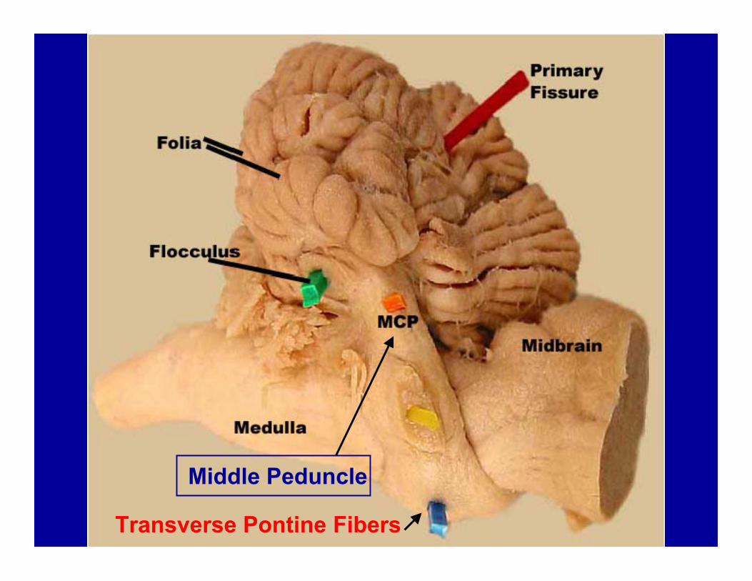
Cerebellar Cortex: the surface gray matter of the cerebellum.3 layers:
1. Molecular Layer- most superficial, consisting of axons of granule cells (parallel fibers) and dendrites of PCs 2. Purkinje Cell Layer- middle layer consisting of a single layer of large neuronal cell bodies (Purkinje cells) 3. Granule Cell Layer- deepest layer (next to white matter) consising of small neurons called granule cells

Cell Types and Afferent Fibers of the Cerebellar Cortex1. Purkinje Cells - the only output neuron from the cortex utilizes GABA to inhibit neurons in deep cerebellar nuclei2. Granule Cells- intrinsic cells of cerebellar cortex; use glutamate as an excitatory transmitter; excites Purkinje cells via axonal branches called “parallel fibers”
3. Basket Cells- inhibitory interneuron;utilizes GABA to inhibit Purkinje cells
Basket Cell
Granule Cell
Cerebellar Purkinje cells in sagittal section and in transverse section
Transverse

Granule cellParallel Fibers
Granulecells

Basket Cell
Basket
Basket cell axonforms a basket onPurkinje cell initialsegmentInhibits axonal firing
Basket madeBy axon ofBasket cell

3. Climbing Fibers- axons arising from the olivary nucleus;use glutamate and aspartate to excite Purkinje cell andcerebellar nuclei neurons
4. Mossy Fibers- all other axons that enter the cerebellum;excite granule cells (and neurons in cerebellar nuclei)

Major Cerebellar Inputs (axons entering the cerebellum):
1. Climbing Fiber Inputs = Olivocerebellar fibers-- ariseexclusively from the olivary nucleus of the caudalmedulla; have a powerful excitatory effect on Purkinjecells upon which they synapse.
OlivaryNucleus
Pyramid
Olivo-CerebellarFibers
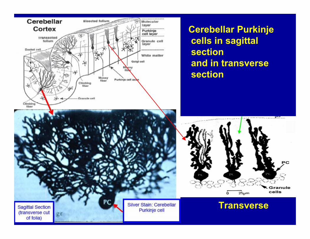
2. Mossy fiber Inputs: A. Vestibulocerebellar fibers--arise mainly from thevestibular nerve and vestibular nuclei; project to flocculo-nodular lobe and fastigial nucleus (coordinate head and eyemovement. B. Spinocerebellar Fibers- arise from spinal cord -->viaspinocerebellar tracts-->go to rostral lobe; makescerebellum aware ofongoing movementsvia proprioceptiveinput from musclespindles and jointreceptors.
8thNerve
VestibularNuclei
F
Nodulus
Dorsal spinocerebellar Tr

C. Cerebropontocerebellar fibers--arise from pyramidalcells in the cerebral cortex, synapse on pontine nucleiwhich send their axons to the contralateral cerebellarcortex via pontocerebellar fibers (form middle peduncle)-Alerts cerebellum regarding anticipated movements.
MiddleCerebellarPeduncle
Neuron in Pontine Nuclei
Ponto-CerebellarAxon
Axon fromCerebral cortex

From Lab Guide, page 57
Cerebellar Output (efferents)Major CerebellarOutputs (arise fromneurons in deepcerebellar nuclei):
1. Fastigial NucleusProjections: (viacaudal peduncle)-->vestibular nuclei andreticular formation-->vestibulospinal &reticulospinal tractsinfluence spinalmotor neurons-->effect extensormuscles related tomaintaining postureand balance.
Fastigial
Fastigial
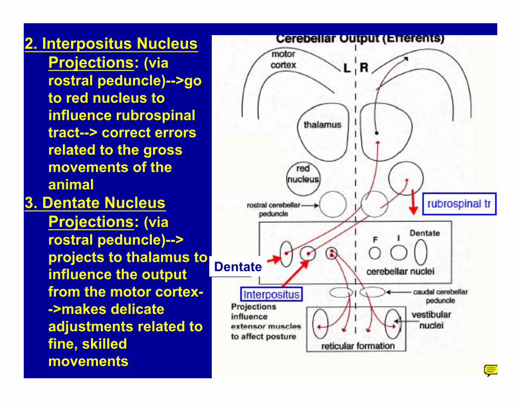
2. Interpositus NucleusProjections: (viarostral peduncle)-->goto red nucleus toinfluence rubrospinaltract--> correct errorsrelated to the grossmovements of theanimal
3. Dentate NucleusProjections: (viarostral peduncle)-->projects to thalamus toinfluence the outputfrom the motor cortex-->makes delicateadjustments related tofine, skilledmovements
Dentate

Clinical Abnormalities:Lesions of the cerebellum (damage to input, output or
cortex) result in symptoms that occur because thecerebellum’s normal function is interrupted-->ataxia,dysmetria, intention tremor occur.
Cerebellar disorders usually result from:1. Tumors (i.e., cerebellar cystic meningioma)2. Viral Infections (encephalitis; distemper)- this may occur
in utero (feline panleukopenia virus)3. Heavy metal poisoning4. Genetic Disorders: cerebellar degeneration, i.e., English
sheepdogs, Gordon setters, Kerry Blue Terriers, ArabianHorses, Yorkshire Pigs; autosomal recessive.


1. Small lesions produce no signs or onlytransient symptoms; small deficits arecompensated for by other parts of the brain
2. Lesions of the cerebellar hemispheres result inloss of muscular coordination and jerkypuppet-like movements of the limbs on theipsilateral side (same side as lesion)
3. Lesions of the vermis result in truncal tremorand gait ataxia (splayed stance and swaying ofthe body while walking)
[signs]

Memories from my trip down south this past summer


SignSeen inAlabama


Outhouse in West Virginia