Cerebellum - Global Anatomy Home Page€¦ · · 2009-08-03539 Cerebellum Finally, we know that...
52
537 Cerebellum We already know quite a bit about the cerebellum from our spinal cord and brain stem discussions. Let’s review our CURRENT STATE of knowledge. CEREBELLAR PEDUNCLES (bundles of fibers connecting the cerebellum with the underlying brain stem). We know that the inferior cerebellar peduncle (restiform body) contains the dorsal spinocerebellar tract (DSCT) fibers. These fibers arise from cells in the ipsilateral Clarke’s column in the spinal cord (C8-L3). We also know that this peduncle contains the cuneocerebellar tract (CCT) fibers. These fibers arise from the ipsilateral accessory cuneate nucleus. The largest component of the inferior cerebellar peduncle consists of the olivocerebellar tract (OCT) fibers. These fibers arise from the contralateral inferior olive. Finally, some of you might remember from Point 13 that vestibulocerebellar tract (VCT) fibers arise from cells in both the vestibular ganglion and the vestibular nuclei and pass in the inferior cerebellar peduncle to reach the cerebellum. Cerebellum
Transcript of Cerebellum - Global Anatomy Home Page€¦ · · 2009-08-03539 Cerebellum Finally, we know that...

537Cerebellum
We already know quite a bit about the cerebellum from our spinal cord and brain stemdiscussions. Let’s review our CURRENT STATE of knowledge.
CEREBELLAR PEDUNCLES (bundles of fibers connecting the cerebellum with the underlyingbrain stem).
We know that the inferior cerebellar peduncle (restiform body) contains the dorsalspinocerebellar tract (DSCT) fibers. These fibers arise from cells in the ipsilateral Clarke’scolumn in the spinal cord (C8-L3). We also know that this peduncle contains the cuneocerebellartract (CCT) fibers. These fibers arise from the ipsilateral accessory cuneate nucleus. The largestcomponent of the inferior cerebellar peduncle consists of the olivocerebellar tract (OCT) fibers.These fibers arise from the contralateral inferior olive. Finally, some of you might remember fromPoint 13 that vestibulocerebellar tract (VCT) fibers arise from cells in both the vestibularganglion and the vestibular nuclei and pass in the inferior cerebellar peduncle to reach thecerebellum.
Cerebellum

538Cerebellum
We also know that the middle cerebellar peduncle (brachium pontis) contains thepontocerebellar tract (PCT) fibers. These fibers arise from the contralateral pontine grey.

539Cerebellum
Finally, we know that thesuperior cerebellar peduncle(brachium conjunctivum; Point18) is the primary efferentpeduncle of the cerebellum. Itcontains fibers that arise fromseveral deep cerebellar nuclei.These fibers pass rostrally for awhile (ipsilaterally) and thencross at the level of the inferiorcolliculus to form the decussationof the superior cerebellarpeduncle. These fibers thencontinue rostrally to terminate inthe red nucleus and the motornuclei of the thalamus (VA, VL).
LET’S REVIEW
INFERIOR CEREBELLAR PEDUNCLE = DSCT, CCT, OCT, VCT
MIDDLE CEREBELLAR PEDUNCLE = PONTOCEREBELLARS
SUPERIOR CEREBELLAR PEDUNCLE = EFFERENT FIBERS

540Cerebellum
Try the questions below to see how you stand regarding the identity and components of thethree cerebellar peduncles. Make sure you do this before moving on in the module.
1. A. Name the peduncle that lies adjacent to the fourth ventricle in the section below
B. Where are the cells of origin of the axons in this peduncle?
2. A. Name the peduncle present in the dorsal part of the section below
B. Name four fiber tracts that contribute to this peduncle

541Cerebellum
3. A. Name the peduncle present in the section below
B. Where are the cells of origin of the axons in this peduncle
542Cerebellum
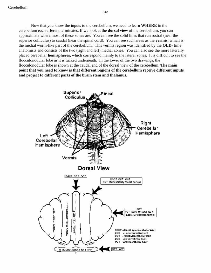
Now that you know the inputs to the cerebellum, we need to learn WHERE in thecerebellum each afferent terminates. If we look at the dorsal view of the cerebellum, you canapproximate where most of these zones are. You can see the solid lines that run rostral (near thesuperior colliculus) to caudal (near the spinal cord). You can see such areas as the vermis, which isthe medial worm-like part of the cerebellum. This vermis region was identified by the OLD- timeanatomists and consists of the two (right and left) medial zones. You can also see the more laterallyplaced cerebellar hemispheres, which correspond mainly to the lateral zones. It is difficult to see theflocculonodular lobe as it is tucked underneath. In the lower of the two drawings, theflocculonodular lobe is shown at the caudal end of the dorsal view of the cerebellum. The mainpoint that you need to know is that different regions of the cerebellum receive different inputsand project to different parts of the brain stem and thalamus.

543Cerebellum
Zones of the cerebellum
The various cerebellar afferents that we have learned distribute to different zones of thecerebellum. Four different zones need to be considered. These are the 1) medial zone, 2)intermediate zone, 3) lateral zone and 4) flocculonodular zone (or lobe). Let’s consider thepathways that end in each zone or lobe.
1. olivocerebellar fibers—distribute to the ENTIRE cerebellum2. dorsal spinocerebellar and cuneocerebellar fibers—distribute to the medial andintermediate zones3. pontocerebellar fibers—distribute to the intermediate and lateral zones
A. Those pontine grey neurons that project to the intermediate zone of thecerebellum receive their corticopontine input from the primary motor cortex(area 4; execution).
B. Those pontine grey neurons that project to the lateral zone of the cerebellumreceive their corticopontine input from posterior parietal cortex (PMl and SMA).In comparison with the primary motor cortex,these areas are involved in planningthe motor act.
4. vestibulocerebellar fibers—(arise from the vestibular ganglion and nuclei) distributeprimarily to the flocculonodular lobe

544Cerebellum
LET’S REVIEW
OLIVOCEREBELLAR FIBERSThe distribution of olivocerebellar fibers onto the flattened view of the cerebellum is shown. This iseasy, since the fibers project to the entire cerebellum.
545Cerebellum
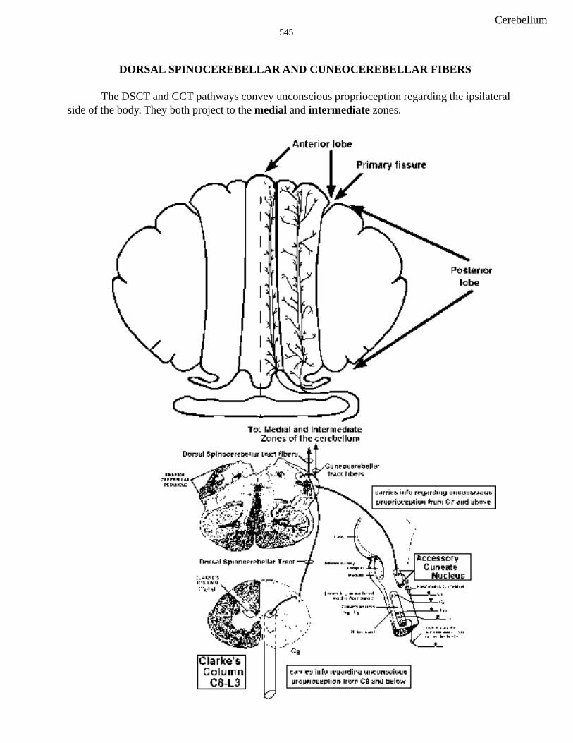
DORSAL SPINOCEREBELLAR AND CUNEOCEREBELLAR FIBERS
The DSCT and CCT pathways convey unconscious proprioception regarding the ipsilateralside of the body. They both project to the medial and intermediate zones.
546Cerebellum
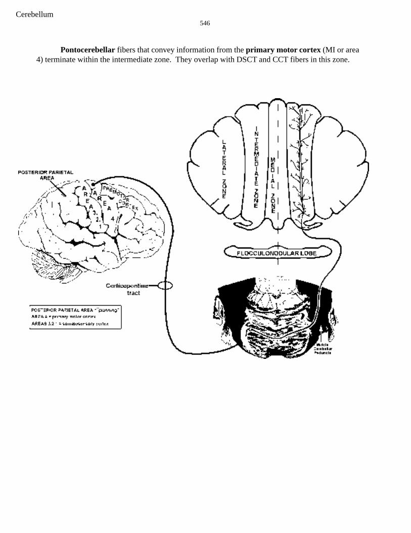
Pontocerebellar fibers that convey information from the primary motor cortex (MI or area4) terminate within the intermediate zone. They overlap with DSCT and CCT fibers in this zone.

547Cerebellum
Pontocerebellar fibers that convey information from the posterior parietal, PMl and SMA(planning) areas of the cortex terminate within the lateral zone of the cerebellum.
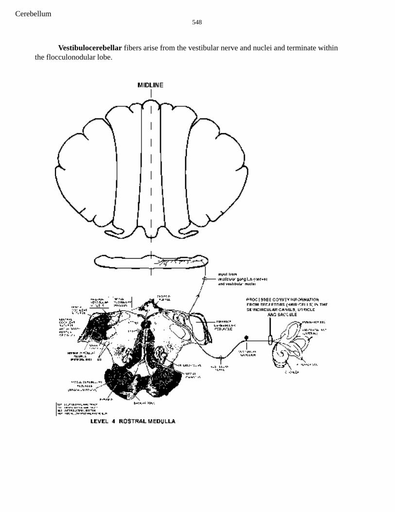
548Cerebellum
Vestibulocerebellar fibers arise from the vestibular nerve and nuclei and terminate withinthe flocculonodular lobe.

549Cerebellum
Now let’s do some practice questions to make sure we understand the organization of inputs to thecerebellum. Then we can move on to the deep cerebellar nuclei.
4. A. In the figure below, name the zones of the cerebellum that receive input from axons travelingwithin the structure labeled A. ________, ________, _________, ________
B. In the figure below, name the zones of the cerebellum that receive input from the structuredlabeled B. ____________, ____________

550Cerebellum
5. The following questions relate to the labeled drawing below.
A. Axons from B travel in which cerebellar peduncle?_______________
B. Does A project to the same side of the pons and spinal cord?____________
C. Which cortical area(s) are is involved in “higher” planning of motor movements?______
D. Which cortical area(s) send(s) information (via the pons) to the lateral zone of thecerebellum?_________
E. Which cortical area (A or B) send(s) information (via the pons) to the intermediate zone of thecerebellum?_________
F. Does either cortical area A or B send information (via the pons) to the medial zone of thecerebellum? YES NO
G. Does either cortical area A or B send information (via the pons) to the flocculonodular zone (orlobe) of the cerebellum? YES NO

551Cerebellum
6. The following questions relate to the TWO labeled drawings below.
A. What cortical areas project to D?
B. What zone(s) of the cerebellum are innervated by axons of the cells in A? (Clarke’s column)
C. A complete lesion of B results in loss of pain and temperature (DON’T FORGET THE SPINALCORD) from what level(s)?
D. Axons in E terminate in which zones of the cerebellum? ___________and___________
E. Do axons with cell bodies in the motor cortex travel in E? _______

552Cerebellum
7. The following questions relate to the labeled drawing below.
A. Fibers from cells that lie in nucleus A project to which zone of the cerebellum?__________________
B. Cells in A project to the lateral zone of the right cerebellum via the middle cerebellar peduncle. True or False?
C. B is the ___________ cerebellar peduncle or ____________body.
D. Vestibular input reaches the cerebellum via 2 pathways; peripherally from the__________________ and centrally from the ____________________.
E. True or False; axons of cells in E project directly to the cerebellum.

553Cerebellum
Deep Cerebellar Nuclei
The inputs to the cerebellum just discussed take part in some very complicated internalcircuitry. An optional overview of this internal circuitry can be found at the end of this lecture, but itis not necessary for a good understanding of how the cerebellum influences movement. All youreally need to know is that after all is said and done, the inputs to the cerebellum ultimately affectPurkinje cells of the cerebellar cortex and the deep cerebellar nuclei. The Purkinje cells inhibit thedeep cerebellar nuclei and ultimately affect the output of the cerebellum. Let’s look at thedistribution of Purkinje cell axons. Purkinje cells in the four different zones, MEDIAL,INTERMEDIATE, LATERAL AND FLOCCULONODULAR, project to different deep cerebellarnuclei. There are four deep cerebellar nuclei, fastigial, globose, emboliform and dentate (feel goodevery day) from medial to lateral.Three of the four deep cerebellarnuclei can be seen below in an oldfriend from the brain stem, level 5.You have not seen the dorsal part ofthis section before, but it shows threeof the four sets of deep cerebellarnuclei. The distribution of Purkinjecell axons to the cerebellar nuclei isactually quite easy to rememberbecause it is topographicallyorganized. The most lateral deepcerebellar nucleus, the dentate,receives its Purkinje cell input fromthe lateral zone of the cerebellum.Remember that this lateral zone of thecerebellum receives its input fromolivocerebellar and pontocerebellarfibers carrying planning information.The interpositus nucleus (actually twonuclei, globose and emboliform)receives its Purkinje cell input fromthe intermediate zone of thecerebellum. The intermediate zone ofthe cerebellum receives its input fromolivocerebellar and pontocerebellarfibers carrying information fromprimary motor cortex (area 4) ANDfrom the DSCT and CCT. ThePurkinje cells that innervate themedially located fastigial nucleus liein the medial zone of the cerebellumwhich, as you remember, receivesinput from the DSCT, CCT and OCT.
554Cerebellum
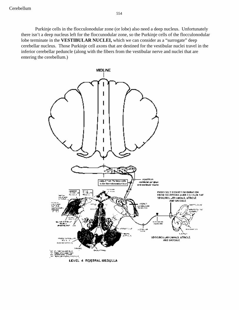
Purkinje cells in the flocculonodular zone (or lobe) also need a deep nucleus. Unfortunatelythere isn’t a deep nucleus left for the floccunodular zone, so the Purkinje cells of the flocculonodularlobe terminate in the VESTIBULAR NUCLEI, which we can consider as a “surrogate” deepcerebellar nucleus. Those Purkinje cell axons that are destined for the vestibular nuclei travel in theinferior cerebellar peduncle (along with the fibers from the vestibular nerve and nuclei that areentering the cerebellum.)

555Cerebellum
The signals from the Purkinje cells that terminate in the deep cerebellar nuclei are inhibitory.Think of the inhibitory signal as increasing or decreasing depending upon what is happening in thecerebellum (see intrinsic circuitry at the end of section if interested). Note that the deepcerebellar nuclei also receive EXCITATORY inputs from the collaterals of both mossy and climbingfibers as they pass through the deep white matter on their way to the overlying cerebellar cortex. Theinterplay of the inhibitory (Purkinje cell) and excitatory (mossy and climbing fiber) inputs to thedeep nuclei determines their output signal to the other parts of the brain that we will discuss next.

556Cerebellum
LET’S REVIEWPURKINJE CELLS IN THE:
• LATERAL ZONE OF CEREBELLUM FEED INTO DENTATE
• INTERMEDIATE ZONE FEED INTO INTERPOSITUS (globose and emboliform)• MEDIAL ZONE FEED INTO FASTIGIAL
• FLOCCULONODULAR LOBE FEED INTO VESTIBULAR NUCLEI

557Cerebellum
Now we can take the information out of the deep cerebellar nuclei and the vestibular nuclei towhere it can do some good, i.e., to the spinal cord, brain stem and thalamus. We already know a lotof this from our discussions of the Superior Cerebellar Peduncle (Point 18) and the Vestibular Nuclei(Point 13).
The dentate nucleus projects primarily to the contralateral VA/VL nuclei of the thalamus viathe superior cerebellar peduncle. Cells in VA/VL project to the premotor area (PM) and thesupplementary motor area (SMA), both of which are motor “planning” areas. These corticalplanning areas then project to primary motor cortex (MI or area 4) and from there the information isconveyed to the corticospinal system.
558Cerebellum
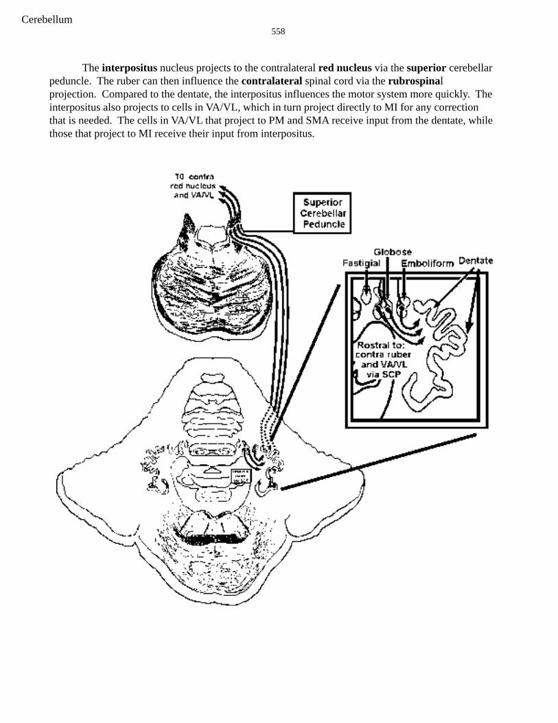
The interpositus nucleus projects to the contralateral red nucleus via the superior cerebellarpeduncle. The ruber can then influence the contralateral spinal cord via the rubrospinalprojection. Compared to the dentate, the interpositus influences the motor system more quickly. Theinterpositus also projects to cells in VA/VL, which in turn project directly to MI for any correctionthat is needed. The cells in VA/VL that project to PM and SMA receive input from the dentate, whilethose that project to MI receive their input from interpositus.

559Cerebellum
Now for the FASTIGIAL nucleus. To exit the cerebellum, the axons of cells in the fastigialnuclei take two routes that we did not discuss in the brain stem or spinal cord series of lectures.Crossing axons from the fastigial pass to the vestibular nuclei and reticular formation of the ponsand medulla (not illustrated below). The greater number of fibers leaving the fastigial nucleus areuncrossed and pass via the inferior cerebellar peduncle to reach the vestibular nuclei and reticularformation. The reticular formation consists of cells that we did not identify in the brain stemseries of discussions. They are kind of “left over”, but nonetheless important, cells that project to thespinal cord as reticulospinal fibers. These fibers lie in the ventral funiculus. I have only illustratedthe medullary reticular formation below. Don’t worry about the pontine.

560Cerebellum
Finally, you already know that the vestibular nuclei (deep nuclei of the flocculonodular lobe)project to the PPRF and the spinal cord (via the MVST and LVST).

561Cerebellum
Putting it all together
Now, let’s figure out what cerebellar circuitry might accomplish. I will use the example ofthe visually guided movement of reaching and grasping an apple from an apple tree. The visualinformation about the apple is carried from the retina to the cerebral cortex via pathways that we willdiscuss later in the course. This information eventually is sent to the posterior parietal cortex.Information from the PPC is conveyed to the PMl and in turn to the SMA. All of these planningareas project to the pons (corticopontine). The pontine grey neurons then relay the planninginformation to the contralateral lateral zone of the cerebellum via pontocerebellar fibers, whicheveryone knows by now are mossy fibers. The messages from these planning areas goes through thecerebellar circuitry. The Purkinje cell then sends a message to the dentate and the dentate projects tothe VA/VL. This new message headed for VA/VL contains planning information for reaching outand grasping the apple. The message in VA/VL is then relayed back to the PMl and SMA which inturn feed into the primary motor cortex (area 4; the cells of origin of the corticospinal tract).Remember all of this happens before any movement has taken place.
You might ask, “what does the lateral cerebellum contribute to the planning that is notprocessed by PPC, PMl amd SMA? One hypothesis is that the lateral zone is involved in the storageand/or retrieval of long-term memory for motor skills. That is, circuitry in the lateral zone “learned”something about the movement that can now assist the PMl and SMA. Thus, when SMA, PPC andPMl information reaches the lateral zone (via corticopontine and then pontocerebellar fibers),specific cerebellar circuits that were laid down when the movement was initially learned areactivated. So, information about the planned movement is contained not only in cortical areas butalso in the lateral zone of the cerebellum.
Lesions of the lateral zone of the cerebellum and dentate result in errors in the direction,force, speed, and amplitude of movements. (Interestingly, these uncoordinated movements arebeing carried out by a healthy corticospinal tract. It is just that the corticospinal tract is receiving badinformation). The “priming” of the corticospinal tract by the dentate is lost. It takes longer to get themovement going, the appropriate muscles contract for too long, and the movement stops too late.The result is incoordination or ataxia, which includes several symptoms or deficits. First there isDecomposition of movements, which means that instead of a nice smooth movement, themovement consists of jerky parts of the movement. Dysmetria, also called past-pointing, occurswhen the patient tries to touch their nose with their finger. There is an inability to perform rapidalternating movements (pronation/supination) called dysdiadochokinesis. Rebound phenomenacan be seen when you hold the patients tensed arm away from his/her sternum and chest and then letgo. It will fly into their sternum. There also is tremor during the uncoordinated movement. This iscalled movement, or intention tremor. It is hard for me to explain why this might occur with lateralzone lesions but it might be due to the fact that there are problems in correctly stopping movements.This results in the movement going too far, after which there is a correction and then oscillationsoccur.
Now we have the planning signals from the lateral zone to MI. Information from MI isconveyed to cells in the spinal cord via the corticospinal tract. This information that is sent to thespinal cord from MI is also conveyed to the pontine grey (corticopontine fibers) and in turn to theintermediate zone of the cerebellum. As the movement to reach and grasp the apple evolves, thedorsal spinocerebellar and cuneocerebellar pathways convey what the muscles are really doing as

562Cerebellum
they reach out and grasp the apple. This incoming sensory information (what the muscles areactually doing) is then compared to the MI-corticospinal signal (conveyed to the intermediate zonevia the corticopontine-pontocerebellar fibers) regarding what the muscles are supposed to be doing.If necessary, corrections are conveyed from the intermediate zone, via nucleus interpositus, to the rednucleus (rubrospinal tract, fast corrections) and MI (corticospinal tract).
The computations occurring in the intermediate zone involve mossy fiber barrages associatedwith the pontocerebellar fibers carrying information from MI about the intended movement. Thisinformation is somehow “compared” with information conveyed via the dorsal and cuneocerebellarfibers (what is really happening to the muscles). The Purkinje cell signals being sent to interpositusmix with the direct signals from mossy and climbing fibers, and an ongoing correction signal is sentto the red nucleus (quick adjustments via the rubrospinal tract) and back to MI (slower adjustments).This is carried from interpositus via the superior cerebellar peduncle.
Lesions of the intermediate zone and nucleus interpositus are thought to result in similardeficits as those in the lateral zone and the dentate. Such lesions interrupt the ability of thecerebellum to correct movements once they are started. In contrast to cells in the dentate nucleus,cell in the interpositus fire after the movement has begun. Such cells are involved in updatingongoing movements versus planning such movements (dentate).
The medial zone receives mainly dorsal spinocerebellar and cuneocerebellar information,and projects to the reticular nuclei and the vestibular nuclei. Lesions of this zone will result in a lossof fastigial excitatory drive to the ipsilateral vestibular nuclei, as well as the reticular nuclei. Thismeans that the opposite vestibular nuclei are dominating. You know what that means as far asstumbling and nystagmus. CLUE-ipsilateral stumbling and contralateral nystagmus. In addition tothe balance problems, the loss of inputs to the reticular nuclei has a powerful affect on the control ofpostural muscles which causes instability of the axial musculature.
Finally, The influence of the vestibulocerebellum (flocculonodular lobe) on the vestibularnuclei also plays a role in posture and balance. Remember that the Purkinje cells in theflocculonodular lobe are inhibiting the vestibular nuclei (while neurons in the fastigial nucleus areexciting them). Therefore a lesion of the right vestibular zone (or lobe) will “release” the rightvestibular nuclei from inhibition. Thus the right vestibular nuclei are in dominance. You can take itfrom here regarding the directions of stumbling and nystagmus.
A classic sign of cerebellar damage is a decrease in tone (hypotonia). While this hypotoniais thought to result from malfunctioning of the descending control over the gamma efferent system, Iwill not discuss this any further.
Finally, there is incoordination of speech following cerebellar damage. This is calleddysarthria.
563Cerebellum
WHAT ABOUT THE OLIVE?
Ever since the inferior olive first appeared back there in the brain stem, we have said “youwill learn more about the olive later in the course.” Well, here we are!! You already know that itprojects to the contralateral cerebellum and the axons end as climbing fibers. Also, you know thatolivocerebellar fibers terminate throughout the cerebellar cortex i.e., they reach all four zones.You even know a little bit about the circuitry of climbing fibers, in that they target deep nuclei andclimb right up those Purk cells.
There is evidence that climbing fibers in the flocculonodular lobe might play a role in theplasticity of the vestibulo-ocular reflex (VOR; remember Point #13?). In this instance, plasticityrefers to changes in the circuitry/functioning of the VOR. Hopefully you remember that the VORhelps stabilize the visual image on the retina as the head moves. So, a quick head movement to theright results in a compensatory horizontal conjugate eye movement to the left.
Now, consider what happens when you get a new pair of glasses. Say that they magnifythings more than your old pair. This means that a larger compensatory eye movement is needed forthe same degree head movement. Immediately after putting them on you notice things are a bitblurry and you might get dizzy. Remember, everything in the visual field is now bigger than before,so that during a head movement (over a certain number of degrees) to the right, the spot you arefixating on will move more to the left than before. Thus the eye movement to the left that is inreaction to the head movement to the right will NOT keep the image stable on the retina. The VORis functioning as if you still have on your old glasses and the “gain” of the VOR is not large enoughto move the eyes far enough to the left to stabilize the image. (Gain just refers to how much the eyesmove in response to a certain head movement.
As you know, you do adjust after wearing the new lenses for a few hours/days. It is thoughtthat this adjustment or adaptation of the gain is carried out by the olivocerebellar fibers that reach theflocculonodular lobe. For instance, a person who wore a new pair of glasses that were 2X themagnifying power of his/her old ones was found to have a VOR gain of 1.8 after a few days (1.0 isnormal). How did this happen? You know that the vestibular nuclei receive information from thesemicircular canals/CN VIII. They send the information to the flocculonodular lobe of thecerebellum via mossy fibers and also project to the contralateral PPRF, which in turn projects to theabducens nucleus. Thus, head right, increased firing of right CN VIII, left PPRF and left abducens;both eyes then go left.

564Cerebellum
When the eyes move to the left, cells in the retina send information to an area of the rostralbrain stem called the pretectum. These pretectal cells can tell if the eyes and head movementsmatch. If they don’t match, there is slippage/blurring of the image and the pretectal cells know it.They in turn tell the inferior olive about the slippage and the climbing fibers comprise a “teachingline” to instruct the Purkinje cells to fire LESS. They would do this by modulating the efficiency ofthe mossy fiber/granule cell input from the vestibular nuclei. If the gain of the VOR is too small forthe head movement (new glasses), the climbing fiber input would somehow decrease the efficiencyof the vestibular mossy fiber/granule cell/parallel fiber input to the cerebellum and the Purks wouldfire less. This would means less inhibition on the vestibular nuclei and a slightly biggercompensatory eye movement to the left (the degrees of head movement did not change). This wouldstabilize the image on the retina during the head movement. It has been found that no suchadaptation of the VOR can occur following lesions of the flocculonodular lobe.
Granted, this is just the flocculonodular lobe and nothing has been said about olivocerebellarsin the other three zones. Well, studies are in progress, but right now you have to be satisfied with theflocculonodular lobe.
REMEMBER—CLIMBING FIBERS=MOTOR LEARNING

565Cerebellum
8. Which of the following statements is correct?
A. a Babinski sign is seen following cerebellar lesionsB. cerebellar lesions result in atrophyC. a cardinal sign of cerebellar damage is hypotoniaD. there are no speech problems following cerebellar lesionsE. dysdiadochokinesia is another name for muscle weakness
9. Which of the following statements is correct?
A. rebound phenomena refers to the inability to grab a rebound above the rimB. past pointing is a cardinal sign of corticospinal damageC. intention tremor is seen following lesions of the substantia nigraD. resting tremor is seen following lesions of the cerebellumE. atrophy is not a sign of cerebellar disease
10. Which of the following statements is correct?
A. hypotonia could never result from defects in the gamma efferent systemB. spasticity is associated with lesions of the lateral cerebellumC. both cerebral cortex and cerebellar deficits involve the ipsilateral side of the bodyD. weakness is not observed in cerebellar diseaseE. ataxia is a cardinal sign of cerebellar disease
11. Which of the following statements is correct?
A. the fastigial nucleus projects to the red nucleusB. the presence of nystagmus suggests damage to the corticospinal tractC. the inferior olive is the sole source of climbing fibersD. the interpositus nucleus projects to the vestibular nuclei via the inferior cerebellar peduncleE. nystagmus is a sign of lower motor neuron disease
12. Which of the following statements is correct?
A. a lesion of the right side of the flocculonodular lobe will result in left nystagmusB. a lesion of the right side of the flocculonodular lobe will result in staggering to the rightC. a lesion of the right fastigial nucleus will result in staggering to the leftD. a lesion of the right fastigial nucleus will result in right nystagmusE. a lesion of the right fastigial nucleus and the right vestibular nuclei will (in different cases)result in nystagmus of the same direction

566Cerebellum
13. Which of the following associations are true?
A-planningA-updatingA-posterior parietal cortexA-VA/VLA-PMl, PPC and supplementary motor cortexA-reticular formationA-vestibular nucleiA-fires after movementB-ruberB-updatingB-reticular formationB-primary motor cortexB-vestibular nucleiB-comparing and updatingB-fires before movementC-nystagmusC-reticular formationC-ruberC-VA/VLC-bilateral efferent projection (but mostly ipsi.)

567Cerebellum
14. Which of the following is associated with cerebellar lesions?
A. left nystagnmus following a lesion of the right flocculonodular lobeB. Babinski signC. right nystagnmus following a lesion of the right fastigial nucleusD. hyperreflexiaE. hemiplegiaF. resting tremorG. apraxiaH. rigidityI. bradykinesiaJ. hemiballismK. choreaL. atrophyM. anesthesiaN. analgesiaO. pronator driftP. intention (movement) tremorQ. reboundR. dysdiadochokinesiaS. past-pointing
15. Which of the following statements regarding climbing fibers is TRUE?
A. those ending in the flocculonodular zone arise from the inferior oliveB. those ending in the medial zone arise from the inferior oliveC. those ending in the intermediate zone arise from the inferior oliveD. those ending in the lateral zone arise from the inferior oliveE. all of the above are TRUE
16. Which of the following statements regarding mossy fibers is TRUE?
A. those reaching lateral zone are solely pontocerebellarB. those reaching intermediate zone are pontocerebellar, DSCT and CCTC. those reaching medial zone solely DSCTand CCTD. those reaching the flocculonodular zone are from the vestibular ganglion and vestibularnucleiE. all of the above are TRUE
17. Which of the following statements is TRUE?
A. Purkinje cells in the lateral zone excite the dentateB. Purkinje cells in the medial zone inhibit the vestibular nucleiC. Purkinje cells in the intermediate zone excite the fastigialD. Purkinje cells in the flocculonodular zone inhibit the fastigialE. Purkinje cells inhibit deep nuclei

568Cerebellum
18. Which of the following statements is TRUE?
A. deep cerebellar nuclei excite their targetsB. deep cerebellar nuclei inhibit their targetsC Purkinje cells excite their targetsD. deep cerebellar nuclei are inhibited by incoming climbing fibersE. deep cerebellar nuclei are inhibited by incoming mossy fibers
19. A 60 y.o. male presents with complaints of an unstable gait and some swaying of the trunk. Hishistory is unremarkable except that for the past 15 years he has had increasing use of alcohol to hispresent level of approximately four hard liquor drinks per day. Most of his neurological exam isunremarkable. His walking gait is broad based, with short regular steps and his trunk is slightlyinclined forward. A test of reflexes demonstrates some hypotonia in the proximal muscles, butnearly normal reflexes. He claims that he walks fine on flat ground, but has difficulty and stumbles alot when he is on uneven ground. He says that people have commented about his gait for manyyears, but he is just beginning to think something may be wrong. Which of the following could betrue of this patient?
A. he probably has a tumor pressing on the superior cerebellar peduncleB. he probably has degeneration of the vermal regions of the cerebellum due to alcoholconsumptionC. an MRI of his cerebellum would show increased space between the folia of the vermal regionand the anterior lobeD. all of his signs indicate damage to the lateral and intermediate zones of the cerebellumE. two of the above are true
20. A 45 y.o. woman presents with nausea, and tremor. Her history states she has been mildlyhypertensive for many years, and has recently had TIAs (transient ischemic attacks). Her bloodpressure is 180/120. A neurological exam shows a slight Horner’s syndrome on the right,reduced pain and temperature sensation on the left, but all other sensations are normal. A motorexam demonstrates reduced tone in the right upper and lower limbs, dysmetria and past pointing,and an intention tremor on the right side. Movements of her right limbs seem to be most affectedand when asked to perform a rapidly alternating movement with her right hand, she clumsilyperforms the task. She has a normal flexor plantar reflex bilaterally and nearly normal strengthin all her limb muscles. Which of the following could be true of this patient?
A. the Horner’s syndrome and altered pain and temp sensations suggest damage to the ALS inthe brainstem.B. this patient probably sustained a vascular accident affecting primarily the intermediate andlateral zones of the cerebellum.C. the inability to perform rapidly alternating movements is called dysdiadochokinesis.D. two of the above are trueE. three of the above are true

569Cerebellum
CEREBELLAR PROBLEM SOLVING ANSWERS
1. A. superior cerebellar peduncleB. ipsilateral deep cerebellar nuclei
2. A. inferior cerebellar peduncleB. OCT, DSCT, CCT, VCT
3. A. middle cerebellar peduncleB. contralateral pontine grey
4. A. flocculonodular, medial, intermediate, lateralB. intermediate, lateral
5. A. none B. no C. B and D D. B and D E. A F. no G. no
6. A. motor, posterior parietal, premotor (SMA and PMl)B. medial, intermediateC. T3 and below (contra)D. intermediate, lateralE. no
7. A. flocculonodularB. falseC. inferior, restiformD. vestibular ganglion, vestib. nuc.E. true
8. C 13. A. T; F; T; T; T; F; F; F; 17. E B. T; T; F; T; F; T; F;
9. E C. T; T; F; F; T 18. A
10. E 14. P,Q,R,S 19. E (B and C)
11. C 15. E 20. E
12. E 16. E

570Cerebellum
Your colleague’s who have taken this course before you have been so intrigued by the nervoussystem that many have written unforgettable songs. I have included one below and hopefully theauthors or their friends will sing them sometime during this module.
YESTERDAY
Micheal Gelman and Alex RomashkoClass of 2000
Yesterday,My cerebellum said “Come on let’s play”.But now my lateral zone’s gone away.No input there from pontine grey.
Suddenly,I’ve got no info from my PPC.I can’t wake up my CST.My movements all come jerkily.
I don’t plan too well, with VL or with VA.Past-point, and I’ve found I rebound when held away.
There’s no doubt,In my premotor all lights are out.It gets no news to think about.My OCT just has no clout.
When I move, I go dysdiado-chokinetically.Dentate cells, I’ve heard, get no word from Purkinje
Yesterday,I moved smoothly, didn’t go astray.Now I can’t get through the SMA.Intention tremor’s here to stay.
Mmm mmm mmm mmm mmm mmm mm…

571Cerebellum
The following pages are a detailed account of the current understanding regarding theintrinsic circuitry of the cerebellum. I have deliberately left it out of the discussion because it is notnecessary for a good understanding of what the cerebellum does for motor control. I encourage youto read it for its academic merit, but you will not be tested on this information.
Circuitry (optional)
The internal circuitry of the cerebellum has been worked out in exquisite detail. Although itis not necessary for an understanding of cerebellar function, it may aid in your overall understandingof the complexity of the nervous system. The following is a complete description of this internalprocessing.
Before looking at the internal cerebellar circuitry, we need to examine how all of the littlecerebellar folds or gyri, called folia (singular = folium) are organized. In the drawing below to theleft are 3 isolated cerebellar folia. From the extracted tissue block at the right you can see that thesefolia can be sectioned either PARALLEL to their LONG axes or TRANSVERSE to their LONGaxes. Each single folium is comprised of an outer cerebellar CORTEX (bark, peel, husk), whichcontains three cell layers, molecular, Purkinje, and granule. This cerebellar cortex overlies a deepzone of efferent and afferent fibers that we just call white matter. Within this dense aggregation ofmillions of fibers lie three sets of deep cerebellar nuclei (dentate, interpositus and fastigial). Thesedeep nuclei ARE NOT shown in the drawing below, but we have already heard about the dentate andinterpositus nuclei when discussing the superior cerebellar peduncle (Point 18). We will return tothese nuclei later in the story.
You can see that the dendrites of the Purkinje cells (the most popular cells in the cerebellarcortex) are oriented TRANSVERSE or perpendicular to the LONG axis of the folium. Spread yourfingers out and look at your palm. You are now looking at how a Purkinje cell dendritic tree lookswhen the folium is sectioned TRANSVERSE to its LONG axis.

572Cerebellum
Now, rotate your hand 90o and see how the dendritic tree of that same Purkinje cell lookswhen the folium is sectioned PARALLEL to its LONG axis. Sadly, you do not see the full extent ofthe Purkinje cell dendritic tree from this view. This is shown below.

573Cerebellum
Lets now schematize the basics of the internal cerebellar circuitry. First, we can look at anold friend who we first met at level 3 in the brain stem—THE INFERIOR OLIVE. Rememberthat, Dr. Harting told you that cells in the olive have axons that pass to the contralateral cerebellumas climbing fibers. These fibers go to all parts of the cerebellum, that is, they are not restricted to aparticular zone. The drawing above shows that a climbing fiber sends a collateral to the deepcerebellar nuclei, which is excitatory, and then “climbs” up and like ivy, entwines and synapses allover the dendrites of the Purkinje cell. Each Purkinje cell receives input from only 1 climbing fiberaxon, but each climbing fiber axon can split to innervate several Purkinje cells. These climbingfiber-Purkinje cell synapses are excitatory.

574Cerebellum
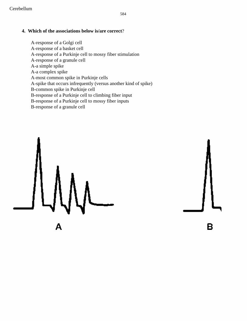
Since climbing fibers have synapses all over the dendritic tree of a Purkinje cell, their totalexcitatory action is extremely strong. In fact, the synaptic connection between the climbing fiber andthe Purkinje cell is one of the most powerful in the nervous system. A single action potential in aclimbing fiber elicits a burst of action potentials in the Purkinje cells that it contacts. This burst ofaction potentials exhibited by a Purkinje cell is called a complex spike. Climbing fibers are “lazy”(but strong), thus Purkinje cells exhibit complex spikes at a rate of about 1 per second. Theillustration above depicts an intracellular recording from a Purkinje cell that has just been turned onby stimulating the climbing fiber a single time. This single climbing fiber stimulus has a powerfuleffect in that it results in 4 action potentials (i.e., complex spike) of varying amplitudes in thePurkinje cell.

575Cerebellum
Now, what about branches of the DSCT, CCT, VCT and PCT that are destined for thecerebellar cortex. The axons of these inputs are called MOSSY fibers. Like climbing fibers, theinformation carried by mossy fibers is heading for the Purkinje cells. However, unlike climbingfibers, mossy fibers DO NOT go directly to the Purkinje cell. Each mossy fiber branches profuselyin the white matter and has multiple (up to 50) swellings (resembling moss to the old timeneuroanatomists) that contain round vesicles and synaptic thickenings. Each swelling, called a“rosette”, is a synapse of the mossy fiber onto the dendrite of a granule cell. In the detail above youcan see two rosettes contacting two different dendrites of the same granule cell. These are excitatorysynapses. A rosette can also occur where the dendrites of several (up to 15) granule cells arecontacted. Each mossy fiber can have up to 50 rosettes. You can see that there is considerabledivergence of the mossy fiber signal.

576Cerebellum
Granule cells have long axons that pass dorsally through the granule and Purkinje, cell layersto reach the molecular layer of the cerebellar cortex, where they bifurcate and run PARALLEL tothe long axis of the folium. These fibers, which are called parallel fibers, travel at right angles tothe dendrites of the Purkinje cells (think of telephone lines running through a row of (flattened) treesin the fall after peak color). Each parallel fiber synapses upon and excites the dendritic spines ofnumerous Purkinje cells, but the synaptic effect of a single parallel fiber upon a Purkinje cell isextremely weak (contrast this with a climbing fiber). How then can the mossy fiber input fire thePurkinje cells? Well, what is needed is for many mossy fibers to fire rapidly and together, whichcauses many granule cells to fire together, which turns on lots of parallel fibers which then exciteenough of the spines on a Purkinje cell to result in an action potential. When this occurs, thePurkinje cell exhibits what is called a simple spike. Such a spike in a Purkinje cell is shown below.In contrast to those lazy climbing fibers that fire about 1 per second (yet have a very powerful effectupon the Purkinje cell resulting in the complex spike), mossy fibers are really “gunners” (I use theterm affectionately) in that they are always working. Thus they fire spontaneously and rapidly (50-100 per second) and cause (via the granule cells and parallel fibers of course) Purkinje cells to firesimple spikes at the same frequency.
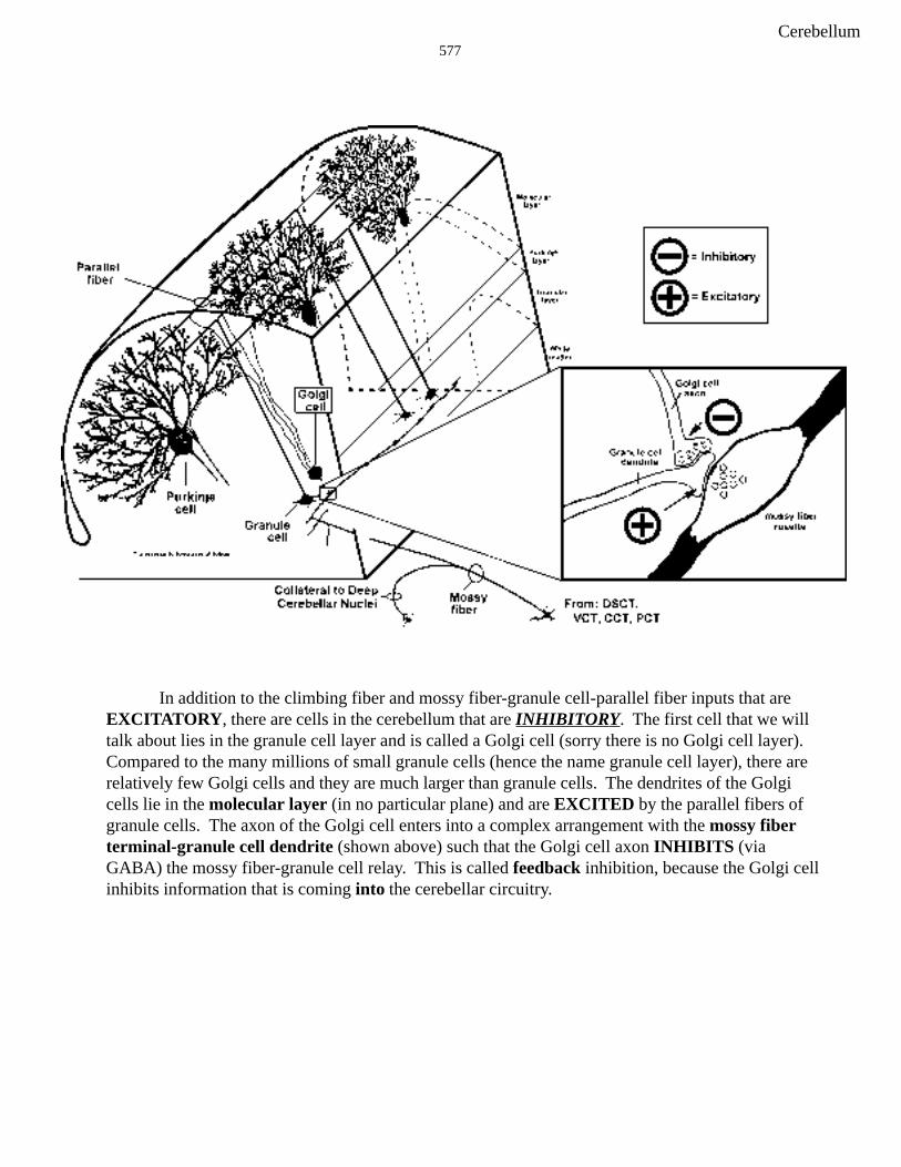
577Cerebellum
In addition to the climbing fiber and mossy fiber-granule cell-parallel fiber inputs that areEXCITATORY, there are cells in the cerebellum that are INHIBITORY. The first cell that we willtalk about lies in the granule cell layer and is called a Golgi cell (sorry there is no Golgi cell layer).Compared to the many millions of small granule cells (hence the name granule cell layer), there arerelatively few Golgi cells and they are much larger than granule cells. The dendrites of the Golgicells lie in the molecular layer (in no particular plane) and are EXCITED by the parallel fibers ofgranule cells. The axon of the Golgi cell enters into a complex arrangement with the mossy fiberterminal-granule cell dendrite (shown above) such that the Golgi cell axon INHIBITS (viaGABA) the mossy fiber-granule cell relay. This is called feedback inhibition, because the Golgi cellinhibits information that is coming into the cerebellar circuitry.

578Cerebellum
The parallel fibers of granule cells (which travel in the molecular layer) also excite thedendrites of basket cells. Both the dendrites and somas of the basket cells lie in the molecular layer.These dendrites, like those of Purkinje cells, lie in a plane that is transverse to the long axes of thefolia. The axons of basket cells also run in this plane (transverse to the long axis of the folia) andterminate on the somas of the Purkinje cells. The inhibition of Purkinje cells by the basket cellaxons is called feedforward inhibition. Remember, inhibition of the input=feedback (Golgi cellaxon-mossy fiber-granule cell relay) while inhibition of the output=feedforward (basket cell axon-Purkinje cell initial segment). Another way to look at these types of inhibition is whether theinhibiting cell is acting on an “earlier” or “later” cell in the cerebellar circuitry.
579Cerebellum
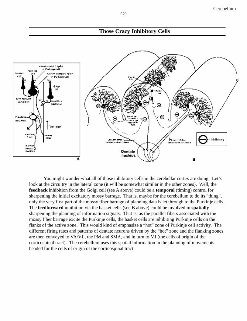
Those Crazy Inhibitory Cells
You might wonder what all of those inhibitory cells in the cerebellar cortex are doing. Let’slook at the circuitry in the lateral zone (it will be somewhat similar in the other zones). Well, thefeedback inhibition from the Golgi cell (see A above) could be a temporal (timing) control forsharpening the initial excitatory mossy barrage. That is, maybe for the cerebellum to do its “thing”,only the very first part of the mossy fiber barrage of planning data is let through to the Purkinje cells.The feedforward inhibition via the basket cells (see B above) could be involved in spatiallysharpening the planning of information signals. That is, as the parallel fibers associated with themossy fiber barrage excite the Purkinje cells, the basket cells are inhibiting Purkinje cells on theflanks of the active zone. This would kind of emphasize a “hot” zone of Purkinje cell activity. Thedifferent firing rates and patterns of dentate neurons driven by the “hot” zone and the flanking zonesare then conveyed to VA/VL, the PM and SMA, and in turn to MI (the cells of origin of thecorticospinal tract). The cerebellum uses this spatial information in the planning of movementsheaded for the cells of origin of the corticospinal tract.
580Cerebellum
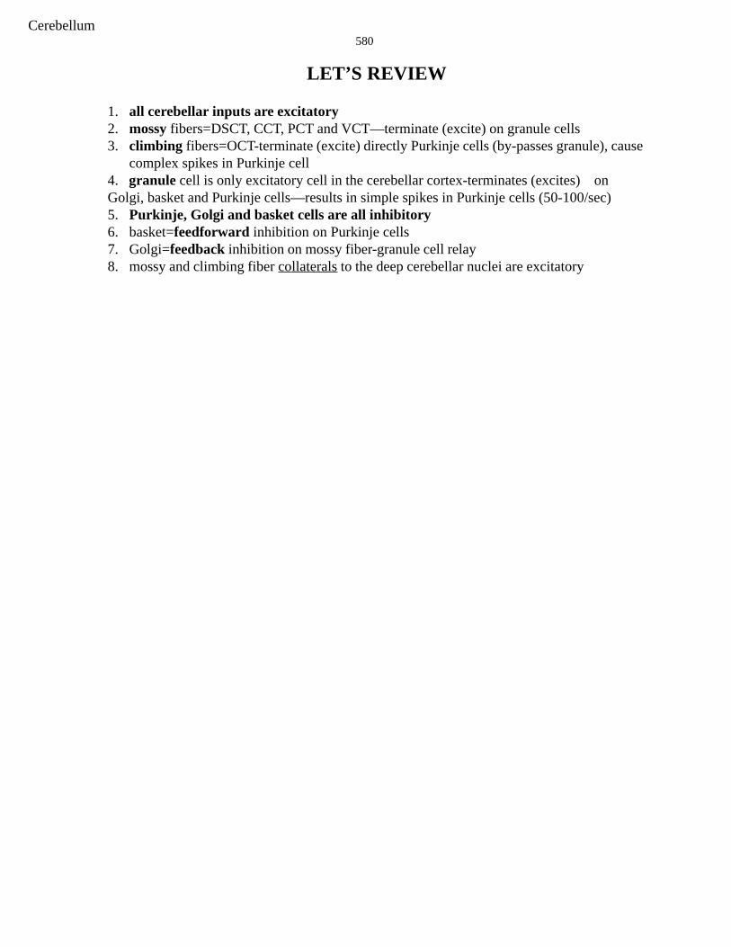
LET’S REVIEW
1. all cerebellar inputs are excitatory2. mossy fibers=DSCT, CCT, PCT and VCT—terminate (excite) on granule cells3. climbing fibers=OCT-terminate (excite) directly Purkinje cells (by-passes granule), cause
complex spikes in Purkinje cell4. granule cell is only excitatory cell in the cerebellar cortex-terminates (excites) onGolgi, basket and Purkinje cells—results in simple spikes in Purkinje cells (50-100/sec)5. Purkinje, Golgi and basket cells are all inhibitory6. basket=feedforward inhibition on Purkinje cells7. Golgi=feedback inhibition on mossy fiber-granule cell relay8. mossy and climbing fiber collaterals to the deep cerebellar nuclei are excitatory
581Cerebellum
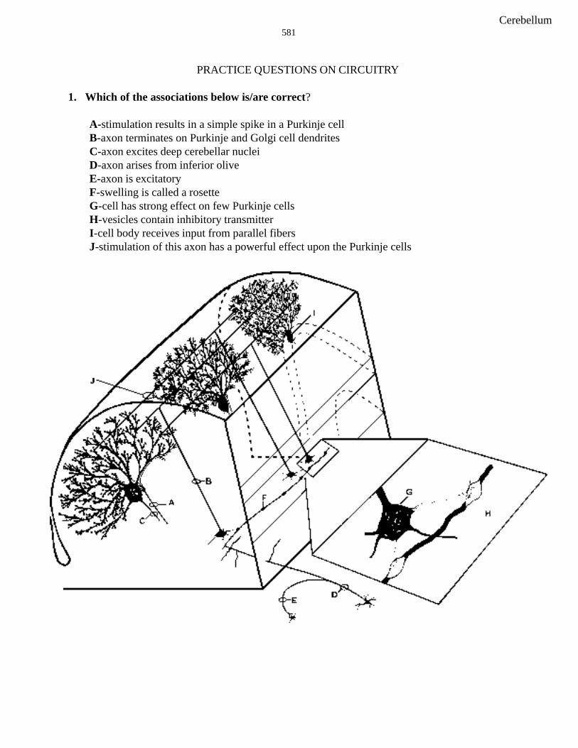
PRACTICE QUESTIONS ON CIRCUITRY
1. Which of the associations below is/are correct?
A-stimulation results in a simple spike in a Purkinje cellB-axon terminates on Purkinje and Golgi cell dendritesC-axon excites deep cerebellar nucleiD-axon arises from inferior oliveE-axon is excitatoryF-swelling is called a rosetteG-cell has strong effect on few Purkinje cellsH-vesicles contain inhibitory transmitterI-cell body receives input from parallel fibersJ-stimulation of this axon has a powerful effect upon the Purkinje cells

582Cerebellum
2. Which of the associations below is/are correct?
A-cell is involved in feedforward inhibitionB-axon is excitatoryC-cell inhibits Golgi cellsD-Purkinje cell axonE-axon innervates over 100 Purkinje cellsF-axon could arise from vestibular ganglionG-swelling is part of climbing fiberH-dendrite lies in the granule cell layerI-axon synapses upon basket cell dendritesJ-cell gives a complex spike upon climbing fiber stimulation

583Cerebellum
3. Which of the associations below is/are correct?
A-cells receive inhibitory input from mossy and climbing fibers and excitatory input fromPurkinje cellsB-cell inhibits Gogli cellsC-“basket”D-stimulation of axon excites basket cellE-dendritic tree of cell lies in plane that is parallel to the long axis of the folium
584Cerebellum
4. Which of the associations below is/are correct?
A-response of a Golgi cellA-response of a basket cellA-response of a Purkinje cell to mossy fiber stimulationA-response of a granule cellA-a simple spikeA-a complex spikeA-most common spike in Purkinje cellsA-spike that occurs infrequently (versus another kind of spike)B-common spike in Purkinje cellB-response of a Purkinje cell to climbing fiber inputB-response of a Purkinje cell to mossy fiber inputsB-response of a granule cell

585Cerebellum
5. Which of the following associations is/are correct regarding the drawing below?
A-nucleus is the sole source of climbing fibersA-nucleus receives direct input from Purkinje cells in the medial zone of cerebellar cortexA-nucleus lies in the granule layer of the cerebellumB-nucleus receives direct excitatory input from Purkinje cells in the intermediate zoneB-nucleus receives direct input from basket cellsB-nucleus receives direct input from granule cellsB-nucleus receives direct input from Golgi cellsB-nucleus receives direct input from parallel fibersC-nucleus receives direct input from Purkinje cells in medial zone of cerebellar cortexC-nucleus receives direct excitatory input from Purkinje cells in the lateral zoneC-nucleus receives direct input from basket cellsC-nucleus receives direct input from granule cellsC-nucleus receives direct input from Golgi cellsC-nucleus receives direct input from mossy fiber rosette

586Cerebellum
CEREBELLAR CIRCUITRY PROBLEM SOLVING ANSWERS
1. A. F; B. T; C. F; D. F; E. T; 4. A. F; F; F; F; F; T; F; T F. T; G. F; H. F; I. F; J. F B. T; F; T; F
2. A. F; B. F; C. F; D. T; E. F; 5. A. F; F; F F. T; G. F; H. T; I. T; J. T B. F; F; F; F; F
C. T; F; F; F; F; F3. A. F; B. F; C. T; D. T; E. F

587Cerebellum
SELF LEARNING Thursday, March 4, 11AM-12Well, all of the lectures on Motor Systems have been given. Hopefully you will use this two-hourblock of time to review. Finish those cerebellum questions and know those four cerebellar zones!!!Watch the review CD-ROM on Motor Systems again.
HOPEFULLY, YOU ARE BRINGING THINGS TOGETHER!!
Sorry to keep harping but you should have read and understood the www reading regardingParkinson’s, Tourette Syndrome and Deep Brain Stimulation. Moreover, the old stuff should befixed in your brains. They are: 1) muscular dystrophy, 2) myasthenia gravis, 3) Guillain-Barre, 4) S1radiculopathy, 5) amyotrophic lateral sclerosis (ALS), 6) Brown Sequard syndrome (spinal cordhemisection), 7) facial colliculus-vestibulo-cochlear, 8) lateral medullary (Wallenberg’s) syndrome,9) acoustic neuroma, 10) Weber Syndrome, 11) syringomyelia and 12) subacute combined systemsdisease. Finally, those power points on the “Integrated Motor Systems”power point for practice quiz#6 should be familiar!
YOU CAN ALSO GO UP TO THE LABS AND LOOK AT THE BRAIN IN ORDER TOVISUALIZE THE BASAL GANGLIA CEREBELLUM WE WILL BE UP THERE WAITINGFOR YOU!!!!!!!
588Cerebellum
PREPARATION FOR QUIZ 6
Go to WEB-CT and do the Practice Questions for Quiz #6. This should be a pretty good test of yourknowledge of the motor Systems. Work hard on those on-line quizzes so as to estimate your com-mand of the information. Make sure you understand the IMPORTANT www reading onParkinson’s, Tourette Syndrome and Deep Brain Stimulation. You should remember the mainpoints about 1) muscular dystrophy, 2) myasthenia gravis, 3) Guillain-Barre, 4) S1 radiculopathy, 5)amyotrophic lateral sclerosis (ALS), 6) Brown Sequard syndrome (spinal cord hemisection), 7) facialcolliculus-vestibulo-cochlear, 8) lateral medullary (Wallenberg’s) syndrome, 9) acoustic neuroma,10) Weber Syndrome, 11) syringomyelia and 12) subacute combined systems disease. Finally, thosepower points on “Motor Systems power point for quiz #6” should be familiar!