Cerebellar Contributions to Adaptive Control of Saccades in Humans
10
Behavioral/Systems/Cognitive Cerebellar Contributions to Adaptive Control of Saccades in Humans Minnan Xu-Wilson, 1 Haiyin Chen-Harris, 1 David S. Zee, 2 and Reza Shadmehr 1 Departments of 1 Biomedical Engineering and 2 Neurology, Johns Hopkins School of Medicine, Baltimore, Maryland 21205 The cerebellum may monitor motor commands and through internal feedback correct for anticipated errors. Saccades provide a test of this idea because these movements are completed too quickly for sensory feedback to be useful. Earlier, we reported that motor com- mands that accelerate the eyes toward a constant amplitude target showed variability. Here, we demonstrate that this variability is not random noise, but is due to the cognitive state of the subject. Healthy people showed within-saccade compensation for this variability with commands that arrived later in the same saccade. However, in people with cerebellar damage, the same variability resulted in dysmetria. This ability to correct for variability in the motor commands that initiated a saccade was a predictor of each subject’s ability to learn from endpoint errors. In a paradigm in which a target on the horizontal meridian jumped vertically during the saccade (resulting in an endpoint error), the adaptive response exhibited two timescales: a fast timescale that learned quickly from endpoint error but had poor retention, and a slow timescale that learned slowly but had strong retention. With cortical cerebellar damage, the fast timescale of adaptation was effectively absent, but the slow timescale was less impaired. Therefore, the cerebellum corrects for variability in the motor commands that initiate saccades within the same movement via an adaptive response that not only exhibits strong sensitivity to previous endpoint errors, but also rapid forgetting. Introduction It is thought that saccades are highly practiced, optimized exam- ples of ballistic movements. Yet, the motor commands that initi- ate an eye movement to a given target are variable, and this variability is not random noise. For example, people make sac- cades with higher velocities in anticipation of seeing a more in- teresting visual stimulus (Xu-Wilson et al., 2009). Repeating a visual target or reducing the reward associated with a target reduces saccade velocities (Straube et al., 1997; Chen-Harris et al., 2008). On the other hand, increasing the reward associated with the target or making the target the goal of both the eye and the arm movements increases saccade velocities (van Donkelaar, 1997; Snyder et al., 2002; Takikawa et al., 2002). Despite this variability in the motor com- mands that initiate saccades, the brain accurately guides the eyes to the target. How is this accomplished? Perhaps endpoint accuracy is possible because the brain in- corporates an internal feedback process that monitors the motor commands and corrects them online (Robinson, 1975). If the internal feedback is intact, variability in the commands that ini- tiate the saccade might be compensated via commands that arrive later during the same saccade. However, for this internal feedback to be effective, it needs to be adaptive and learn from endpoint errors. A computational framework that captures these ideas is one in which motor commands are monitored via a forward model, predicting sensory consequences and allowing for within- saccade compensation. Endpoint errors should produce adapta- tion in the forward model because they reflect a prediction error. If there is such an internal feedback process, damage to it might produce both an inability to compensate for the variability in the motor commands that initiated the movement, and an inability to learn from endpoint errors. That is, the two abilities should be correlated. Previously, we observed that when a visual target on the hor- izontal meridian was moved vertically as a saccade was made toward it (cross-axis paradigm), saccades became curved (Chen- Harris et al., 2008). This suggested that the forward model was learning from endpoint errors, steering the saccade to the target. Here, we looked for the neural basis of this hypothetical internal feedback process by examining saccades of cerebellar patients, as their saccades are dysmetric, and the cerebellum has long been hypothesized to function as an internal feedback pathway that “steers” the eyes to the target (Zee et al., 1976; Optican and Robinson, 1980; Quaia et al., 1999). Our subjects were a group of patients who suffered from spinocerebellar ataxia type 6 (SCA-6), a neuro-degenerative dis- ease that targets the Purkinje cells of the cerebellum (Sasaki et al., 1998; Honjo et al., 2004; Koeppen, 2005). Our experiment (the cross-axis paradigm) involved repetition of a visual target, some- thing that we had found to produce structured variability in the motor commands that initiate the saccade (Chen-Harris et al., 2008). We wondered whether this natural variability would also be present in the motor commands that initiated saccades of Received July 1, 2009; revised Aug. 25, 2009; accepted Aug. 29, 2009. This work was supported by grants from the National Institutes of Health (NS37422, EY19581, and EY01849). Minnan Xu-Wilson and Haiyin Chen-Harris were both supported by National Research Service Award predoctoral fellowships from the National Institute of Neurological Disorders and Stroke. We thank Dale Roberts and Adrian Lasker for their superb technical support. We thank the reviewers who provided thorough and insightful comments that greatly improved this manuscript. Correspondence should be addressed to Minnan Xu, Johns Hopkins School of Medicine, 416 Traylor Building, 720 Rutland Avenue, Baltimore, MD 21205. E-mail: [email protected]. DOI:10.1523/JNEUROSCI.3115-09.2009 Copyright © 2009 Society for Neuroscience 0270-6474/09/2912930-10$15.00/0 12930 • The Journal of Neuroscience, October 14, 2009 • 29(41):12930 –12939
Transcript of Cerebellar Contributions to Adaptive Control of Saccades in Humans

Behavioral/Systems/Cognitive
Cerebellar Contributions to Adaptive Control of Saccadesin Humans
Minnan Xu-Wilson,1 Haiyin Chen-Harris,1 David S. Zee,2 and Reza Shadmehr1
Departments of 1Biomedical Engineering and 2Neurology, Johns Hopkins School of Medicine, Baltimore, Maryland 21205
The cerebellum may monitor motor commands and through internal feedback correct for anticipated errors. Saccades provide a test ofthis idea because these movements are completed too quickly for sensory feedback to be useful. Earlier, we reported that motor com-mands that accelerate the eyes toward a constant amplitude target showed variability. Here, we demonstrate that this variability is notrandom noise, but is due to the cognitive state of the subject. Healthy people showed within-saccade compensation for this variability withcommands that arrived later in the same saccade. However, in people with cerebellar damage, the same variability resulted in dysmetria.This ability to correct for variability in the motor commands that initiated a saccade was a predictor of each subject’s ability to learn fromendpoint errors. In a paradigm in which a target on the horizontal meridian jumped vertically during the saccade (resulting in anendpoint error), the adaptive response exhibited two timescales: a fast timescale that learned quickly from endpoint error but had poorretention, and a slow timescale that learned slowly but had strong retention. With cortical cerebellar damage, the fast timescale ofadaptation was effectively absent, but the slow timescale was less impaired. Therefore, the cerebellum corrects for variability in the motorcommands that initiate saccades within the same movement via an adaptive response that not only exhibits strong sensitivity to previousendpoint errors, but also rapid forgetting.
IntroductionIt is thought that saccades are highly practiced, optimized exam-ples of ballistic movements. Yet, the motor commands that initi-ate an eye movement to a given target are variable, and thisvariability is not random noise. For example, people make sac-cades with higher velocities in anticipation of seeing a more in-teresting visual stimulus (Xu-Wilson et al., 2009). Repeating avisual target or reducing the reward associated with a target reducessaccade velocities (Straube et al., 1997; Chen-Harris et al., 2008). Onthe other hand, increasing the reward associated with the target ormaking the target the goal of both the eye and the arm movementsincreases saccade velocities (van Donkelaar, 1997; Snyder et al., 2002;Takikawa et al., 2002). Despite this variability in the motor com-mands that initiate saccades, the brain accurately guides the eyes tothe target. How is this accomplished?
Perhaps endpoint accuracy is possible because the brain in-corporates an internal feedback process that monitors the motorcommands and corrects them online (Robinson, 1975). If theinternal feedback is intact, variability in the commands that ini-tiate the saccade might be compensated via commands that arrivelater during the same saccade. However, for this internal feedback
to be effective, it needs to be adaptive and learn from endpointerrors. A computational framework that captures these ideas isone in which motor commands are monitored via a forwardmodel, predicting sensory consequences and allowing for within-saccade compensation. Endpoint errors should produce adapta-tion in the forward model because they reflect a prediction error.If there is such an internal feedback process, damage to it mightproduce both an inability to compensate for the variability in themotor commands that initiated the movement, and an inabilityto learn from endpoint errors. That is, the two abilities should becorrelated.
Previously, we observed that when a visual target on the hor-izontal meridian was moved vertically as a saccade was madetoward it (cross-axis paradigm), saccades became curved (Chen-Harris et al., 2008). This suggested that the forward model waslearning from endpoint errors, steering the saccade to the target.Here, we looked for the neural basis of this hypothetical internalfeedback process by examining saccades of cerebellar patients, astheir saccades are dysmetric, and the cerebellum has long beenhypothesized to function as an internal feedback pathway that“steers” the eyes to the target (Zee et al., 1976; Optican andRobinson, 1980; Quaia et al., 1999).
Our subjects were a group of patients who suffered fromspinocerebellar ataxia type 6 (SCA-6), a neuro-degenerative dis-ease that targets the Purkinje cells of the cerebellum (Sasaki et al.,1998; Honjo et al., 2004; Koeppen, 2005). Our experiment (thecross-axis paradigm) involved repetition of a visual target, some-thing that we had found to produce structured variability in themotor commands that initiate the saccade (Chen-Harris et al.,2008). We wondered whether this natural variability would alsobe present in the motor commands that initiated saccades of
Received July 1, 2009; revised Aug. 25, 2009; accepted Aug. 29, 2009.This work was supported by grants from the National Institutes of Health (NS37422, EY19581, and EY01849).
Minnan Xu-Wilson and Haiyin Chen-Harris were both supported by National Research Service Award predoctoralfellowships from the National Institute of Neurological Disorders and Stroke. We thank Dale Roberts and AdrianLasker for their superb technical support. We thank the reviewers who provided thorough and insightful commentsthat greatly improved this manuscript.
Correspondence should be addressed to Minnan Xu, Johns Hopkins School of Medicine, 416 Traylor Building, 720Rutland Avenue, Baltimore, MD 21205. E-mail: [email protected].
DOI:10.1523/JNEUROSCI.3115-09.2009Copyright © 2009 Society for Neuroscience 0270-6474/09/2912930-10$15.00/0
12930 • The Journal of Neuroscience, October 14, 2009 • 29(41):12930 –12939

cerebellar patients, and if so, whether these subjects would showa reduced ability to compensate for that variability.
A second part of our experiment was the endpoint errors thatwere caused by jumping of the target. We had earlier found thatthe adaptive response to errors appeared to involve two or moretimescales: a fast timescale that learned a great deal from error buthad poor retention, and a slower timescale that learned little fromerror but had good retention (Ethier et al., 2008). Cerebellardamage is known to profoundly impair the ability to adaptivelycontrol saccades (Straube et al., 2001; Golla et al., 2008). Here, wewondered whether these two timescales were uniformly affectedby cortical cerebellar damage, or was there a greater impairmentin one timescale of the adaptive process than in the other.
Materials and MethodsExperimental setup and design. Subjects sat in a dark room with their headrestrained using a dental bite-bar. We used a scleral search coil system(Skalar Medical) to record horizontal and vertical eye movements from ei-ther the right or the left eye (Robinson, 1963). Raw eye position informationfrom the coils was filtered in hardware (90 Hz low-pass Butterworth), digi-tized (1000 Hz), and saved on computer for later analysis. A 0.2° red laserbeam was rear-projected onto a translucent screen located 1 m in front of thesubject. Target position was varied by a galvo-controlled mirror.
The experiment is summarized in Figure 1A. It consisted of 5 blocks:preadaptation oblique (112 trials), preadaptation error-clamp (error-clampI, 60 trials), counterclockwise (CCW) cross-axis adaptation (500 trials),clockwise (CW) deadaptation trials (80 trials), and postadaptation error-clamp trials (error-clamp II, 140 trials). The experimental blocks are ex-
plained in detail below. Each block was furtherdivided into short sets of �60 trials (1.5 s inter-trial interval). On each trial, the saccade crossedthe vertical meridian. The sets were separatedwith a break typically of 30 s. During set breaksthe subjects sat quietly, usually with their eyesclosed, except for occasional breaks that were 2–3min long to administer eye drops to the subject.
Oblique trials. The experiment began withtwo sets of oblique trials (Fig. 1 A). The targetlocations were always 15° away from fixation inthe horizontal direction and 0, 1, 2, or 3° aboveor below the meridian vertically. The targetsappeared randomly within the set. Saccades al-ternated rightward and leftward symmetricabout the midline.
Error-clamp trials. The target T1 was pre-sented at 15° to the left or right of fixation.Once the saccade began, T1 disappeared. At500 ms later, a fixation point appeared with ahorizontal position the same as that of T1 and avertical position of the eye from 10 ms prior.Therefore, in error-clamp trials we attempt toassay the motor output after adaptation whilepreventing further learning by minimizing theendpoint errors. Because in the cross-axis par-adigm, learning takes place in the vertical di-rection, the error-clamp trials “clamp” thevertical error to zero but do not affect the hor-izontal error. To prevent accumulation of alarge vertical offset, we restricted the target po-sition to within �10° range vertically. Onceoutside of this range, the vertical position of thetarget was reset to 0° for the next trial. Therewere two error-clamp sets of trials, one beforethe adaptation sets (error-clamp I) and one af-ter (error-clamp II).
Cross-axis trials. The pattern of target posi-tions is shown in Figure 1 A. A target was pro-jected at 15° with respect to fixation (T1). As
soon as the saccade began, the target jumped 5° vertically to a new loca-tion (T2). In the adaptation trials, the jump direction was consistentlycounterclockwise to the orientation of T1. T2 then served as the fixationpoint (F) for the next trial. In deadaptation trials, the jump direction wasclockwise to the orientation of T1. The primary saccade to T1 was alwaysfollowed by a secondary “corrective” saccade that brought the eyes to T2. Thedata presented here represents characteristics of only the primary saccade.
The transition between CCW to CW adaptation trials occurred mid-set without a break: the ninth adaptation set began with 20 CCW trialsbut then suddenly changed to CW training (40 trials). Similarly, thetransition from CW to error-clamp was mid-set without a break: the 10thadaptation set began with 40 CW trials but then suddenly changed toerror-clamp trials (20 trials).
Subjects. Nine individuals with cerebellar degeneration participated inthe study (Table 1). All but one were diagnosed with SCA-6, a neuro-degenerative disease that primarily affects the Purkinje cells of the cere-bellum, particularly in the vermis (Sasaki et al., 1998; Honjo et al., 2004).Magnetic resonance imaging scans confirmed that these patients hadglobal degeneration of the cerebellum including the vermis and thehemispheres. The one patient without a genetic diagnosis (P2) had aclinical picture indistinguishable from the others with a genetically con-firmed diagnosis. SCA-6 is caused by mutations in a gene that encodes acalcium channel in Purkinje cells. In the cell culture models of this mu-tation, the result is premature death of the Purkinje cells. In the survivingcells, excitability is decreased. Postmortem examination of the brainshows a severe loss of Purkinje cells, with very mild loss of granule,stellate, and basket cells, as well as little or no loss of cells in the inferiorolive (Ishikawa et al., 1999). The mutation is a trinucleotide repeat ofCAG, with longer repeats resulting in symptoms beginning at an
Figure 1. Experimental procedures. A, Subjects were trained on a cross-axis adaptation task. The experiment consisted of fiveblocks: trials in which saccade targets were presented at various oblique angles, 60 preadapt error-clamp trials in which targetswere always at 15° horizontal (error-clamp I), 500 adaptation trials (target jump is counter-clockwise), 80 deadaptation trials(target jump is clockwise), and 140 postadapt error-clamp trials (error-clamp II, targets again at 15° horizontal). During error-clamp trials, the target did not jump but disappeared after saccade onset and reappeared 500 ms later at the current eye position.The dashed lines indicate axes centered straight ahead. B, Adaptation trials. Filled circles indicate current laser position. Arrow-heads indicate when a saccade began. A target was projected 15° away from fixation (T1). As soon as the saccade began, the targetjumped 5° vertically to a new location (T2). The jump direction was consistently counterclockwise to the orientation of T1. T2 thenserved as the fixation point (F) for the next trial. In deadaptation trials, the jump direction was clockwise to the orientation ofT1. C, Error clamp trials. T1 was presented at 15° to the left or right of fixation. Once the saccade began, T1 disappeared.Five-hundred milliseconds later, a fixation point appeared with a horizontal position the same as T1 and a vertical positionof the eye from 10 ms prior.
Xu-Wilson et al. • Cerebellum and the Adaptive Control of Saccades J. Neurosci., October 14, 2009 • 29(41):12930 –12939 • 12931

earlier age (Matsuyama et al., 1997). Table 1 summarizes the patientinformation.
Control 1. Eleven age matched control subjects also took part in ourexperiment (six female and five male; mean age 57 years, range 45– 69).All subjects gave written consent to protocols approved by the JohnsHopkins Institution Review Board.
Control 2. In our age-matched control group 1 we noted a trend inwhich the ability to control for horizontal variability correlated with theability to learn in the cross-axis adaptation paradigm. To improve ourpower to detect such a pattern, we enlarged our control group by addingsix non-age-matched control subjects (age range 20 – 43 years). Thisyounger group allowed us to validate the previous correlations that wehad seen in Control 1. The authors M.X. and R.S. were part of the Control2 group.
Data analysis. The beginning and end of saccades were determined bya 16°/s speed threshold. Criteria for including saccades in analysis were asfollows: (1) Saccade amplitude must be �50% of the target displacement.(2) Saccade duration must be within 50 –150 ms. (3) Saccade reactiontime must lie within 100 –500 ms. (4) Peak horizontal velocity must be�100°/s. On average, control subjects had 13% of their saccades ex-cluded while patients, with their more variable saccades, had 26%excluded.
We use a constant bin width (bw) of 4 to show the effect of stimulusrepetition and learning that evolves over the entire course of the experi-ment (see Figs. 4 A, D, 5A, 6 A). We then used a smaller bin width of 2 toshow the rapid changes that take place at set breaks and sudden changesin trial-type (see Figs. 4 B, C, E, F, 5C,D, 6C,D).
The amount of adaptation was assessed by comparing (using a paired,2-tailed t test) the vertical eye movement averaged over all the baselineerror-clamp trials with the vertical movement averaged over the last60-trial set of the CCW cross-axis adaptation block. Likewise, the recov-ery of the previous adaptation that occurred following the brief deadap-tation block was assessed by comparing the vertical movement in thebaseline trials with the vertical movement averaged over the first set ofpostadaptation error clamp trials. We used a significance level of p � 0.05for each pairwise comparison. We used a 2 � 1 repeated-measureANOVA design to compare the effect of group (control and cerebellar)and the within-subject factor of set number on saccade horizontal pa-rameters (peak horizontal velocity and amplitude). Significance level wasset at p � 0.05. Two-sided unpaired t tests were used to assess the differ-ences in adaptation and recovery levels between groups.
The vertical bias (in Fig. 7C) was calculated as the difference betweenthe absolute of the saccade’s vertical endpoint and the absolute of thetarget’s vertical position. For example, a vertical endpoint of �2.5° to atarget with vertical eccentricity of �3° had a bias of �0.5°, which isundershooting.
The SE of various parameters (Fig. 6, error bars) were calculated asfollows. The SE of the adaptation amount s is approximated as s/�n,where n is the number of trials used to calculate this measure. The SEs ofhorizontal and vertical variability as measured by SD � are approximatedas 0.71�/�n.
ResultsWe will first focus on the horizontal component of eye move-ments to illustrate that there is variability in the motor com-
mands that initiate saccades, and that the cerebellum plays a rolein the within-saccade compensation for this variability. We willthen focus on the vertical direction and the process of adaptation.
A role for the cerebellum in compensating for the variabilityin the motor commands that initiate saccadesFigure 2A shows the horizontal component of the saccade’s ki-nematics from the first and last error-clamp sets for a typicalhealthy control (C5) and a representative cerebellar patient (P4).In the control subject, the amplitude of the movement was thesame in the first and last sets ( p � 0.34). However, the amplitudedropped by 2.2° for the cerebellar patient ( p � 0.001). The peakvelocity dropped in both subjects from the first to the last set: thecontrol subject’s saccades slowed by 68.3°/s or 21% ( p � 0.001)and the cerebellar patient’s saccades slowed by 68°/s or 24% ( p �0.001). In the control subject, the saccades in the last set startedwith reduced peak velocities but then were completed with anincreased velocity late in the same saccade (see arrow). In thecerebellar patient, the reduction in peak velocity was left uncom-pensated. Saccade peak accelerations showed similar drops forboth subjects: 20% ( p � 0.001) for the control subject and 18%
Table 1. Cerebellar patients
Patient Age Gender Ataxia type Disease duration (years)
P1 45 F SCA-6 7P2 35 F SCA, type unknown 13P3 53 F SCA-6 11P4 55 F SCA-6 6P5 74 M SCA-6 35P6 59 F SCA-6 14P7 63 M SCA-6 12P8 67 F SCA-6 15P9 64 F SCA-6 4
Seven patients are female; mean age 57 years, range 35–74 years. F, Female; M, male.
Figure 2. Cerebellar patients could not correct for variability in the motor commands thatinitiated saccades. A, The average horizontal amplitude, velocity, and acceleration traces fromthe first and last error-clamp blocks (error clamp I and II), in response to a target at 15°, for tworepresentative subjects. In the last block, the saccades of both the control subject (C5) and thecerebellar patient (P4) were initiated with reduced velocities, but the control subject compen-sated later during the same saccade. Shading indicates SD. B, Group data for horizontal peakvelocity and amplitude changes. Percentage change is with respect to the first error-clampblock (error clamp I). Each point is the average from one set of 60 trials. Error bars indicate SEM.
12932 • J. Neurosci., October 14, 2009 • 29(41):12930 –12939 Xu-Wilson et al. • Cerebellum and the Adaptive Control of Saccades

( p � 0.001) for the cerebellar patient. The deceleration pattern,however, was strikingly different. The peak value of decelerationof the control subject was less and occurred later than the cere-bellar patient. Therefore, from the first to the last set, the magni-tude of the motor commands that initiated saccades dropped forboth subjects, but only the control subject was able to maintainamplitude by compensating late in the saccade’s time course.
Figure 2B summarizes some of these changes in the horizontaldirection through the course of the experiment. Both groupsmade slower saccades as the experiment proceeded. In controlsubjects, saccades slowed by 11.1% or 33°/s ( p � 0.005). In cer-ebellar patients, saccades slowed by 12.9% or 40°/s ( p � 0.01).Repeated measure ANOVA on peak velocity showed an effect ofset ( p � 0.001), no effect of group, and no interaction betweengroup and set. Therefore, saccades slowed by a similar amount inthe two groups. Repeated measure ANOVA of horizontal ampli-tude showed an effect of set ( p � 0.01), group ( p � 0.05), andgroup by set interaction ( p � 0.05). Control subjects were able tomaintain endpoint amplitude from first to last set ( p � 0.43),while cerebellar patients showed a drop in amplitude by 9.0% or1.06° ( p � 0.05).
An analysis of saccade timing parameters demonstrated thatcontrol and cerebellar subjects differed most in the decelerationphase of saccades (Fig. 3). The time of peak acceleration remainedunchanged in both groups ( p � 0.45 for controls and p � 0.59 forpatients). In the control group, the time of peak velocity (shift �1.7 ms, p � 0.01) and time of peak deceleration (shift � 6.5 ms,p � 0.01) shifted to a later time, and duration of the saccade(shift � 8.4 ms, p � 0.001) increased. For cerebellar patients,however, time of peak deceleration showed less than half thechange seen in controls (shift � 2.9 ms, p � 0.01) while time ofpeak velocity and duration of saccades showed no significant
changes. As a result, while in both groups the commands thataccelerated the eyes along the horizontal dimension decreasedfrom the first to the last set of the experiment, the healthy subjectswere able to maintain horizontal amplitude by compensatinglater in the saccade.
In the above data we averaged saccade parameters in each set,and then displayed the results across sets. However, there werealso consistent changes that occurred within each set. Each setconsisted of 60 trials (intertrial interval of �1.5 s). Between thesets our subjects rested for �30 s and closed their eyes. As in ourprevious studies of cross-axis adaptation (Chen-Harris et al.,2008), on-axis adaptation (Ethier et al., 2008), or simply controlstudies in which targets did not jump (Chen-Harris et al., 2008),we found that the peak horizontal velocity dropped within eachset and then sharply increased in the first saccade after the setbreak (Fig. 4A). On average, the healthy subjects showed 49.8°/sor 16.3% increase in peak horizontal velocity (last two saccadesbefore set break vs first two saccades after set break, p � 0.001)and 10.0 ms or 15.4% decrease in duration ( p � 0.001), as shownin Figure 4B. The set breaks also produced a small increase in thehorizontal amplitude (0.6° or 4.6%, p � 0.05) (supplemental Fig.1, available at www.jneurosci.org as supplemental material) and asmall decrease in saccade latencies ( p � 0.05) (supplemental Fig.1, available at www.jneurosci.org as supplemental material). Thesmall but significant increase in amplitude at set breaks suggeststhat even in healthy people, some of the variability in motorcommands that accelerated the eyes was left uncompensated.This is a crucial finding for us as we will later show that theamount of within-saccade compensation in the horizontal direc-tion is a predictor of the ability of that subject to learn fromendpoint errors in the vertical direction.
Similar to control subjects, cerebellar patients showed an ob-vious structure in their saccade velocities: set breaks induced anincrease of 28.0°/s or 9.3% in peak horizontal velocity ( p � 0.01)(Fig. 4E). This was a clear finding, as set breaks induced an in-crease in peak horizontal velocity in 7 of 9 cerebellar subjects (5subjects with p � 0.05 and 2 subjects with p � 0.1), and in 7 of 9set breaks (supplemental data and supplemental Fig. S2, available atwww.jneurosci.org). However, unlike controls, the cerebellar pa-tients did not decrease the saccade durations to compensate forthis increased velocity ( p � 0.92).
In summary, motor commands that accelerated the eyes alongthe horizontal dimension were affected by two forms of variabil-ity: set breaks produced a sharp increase, while target repetitionproduced a gradual decrease. In healthy people, this variabilityappeared to be corrected within the same saccade, whereas in thecerebellar subjects, the variability produced dysmetria.
What caused the variability in the motor commands thatinitiated the saccades?It is possible that the experiment induced use-dependent fatiguein the neuronal or muscular structures of the oculomotor system,and the set breaks allowed recovery from this fatigue. However,data from a crucial component of our experiment argued againstthis possibility: after 20 trials in the final adaptation set, the targetsequence unexpectedly switched from a counter-clockwise to aclockwise sequence (start of deadaptation, first red line in Fig.4A). If fatigue is a form of habituation in the sensory neurons thatconvey target information to the motor system, then the transi-tion from adaptation to deadaptation should not produce a re-covery because the stimuli that elicited horizontal saccadesactivated precisely the same retinal location as before. Similarly, iffatigue is a form of use-dependent reduction in the response of
Figure 3. Saccades of control and cerebellar subjects differed most in the decelerationphase. The plots show the changes in the timing of saccade parameters. Position, velocity, andacceleration refer to horizontal components of the movement. Each point is the average fromone set of 60 trials. Error bars indicate SEM.
Xu-Wilson et al. • Cerebellum and the Adaptive Control of Saccades J. Neurosci., October 14, 2009 • 29(41):12930 –12939 • 12933

the extra-ocular muscles, then thereshould be no recovery because the transi-tion from adaptation to deadaptation didnot include a rest period. However, if thefatigue is due to a top-down factor, forexample, a decline in an attentional state,then the surprising event should producerecovery.
The unexpected change in the positionof a vertical target produced an immediateand robust recovery of velocities in re-sponse to the subsequent horizontal tar-get. In Figure 4, C and F, the saccadeparameters are aligned to the first trial af-ter the target sequence changed fromcounterclockwise to clockwise targetjumps. Changes in percentage are calcu-lated with respect to the trial before thesequence change. In controls, we found anincreased velocity of 32.9°/s or 11.8%( p � 0.05) and decreased duration of 12.5ms or 16.9% ( p � 0.01). Horizontal dis-placement ( p � 0.64) and reaction time( p � 0.20) did not change significantly(supplemental Fig. 1C, available at www.jneurosci.org as supplemental material).Importantly, this recovery was smaller thesecond time the target sequence changed(at the end of the deadaptation block, sec-ond red line in Fig. 4A): controls showed a20.4°/s or 7.7% increase in peak velocity( p � 0.15). That is, the change in the tar-get sequence produced a sharp increase invelocities when it first occurred, but asmaller one when it repeated.
In cerebellar patients, the unexpectedchange in target sequence produced24.9°/s or 7.3% increase in peak velocities, but these changes didnot reach significance ( p � 0.22). There were no significantchanges in duration ( p � 0.57), horizontal amplitude ( p �0.51), or reaction time ( p � 0.53). Therefore, the data from thecontrol subjects suggested that the changes in the commands thatinitiated the saccades were probably not due to a neuronal ormuscular fatigue process that required passage of time for recov-ery. Rather, the fact that an unexpected change in the stimulusrestored peak velocities suggests that the decline in velocity wasdue to reduced motivation, boredom, or a similar top-downeffect.
Adaptation and the multiple timescales of memoryOur experiment was designed to not only quantify adaptationcapabilities of healthy and cerebellar patients, but to potentiallyunmask the multiple timescales that underlie this adaptation. Weconsidered a common paradigm in which a long period of adap-tation was followed by a brief period of deadaptation (Kojima etal., 2004; Criscimagna-Hemminger and Shadmehr, 2008; Ethieret al., 2008). If there are multiple timescales that support adapta-tion, then behavior should exhibit signatures of these timescales(Smith et al., 2006; Kording et al., 2007). For example, a fast time-scale of adaptation should produce forgetting with passage of time(during set breaks), and forgetting with removal of the adaptation-driving errors (during error-clamp trials). This fast timescaleshould also produce rapid learning in the presence of error. A
slow timescale of adaptation should resist deadaptation (whenerrors reverse direction, termed “extinction”) and produce spon-taneous recovery toward the previously adapted state in the pos-tadaptation error-clamp period. In the cerebellar patients, is thedamage predominantly affecting one timescale of adaptation?
During the adaptation block, as the saccade was initiated thetarget at the horizontal meridian jumped vertically (in a counter-clockwise direction), resulting in an endpoint error which wascorrected with a second saccade. In response to this endpointerror, both the control and the cerebellar groups learned to pro-duce primary saccades that had increasing vertical motor com-mands, but the learning was significantly smaller in the cerebellarpatients (Fig. 5A). For example, the change in vertical endpointfrom the preadapt error-clamp trials to the last set of adaptationwas 2.01° in control subjects ( p � 10�4), and 0.56° in cere-bellar patients ( p � 0.05), with the change being significantlysmaller in the cerebellar patients (Fig. 5B).
Despite the significant amount of adaptation in the saccadesmade by the cerebellar patients, their adapted response was miss-ing a fundamental characteristic. The control subjects exhibitedrobust forgetting during each set break: the vertical endpoint ofcontrol saccades suddenly decreased (Fig. 5A, first arrow). Thisset structure was prominent when we plotted the changes in sac-cade parameters with respect to the last bin (last two saccades) ofeach set (Fig. 5C). On average, the vertical endpoint declined by0.43 o or 26% at set start ( p � 0.005, set start vs previous set end)
Figure 4. Effect of set breaks on saccade horizontal velocities and durations. A, D, Peak vertical velocity and duration, averagedacross each group. Dotted vertical lines mark set boundaries. Each set consisted of 60 trials. Red lines mark sudden changes in targetsequence that occurred within sets without breaks. B, E, Within-subject changes in peak velocity and duration, aligned to setrestart. The plots show percentage change with respect to the last bin of each set. The amount of recovery is calculated as thedifference between the last and first bins (t test, *p � 0.05, **p � 0.01, ***p � 0.001). Shading indicates across subject SEM.C, F, Within-subject changes in peak velocity and duration, aligned to the sudden change in sequence of targets from CCW to CWcross-axis target jumps. A–C are control data, and D–F are cerebellar data.
12934 • J. Neurosci., October 14, 2009 • 29(41):12930 –12939 Xu-Wilson et al. • Cerebellum and the Adaptive Control of Saccades

and the peak vertical velocity declined by 5.8°/s or 14.3% ( p �0.05). By the sixth saccade after set start the vertical endpointsand velocities had recovered to the magnitude of the previous set(Fig. 5C). That is, the short break produced forgetting, and the setrestart produced rapid relearning. When viewed as a group, boththe forgetting and the rapid relearning were absent in the sac-cades of the cerebellar patients.
When we analyzed the data in terms of individual subjects, wefound a strong correlation between a subject’s tendency to forgetat set breaks and the ability to learn rapidly after set start. For
example, Figure 5E shows the change invertical endpoint position during the first6 trials after the set start (relearning) as afunction of change in the same index dur-ing the set break (forgetting). These twomeasures were strongly correlated in the con-trolsubjects(r�0.79,p�0.004)andmargin-ally significant in the cerebellar subjects (r �0.63, p � 0.068). The one cerebellar subjectwho showed forgetting at set break alsoshowed rapid relearning at set start.
It is possible that the rapid changes inperformance after set start are not due torapid relearning, but a contextual effect inwhich there is remanifestation of a previ-ously learned state. There is a simple wayto check for this. If the forgetting andrapid relearning are both due to a fastadaptive process, then the same fast learn-ing should be present at the very first ad-aptation set as well as after each set break.On the other hand, if the rapid changeafter set start was due to revisiting a previ-ous context, then it should be absent inthe first set as that context had not beenrepeated before. In the control group, wefound rapid learning in the very first set:the change in vertical velocity by trial 6was 12.56°/s ( p � 0.05), which was nodifferent from the average change ob-served after set breaks (within-subject ttest, p � 0.33). Similarly, the vertical end-point changed by 0.34° ( p � 0.05) in thefirst 6 trials of the first set, which was nodifferent from changes seen after setbreaks (within-subject t test, p � 0.57).This is consistent with the idea that setbreaks induced forgetting, and set restartinduced relearning; both of which are sig-natures of a fast adaptive process.
We were concerned that for the cere-bellar subjects, we could not detect forget-ting during the set-breaks because theyhad learned only a small amount. There-fore, we focused on the last three adapta-tion sets as during these sets the responseshowed significant adaptation. Despitethis, we could not detect a robust change inthe cerebellar saccades at set start in our mea-sures of adaptation ( p � 0.13 for vertical end-points and p � 0.12 for vertical velocity).
An interesting prediction of the ideathat learning in healthy people is sup-
ported by two timescales is that when adaptation is followed bydeadaptation, the direction of forgetting should reverse (Ethier etal., 2008). To explain this, consider that during the deadaptationperiod a competition may be formed between a fast adaptivemechanism that learns the CW perturbation, and the slow adap-tive mechanism that previously has learned the CCW perturba-tion. During the set break in the deadaptation block the fastmechanism should forget, and the behavior should revert to whatthe slow mechanism had learned. Whereas during the adaptation
Figure 5. The multiple timescales of adaptation. A, The plots show the vertical endpoint of the primary saccade and its peakvertical velocity. Cerebellar patients (red) are impaired in adapting to cross-axis target jumps compared with controls, but never-theless show significant adaptation. In the deadapt period, the behavior of both groups returned to baseline, but in the followingerror-clamp trials, there was partial recovery. Dashed vertical lines denote set breaks. Solid vertical lines denote changes of trialtypes. Note that in the control group, there is forgetting (first arrow) at each set break followed by rapid relearning. Also note thatforgetting reverses direction (second arrow) in the deadaptation period. B, Summary of the performance at the final set ofadaptation and during the first 60 trials of error-clamp II in controls and patients (*p�0.05, **p�0.01, ***p�0.001). Error barsindicate SEM. C, Vertical endpoint and velocity aligned on set start, as in Figure 4 B. The forgetting followed by rapid relearning ispresent in controls but absent in patients. D, Vertical endpoint and velocity at the set break in the deadaptation block. Only thecontrols exhibit reverse-forgetting. Error bars indicate SEM. E, Rapid learning within the first 6 trials of each adaptation set andforgetting at set breaks for individual subjects. The control group showed a strong correlation of r 2 � 0.62 with p � 0.004. Thecerebellar subjects showed a marginally significant correlation of r 2 � 0.40 with p � 0.067.
Xu-Wilson et al. • Cerebellum and the Adaptive Control of Saccades J. Neurosci., October 14, 2009 • 29(41):12930 –12939 • 12935

period forgetting during set breaks was to-ward baseline, now in the deadaptation pe-riod forgetting should be away frombaseline toward the CCW value stored bythe slow system.
Indeed, for the control subjects the setbreak in the deadaptation period pro-duced “reverse-forgetting” (recovery of0.45° in vertical endpoint and 12.3°/s invertical velocity, p � 0.05, second arrowin Fig. 5A, close-up of the deadapt periodin Fig. 5D), but this pattern was missing inthe cerebellar subjects ( p � 0.25). Thisresult further confirms that the fast time-scale of adaptation was present in the con-trol subjects but missing in the patients.
Our simple two-timescale model couldnot account for one aspect of the data. Ifthe return of performance to baseline (i.e.,washout or extinction) during deadapta-tion was solely due to a competition be-tween two timescales of adaptation, and ifcerebellar subjects were impaired in thefast timescale, these subjects should showa slower than normal rate of deadapta-tion. This was not the case. In the deadapta-tion period, vertical endpoint and velocityof saccades in cerebellar subjects returned tobaseline even faster than controls (Fig. 5A).This possibly indicates that deadaptation incerebellar subjects benefited from the abilityto inhibit a previously learned pattern,rather than set up a competition between afast and a slow adaptive process.
The deadaptation block was followedby an error-clamp block. In this block,both groups exhibited spontaneous re-covery of their previously adapted behav-ior. In controls, vertical endpoints andvelocities were significantly greater than baseline ( p � 0.001) (Fig.5B). Similarly, in the cerebellar patients vertical endpoints and ve-locities were significantly greater in the final error-clamp block thanbaseline ( p � 0.05). Spontaneous recovery is a signature of the slowadaptive system that resists “unlearning” during the deadaptationperiod (Ethier et al., 2008). On average, the magnitude of the spon-taneous recovery in controls was 28% of the state achieved duringthe adaptation block. In the cerebellar patients, the magnitude of thespontaneous recovery was 33% of the state achieved during the ad-aptation block. Importantly, there were no significant differences inthe magnitude of percent spontaneous recovery in the two groups.
Saccade curvatureA prominent feature of saccades in cross-axis adaptation is cur-vature (Chen-Harris et al., 2008); i.e., motor commands thatinitiate the saccade appear to adapt by a smaller amount thanthose that terminate the saccade. A proxy for curvature is thedifference in the slopes of the saccade near its start and finish. Wedivided each saccade into four equal horizontal segments andmeasured the slope of each segment. Figure 6A shows the slope atsaccade start (termed S1) and the slope at saccade end (termedS4). In both groups, S1 increased significantly during the adap-tation block (0.12, p � 10�4 in controls, 0.028, p � 0.05 incerebellar patients), with the changes being significantly smaller
in cerebellar patients ( p � 0.001). In both groups, S4 was largerthan S1, resulting in saccades that curved toward the target(within group comparison, last set of adaptation, 0.19, p �10�5 in controls, 0.073, p � 0.01, in cerebellar patients). Whenthe trial-to-trial changes in slope were aligned on set starts, weonce again observed the fast timescales of learning in the healthycontrols (S1 dropped by 0.039 or 35%, p � 0.01, S4 dropped by0.041 or 26%, p � 0.01), but not clearly in the cerebellar group: S1showed no drop at set break ( p � 0.3), and while S4 dropped by0.026, 42% at set break, it showed similar drops both before theset break and after the set break. Similarly, Figure 6D illustratesthat the set-break during deadaptation produced reverse-forgetting in the control subjects (recovery of 0.34 for S1, p �0.05, recovery of 0.028 for S4, p � 0.05), but not in the cerebellargroup ( p � 0.4 for S1 and p � 0.15 for S4).
Ability to compensate for variability predicts ability to learnfrom endpoint errorsFinally, we asked whether a subject’s ability to control endpointaccuracy during the preadaptation control trials was a predictorof their ability to learn from endpoint errors during adaptationtrials. Our proxy for the ability to control accuracy was endpointvariability (SD) and endpoint bias in the oblique control trials,before the adaptation trials began. Our proxy for the ability to
Figure 6. Early versus late part of single saccades. We divided each saccade into four equal horizontal segments andmeasured the slope of each segment. A, The slope at saccade start is termed S1 and the slope at saccade end S4. B, Summaryof S1 and S4 at the final set of adaptation and the recovery seen during the first 60 trials of error-clamp II in controls andpatients (*p � 0.05, **p � 0.01, ***p � 0.001). C, Within-subject change in S1 and S4 with respect to the end of theprevious set. D, Within-subject change at the set break in the deadaptation block. Error bars indicate SEM.
12936 • J. Neurosci., October 14, 2009 • 29(41):12930 –12939 Xu-Wilson et al. • Cerebellum and the Adaptive Control of Saccades

adapt was the vertical endpoint achieved in the final set of theadaptation block. As a group, the cerebellar patients had largervertical bias (negative value means undershooting, p � 0.001)(Fig. 7A), more horizontal endpoint variability ( p � 0.01) (Fig.7B), and more vertical endpoint variability ( p � 0.05) (Fig. 7C)than controls. Inclusion of the cerebellar group with healthy controlsin a regression analysis would, of course, produce a significantcorrelation between control of saccade accuracy and adapta-tion. However, a more interesting question is whether within thecontrol subjects, the ability to control endpoint variability was apredictor of the ability to adapt. Indeed, in healthy subjects theability to control saccades during the oblique trials was a predic-tor of the ability to adapt to errors during the adaptation trials(blue circles, Fig. 7A–C, vertical bias: r 2 � 0.43, p � 0.05; hori-zontal variability: r 2 � 0.50, p � 0.05; vertical variability was closeto significance: r 2 � 0.32, p � 0.06). (In comparison, we foundno correlation between horizontal bias and amount of learning.)
To test the strength of the correlation between endpoint vari-ability and learning, we considered a cross-validation procedure.We added to our analysis six additional control subjects (Fig. 7,black circles) who were not age-matched to the patients. Within
this larger control group, we found an even stronger relationshipbetween the trial to trial control of saccade accuracy and verticaladaptation (vertical bias: r 2 � 0.51, p � 0.005; vertical variability:r 2 � 0.42, p � 0.005; horizontal variability: r 2 � 0.50, p � 0.001).
To further test the idea that the ability to control endpointvariability predicts the ability to adapt, we considered other mea-sures of within-trial saccadic control. For example, as saccadehorizontal velocity decreases, durations should increase to main-tain horizontal amplitude. This implies that for a given subject, anegative correlation between peak horizontal velocity and dura-tion is indicative of better endpoint control. Indeed, this measurewas also a predictor of the ability to learn from endpoint errors:the more negative the correlation between peak horizontal veloc-ity and duration, the better the ability of that subject to learn fromvertical endpoint errors (r 2 � 0.27, p � 0.05 for all controls). Inanother example of within-saccade control, consider that as sac-cade horizontal velocity changes, the more positive the correla-tion with horizontal amplitude, the less perfect the compensation(e.g., if a decrease in velocity is not compensated, the result is adecrease in amplitude). Indeed, people who exhibited a positivecorrelation between horizontal velocity and amplitude were gen-erally the subjects who also learned the least from the verticalendpoint errors (r 2 � 0.30, p � 0.05 for all controls).
In summary, we found that a healthy subject’s ability to controlendpoint accuracy during the preadaptation control trials predictedtheir ability to learn from endpoint errors during adaptation trials.
DiscussionSome three decades ago, David Robinson proposed that saccadicmotor commands are monitored and corrected to steer the eyesto the target (Robinson, 1975). Later work suggested the cerebel-lum was central to this monitoring process (Quaia et al., 1999). Inthe current computational view of motor control (Shadmehr andKrakauer, 2008) the cerebellum may be a forward model (Pasalaret al., 2006) that uses efferent copy to predict consequences ofmotor commands and contributes to the online correction ofmovement. Presumably, the forward model learns from end-point errors to maintain accuracy. Saccades (compared withreaching movements) are particularly useful for testing the the-ory of forward models because these eye movements are com-pleted too quickly for visual or proprioceptive feedback to play arole in control. Here, we wished to quantify the influence of thecerebellum on both the within-saccade compensation of thecommands that initiated the movement, and the longer-termlearning that compensated for persistent errors.
Our subjects were healthy people and patients with a neuro-degenerative disorder (SCA-6) that affected the Purkinje cells ofthe cerebellum. As noted before (Chen-Harris et al., 2008), rep-etition of a target on the horizontal meridian gradually reducedhorizontal saccade velocities, and short breaks rapidly increasedthese velocities. In healthy people, this variability was generallycompensated via corrective motor commands that arrived laterin the same saccade. However, the compensation was missing incerebellar subjects. In healthy people, the within-saccade abilityto compensate for the variability in the horizontal velocity was apredictor of the ability to adapt to errors in the vertical direction.The adaptation relied on a memory that exhibited multiple time-scales: a fast process that learned quickly from endpoint errorsbut showed forgetting during the short set breaks, and a slowprocess that learned gradually, resisted unlearning, and becamelatent during a brief period of deadaptation but reemerged aftercessation of the deadapting stimulus. Despite significant adapta-tion in the cerebellar subjects, their adapted saccades were miss-
Figure 7. The ability to compensate for internal sources of variability correlates with theability to compensate for external sources of error. A, Correlation between bias in the verticaldirection for oblique trials and learning along the vertical direction during adaptation. The bestfit line is for control subjects. Error bars are SEM. B, C, Horizontal (or vertical) endpoint variabilitybefore adaptation is plotted on the x-axis and the ability to adapt to endpoint errors duringadaptation trials is plotted on the y-axis. The best fit line is for all control subjects. Error bars forhorizontal and vertical variability are SEs of the SD estimate.
Xu-Wilson et al. • Cerebellum and the Adaptive Control of Saccades J. Neurosci., October 14, 2009 • 29(41):12930 –12939 • 12937

ing the forgetting and the rapid-relearning, suggesting that thedamage to the cerebellar cortex had produced a deficit thatmostly affected the fast timescale of adaptation.
A source of internal variability in the motor commands thatinitiate a saccadeIn this study, only healthy controls were able to compensate forvariability due to repetition (gradual decline of saccade horizon-tal velocity and its rapid recovery after set breaks). This compen-sation was via a change in the time course of the decelerationphase, late in the saccade. Therefore, while the source of the vari-ability appeared to be outside the cerebellar cortex (as it waspresent in both the patients and the controls), only subjects witha healthy cerebellum corrected for the variability.
Saccade slowing could be due to fatigue of the oculomotorplant or cognitive factors. Here, we gained new insights into thesource of this repetition attenuation of saccade velocities: we foundthat an unexpected change in the repeating target produced imme-diate recovery of the horizontal velocities. This suggests that theslowing of the saccades and its recovery at set breaks were unrelatedto neuromuscular fatigue in the oculomotor plant, as recoverywould require passage of time. Sensory neurons that encode someparticular attribute of a stimulus usually show progressively smallerresponses when that attribute is repeated (Miller and Desimone,1994; Kohn and Movshon, 2003). This “repetition suppression” hasbeen observed in area V1 of the occipital cortex (Grill-Spector et al.,2006) and in the superficial layer of the superior colliculus(Boehnke et al., 2007). Because the receptive field of these cellsis retinotopic and the unexpected change in the repeatingstimuli did not alter its positions in retinotopic coordinates,the recovery of velocity cannot be explained by recruitment ofnew sensory neurons with different receptive fields. That is,the repetition attenuation is probably not a “bottom-up” phe-nomenon. However, it is known that the brain directs attentionto a visual stimulus that has behaved differently than expected(Itti and Baldi, 2009). It seems likely that the repetition attenua-tion in saccade velocities was due to top-down cognitive factorslike attention.
Our observation agrees well with the data from Golla et al. (2008)who also noted a drop in saccade velocities that went uncompen-sated in cerebellar patients. A possible explanation is that the cere-bellum receives a copy of the motor commands and then makesadjustments to steer the eyes to the target (Quaia et al., 1999). Indeed,cells in the cerebellar vermis and caudal fastigial nucleus show dis-charges with timing that could be used to adjust motor commandsduring the deceleration phase of the saccade (Ohtsuka and Noda,1992; Fuchs et al., 1993; Catz et al., 2008).
The multiple timescales of adaptationIn healthy people, adaptation appeared to consist of two distinctprocesses: a fast process that exhibited forgetting and relearningat set breaks, and a slow process that exhibited little forgettingand strong resistance to unlearning when the endpoint errorssuddenly reversed direction (the deadaptation period). We hadearlier observed a similar ‘“two-state” process in a gain adapta-tion paradigm (Ethier et al., 2008), and in a reaching paradigm(Smith et al., 2006; Criscimagna-Hemminger and Shadmehr,2008). In cerebellar patients, the fast adaptation process appearedto be impaired as the learned vertical component did not exhibitforgetting during rest periods. In contrast, the slow process was atleast partially spared in the patients as the learned vertical com-ponent exhibited recovery during the error clamp period thatfollowed the brief deadaptation stimulus.
Is the lack of evidence for a fast adaptive process in cerebellarpatients due to increased noise in these subjects? Indeed, it is possiblethat our inability to observe a component of adaptation in our sub-ject group was due to sample size and increased variability in thatgroup. However, note that we found significant changes in horizon-tal velocities in the saccades of the same patients at set breaks, but noforgetting in the vertical velocities (Fig. 4C). Changes in horizontalvelocities are unrelated to adaptation (Chen-Harris et al., 2008), andare examples of fast cognitive or attentional processes that affectedsaccades of both patients and healthy controls.
There are experimental results that support the idea that the cer-ebellar cortex may be specialized for fast processes that underlie mo-tor memory, whereas the deep cerebellar nuclei may specialize in theslow processes. For example, in classical conditioning of the eye blinkreflex, washout of the previous learning still results in a more rapidreacquisition than when the subject is naive, a phenomenon termedsavings. Medina et al. (2001) have attributed this to different time-scales of learning in the cerebellar cortex and nuclei. Barash et al. (1999)suggested that there are two processes that adjust the saccade gain (eyedisplacement/target amplitude): the rapid gain adjustment mechanismdependsonthecerebellarcortexwhilethesloweradjustmenttakesplaceoutside of the cerebellar cortex. Similar suggestions have been made forthe role of the cerebellum in other types of oculomotor learning(Ohyama et al., 2006; Shutoh et al., 2006; Masuda and Amari, 2008).
Are the changes in the vertical motor commands an adaptiveresponse related to deficits in the horizontal motor commands?At set starts, we found that the internal/cognitive perturbation iseffectively absent since horizontal saccade velocity recovered. At thesame time, vertical motor commands showed a large reduction incontrol subjects. That is, when there was little need to correct fordeficits in the motor commands that initiated the saccade, there wasalso a reduced ability to express learning, i.e., an apparent forgetting.The cerebellar subjects, however, displayed neither the abilityto correct for changes in the horizontal motor commands nor theforgetting. One hypothesis to explain this observation is that thecerebellum learns a compensatory response in the vertical direction,but this response can only be expressed when there is a deficit in thehorizontal motor commands that initiate the saccade. In this view,the term “forgetting” is unjustified because the changes in set breaksare not due to decay or loss of a previously learned response, butsimply a lack of necessity to express it.
Although this is an attractive hypothesis, there are a numberof patterns in our data that are inconsistent with it. First, the rapidlearning that follows set breaks has a much faster rate (achievingpreviously learned level within 6 trials) than the repetition-induced drop in the horizontal velocity, which slows over thecourse of many more trials. That is, changes in the horizontalvelocities alone could not explain the forgetting and rapid re-learning in the vertical direction. Second, at set start we foundrobust reductions in not just the vertical commands that termi-nated the saccade, but also the vertical commands that initiated it(Fig. 6C). Therefore, forgetting in the vertical motor commandsoccurred too early into a saccade to have been a response tochanges in horizontal motor commands. Finally, if set breaks didnot produce forgetting, then the rapid relearning after set start isa remanifestation of a previously learned state, which should notbe present in the first adaptation set. In fact, rapid learning waspresent in the first adaptation set. Together, the data are moreconsistent with the idea that the time passage during set breaksinduced forgetting, the large errors at set restart induced relearn-ing, and damage to the cerebellar cortex primarily influencedthese fast timescales of adaptation.
12938 • J. Neurosci., October 14, 2009 • 29(41):12930 –12939 Xu-Wilson et al. • Cerebellum and the Adaptive Control of Saccades
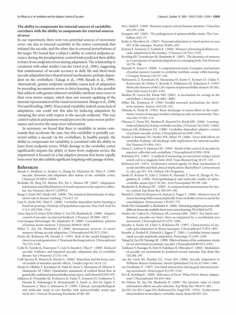
The ability to compensate for internal sources of variabilitycorrelates with the ability to compensate for external sourcesof errorIn our experiment, there were two potential sources of movementerror: one due to internal variability in the motor commands thatinitiated the saccade, and the other due to external perturbations ofthe target. We found that a subject’s ability to control endpoint ac-curacy during the preadaptation control trials predicted their abilityto learn from endpoint errors during adaptation. The relationship isconsistent with other studies (van der Geest et al., 2006), suggestingthat maintenance of saccade accuracy in daily life and short-termsaccade adaptation have shared neural mechanisms, perhaps depen-dent on the cerebellum (Takagi et al., 1998; Barash et al., 1999).Alternatively, greater endpoint variability causes lack of adaptationby providing inconsistent errors to drive learning. It is also possiblethat subjects with greater inherent variability attribute more error totheir own motor output, which does not warrant updating of theinternal representation of the visual environment (Burge et al., 2008;Wei and Kording, 2009). If increased variability indeed causes lack ofadaptation, one could test this idea by inducing adaptation byclamping the error with respect to the saccade endpoint. This waycontrol subjects and patients would perceive the same motor perfor-mance and receive the same error information.
In summary, we found that there is variability in motor com-mands that accelerate the eyes, but this variability is partially cor-rected within a saccade via the cerebellum. In healthy people, theability to compensate for variability is correlated with the ability tolearn from endpoint errors. While damage to the cerebellar cortexsignificantly impairs the ability to learn from endpoint errors, theimpairment is focused on a fast adaptive process that learns rapidlyfrom error but also exhibits significant forgetting with passage of time.
ReferencesBarash S, Melikyan A, Sivakov A, Zhang M, Glickstein M, Thier P (1999)
Saccadic dysmetria and adaptation after lesions of the cerebellar cortex.J Neurosci 19:10931–10939.
Boehnke SE, Berg DJ, Marino RA, Itti L, Munoz DP (2007) Adaptation,habituation and dishabituation of visual responses in the superior collicu-lus. Soc Neurosci Abs 617.12/PP14.
Burge J, Ernst MO, Banks MS (2008) The statistical determinants of adap-tation rate in human reaching. J Vis 8:1–19.
Catz N, Dicke PW, Thier P (2008) Cerebellar-dependent motor learning isbased on pruning a Purkinje cell population response. Proc Natl Acad SciU S A 105:7309 –7314.
Chen-Harris H, Joiner WM, Ethier V, Zee DS, Shadmehr R (2008) Adaptivecontrol of saccades via internal feedback. J Neurosci 28:2804 –2813.
Criscimagna-Hemminger SE, Shadmehr R (2008) Consolidation patternsof human motor memory. J Neurosci 28:9610 –9618.
Ethier V, Zee DS, Shadmehr R (2008) Spontaneous recovery of motormemory during saccade adaptation. J Neurophysiol 99:2577–2583.
Fuchs AF, Robinson FR, Straube A (1993) Role of the caudal fastigial nu-cleus in saccade generation. I. Neuronal discharge pattern. J Neurophysiol70:1723–1740.
Golla H, Tziridis K, Haarmeier T, Catz N, Barash S, Thier P (2008) Reducedsaccadic resilience and impaired saccadic adaptation due to cerebellardisease. Eur J Neurosci 27:132–144.
Grill-Spector K, Henson R, Martin A (2006) Repetition and the brain: neu-ral models of stimulus-specific effects. Trends Cogn Sci 10:14 –23.
Honjo K, Ohshita T, Kawakami H, Naka H, Imon Y, Maruyama H, Mimori Y,Matsumoto M (2004) Quantitative assessment of cerebral blood flow ingenetically confirmed spinocerebellar ataxia type 6. Arch Neurol 61:933–937.
Ishikawa K, Watanabe M, Yoshizawa K, Fujita T, Iwamoto H, Yoshizawa T,Harada K, Nakamagoe K, Komatsuzaki Y, Satoh A, Doi M, Ogata T,Kanazawa I, Shoji S, Mizusawa H (1999) Clinical, neuropathological,and molecular study in two families with spinocerebellar ataxia type6a(SCA6). J Neurol Neurosurg Psychiatry 67:86 – 89.
Itti L, Baldi P (2009) Bayesian surprise attracts human attention. Vision Res49:1295–1306.
Koeppen AH (2005) The pathogenesis of spinocerebellar ataxia. The Cere-bellum 4:62–73.
Kohn A, Movshon JA (2003) Neuronal adaptation to visual motion in areaMT of the macaque. Neuron 39:681– 691.
Kojima Y, Iwamoto Y, Yoshida K (2004) Memory of learning facilitates sac-cadic adaptation in the monkey. J Neurosci 24:7531–7539.
Kording KP, Tenenbaum JB, Shadmehr R (2007) The dynamics of memoryas a consequence of optimal adaptation to a changing body. Nat Neurosci10:779 –786.
Masuda N, Amari S (2008) A computational study of synaptic mechanismsof partial memory transfer in cerebellar vestibulo-ocular-reflex learning.J Comput Neurosci 24:137–156.
Matsuyama Z, Kawakami H, Maruyama H, Izumi Y, Komure O, Udaka F,Kameyama M, Nishio T, Kuroda Y, Nishimura M, Nakamura S (1997)Molecular features of the CAG repeats of spinocerebellar ataxia 6 (SCA6).Hum Mol Genet 6:1283–1287.
Medina JF, Garcia KS, Mauk MD (2001) A mechanism for savings in thecerebellum. J Neurosci 21:4081– 4089.
Miller EK, Desimone R (1994) Parallel neuronal mechanisms for short-term memory. Science 263:520 –522.
Ohtsuka K, Noda H (1992) Burst discharges of mossy fibers in the oculo-motor vermis of macaque monkeys during saccadic eye movements. Neu-rosci Res 15:102–114.
Ohyama T, Nores WL, Medina JF, Riusech FA, Mauk MD (2006) Learning-induced plasticity in deep cerebellar nucleus. J Neurosci 26:12656 –12663.
Optican LM, Robinson DA (1980) Cerebellar-dependent adaptive controlof primate saccadic system. J Neurophysiol 44:1058 –1076.
Pasalar S, Roitman AV, Durfee WK, Ebner TJ (2006) Force field effects oncerebellar Purkinje cell discharge with implications for internal models.Nat Neurosci 9:1404 –1411.
Quaia C, Lefevre P, Optican LM (1999) Model of the control of saccades bysuperior colliculus and cerebellum. J Neurophysiol 82:999 –1018.
Robinson DA (1963) A method of measuring eye movement using a scleralsearch coil in a magnetic field. IEEE Trans Biomed Eng 10:137–145.
Robinson DA (1975) Oculomotor control signals. In: Basic mechanisms ofocular motility and their clinical implications (BachyRita P, LennerstrandG, eds), pp 337–374. Oxford, UK: Pergamon.
Sasaki H, Kojima H, Yabe I, Tashiro K, Hamada T, Sawa H, Hiraga H, Na-gashima K (1998) Neuropathological and molecular studies of spino-cerebellar ataxia type 6 (SCA6). Acta Neuropathol 95:199 –204.
Shadmehr R, Krakauer JW (2008) A computational neuroanatomy for mo-tor control. Exp Brain Res 185:359 –381.
Shutoh F, Ohki M, Kitazawa H, Itohara S, Nagao S (2006) Memory trace ofmotor learning shifts transsynaptically from cerebellar cortex to nuclei forconsolidation. Neuroscience 139:767–777.
Smith MA, Ghazizadeh A, Shadmehr R (2006) Interacting adaptive processes withdifferent timescales underlie short-term motor learning. PLoS Biol 4:e179.
Snyder LH, Calton JL, Dickinson AR, Lawrence BM (2002) Eye-hand coor-dination: saccades are faster when accompanied by a coordinated armmovement. J Neurophysiol 87:2279 –2286.
Straube A, Fuchs AF, Usher S, Robinson FR (1997) Characteristics of sac-cadic gain adaptation in rhesus macaques. J Neurophysiol 77:874 – 895.
Straube A, Deubel H, Ditterich J, Eggert T (2001) Cerebellar lesions impairrapid saccade amplitude adaptation. Neurology 57:2105–2108.
Takagi M, Zee DS, Tamargo RJ (1998) Effects of lesions of the oculomotor vermison eye movements in primate: saccades. J Neurophysiol 80:1911–1931.
Takikawa Y, Kawagoe R, Itoh H, Nakahara H, Hikosaka O (2002) Modulationof saccadic eye movements by predicted reward outcome. Exp Brain Res142:284–291.
van der Geest JN, Haselen GC, Frens MA (2006) Saccade Adaptation inWilliams-Beuren Syndrome. Invest Ophthalmol Vis Sci 47:1464 –1468.
van Donkelaar P (1997) Eye-hand interactions during goal-directed point-ing movements. Neuroreport 8:2139 –2142.
Wei K, Kording K (2009) Relevance of Error: What Drives Motor Adapta-tion? J Neurophysiol 101:655– 664.
Xu-Wilson M, Zee DS, Shadmehr R (2009) The intrinsic value of visualinformation affects saccade velocities. Exp Brain Res 196:475– 481.
Zee DS, Yee RD, Cogan DG, Robinson DA, Engel WK (1976) Ocular motorabnormalities in hereditary cerebellar ataxia. Brain 99:207–234.
Xu-Wilson et al. • Cerebellum and the Adaptive Control of Saccades J. Neurosci., October 14, 2009 • 29(41):12930 –12939 • 12939