Cerebellar Hematoma
13
8/23/2015 www.medscape.com/viewarticle/761751_print http://www.medscape.com/viewarticle/761751_print 1/13 www.medscape.com Abstract and Introduction Abstract Evidencebased guidelines for the management of hemorrhagic and ischemic cerebellar stroke are sparse, and most available data come from Class III studies. As a result, opinions and practices regarding the nature and role of neurosurgical intervention vary widely. A comprehensive literature review was conducted to adjudicate several contentious issues, such as the difference in the management of cerebellar hemorrhage versus infarction, criteria for imaging to exclude an underlying structural lesion, the value of MRI for patient selection, the role of external ventricular drainage, the indications for operative management, the timing of surgical intervention, and various options of surgical technique, among others. Treatment algorithms proposed in several different studies are compared and contrasted. This analysis is concluded by a summary of the recommendations from the American Stroke Association, which advises that patients with cerebellar hemorrhage who experience neurological deterioration or who have brainstem compression and/or hydrocephalus due to ventricular obstruction should undergo surgical evacuation of the hemorrhage as soon as possible, and that initial treatment of such patients with ventricular drainage alone rather than surgical removal of the hemorrhage is not recommended. Introduction The management of cerebellar hemorrhage has endured controversy ever since Sir Charles Ballance reported the first successful surgical evacuation in 1906. [2] Institutional and individual variations abound in the management of cerebellar hemorrhage. To a degree, this debate is due to the relatively flimsy quality of the medical literature in support of different practices. For instance, in advocating against external ventricular drainage alone in the treatment of patients with cerebellar hemorrhage who are deteriorating neurologically or who have brainstem compression and/or hydrocephalus from ventricular obstruction, the American Stroke Association relies on evidence rated as Level C, the weakest category in effect at the time (consensus opinion of experts, case studies, or standard of care). [24] Similarly, their prior recommendation that angiography is not required for older, hypertensive patients with cerebellar hemorrhage in whom CT findings do not suggest a structural lesion is based upon Level V evidence, the weakest category in effect at the time (data from anecdotal case series only). [4] Several randomized trials comparing early surgery with initial conservative management for ICH have been conducted, including the recent Surgical Trial in Intracerebral Hemorrhage (STICH). [23] Overall, these studies have largely shown no benefit to surgery, although post hoc subgroup analysis reveals some exceptions. Patients with cerebellar ICH have been excluded from all these randomized trials, because clinical equipoise was not believed to be present. [24] As one expert commented about the related condition of cerebellar infarction, "the results of surgery have been so consistently favorable in patients who clearly were progressively deteriorating that it seems fair to say that this is one surgical indication that does not need the scrutiny of a randomized study." [12] As a result of these biases, data principally consist of uncontrolled, single institution retrospective case series (Class III evidence). Collectively, however, these reports suggest that the benefit of surgery is not so straightforward. Donauer et al. [7] reviewed 21 papers from 1958 to 1993 and performed a metaanalysis comparing medical versus operative treatment of cerebellar ICH. In the cohort of 357 patients who underwent surgery, the mortality rate was 49%, while that in the 269 patients treated conservatively was 50%. Similarly, Hankey and Hon [10] reviewed 8 prior series of surgery for infratentorial hemorrhage comprising a total of 405 patients. One study suggested overall benefit, while 2 studies reported benefit only in certain subgroups (conscious or drowsy but deteriorating patients), and the remaining 5 studies were either inconclusive or showed no benefit of surgery. In an effort to provide more concreteness to this issue and to elucidate related concepts in the management of cerebellar hemorrhage and infarction, this article reviews relevant studies from the past century. It begins with an overview of the pathogenesis and natural history, which form the foundation and rationale for all treatment. Next, 9 separate areas of controversy are explored in detail. The review concludes with a summary of the recommendations from the American Stroke Association, whose position statements have evolved considerably from their first publication in 1999 to 2010. [3,4,24] Pathophysiology and Natural History Spontaneous cerebellar hematomas represent approximately 10%–15% of all ICH. [11,18,35] As with cerebellar infarction, cerebellar hemorrhage occurs most frequently in the 5th through the 8th decades of life and with greater frequency in males than Controversies in the Neurosurgical Management of Cerebellar Hemorrhage and Infarction Arun Paul Amar, M.D. Neurosurg Focus. 2012;32(4):e1
-
Upload
bill-jones-tanawal -
Category
Documents
-
view
27 -
download
4
description
perdarahan serebelum
Transcript of Cerebellar Hematoma
8/23/2015 www.medscape.com/viewarticle/761751_print
http://www.medscape.com/viewarticle/761751_print 1/13
www.medscape.com
Abstract and IntroductionAbstract
Evidencebased guidelines for the management of hemorrhagic and ischemic cerebellar stroke are sparse, and most availabledata come from Class III studies. As a result, opinions and practices regarding the nature and role of neurosurgical interventionvary widely. A comprehensive literature review was conducted to adjudicate several contentious issues, such as the difference inthe management of cerebellar hemorrhage versus infarction, criteria for imaging to exclude an underlying structural lesion, thevalue of MRI for patient selection, the role of external ventricular drainage, the indications for operative management, the timingof surgical intervention, and various options of surgical technique, among others. Treatment algorithms proposed in severaldifferent studies are compared and contrasted. This analysis is concluded by a summary of the recommendations from theAmerican Stroke Association, which advises that patients with cerebellar hemorrhage who experience neurological deteriorationor who have brainstem compression and/or hydrocephalus due to ventricular obstruction should undergo surgical evacuation ofthe hemorrhage as soon as possible, and that initial treatment of such patients with ventricular drainage alone rather thansurgical removal of the hemorrhage is not recommended.
Introduction
The management of cerebellar hemorrhage has endured controversy ever since Sir Charles Ballance reported the first successfulsurgical evacuation in 1906.[2] Institutional and individual variations abound in the management of cerebellar hemorrhage. To adegree, this debate is due to the relatively flimsy quality of the medical literature in support of different practices. For instance, inadvocating against external ventricular drainage alone in the treatment of patients with cerebellar hemorrhage who aredeteriorating neurologically or who have brainstem compression and/or hydrocephalus from ventricular obstruction, the AmericanStroke Association relies on evidence rated as Level C, the weakest category in effect at the time (consensus opinion of experts,case studies, or standard of care).[24] Similarly, their prior recommendation that angiography is not required for older,hypertensive patients with cerebellar hemorrhage in whom CT findings do not suggest a structural lesion is based upon Level Vevidence, the weakest category in effect at the time (data from anecdotal case series only).[4]
Several randomized trials comparing early surgery with initial conservative management for ICH have been conducted, includingthe recent Surgical Trial in Intracerebral Hemorrhage (STICH).[23] Overall, these studies have largely shown no benefit tosurgery, although post hoc subgroup analysis reveals some exceptions. Patients with cerebellar ICH have been excluded from allthese randomized trials, because clinical equipoise was not believed to be present.[24] As one expert commented about therelated condition of cerebellar infarction, "the results of surgery have been so consistently favorable in patients who clearly wereprogressively deteriorating that it seems fair to say that this is one surgical indication that does not need the scrutiny of arandomized study."[12]
As a result of these biases, data principally consist of uncontrolled, single institution retrospective case series (Class IIIevidence). Collectively, however, these reports suggest that the benefit of surgery is not so straightforward. Donauer et al.[7]
reviewed 21 papers from 1958 to 1993 and performed a metaanalysis comparing medical versus operative treatment ofcerebellar ICH. In the cohort of 357 patients who underwent surgery, the mortality rate was 49%, while that in the 269 patientstreated conservatively was 50%. Similarly, Hankey and Hon[10] reviewed 8 prior series of surgery for infratentorial hemorrhagecomprising a total of 405 patients. One study suggested overall benefit, while 2 studies reported benefit only in certain subgroups(conscious or drowsy but deteriorating patients), and the remaining 5 studies were either inconclusive or showed no benefit ofsurgery.
In an effort to provide more concreteness to this issue and to elucidate related concepts in the management of cerebellarhemorrhage and infarction, this article reviews relevant studies from the past century. It begins with an overview of thepathogenesis and natural history, which form the foundation and rationale for all treatment. Next, 9 separate areas of controversyare explored in detail. The review concludes with a summary of the recommendations from the American Stroke Association,whose position statements have evolved considerably from their first publication in 1999 to 2010.[3,4,24]
Pathophysiology and Natural History
Spontaneous cerebellar hematomas represent approximately 10%–15% of all ICH.[11,18,35] As with cerebellar infarction,cerebellar hemorrhage occurs most frequently in the 5th through the 8th decades of life and with greater frequency in males than
Controversies in the Neurosurgical Management of CerebellarHemorrhage and InfarctionArun Paul Amar, M.D.Neurosurg Focus. 2012;32(4):e1
8/23/2015 www.medscape.com/viewarticle/761751_print
http://www.medscape.com/viewarticle/761751_print 2/13
in females.[11,18] Between 60% and 90% of all spontaneous cerebellar hemorrhages occur in hypertensive patients. Vascularmalformations, coagulopathies (including the use of anticoagulants), neoplasms, aneurysms, cerebral amyloid angiopathy, andtrauma account for the remainder.[11,17] In younger patients, underlying structural conditions are the prevailing causes.
Cerebellar infarctions, on the other hand, may result from cardiac emboli, traumatic injury to the vertebral arteries, and othercauses.[11,14,26] The majority of patients also have hypertension.[13,26] The infarction most frequently occurs in the vasculardistribution of the posterior inferior cerebellar artery, but the anterior inferior cerebellar artery and/or superior cerebellar arteryterritories can also be involved.[14,26] Cerebellar infarctions are approximately twothirds as common as cerebellar hemorrhage.[11]
In hypertensive patients, cerebellar hematoma is believed to result from rupture of microaneurysms, as first proposed by Charcotand Bouchard, and recently confirmed.[31] Typically, these hemorrhages begin in the area of the dentate nucleus and thenspread throughout the ipsilateral hemisphere.[15] They may also extend across the vermis to the contralateral side. Although theycommonly spread into the cerebellar peduncles or rupture into the fourth ventricle, only rarely do they directly involve thebrainstem.[11] Dizziness, headache, nausea, vomiting, loss of balance, and difficulty walking are the most common presentingsymptoms of both cerebellar hemorrhage and infarction.[1,11,14,26,30]
Clinical deterioration befalls up to 50% of patients with cerebellar ICH.[25] In its mild form, deterioration manifests as irritability,confusion, or somnolence, while the more severe form presents as coma, stupor, posturing, and hemodynamic or respiratoryinstability due to loss of brainstem regulation.[13] The peak incidence of deterioration is 3 days after onset, although it may occurwithin hours or even weeks later. When deterioration occurs, mortality has been reported to be high (25%–100%), regardless oftreatment.[1,7,17,18,30] Deterioration can occur unpredictably, even in patients who appear to have reached a clinical plateau.[11,17] Ott et al.[25] reported that 50% of patients who remained alert and relatively stable for 2 days degenerated into coma overthe course of the next several days, and a disconcerting 25% of patients who remained awake for 7 days subsequentlydeteriorated.
The causes of deterioration are protean and include increased mass effect from surrounding edema or expansion of thehematoma from repeat bleeding. Either mechanism can cause direct brainstem compression, which leads to upwards herniationthrough the tentorial incisura or downward tonsillar herniation through the foramen magnum. Obstructive hydrocephalus, causedby intraventricular extension of the hemorrhage or by compression of the fourth ventricle, is another mechanism of clinicaldecline.
In cerebellar infarction, brain swelling results from both cytotoxic and vasogenic edema. Initially, brain ischemia disrupts cellmembrane integrity, which causes the accumulation of intracellular fluid. Later, vasogenic edema results from the diffusion ofproteinbound fluid across a damaged bloodbrain barrier.[5,19] With progressive mass effect caused by the infarct andsurrounding edema, brainstem compression and/or fourth ventricle compression can result. The range of time that can elapsebetween symptom onset and further neurological deterioration is typically 1–7 days, with a median and mode of 3 days.[5,13,14]
However, the likelihood of deterioration has been reported to be lower in cerebellar infarct (7%–32%) than in hemorrhage.[1,14]
Whereas hydrocephalus and brainstem compression can both cause decreased level of consciousness, the latter is alleged tohave associated focal neurological signs.[11,13,29] Early compression of the dorsal pons results in ipsilateral sixth nerve paresis ofvoluntary lateral gaze that can be overcome with caloric stimulation. Later, as the compression progresses, conjugate gazeparesis that is unresponsive to caloric stimulation occurs from pressure upon the horizontal gaze center. At this stage, ipsilateralperipheraltype facial paresis is usually present due to concomitant compression of the facial colliculus. Babinski signs, Hornersyndrome, and hemiparesis are all late signs of brainstem compression.[11,13,29] Recognition of these findings, along withneuroimaging, can help distinguish between altered level of consciousness due to hydrocephalus versus that due to directbrainstem compression. Appropriate therapy (for example, ventricular drainage versus surgical decompression) can then betargeted to the underlying mechanism.
The tenets of medical management of cerebellar hemorrhage are similar to those of supratentorial ICH.[24] Patients are generallymonitored in a critical care setting, with frequent neurological assessment. Those with severe coagulation factor deficiency orthrombocytopenia should receive transfusion of appropriate blood products to correct the disorder. Patients whose hemorrhage iscaused by oral anticoagulation therapy should receive intravenous vitamin K as well as therapy to replace the vitamin K–dependent factors. Prothrombin complex concentrates have not been proven to improve outcome compared with freshfrozenplasma, but may have fewer complications.[24] Recombinant factor VIIa is not routinely recommended as the sole agent forreversal of oral anticoagulation therapy.[24] All patients should undergo intermittent pneumatic compression for prevention ofvenous thromboembolism in addition to elastic stockings. After documentation of cessation of bleeding, lowdose subcutaneousheparin formulations may be considered as well.[24] Glucose should be monitored closely, and normoglycemica is recommended.The management of blood pressure remains disputed, without clear guidelines or target parameters, but in patients presentingwith systolic blood pressure of 150 to 220 mm Hg, acute lowering to 140 mm Hg is probably safe.[24] Therapeutic cooling has notbeen adequately studied in cerebellar ICH, although most practitioners favor avoidance of hyperthermia.
8/23/2015 www.medscape.com/viewarticle/761751_print
http://www.medscape.com/viewarticle/761751_print 3/13
Hemorrhagic versus Ischemic Cerebellar Stroke
Since the first reports of decompressive surgery performed by Fairburn and Oliver[8] and by Lindgren,[20] both in 1956, thepotential value of suboccipital craniectomy and resection of necrotic tissue in cerebellar infarction has been recognized. However,cerebellar hemorrhage and infarction are distinct entities, which calls into question whether the same management principlesshould apply to each.
Mathew et al.[21] compared the neurosurgical management of 48 patients with cerebellar ICH to that of 71 patients withcerebellar infarction. They found that patients with hematoma were more likely to be in a coma and more likely to havebrainstem compression upon presentation than those with infarction. This explains why 75% of their patients with ICH requiredsurgery, while it was necessary in only 24% for infarction.
In both cerebellar hemorrhage and infarction, perilesional edema can aggravate the spaceoccupying effect within the confines ofthe posterior fossa. However, a condition unique to ICH is the toxic effects of blood products and associated inflammation, whichmight provide impetus for its removal regardless of the mechanical compression of adjacent tissue. Furthermore, cerebellarhemorrhage may extend into the ventricle system, thus providing an additional mechanism of hydrocephalus besides fourthventricle effacement. Conversely, cerebellar ICH only rarely extends directly into the brainstem.[17]
By comparison, cerebellar infarction does not lead to intraventricular hemorrhage and is thus less likely to cause hydrocephalusthan cerebellar hematoma. In the patient series of Auer et al.,[1] occlusive hydrocephalus developed in 75% of patients withcerebellar hemorrhage but only 23% of those with cerebellar infarction. Patients with cerebellar ICH also had a higher incidenceof hydrocephalus than those with cerebellar infarct in the series by Mathew et al.[21] However, cerebellar infarction is more likelyto directly involve the brainstem than cerebellar ICH due to shared vascular territory; this occurred in 2 of 40 patients in the seriesof Auer et al.[1]
Emerging data suggest that in some circumstances, the area of restricted diffusion apparent on MRI, once believed to representpermanent damage, may be reversible.[16] Therefore, it is conceivable that resection of this presumed necrotic tissue incerebellar infarction may actually compromise recovery.
In light of these considerations, a policy that limits the extent of resection of apparent necrotic tissue to the mininum needed toachieve adequate decompression appears reasonable, although the data in support of this practice are not robust.
Criteria for CT Angiogram or Catheter Angiogram
Although most cases of spontaneous cerebellar hemorrhage are the result of hypertension, some are caused by underlyinglesions. In Kobayashi et al.'s series of 110 patients,[18] for example, 5 hemorrhages resulted from a cerebellar AVM, 2 resultedfrom a cerebellar tumor, and the remaining 103 were believed to be caused by hypertension on the basis of prior history and/ornegative angiographic studies.
Even in the presence of preexisting hypertension, however, as many as 36% of all ICH cases are associated with secondarycauses.[35] The indications, nature, and diagnostic accuracy of imaging for an underlying structural lesion in spontaneouscerebellar hemorrhage remains controversial. The presence of subarachnoid blood, calcification, prominent vascular structures, oredema out of proportion to the size and age of the hemorrhage might suggest the presence of an underlying lesion. Similarly, ahemorrhage that has an unusual (geographic or noncircular) shape or is located in an unusual location, such as an epicenterremote from the dentate nucleus, might prompt further study (Fig. 1). However, features of CT in isolation had a sensitivity ofonly 77% and specificity of only 84% in 1 study.[9] Clinical features such as age and history of preexisting hypertension also affectthe decision to pursue advanced imaging.

8/23/2015 www.medscape.com/viewarticle/761751_print
http://www.medscape.com/viewarticle/761751_print 4/13
Figure 1.
Images obtained in a 77yearold woman with a medical history significant for hypertension, heart murmur, and Crohn diseaserequiring large doses of aspirin. She awoke with headache, dizziness, and incoordination of the right arm. A: Unenhanced axialCT scans of her brain reveal acute hemorrhage of the right cerebellum extending to the tentorial surface and across the vermis tothe left hemisphere. B: Although she had multiple risk factors for spontaneous intracerebral hemorrhage (such as hypertension,possible cardiac embolism related to the cause of her heart murmur, aspirin use, and possible agerelated amyloid angiopathy),the unusual location of the hemorrhage and questionable prominence of vessels near the vein of Galen prompted furtherimaging. This frontal view of a left vertebral catheter angiogram reveals an AVM of the cerebellar vermis supplied primarily bybranches of the bilateral superior cerebellar arteries. High risk features include feeding artery aneurysms bilaterally and venousoutflow restriction of the draining vein coursing to the straight sinus. C: The patient underwent a series of staged liquid adhesiveembolization sessions to protect against rehemorrhage. This frontal view of left vertebral catheter angiography after embolizationreveals elimination of the feeding artery aneurysms and significantly reduced flow through the nidus.
Halpin et al.[9] performed a prospective evaluation of catheter cerebral angiography in the workup of 102 patients withspontaneous cerebral hematoma. Both supratentorial and cerebellar hemorrhages were included in the analysis. Overall, ananeurysm or AVM was the cause of the hemorrhage in 12.8% of hypertensive patients and in 18.2% of those with posterior fossahemorrhage. The authors prospectively stratified the patients into 2 groups: those suspected to have a high likelihood of anunderlying structural lesion based on CT features (Group 1) and those without such findings (Group 2). Catheter angiography waspositive for AVM or aneurysm in 84% of the highsuspicion cohort and 24% in the lowsuspicion one.

8/23/2015 www.medscape.com/viewarticle/761751_print
http://www.medscape.com/viewarticle/761751_print 5/13
In another study, Zhu et al.[35] reviewed 206 consecutive patients with spontaneous ICH to determine the diagnostic yield ofcerebral angiography. Both supra and infratentorial hemorrhages were included. Patients in whom severe coagulopathyaccounted for the ICH, those with bleeding into tumor, or those with predominant subarachnoid hemorrhage were excluded.Overall, the angiographic yield was significantly higher in patients less than the median age of 45 years and those without priorhypertension. In 15 patients with posterior fossa hemorrhage, 5 (33%) were found to have an underlying AVM. All 5 werenormotensive, and the oldest patient in this group was 39. Another 6 patients had preexisting hypertension, the youngest ofwhom was 48. None of those 6 patients had positive angiography. The authors amalgamated hemorrhages in the putamen,thalamus, or posterior fossa into a single group for analysis. In this collective, the angiographic yield in patients with younger ageand without preexisting hypertension was 48%, while in hypertensive patients the yield was 0%. They concluded that diagnosticangiography should not be routinely performed in patients with cerebellar hemorrhage over 45 years old with preexistinghypertension.
Although CT angiography and catheter angiography are potentially useful in the workup of spontaneous cerebellar ICH, neither iscompletely reliable. In some cases, compression of adjacent vessels by the hematoma can give the false appearance of avascular malformation, thus reducing the specificity of these tests (Fig. 2). Conversely, the mass effect can conceal an underlyinglesion, thus reducing the sensitivity of vascular studies performed acutely. In the series of Halpin et al.,[9] for example, followupangiography at 3 months showed an AVM in 1 of 7 patients in the highsuspicion group, even though the original study resultswere normal. Thus, when clinically warranted, vascular studies should be repeated in a delayed fashion even if the initial workupis negative.
8/23/2015 www.medscape.com/viewarticle/761751_print
http://www.medscape.com/viewarticle/761751_print 6/13
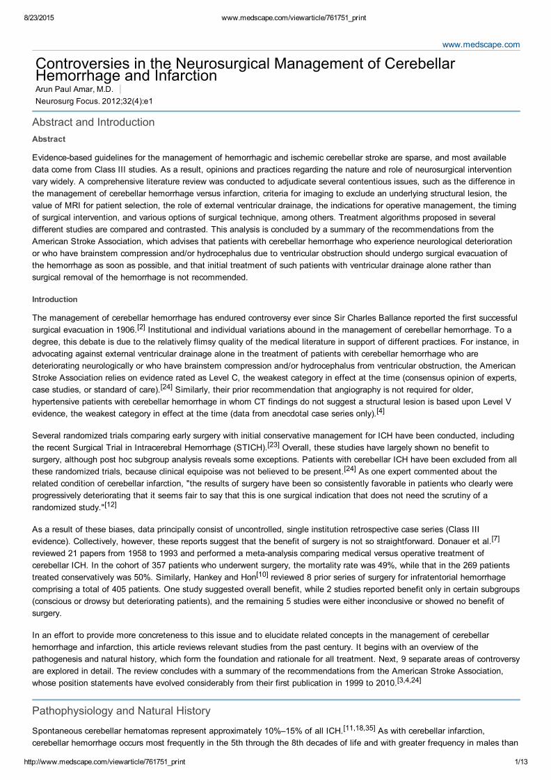
Figure 2.
Images obtained in a 43yearold man with no prior medical history, who awoke with headache, nausea, and dizziness, andwhose blood pressure was 109/54 mm Hg. A: Axial CT scans without contrast demonstrate a large hemorrhage of the right
8/23/2015 www.medscape.com/viewarticle/761751_print
http://www.medscape.com/viewarticle/761751_print 7/13
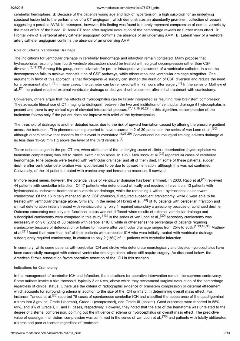
cerebellar hemisphere. B: Because of the patient's young age and lack of hypertension, a high suspicion for an underlyingstructural lesion led to the performance of a CT angiogram, which demonstrates an abundantly prominent collection of vesselssuggesting a possible AVM. In retrospect, however, this finding was found to merely represent compression of normal vessels bythe mass effect of the bleed. C: Axial CT scan after surgical evacuation of the hemorrhage reveals no further mass effect. D:Frontal view of a vertebral artery catheter angiogram confirms the absence of an underlying AVM. E: Lateral view of a vertebralartery catheter angiogram confirms the absence of an underlying AVM.
Role of External Ventricular Drainage
The indications for ventricular drainage in cerebellar hemorrhage and infarction remain contested. Many propose thathydrocephalus resulting from fourth ventricle obstruction should be treated with surgical decompression rather than CSFdiversion.[5,17,33] Among this group, some advocate for pre or intraoperative placement of a ventricular catheter, in case thedecompression fails to achieve reconstitution of CSF pathways, while others renounce ventricular drainage altogether. Oneargument in favor of this approach is that decompressive surgery can shorten the duration of CSF diversion and reduce the needfor a permanent shunt.[5] In many cases, the catheter can be removed within 72 hours after surgery.[5] In the series of Mathew etal.,[21] no patient required external ventricular drainage or delayed shunt placement after initial treatment with craniectomy.
Conversely, others argue that the effects of hydrocephalus can be falsely interpreted as resulting from brainstem compression.They advocate liberal use of CT imaging to distinguish between the two and institution of ventricular drainage if hydrocephalus ispresent and there is any clinical sign of elevated intracranial pressure.[7,17,18,26,29] In this algorithm, decompression of thebrainstem follows only if the patient does not improve with relief of the hydrocephalus.
The threshold of drainage is another debated issue, due to the risk of upward herniation caused by altering the pressure gradientacross the tentorium. This phenomenon is purported to have occurred in 2 of 30 patients in the series of van Loon et al.,[30]
although others believe that concern for this event is overstated.[4,26,29] Conventional neurosurgical training advises drainage atno less than 15–20 mm Hg above the level of the third ventricle.[7]
These debates began in the preCT era, when attribution of the underlying cause of clinical deterioration (hydrocephalus vsbrainstem compression) was left to clinical examination alone. In 1960, McKissock et al.[22] reported 34 cases of cerebellarhemorrhage. Nine patients were treated with ventricular drainage, and all of them died. In some of these patients, suddendecline after ventricular deterioration was hypothesized to be due to upward herniation, although this was not confirmed.Conversely, of the 14 patients treated with craniectomy and hematoma resection, 9 survived.
In more recent series, however, the potential value of ventricular drainage has been affirmed. In 2003, Raco et al.[26] reviewed44 patients with cerebellar infarction. Of 17 patients who deteriorated clinically and required intervention, 13 patients withhydrocephalus underwent treatment with ventricular drainage, while the remaining 4 without hydrocephalus underwentcraniectomy. Of the 13 initially managed using CSF diversion, 5 required subsequent craniectomy, while 8 were able to betreated with ventricular drainage alone. Similarly, in the series of Hornig et al.,[13] of 10 patients with cerebellar infarction andclinical deterioration initially treated with ventriculostomy, only 4 required secondary craniectomy because of continued decline.Outcome concerning mortality and functional status was not different when results of external ventricular drainage andsuboccipital craniectomy were compared in this study.[13] In the series of van Loon et al.,[30] secondary craniectomy wasnecessary in only 6 (20%) of 30 patients with cerebellar ICH, while in other series the percentage of patients requiringcraniectomy because of deterioration or failure to improve after ventricular drainage ranges from 25% to 80%.[1,13,16,30] Mathewet al.[21] found that more than half of their patients with cerebellar ICH who were initially treated with ventricular drainagesubsequently required craniectomy, in contrast to only 2 (18%) of 11 patients with cerebellar infarction.
In summary, while some patients with cerebellar ICH and stroke who deteriorate neurologically and develop hydrocephalus havebeen successfully managed with external ventricular drainage alone, others still require surgery. As discussed below, theAmerican Stroke Association favors operative resection of the ICH in this scenario.
Indications for Craniotomy
In the management of cerebellar ICH and infarction, the indications for operative intervention remain the supreme controversy.Some authors invoke a size threshold, typically 3 or 4 cm, above which they recommend surgical evacuation of the hemorrhageregardless of clinical status. Others use the criteria of radiographic evidence of brainstem compression or cisternal effacement,which accounts for surrounding edema in addition to the size of the ICH or infarct in determining overall mass effect. Forinstance, Taneda et al.[28] reported 75 cases of spontaneous cerebellar ICH and classified the appearance of the quadrigeminalcistern into 3 groups: Grade I (normal), Grade II (compressed), and Grade III (absent). Good outcomes were reported in 88%,69%, and 0% of Grade I, II, and III cases, respectively. However, they noted that the size of the hematoma was unrelated to thedegree of cisternal compression, pointing out the influence of edema or hydrocephalus on overall mass effect. The predictivevalue of quadrigeminal cistern compression was confirmed in the series of van Loon et al.,[30] and patients with totally obliteratedcisterns had poor outcomes regardless of treatment.

8/23/2015 www.medscape.com/viewarticle/761751_print
http://www.medscape.com/viewarticle/761751_print 8/13
Still others discount these radiographic features and emphasize the neurological examination, including level of consciousnessand brainstem reflexes, in determining criteria for surgery. Kobayashi et al.[18] performed a retrospective review of 52 patientswith hypertensive cerebellar ICH. On the basis of this analysis, they proposed new criteria for intervention that were prospectivelyapplied to the next 49 patients for validation and confirmation. Patients with GCS scores of 14 or 15 and with hematoma sizesless than 4 cm in maximal diameter were treated conservatively, while patients with GCS scores of 13 or less at admission orwith a hematoma measuring greater than 4 cm underwent surgical evacuation. For patients with flaccid tetraplegia and absentbrainstem reflexes, intensive therapy was not rendered.
Kirollos et al.[17] developed a different protocol, based on compression of the fourth ventricle as a measure of mass effect, whichthey applied prospectively in the management of 50 consecutive patients with cerebellar ICH. The appearance of the fourthventricle was divided into 3 groups: Grade I (normal size and configuration), Grade II (partially compressed and shifted), andGrade III (completely obliterated). The ICH was evacuated for all patients with Grade III compression and for patients with GradeII compression when the GCS score deteriorated in the absence of untreated hydrocephalus. Patients with Grade I or IIcompression were initially treated using only ventricular drainage if they developed hydrocephalus and clinical deterioration.Stable Grade I and II patients were managed conservatively. Acute deterioration to comatose state occurred in 6 (43%) of the 14patients with Grade III compression who were conscious at presentation; none of them experienced good outcomes. However, 15(60%) of 25 patients with hematomas greater than 3 cm and Grade I or II compression did not require clot evacuation.
In summary, clinical considerations should complement radiographic appearance in the management algorithm, and decisions forsurgical intervention should rarely be made on the basis of imaging findings alone.
Timing of Surgical Intervention
Evidence consistently shows that postoperative outcomes generally correlate with preoperative status.[6,17] For instance, in theseries of Ott et al.,[25] the mortality rate was 17% for patients who were conscious at the time of surgery and 75% for those whowere unconscious. Similarly, in the series of Donauer et al.,[7] patients presenting with GCS scores less than 6 had a 60%mortality rate and Karnofsky Outcome Index total of 26, while those with GCS scores greater than 10 had only a 20% mortalityrate and a Karnofsky Outcome Index total of 66. In the series of Kobayashi et al.,[18] of the 5 patients with GCS scores of 4, 3died and 2 remained vegetative despite surgery. In the series of van Loon et al.,[30] patients with total obliteration of thequadrigeminal cistern had a poor outcome irrespective of treatment.
Furthermore, many patients who experience clinical deterioration improve significantly after surgery. Some have even beenrestored to functional capacity. On this basis, it is natural to question whether patients who remain dependent after surgery wouldhave fared better if intervention had been performed earlier in their course.[12,19]
For these reasons, many recommend surgery prior to clinical deterioration.[12] Yoshida et al.[34] emphasize the importance ofsurgical therapy even for alert patients if the hematoma is larger than 3 cm to avoid delayed deterioration. Similarly, in the seriesof Kirollos et al.,[17] no patient with Grade III compression of the fourth ventricle who was conscious at the outset but thendeveloped a GCS score less than 8 experienced a good outcome. These authors thus advocate aggressive early surgicalevacuation of the hematoma for all Grade III patients, regardless of initial examination results, before deterioration occurs.
Conversely, Dammann et al.[6] reviewed their series of 57 patients who underwent surgical evacuation of spontaneous cerebellarICH. The initial neurological condition proved to be highly predictive of outcome. Based on the excellent results in patients withgood initial clinical condition who underwent surgery due to secondary deterioration, this group advises against the preventiveevacuation of cerebellar hemorrhage. Similarly, in the German Austrian Cerebellar Infarction Study, 84 patients with massivecerebellar infarction were prospectively observed after assignment to 1 of 3 groups: craniotomy and evacuation, ventriculostomy,or medical therapy alone. Treatment was left to the discretion of the provider in this unrandomized trial. In a logistic regressionmodel, there was found to be no benefit to performing surgery in patients who had not yet deteriorated to coma.[14]
Despite the focus on early evacuation of cerebellar ICH, even delayed removal might have benefit. Auer et al.[1] reported 2patients with cerebellar hemorrhage and focal signs of a posterior fossa lesion. Because they had no impairment ofconsciousness, they were initially managed conservatively. Because their symptoms had not shown a tendency to improve,however, they underwent evacuation of the hematoma performed on Days 23 and 54, respectively. Both patients then recoveredfully without neurological deficit.
What Constitutes Futility?
As stated, evidence consistently shows that postoperative outcomes generally correlate with preoperative status. However, thereare numerous anecdotal accounts of good postoperative outcome among comatose patients.[11,12] In some cases, even patientswith fixed and dilated pupils or other absent brainstem reflexes have recovered.[32] In the series of Hornig et al.,[13] 38% ofcomatose patients achieved a good recovery (nondisabled at hospital discharge) after decompressive surgery. In the GermanAustrian Cerebellar Infarction Study, half of all patients who deteriorated into coma and were treated with ventricular drainage or

8/23/2015 www.medscape.com/viewarticle/761751_print
http://www.medscape.com/viewarticle/761751_print 9/13
decompressive craniotomy experienced a meaningful recovery (modified Rankin score of 2 or less).[14] Similarly, Kobayashi et al.[18] reported 2 patients with ruptured cerebellar AVMs who had flaccid tetraplegia and apnea at admission. After emergencysurgery, both recovered to enjoy "a useful life."[18]
Furthermore, pathoanatomical studies reveal surprisingly few structural changes due to brainstem compression in patients withfatal spaceoccupying cerebellar infarcts.[27] Similarly, there is no intrinsic damage to supratentorial telencephalic structures incerebellar ICH and infarct, which suggests the possibility of full intellectual and cognitive recovery in some cases.[7,12] In light ofthese considerations, it is reasonable to question whether any patient's condition is "too poor" to forego surgical intervention andwhat constitutes futile treatment. From a practical standpoint, surgery might be considered, even if the situation appears"hopeless."
Value of Preoperative MRI
Yanaka et al.[33] studied the prognostic value of postoperative MRI in 31 patients, all with GCS scores of 8 or less, whounderwent surgical evacuation of cerebellar ICH. The patients were divided into 2 groups based on outcome. Good recovery oronly moderate disability was achieved in 8 patients, while the remaining 23 died or became severely disabled/vegetative. Therewere no significant differences between the 2 groups in preoperative CT findings such as hematoma size, presence ofhydrocephalus, fourth ventricular compression, or obliteration of the perimesencephalic cistern. However, the incidence of highsignal intensity in the pons and midbrain on T2weighted MRI, indicating brainstem damage, was significantly higher in the pooroutcome group. These intriguing results raise the question of whether preoperative MRI can be used as a predictive tool toscreen patients for brainstem injury, thus improving patient selection for aggressive therapy. However, no study has yetaddressed this issue, possibly because of logistical impediments to performing MRI scans acutely in critically ill patients. Theabsence of brainstem injury, confirmed by preoperative MRI, might provide impetus for surgical intervention in patients whootherwise might have been considered "hopeless."
Technical Aspects of Surgery
Numerous technical considerations in the operative management of cerebellar hemorrhage and infarction remain in the realm ofindividual preference. These include the size of the suboccipital bone removed and whether to fixate the bone flap (craniotomy)or float it or abandon it (craniectomy) at the end of the procedure. Other adjuncts such as the removal of the arch of the firstcervical vertebra remain optional. In the German Austrian Cerebellar Infarction Study, for instance, decompressive surgeryconsisted of a large craniotomy, duraplasty, and resection of the posterior atlas arch if tonsillar herniation was apparent, butresection of necrotic tissue was not mandatory.[14] In other series, however, craniectomy with resection of the infarcted tissuewas applied, including possible resection of cerebellar tonsils.[13,25] One risk of too large a craniectomy is subsequent sagging ofthe cerebellar hemispheres. Conversely, a bone flap that is too small and then replaced may fail to achieve adequatedecompression (Fig. 3). Because the degree of mass effect is different in each patient, intraoperative judgement must beexercised in determining the extent of bone removal necessary to achieve decompression, and no rigid guidelines can be offeredabout a prespecified size threshold.
8/23/2015 www.medscape.com/viewarticle/761751_print
http://www.medscape.com/viewarticle/761751_print 10/13
Figure 3.
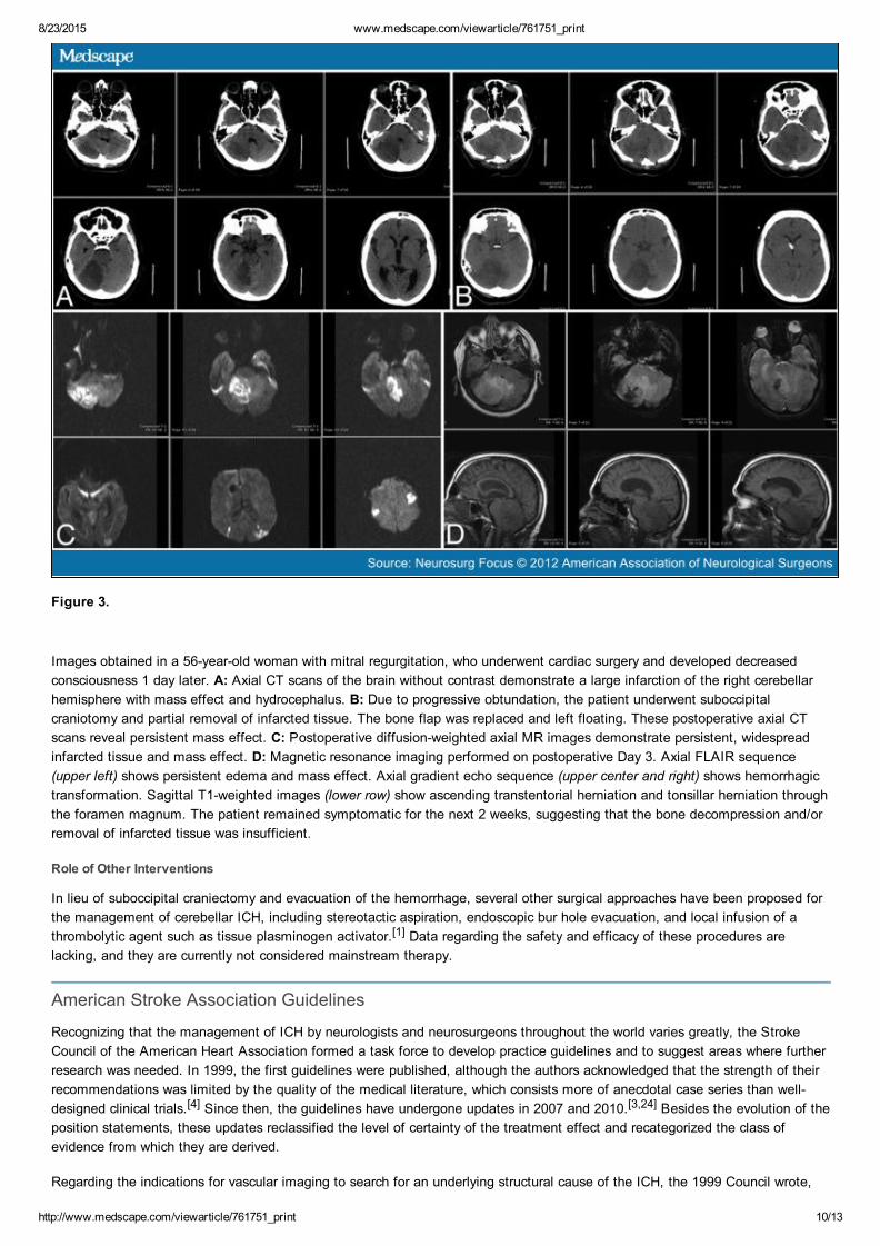
Images obtained in a 56yearold woman with mitral regurgitation, who underwent cardiac surgery and developed decreasedconsciousness 1 day later. A: Axial CT scans of the brain without contrast demonstrate a large infarction of the right cerebellarhemisphere with mass effect and hydrocephalus. B: Due to progressive obtundation, the patient underwent suboccipitalcraniotomy and partial removal of infarcted tissue. The bone flap was replaced and left floating. These postoperative axial CTscans reveal persistent mass effect. C: Postoperative diffusionweighted axial MR images demonstrate persistent, widespreadinfarcted tissue and mass effect. D: Magnetic resonance imaging performed on postoperative Day 3. Axial FLAIR sequence(upper left) shows persistent edema and mass effect. Axial gradient echo sequence (upper center and right) shows hemorrhagictransformation. Sagittal T1weighted images (lower row) show ascending transtentorial herniation and tonsillar herniation throughthe foramen magnum. The patient remained symptomatic for the next 2 weeks, suggesting that the bone decompression and/orremoval of infarcted tissue was insufficient.
Role of Other Interventions
In lieu of suboccipital craniectomy and evacuation of the hemorrhage, several other surgical approaches have been proposed forthe management of cerebellar ICH, including stereotactic aspiration, endoscopic bur hole evacuation, and local infusion of athrombolytic agent such as tissue plasminogen activator.[1] Data regarding the safety and efficacy of these procedures arelacking, and they are currently not considered mainstream therapy.
American Stroke Association Guidelines
Recognizing that the management of ICH by neurologists and neurosurgeons throughout the world varies greatly, the StrokeCouncil of the American Heart Association formed a task force to develop practice guidelines and to suggest areas where furtherresearch was needed. In 1999, the first guidelines were published, although the authors acknowledged that the strength of theirrecommendations was limited by the quality of the medical literature, which consists more of anecdotal case series than welldesigned clinical trials.[4] Since then, the guidelines have undergone updates in 2007 and 2010.[3,24] Besides the evolution of theposition statements, these updates reclassified the level of certainty of the treatment effect and recategorized the class ofevidence from which they are derived.
Regarding the indications for vascular imaging to search for an underlying structural cause of the ICH, the 1999 Council wrote,

8/23/2015 www.medscape.com/viewarticle/761751_print
http://www.medscape.com/viewarticle/761751_print 11/13
"Angiography should be considered for all patients without a clear cause of hemorrhage who are surgical candidates, particularlyyoung, normotensive patients who are clinically stable (level of evidence V, grade C recommendation)."[4] Additionally, theywrote, "Angiography is not required for older hypertensive patients who have a hemorrhage in the basal ganglia, thalamus,cerebellum, or brain stem and in whom CT findings do not suggest a structural lesion ….(level of evidence V, grade Crecommendation)."4 Under the definitions in effect at that time, these were the weakest possible recommendations and based onthe lowest quality data. In 2010, the guidelines state that, "…CT angiography, CT venography, contrastenhanced CT, contrastenhanced MRI, magnetic resonance angiography, and magnetic resonance venography can be useful to evaluate for underlyingstructural lesions, including vascular malformations and tumors when there is clinical or radiological suspicion (Class IIa; Level ofEvidence: B)."[24]
Regarding the indications for surgical removal of ICH, the 1999 council wrote:[4]
Patients with cerebellar hemorrhage > 3 cm who are neurologically deteriorating or who have brain stem compression andhydrocephalus from ventricular obstruction should have surgical removal of the hemorrhage as soon as possible (levels ofevidence III through V, grade C recommendation)… Stereotactic aspiration may be associated with better outcomes thanstandard craniotomy for moderatesized cerebellar hemorrhages, but this hypothesis has yet to be tested in a randomizedstudy (no recommendation).
In 2007, there was no change to this recommendation, although the authors revised the categorization of its strength as "Class1, Level of Evidence B," which is an intermediate grade.[3] In 2010, the qualification of the 3cmsize threshold was abandoned,and a new recommendation concerning ventricular drainage was offered:[24]
Patients with cerebellar hemorrhage who are deteriorating neurologically or who have brainstem compression and/orhydrocephalus from ventricular obstruction should undergo surgical removal of the hemorrhage as soon as possible (Class 1;Level of Evidence: B). (Revised from the previous guideline). Initial treatment of these patients with ventricular drainagealone rather than surgical evacuation is not recommended (Class III; Level of Evidence: C). (New recommendation).
Conclusions
The management of cerebellar hemorrhagic and ischemic stroke is controversial. Issues such as the difference in the treatmentalgorithm of cerebellar ICH versus infarction, criteria for imaging to exclude an underlying structural lesion, the value of MRI forpatient selection, the role of external ventricular drainage, the indications for operative management, the timing of surgicalintervention, and various options of surgical technique remain unresolved. Professional society guidelines for these considerationsare sparse and based on relatively poor quality data. Nonetheless, the potential value of neurosurgical intervention remains wellestablished.
References
1. Auer LM, Auer T, Sayama I: Indications for surgical treatment of cerebellar haemorrhage and infarction. Acta Neurochir(Wien) 79:74–79, 1986
2. Ballance HA: Case of traumatic haemorrhage into the left lateral lobe of the cerebellum, treated by operation, withrecovery. Surg Gynecol Obstet 3:223–225, 1906
3. Broderick J, Connolly S, Feldmann E, Hanley D, Kase C, Krieger D, et al: Guidelines for the management ofspontaneous intracerebral hemorrhage in adults: 2007 update: a guideline from the American Heart Association/AmericanStroke Association Stroke Council, High Blood Pressure Research Council, and the Quality of Care and Outcomes inResearch Interdisciplinary Working Group. Circulation 116:e391–e413, 2007
4. Broderick JP, Adams HP Jr, Barsan W, Feinberg W, Feldmann E, Grotta J, et al: Guidelines for the management ofspontaneous intracerebral hemorrhage: a statement for healthcare professionals from a special writing group of the StrokeCouncil, American Heart Association. Stroke 30:905–915, 1999
5. Chen HJ, Lee TC, Wei CP: Treatment of cerebellar infarction by decompressive suboccipital craniectomy. Stroke 23:957–961, 1992
6. Dammann P, Asgari S, Bassiouni H, Gasser T, Panagiotopoulos V, Gizewski ER, et al: Spontaneous cerebellarhemorrhage— experience with 57 surgically treated patients and review of the literature. Neurosurg Rev 34:77–86, 2011
7. Donauer E, Loew F, Faubert C, Alesch F, Schaan M: Prognostic factors in the treatment of cerebellar haemorrhage. ActaNeurochir (Wien) 131:59–66, 1994
8. Fairburn B, Oliver LC: Cerebellar softening; a surgical emergency. BMJ 1:1335–1336, 1956
8/23/2015 www.medscape.com/viewarticle/761751_print
http://www.medscape.com/viewarticle/761751_print 12/13
9. Halpin SF, Britton JA, Byrne JV, Clifton A, Hart G, Moore A: Prospective evaluation of cerebral angiography andcomputed tomography in cerebral haematoma. J Neurol Neurosurg Psychiatry 57:1180–1186, 1994
10. Hankey GJ, Hon C: Surgery for primary intracerebral hemorrhage: is it safe and effective? A systematic review of caseseries and randomized trials. Stroke 28:2126–2132, 1997
11. Heros RC: Cerebellar hemorrhage and infarction. Stroke 13: 106–109, 1982
12. Heros RC: Surgical treatment of cerebellar infarction. Stroke 23:937–938, 1992
13. Hornig CR, Rust DS, Busse O, Jauss M, Laun A: Spaceoccupying cerebellar infarction. Clinical course and prognosis.Stroke 25:372–374, 1994
14. Jauss M, Krieger D, Hornig C, Schramm J, Busse O: Surgical and medical management of patients with massivecerebellar infarctions: results of the GermanAustrian Cerebellar Infarction Study. J Neurol 246:257–264, 1999
15. Kanno T, Sano H, Shinomiya Y, Katada K, Nagata J, Hoshino M, et al: Role of surgery in hypertensive intracerebralhematoma. A comparative study of 305 nonsurgical and 154 surgical cases. J Neurosurg 61:1091–1099, 1984
16. Kidwell CS, Alger JR, Saver JL: Beyond mismatch: evolving paradigms in imaging the ischemic penumbra withmultimodal magnetic resonance imaging. Stroke 34:2729–2735, 2003
17. Kirollos RW, Tyagi AK, Ross SA, van Hille PT, Marks PV: Management of spontaneous cerebellar hematomas: aprospective treatment protocol. Neurosurgery 49:1378–1387, 2001
18. Kobayashi S, Sato A, Kageyama Y, Nakamura H, Watanabe Y, Yamaura A: Treatment of hypertensive cerebellarhemorrhage— surgical or conservative management? Neurosurgery 34:246–251, 1994
19. Koh MS, Goh KY, Tung MY, Chan C: Is decompressive craniectomy for acute cerebral infarction of any benefit? SurgNeurol 53:225–230, 2000
20. Lindgren SO: Infarctions simulating brain tumours in the posterior fossa. J Neurosurg 13:575–581, 1956
21. Mathew P, Teasdale G, Bannan A, OluochOlunya D: Neurosurgical management of cerebellar haematoma and infarct. JNeurol Neurosurg Psychiatry 59:287–292, 1995
22. McKissock W, Richardson A, Walsh L: Spontaneous cerebellar haemorrhage: a study of 34 consecutive cases treatedsurgically. Brain 83:1–9, 1960
23. Mendelow AD, Gregson BA, Fernandes HM, Murray GD, Teasdale GM, Hope DT, et al: Early surgery versus initialconservative treatment in patients with spontaneous supratentorial intracerebral haematomas in the International SurgicalTrial in Intracerebral Haemorrhage (STICH): a randomised trial. Lancet 365:387–397, 2005
24. Morgenstern LB, Hemphill JC III, Anderson C, Becker K, Broderick JP, Connolly ES Jr, et al: Guidelines for themanagement of spontaneous intracerebral hemorrhage: a guideline for healthcare professionals from the American HeartAssociation/American Stroke Association. Stroke 41:2108– 2129, 2010
25. Ott KH, Kase CS, Ojemann RG, Mohr JP: Cerebellar hemorrhage: diagnosis and treatment. A review of 56 cases. ArchNeurol 31:160–167, 1974
26. Raco A, Caroli E, Isidori A, Salvati M: Management of acute cerebellar infarction: one institution's experience.Neurosurgery 53:1061–1066, 2003
27. Sypert GW, Alvord EC Jr: Cerebellar infarction. A clinicopathological study. Arch Neurol 32:357–363, 1975
28. Taneda M, Hayakawa T, Mogami H: Primary cerebellar hemorrhage. Quadrigeminal cistern obliteration on CT scans as apredictor of outcome. J Neurosurg 67:545–552, 1987
29. Tulyapronchote R, Malkoff MD, Selhorst JB, Gomez CR: Treatment of cerebellar infarction by decompression suboccipitalcraniectomy. Stroke 24:478–480, 1993 (Letter)
30. van Loon J, Van Calenbergh F, Goffin J, Plets C: Controversies in the management of spontaneous cerebellarhaemorrhage. A consecutive series of 49 cases and review of the literature. Acta Neurochir (Wien) 122:187–193, 1993

8/23/2015 www.medscape.com/viewarticle/761751_print
http://www.medscape.com/viewarticle/761751_print 13/13
Abbreviations used in this paper AVM = arteriovenous malformation; GCS = Glascow Coma Scale; ICH = intracerebral hemorrhage.
Neurosurg Focus. 2012;32(4):e1 © 2012 American Association of Neurological Surgeons
31. Wakai S, Nagai M: Histological verification of microaneurysms as a cause of cerebral haemorrhage in surgical specimens.J Neurol Neurosurg Psychiatry 52:595–599, 1989
32. Yanaka K, Meguro K, Fujita K, Narushima K, Nose T: Immediate surgery reduces mortality in deeply comatose patientswith spontaneous cerebellar hemorrhage. Neurol Med Chir (Tokyo) 40:295–300, 2000
33. Yanaka K, Meguro K, Fujita K, Narushima K, Nose T: Postoperative brainstem high intensity is correlated with pooroutcomes for patients with spontaneous cerebellar hemorrhage. Neurosurgery 45:1323–1328, 1999
34. Yoshida S, Sasaki M, Oka H, Gotoh O, Yamada R: [Hypertensive cerebellar hemorrhage with signs of lower brainstemcompression—a case report (author's transl).] No Shinkei Geka 6:379–383, 1978 (Jpn)
35. Zhu XL, Chan MS, Poon WS: Spontaneous intracranial hemorrhage: which patients need diagnostic cerebralangiography? A prospective study of 206 cases and review of the literature. Stroke 28:1406–1409, 1997