Ceramics for Medical Applications a Picture for the Next 20 Years 2009 Journal of the European...
11
Available online at www.sciencedirect.com Journal of the European Ceramic Society 29 (2009) 1245–1255 Ceramics for medical applications: A picture for the next 20 years J. Chevalier ∗ , L. Gremillard Université de Lyon, INSA-Lyon, UMR CNRS 5510 (MATEIS), 20 Avenue Albert Einstein, 69621 Villeurbanne Cedex, France Available online 9 October 2008 Abstract High-tech ceramics have always been associated to medical devices: they are used today as femoral heads and acetabular cups for total hip replacement, dental implants and restorations, bone fillers and scaffolds for tissue engineering. Here, we describe their current clinical use and propose a picture of their evolutions for the next 20 years. The need for tough, strong and stable bioinert ceramics should be met by either nano-structured, alumina and zirconia based ceramics and composites or by non-oxide ceramics. Nano-structured calcium phosphate ceramics and porous bioactive glasses, possibly combined with an organic phase should present the desired properties for bone substitu- tion and tissue engineering. The position of ceramics in a gradual medical approach, from tissue regeneration to conventional implants, is discussed. © 2008 Elsevier Ltd. All rights reserved. Keywords: Biomedical applications; Toughness and toughening; Nano-composites; Composites; Lifetime 1. Introduction: clinical use of ceramics, historical highlights and current solutions Ceramics are generally defined as ‘inorganic, non-metallic materials’. Biomaterials are now defined as ‘natural or synthetic materials suitable for introduction into living tissue especially as part of a medical device’. Considering these definitions, we may argue that ceramics have been used as biomaterials for millenia. In 1972, indeed, Amadeo Bobbio discovered Mayan skulls, some of then more than 4000 years old, in which missing teeth had been replaced by nacre substitutes. 1 Nacre is a natural com- posite consisting of 95–98 wt.% of calcium carbonate (aragonite, the ‘ceramic’ phase) and 2–5 wt.% of organic matter (fibrous proteins, polysaccharides). In clinical practice, the controlled implantation of bioceramics started late 18th century in dentals with the use of porcelain for crowns and late 19th in orthope- dics with the use of Plaster of Paris, or gypsum (calcium sulfate dihydrate) for bone filling. 2 With the advances in the ceramic technology, the 20th century saw more and more ‘high-tech’ ceramics available for medical purpose. 3 Tricalcium phosphate was first proposed in 1920 as a bioresorbable substance to fill bone gaps. However, tricalcium phosphate (TCP) and plaster are ∗ Corresponding author. Tel.: +33 4 72 43 61 25. E-mail address: [email protected] (J. Chevalier). weak ceramics, unable to sustain significant loading. The need for tough and strong ceramics was not met before 1965, when the first alumina (Al 2 O 3 ) material dedicated to hip joints was patented. 4 Synthetic calcium phosphate ceramics (together with calcium and/or phosphorus containing ceramics and glasses) and zirconia were then proposed as alternatives to TCP and alumina, respectively. After roughly 100 years of clinical use, we come to the conclusion that there is, so far, no tough and strong ceramic able to create a strong, biologically relevant interface with bone. On the other hand, ceramics and glasses able to promote direct bone-implant adhesion without fibrous soft tissue interlayer are all unable to be used as loaded devices. Bioceramics are there- fore generally separated in two families, so-called ‘bioinert’ and ‘bioactive’. Alumina (and zirconia) ceramics are most often considered as ‘bioinert’ (although a material should never be considered as totally inert), since no direct bone–material interface is cre- ated. A soft tissue interlayer always shields the bone from the implant. This biological shielding unfortunately leads to mechanical (stress) shielding, known to promote micro-motion and subsequent aseptic implant loosening. Only under compres- sion, with a porous structure and with a good fit with bone cavity (avoiding relative micro-motion), the fibrous tissue at the inter- face is thin enough and a successfull bone ingrowth is achieved. Given these restrictions, ‘bioinert’ ceramics are hardly used as bone fillers. 0955-2219/$ – see front matter © 2008 Elsevier Ltd. All rights reserved. doi:10.1016/j.jeurceramsoc.2008.08.025
-
Upload
nguyen-cong-hanh -
Category
Documents
-
view
5 -
download
2
description
Ceramics-for-medical-applications-A-picture-for-the-next-20-years_2009_Journal-of-the-
Transcript of Ceramics for Medical Applications a Picture for the Next 20 Years 2009 Journal of the European...

A
Hraectd©
K
1h
mmpaIshptpiwddtcwb
0d
Available online at www.sciencedirect.com
Journal of the European Ceramic Society 29 (2009) 1245–1255
Ceramics for medical applications: A picture for the next 20 years
J. Chevalier ∗, L. GremillardUniversité de Lyon, INSA-Lyon, UMR CNRS 5510 (MATEIS), 20 Avenue Albert Einstein,
69621 Villeurbanne Cedex, France
Available online 9 October 2008
bstract
igh-tech ceramics have always been associated to medical devices: they are used today as femoral heads and acetabular cups for total hipeplacement, dental implants and restorations, bone fillers and scaffolds for tissue engineering. Here, we describe their current clinical usend propose a picture of their evolutions for the next 20 years. The need for tough, strong and stable bioinert ceramics should be met byither nano-structured, alumina and zirconia based ceramics and composites or by non-oxide ceramics. Nano-structured calcium phosphate
eramics and porous bioactive glasses, possibly combined with an organic phase should present the desired properties for bone substitu-ion and tissue engineering. The position of ceramics in a gradual medical approach, from tissue regeneration to conventional implants, isiscussed.2008 Elsevier Ltd. All rights reserved.
ites; C
wftpczrtaObaf‘
aaat
eywords: Biomedical applications; Toughness and toughening; Nano-compos
. Introduction: clinical use of ceramics, historicalighlights and current solutions
Ceramics are generally defined as ‘inorganic, non-metallicaterials’. Biomaterials are now defined as ‘natural or syntheticaterials suitable for introduction into living tissue especially as
art of a medical device’. Considering these definitions, we mayrgue that ceramics have been used as biomaterials for millenia.n 1972, indeed, Amadeo Bobbio discovered Mayan skulls,ome of then more than 4000 years old, in which missing teethad been replaced by nacre substitutes.1 Nacre is a natural com-osite consisting of 95–98 wt.% of calcium carbonate (aragonite,he ‘ceramic’ phase) and 2–5 wt.% of organic matter (fibrousroteins, polysaccharides). In clinical practice, the controlledmplantation of bioceramics started late 18th century in dentalsith the use of porcelain for crowns and late 19th in orthope-ics with the use of Plaster of Paris, or gypsum (calcium sulfateihydrate) for bone filling.2 With the advances in the ceramicechnology, the 20th century saw more and more ‘high-tech’
eramics available for medical purpose.3 Tricalcium phosphateas first proposed in 1920 as a bioresorbable substance to fillone gaps. However, tricalcium phosphate (TCP) and plaster are∗ Corresponding author. Tel.: +33 4 72 43 61 25.E-mail address: [email protected] (J. Chevalier).
mas(fGb
955-2219/$ – see front matter © 2008 Elsevier Ltd. All rights reserved.oi:10.1016/j.jeurceramsoc.2008.08.025
omposites; Lifetime
eak ceramics, unable to sustain significant loading. The needor tough and strong ceramics was not met before 1965, whenhe first alumina (Al2O3) material dedicated to hip joints wasatented.4 Synthetic calcium phosphate ceramics (together withalcium and/or phosphorus containing ceramics and glasses) andirconia were then proposed as alternatives to TCP and alumina,espectively. After roughly 100 years of clinical use, we come tohe conclusion that there is, so far, no tough and strong ceramicble to create a strong, biologically relevant interface with bone.n the other hand, ceramics and glasses able to promote directone-implant adhesion without fibrous soft tissue interlayer arell unable to be used as loaded devices. Bioceramics are there-ore generally separated in two families, so-called ‘bioinert’ andbioactive’.
Alumina (and zirconia) ceramics are most often considereds ‘bioinert’ (although a material should never be considereds totally inert), since no direct bone–material interface is cre-ted. A soft tissue interlayer always shields the bone fromhe implant. This biological shielding unfortunately leads to
echanical (stress) shielding, known to promote micro-motionnd subsequent aseptic implant loosening. Only under compres-ion, with a porous structure and with a good fit with bone cavity
avoiding relative micro-motion), the fibrous tissue at the inter-ace is thin enough and a successfull bone ingrowth is achieved.iven these restrictions, ‘bioinert’ ceramics are hardly used asone fillers.
1246 J. Chevalier, L. Gremillard / Journal of the European Ceramic Society 29 (2009) 1245–1255
Table 1Mechanical properties of different ceramics
Material Toughness (KIC, MPa m1/2) Threshold (KI0, MPa m1/2) Strength (MPa) Vickers hardness
Alumina 4.2 2.4 400–600 1800–2000Zirconia 5.4 3.5 1000 1200–1300A10Z0Y 5.8 4 700–900 1800Hydroxyapatite 0.9 0.6 50–60 500Tricalcium phosphate 1.3 0.8 50–60 900Mg-PSZ 8 6 600 100012Ce-TZP 7.8 5.1 700 1000–1100Micro-nano-alumina–zirconia 6 5 600 1800Nano-nano-Ce-TZP–alumina 8.4 4.6 900 1300Silicon nitride 10* ? 1000* 2500
T the Ds
krocmwamwubpoirpmufr1nhosfcpfrtpatpatbmo
amTetlYlczsilfa
tcantmrgdan(wfmtc(oom
oughness (KIC) and threshold stress intensity factor (KI0) were measured bytrength by four point bending.
Their major application in orthopedics concerns total hip andnee replacement. The use of bioceramic materials reduces wearates of bearing components and produces negligible amountf ion release. The clinical success associated to the use oferamics led to the implantation of more than 3.5 millions alu-ina components and more than 600,000 zirconia femoral headsorldwide since 1990. There are many reports on fracture rates
ssociated with ceramics, since their intrinsic brittleness is theirajor drawback.5 If, in the pioneering days, the fracture rateas quite high (up to 13% for some series), the in vivo fail-re rate reported by the producer of Biolox® alumina is todayelow 0.01%.6 A comparable failure rate was claimed by theroducer of Prozyr® zirconia heads7 before the critical eventf 2001, discussed below. The current fracture rate of ceram-cs is therefore negligible when compared to the overall failureate of implants (mainly due to aseptic loosening resulting fromarticles release). If the clinical follow up with current alu-ina ceramics is very good, it must be kept in mind that their
se has been restricted so far to a limited number of designsor which the mechanical loading is less demanding. This iselated to their modest mechanical properties (Table 1). In the990s, yttria-stabilised zirconia (Y-TZP) became a popular alter-ative to alumina as structural ceramic because of substantiallyigher fracture toughness and strength. The use of Y-TZP haspened the way towards new implant designs that were not pos-ible with alumina, more brittle. Examples are 22 mm Y-TZPemoral heads, and the development of Y-TZP knees. Biomedi-al grade Y-TZP exhibits the best mechanical properties of singlehase oxide ceramics: this is the consequence of phase trans-ormation toughening, which increases its crack propagationesistance. The stress-induced phase transformation involves theransformation of metastable tetragonal grains to the monoclinichase at the crack tip. It is accompanied by volume expansionnd induces compressive stresses which hinder crack propaga-ion. On the other hand, due to this meta-stability, Y-TZP isrone to low temperature degradation (sometimes referred to asging) in the presence of water.8 Aging occurs by a progressive
etragonal to monoclinic transformation at the surface triggeredy water molecules, which results in surface roughening andicro-cracking. This inevitably impacts the wear performancef hip joint heads, as roughening increases the wear rate of the
Fctm
ouble Torsion method (except for values with *, extracted from Ref. 23) and
ntagonist part of the prosthesis, while the coupled effects oficro-cracking and wear generate pull-out of zirconia grains.he extension of the micro-cracked, transformed zone also gen-rates defects, that may grow with the transformed zone and leado delayed failure. Y-TZP manufacturers considered this prob-em as a minor issue until 2001, when hundreds of failures of-TZP heads were reported within a very short period. Even if
imited in time and number, and clearly identified to be processontrolled, these events have had a negative impact for the use ofirconia in orthopedics. More important, some clinical reportshow that yttria-stabilised zirconia can exhibit a progressive age-ng degradation even under ‘normal’ situation, which limits itsong-term stability. Orthopedic community now faces the needor tough, strong and stable ceramics as alternatives to aluminand Y-TZP.
Dental applications add aesthetic requirements (colour,ranslucency) to the mechanical specifications. White to ivoryolour gives a clear advantage for oxide ceramics versus met-ls, which is the reason why research and development areowadays directed towards metal-free dental prosthetic restora-ions. Indeed, metal-free restorations preserve soft tissue colour
ore similar to the natural one than porcelain fused to metalestorations. Moreover, ceramics do not suffer corrosion and/oralvanic coupling as it can be observed for metals. The clinicalemand for all-ceramic restoration is increasing and ceramicsre becoming important restorative materials in dentistry. Pio-eers like Duchateau had only access to conventional porcelainor more precisely mixture of kaolin, feldspar and quartz), whichere later replaced by more translucent feldspathic glasses rein-
orced by silica inclusions. However, these porcelain basedaterials still lacked mechanical strength. Therefore, during
he last 200 years, a global approach has been to increase theontent of ceramic: from silica to alumina reinforced porcelainin 1960), to glass-infiltrated high strength ceramics (aluminar zirconia) and finally to monolithic ceramics. Translucencyf technical ceramics may be achieved with a very fine (sub-icron) grain size and low porosity content (less than 1%).
ully dense, translucent (yttria-stabilised) zirconia ceramicsan be processed with grain size less than 0.5 �m and meethe demand for both natural-teeth-looking restoration and highechanical strength. For the last 10 years Y-TZP has been

urop
cHebos
catt(bb
birhTpttpassltioctistriTdmbii
gewel(tnmism
2
2
2b
ttassflteieccpaiivYcibctsslbsoTp
(ccmizt5
•
J. Chevalier, L. Gremillard / Journal of the E
onsidered as the ideal solution for most dental applications.owever, long-term in vivo studies on its stability in oral
nvironment are still lacking and few reports on in vitro sta-ility show that aging could also be an issue. As well as forrthopedic applications, alternatives to Y-TZP shall be neededhortly.
Unlike bioinert ceramics, the requirements for bioactiveeramics are to provide favorable surfaces for bone adhesionnd bone ingrowth.9 On the other hand, the specifications inerms of load-bearing capability are less demanding. Most bioac-ive ceramics are thus based on calcium phosphate materialsmainly hydroxyapatite, HAP, and tricalcium phosphate, TCP),ecause their compositions are close to the mineral part ofone.
Since the early 1980s, the most important application ofioactive ceramics has been the coating of orthopedic metalmplants, at locations where a strong interface with bone isequired (i.e. femoral stems and acetabular metal-backs for theip joints and tibial and femoral components for the knee joints).hese systems represent successful alternatives to cementedrostheses, especially for young and active patients, and areherefore generally associated to ceramic–ceramic couplings. Inhe mid 1980s, the osteo-conductive properties of calcium phos-hate led to their use as synthetic bone grafts, as an alternative touto-grafts and allo-grafts.9 Indeed, as compared to auto-grafts,ynthetic bone substitute involve less invasive surgery (a twotep operation is necessary for the former) and are available inarge quantities. As compared to allo-grafts, the risk of rejec-ion is much less important, and the transmission of diseasess avoided. Most of current bone substitutes are porous piecesf biphasic calcium phosphates, i.e. HAP-TCP composites.10 Aareful control of the architecture (volume and morphology ofhe macro- and micro-porosities) is a key issue for a successfulmplant, as the macro-porosity controls the access of the tis-ues and biological fluids to the volume of the substitute, andhe micro-porosity the adhesion of the cells and the resorptionate of the calcium phosphate (thus the availability of Ca and Pons for bone reconstruction). The fast in vivo resorption rate ofCP, as compared to HAP, allows controlling the overall degra-ation rate of the HAP-TCP composite, and thus adapting theaterial to the patient (faster resorption for patients with faster
one reconstruction). Current synthetic bone substitutes markets about 40 million D in Europe, with an expected 12% yearlyncrease (www.frost.com).
However, current calcium phosphate bone substitutes do notive full satisfaction. Micro- and macro-porosities and theirffect on biological properties are not always taken into account,hich results in a large variability of physico-chemical prop-
rties among current commercial substitutes. Another strongimitation is their brittleness associated to low crack resistance11
Table 1), which restricts their use to non-load-bearing applica-ions, and makes them difficult to handle during surgery. Last butot least, their bioactivity should be increased in order to pro-
ote a faster and better bone reconstruction. We should keepn mind that natural HAP crystals are nano-sized while mostynthetic bone substitutes in clinical use are still constituted oficro-sized grains.
ean Ceramic Society 29 (2009) 1245–1255 1247
. Advances on ceramics: options for the next 20 years
.1. The need for tough, strong and stable bioinert ceramics
.1.1. Next 5 years: clinical use of alternative zirconiaased ceramics and composites
Yttria-stabilised zirconia was the ceramic gold standard inerms of strength and toughness (see Table 1), but its lack of long-erm stability is a major issue for medical use. Current researchims at developing zirconia based ceramics and composites thathould benefit from phase transformation toughening withoutuffering surface degradation in the presence of water or bodyuid. Before discussing these new materials, a synthetic descrip-
ion of aging is necessary. Detailed analysis is given in Chevaliert al.12 Aging occurs experimentally in zirconia samples mostlyn humid atmosphere or in water. Therefore, today, there are sev-ral models that attempt to explain how the presence of waterould promote tetragonal to monoclinic transformation in zir-onia. Several experimental results show that water radicalsenetrate inside the zirconia lattice during exposure to humidtmosphere. Most probably, the oxygen of environmental waters located on vacancy sites and the hydrogen is placed on adjacentnterstitial site.13 This emphasizes the primary role of oxygenacancies initially present in zirconia on water diffusion rate. In-TZP, the presence of numerous vacancies due to the trivalentharacter of yttrium makes the diffusion rate of water higher thann other zirconia ceramics (i.e. CeO2 doped ZrO2). After Schu-ert and Frey,13 the penetration of water radicals leads to a latticeontraction, which results in the formation of tensile stresses inhe surface grains that destabilise the tetragonal phase. Marten-itic transformation of some grains (or part of grains) at theurface can then take place. This nucleation of the transformationeads then to a cascade of events occurring neighbour to neigh-our: the transformation of one grain leads to a volume increasetressing up the neighbouring grains and to micro-cracking. Thisffers a path for the water to penetrate down into the specimen.he transformation occurs therefore by a nucleation and growthrocess.
In addition to the current improvement of Y-TZP powdersi.e. TZ3Y-E, with a very small – less than 0.5 wt.% – aluminaontent, which exhibits improved aging resistance), more effi-ient answers to the aging issue lie in stabilisation mechanismsinimizing the quantity of oxygen vacancies and/or minimiz-
ng nucleation and growth kinetics by avoiding contact betweenirconia grains. Two kinds of materials are under developmentoday and may be used clinically at large scale within the next–10 years:
Alumina-zirconia composites (referred to as zirconia-toughened alumina, ZTA). The stabilisation of zirconia grainsin such composites is achieved thanks to the presence of a stiffalumina matrix. Doping zirconia grains with yttria is not nec-
essary, thus no oxygen vacancy is created. Diffusion of waterradicals into the zirconia lattice is therefore strongly reduced.Moreover, for sufficiently low zirconia content (below thepercolation threshold) and sufficiently good dispersion,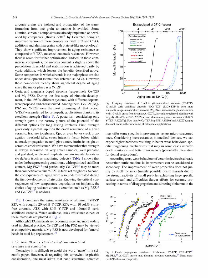
1 uropean Ceramic Society 29 (2009) 1245–1255
•
Zfst
uah
2c
ec
Fig. 1. Aging resistance of 3 mol.% yttria-stabilised zirconia (3Y-TZP),10 mol.% ceria stabilised zirconia (10Ce-TZP) (12Ce-TZP is even moreresistant), magnesia-stabilised zirconia (MgPSZ), zirconia-toughened aluminawith 10 vol.% yttria-free zirconia (A10Z0Y), zirconia-toughened alumina withrYd
moeccf
bstthe strong reactivity of small particles exhibiting large specificsurface areas) and difficulties (larger efforts for ceramic pro-cessing in terms of disaggregation and sintering) inherent to the
248 J. Chevalier, L. Gremillard / Journal of the E
zirconia grains are isolated and propagation of the trans-formation from one grain to another is avoided. Somealumina–zirconia composites are already implanted or devel-oped by companies (Biolox delta® by Ceramtec being animproved version of these composites, with SrO and Cr2O3additions and alumina grains with platelet-like morphology).They show significant improvement in aging resistance ascompared to Y-TZP, and excellent crack resistance. However,there is room for further optimization. Indeed, in these com-mercial composites, the zirconia content is slightly above thepercolation threshold and stabilisation is achieved partly byyttria addition, which lowers the benefits described above.Some composites in which zirconia is the major phase are alsounder development (sometimes referred as ATZ). However,these composites clearly show significant degree of agingsince the major phase is a Y-TZP.Ceria and magnesia doped zirconia (respectively Ce-TZPand Mg-PSZ). During the first stage of zirconia develop-ment, in the 1980s, different systems, with different dopants,were proposed and characterized. Among them, Ce-TZP, Mg-PSZ and Y-TZP were the most promising. At that period,Y-TZP was preferred for orthopedic applications thanks to itsexcellent strength (Table 1). A posteriori, considering onlystrength gave a too narrow picture of the potential of thedifferent options for long lasting implants. First, strengthgives only a partial input on the crack resistance of a givenceramic: fracture toughness, KIC, or even better crack prop-agation threshold (KI0, stress intensity factor below whichno crack propagation occurs) give a more intrinsic insight onceramics crack resistance. We have to remember that strengthis always measured on very small samples, well preparedand polished, while real implants contain inevitably extrin-sic defects (such as machining defects). Table 1 shows thatunder the best processing conditions, with optimized stabilisercontent, Mg-PSZ14 and particularly Ce-TZP15 may be morethan competitive versus Y-TZP in terms of toughness. Second,the consequences of aging were also underestimated duringthe first developments of zirconia. Knowing the critical con-sequences of low temperature degradation on implants, thechoice of aging resistant zirconia ceramics such as Mg-PSZ16
and Ce-TZP17 is obvious.
Fig. 1 compares the aging resistance of alumina, 3Y-TZP,TA with roughly 20 vol.% Y-TZP, ZTA with 10 vol.% yttria-
ree zirconia, ATZ with 80% Y-TZP and 10 mol.% ceriatabilised zirconia. When available, crack resistance curves ofhese materials are plotted in Fig. 2.
Although ZTA materials are becoming more and more widelysed in clinical practice, Ce-TZP and Mg-PSZ may be vieweds competitive materials. Mg-PSZ is now developed for femoraleads in total hip replacement.16
.1.2. Next 10 years: clinical use of nano-structured
eramics and compositesNowadays it is difficult to avoid the word “nano” in a sci-ntific paper. However, disregarding this somewhat despicableonsideration, one must admit that nano-structured ceramics
FMC
oughly 20 vol.% Y-TZP (A20Z2Y) and alumina-toughened zirconia with 80%-TZP [A80Z(Y)]. Note that for Ce-TZP, Mg-PSZ, A10Z0Y and A20Z2Y, agingoes not occur in the timeframe of orthopedic applications.
ay offer some specific improvements versus micro-structurednes. Considering inert ceramics biomedical devices, we canxpect higher hardness resulting in better wear behaviour, spe-ific toughening mechanisms that may in some cases improverack resistance, and better translucency (potentially interestingor dental restorations).
According to us, wear behaviour of ceramic devices is alreadyetter than sufficient, thus its improvement can be considered asecondary. The improvement of wear properties does not jus-ify by itself the risks (mainly possible health hazards due to
ig. 2. Crack propagation resistance of alumina, 3Y-TZP, 12Ce-TZP,15
g-PSZ,14 A10Z0Y, micro-nano-alumina–zirconia composite,19 Nano-nano-e-TZP–alumina composite.

J. Chevalier, L. Gremillard / Journal of the European Ceramic Society 29 (2009) 1245–1255 1249
F micron s alum
dindniobsioeotctbsiaz5awimc
•
•
ig. 3. (a) Schematic drawing of micro-nano-composite, (b) example of suchano-nano-composite, and (d) nano-nano-composite (Matsushita Electric Work
evelopment of nano-structured ceramics. On the other hand,mproving the mechanical properties, which gives access toew implants designs, is highly desirable. From their intro-uction around 20 years ago, the mechanical properties ofano-ceramics have been subject to controversies. The pioneer-ng work of Niihara,18 describing high potential benefits in termsf strength and to a lower extent of toughness, was followedy a general frustration in the 1990s mainly due to the impos-ibility to repeat his experiments at the time. New progressesn powder synthesis, forming and sintering allow a validationf his insights on a larger range of data obtained on differ-nt materials (oxide, non-oxides) systems and different typesf microstructures. From the large variety of systems proposedoday by ceramic researchers, we will extract the case of nano-omposites processed from alumina and zirconia, since they arehe most advanced in terms of process control and since theiocompatibility of each constituent is well established. Twotrategies are currently explored: alumina rich nano-compositesn which zirconia nanoparticles are evenly dispersed in microniclumina grains (referred to as ‘micro-nano-composites’) andirconia rich nano-composites in which both phases are below00 nm (referred to as ‘nano-nano-composites’). Fig. 3 showsschematic drawing of each type of nano-composite, togetherith two examples of microstructures. Both strategies aim at
ncreasing crack propagation threshold, tensile strength andaterials stability as compared to the micro-scale ceramics and
omposites:
Micro-nano-alumina–zirconia composites (increasingcrack propagation threshold of alumina). In micron sizealumina–zirconia composites, the increase in crack resistanceis mainly due to phase transformation toughening and to a
-alumina–nano-zirconia composite (from Ref. 17), (c) schematic drawing ofina–zirconia).
lower extent to crack bridging. In micro-nano-composites,we shift to another toughening mechanism associated tothe presence of large residual compressive stresses aroundthe zirconia nanoparticles (up to 150 MPa compressivestress in the alumina grains, reported in the nano-compositeshown in Fig. 3, with only 1.7 vol.% zirconia intragranularparticles).19 Residual stresses are a function of the volumefraction, the size and the location of the zirconia particles.Zirconia particles at grain boundaries would not give rise tosuch high residual stresses. Therefore, the main challengelies in the difficulty to confine zirconia nano-particles insidealumina grains. This may be achieved by modified colloidalroutes. An example is described in details in Ref. 20 Briefly,the method consists in grafting a zirconia precursor at thesurface of micron-sized alumina grains dispersed in a liquidmedium. The modified powder is then dried and thermallytreated so as to obtain a composite powder with zirconiananoparticles strongly attached to alumina grains, as it isshown in Fig. 4. A strong benefit of this processing route isthat it avoids the manipulation of any nano-powder: zirconiaparticles, when created, are bound to alumina, without riskof contamination of the atmosphere and health issues.Nano-nano-ceria doped zirconia–alumina composites(increasing strength of Ce-TZP). Standard Ce-TZP exhibitsthe largest crack resistance of oxide ceramics but moderatestrength and hardness compared to Y-TZP. This is mainlya consequence of larger grain coarsening during sintering.The mobility of grain boundaries is indeed much higher in
Ce-TZP than in Y-TZP. It is therefore difficult to obtain afine-grained, fully dense Ce-TZP. With the aim of developingultrafine Ce-TZP based ceramics, Nawa21 has developed a10 mol.% Ce-TZP–alumina nano-composite. In this compos-
1250 J. Chevalier, L. Gremillard / Journal of the European Ceramic Society 29 (2009) 1245–1255
Fig. 4. Alumina–zirconia composite via modified colloidal route: (a) zirconium precursor and alumina powder dispersed in liquid medium; (b) after drying, aluminapowder is coated by a zirconium precursor layer; (c and d) during thermal treatments, the layer discomposes and zirconia nanoparticles nucleate and grow on thes afterg partic
tTpbicitaia
amcgifSpacnttc
2o
a(aammimosSoc
nFsbrhB
at
urface of the alumina grains; (e) conventional TEM image of such a powderrain; (f) high resolution TEM of a zirconia grain on the surface of an alumina
ite the Ce-TZP is further doped by small amount of yttria.The sintered material exhibits a grain size around 250 nm forboth phases.
Table 1 summarizes mechanical properties available for thewo nano-composites, as compared to alumina and Y-TZP.oughening by residual compressive stresses, associated to theresence of only 1.7 vol.% zirconia nano-particles, increasesoth toughness and strength as compared to alumina. Moremportant, the micro-nano-composite presents the best slowrack growth resistance (ratio of KI0 to KIC) among oxide ceram-cs. On the other hand, Ce-TZP–alumina nano-composites offerhe best balance between toughness and strength. Only wearnd fatigue testing on real implants followed by long-term clin-cal studies will allow a fine tuning of each material for a givenpplication.
Nano-structured ceramics, more than nano-composites (usu-lly opaque, as it is the case of both previous materials), mayeet the need for translucency of dental restoration. Translu-
ency is driven by grain size and porosity content and size. It isenerally assumed that a grain size well below 1 �m and a poros-ty content much lower than 1% are absolutely necessary. Newast sintering techniques, such as Spark Plasma and Microwaveintering or even fast heating rate furnaces, open an avenue to therocessing of ultrafine, fully dense ceramic materials offeringlternatives to Y-TZP. Examples of transparent or highly translu-ent ceramics (alumina, YAG, etc.) are already published, but
ot dedicated to biomedical application.22 Most of them unfor-unately offer poor crack resistance. Research should focus onhe development of translucent monolithic ceramics able to resistrack propagation.atlo
thermal treatments, showing zirconia nanoparticles on the surface an aluminale.
.1.3. Next 10 years: on the use of non-oxide ceramics inrthopedics?
Non-oxide ceramics, such as silicon carbide or silicon nitride,re considered to be almost not sensitive to slow crack growthKI0 and KIC being roughly the same), which leads to a better reli-bility of structural pieces (lower risk of delayed failure). Theylso possess the best wear properties among ceramics, whichakes them preferred candidates for bearings in industry. Bestechanical properties are reached for microstructures contain-
ng elongated grains able to bridge a propagating crack. Theseaterials are often referred to as in situ toughened, since no sec-
ndary phase is necessary to enhance crack resistance. Table 1ummarizes the optimal strength and toughness data for Si3N4.i3N4 implants for Total Hip Arthroplasty are currently devel-ped by Amedica Corp., Salt Lake City, UT. They are underlinical trials.23
With such interesting characteristics, one may wonder whyon-oxide ceramics were not introduced sooner in orthopedics.irst, we have to remind that validation of a new material byanitary authorities is long and costly and that Y-TZP seemed toe the ultimate choice in the 1990s. Second, non-oxide ceramicsequire more effort during processing: they must be sintered atigher temperatures than oxides and under inert atmosphere.eing harder, they are also more difficult to machine.
Silicon nitride is now approved in terms of biocompatibility,nd Y-TZP has shown its limitation. The use of nano-powders,ogether with the use of innovative fast sintering routes will
llow to sinter these materials under moderate temperatures ando develop near net shape sintering to overcome existing techno-ogical barriers. As a consequence, we expect the developmentf silicon nitride in orthopedics within the next 10 years.
J. Chevalier, L. Gremillard / Journal of the European Ceramic Society 29 (2009) 1245–1255 1251
Fig. 5. (a) 45S5 scaffold processed by the polymer foam replication technique; (b) cross section of a strut of the same scaffold after exposure to simulated body fluid:t calciut
2
2o
vtospacpasbc5
2b
agbtsitdda
otSmictggaomiftttmrppgcb
lwt
TP
he white outer layer is calcium phosphate; the dark grey layer is a silica-rich,he original 45S5 glass.
.2. The need for tunable bioactive ceramics
.2.1. Next 5 years: playing with the nano-scale structuref calcium phosphate ceramics
Current calcium phosphate ceramics are typically processedia sintering of submicron powders, and exhibit for most ofhem a final grain size of more than 1 �m, i.e. one to two ordersf magnitude larger than natural crystallites in bone. Nano-tructuration is therefore essential for better ion exchange, largerrotein adsorption and better cell response. For the same over-ll porosity, it has been shown recently that nano-structuredalcium phosphate bone substitutes (with 200 nm grains)rocessed using Spark Plasma Sintering exhibited strengthnd Young’s modulus up to twice higher than their micro-tructured counterparts.24 Calcium phosphate bioceramicseing already approved by sanitary authorities, clinical appli-ations of their nanostructured version may be expected withinyears.
.2.2. Next 5 years: clinical use of porous bioactive glassesone substitutes
As an alternative to biphasic calcium phosphates, there isrenewed interest on the use of bioactive glasses.25 Bioactivelasses are biocompatible and exhibit an early strong interfacialonding with bone. Their bioactivity is attributed to the forma-ion on their surface of a hydroxycarbonated apatite (HCA) layerimilar to a large extent to the mineral part of bone. This apatites characterized by nanometer size, non-stoichiometric and par-
ially amorphous grains. The rate of tissue bonding appears toepend on the rate of HCA formation, which follows a wellescribed sequence of reactions between the implanted materialnd the surrounding tissues and physiologic fluids.26 The studytccS
able 2hase transformation occurring in Bioglass® versus temperature up to 800 ◦C and the
m and sodium depleted glass, the inner, light grey part has the composition of
f different compositions in the ternary Na2O, SiO2, CaO sys-em with 6 wt.% P2O5 showed that the 45S5 Bioglass® (45 wt.%iO2, 24.5 wt.% Na2O, 24.5 wt.% CaO, 6 wt.% P2O5) is theost bioactive glass. Bioglass® is believed to have the abil-
ty to promote stem cells differentiation into osteoblasts, theells which create bone matrix.27 Moreover it has been shownhat dissolution products of Bioglass® have a direct effect onene expression of osteoblast cells leading to enhanced bonerowth.28 In addition recent work has shown that Bioglass® canlso induce a neovascularisation effect promoting the formationf blood vessels in vitro.29 It is nowadays used successfully asiddle ear and dental implants9 but has the potential to be used
n many more clinical applications, such as porous architecturesor bone substitution and tissue engineering (see Fig. 5). In addi-ion to the possible modification of micro-porosity via sinteringemperatures and macro-porosity via forming methods, bioac-ive glasses offer the opportunity to tailor their bio-activity and
echanical properties via crystallisation process. Indeed, fromoom to melting temperature, Bioglass® undergoes a series ofhase transformation presented in Table 2. Sintered Bioglass®
orous structures may then exhibit different crystallinity ratios,rains sizes (from some nanometers to microns) and evenrystalline phases. The effect of crystallisation processes onioactivity are schematically depicted in Table 2.
Phase transformations occurring during thermal treatmentsead to a high versatility of final component which is not reachedith standard calcium phosphate ceramics. They however make
he control of processing much more complex. Indeed, glass
ransition, immediately followed by glass in glass separation andrystallisation occur at temperatures necessary to release organicompounds generally necessary to process porous architectures.ophisticated processing routes able to create porous architec-ir effect on bioactivity

1 uropean Ceramic Society 29 (2009) 1245–1255
ttm
iin
2b
psaglbosuoibcm
Rfwgeommmt
cpsftpospet
saaanbtp
Fig. 6. Polycaprolactone (PCL) coated calcium phosphate porous composite. (a)Slm
ps
2a
opostloc(cpt
252 J. Chevalier, L. Gremillard / Journal of the E
ures without the need for the critical step of debinding, as it ishe case of freeze casting30 should make the fine tuning of these
aterials easier.There is still no clinical use of porous bioactive glasses
n orthopedics. In vivo evaluation of their expected superior-ty versus calcium phosphate ceramics bone substitutes is stillecessary and time consuming.
.2.3. Next 10 years: clinical use of organic–inorganicone substitutes
Calcium phosphate ceramics and bioactive glasses haveroven good biological properties and clinical successes inome specific applications and will certainly be improved tocertain extent. However, calcium phosphates and bioactive
lasses will always remain brittle, impairing their use foroad-bearing applications and making difficult the handlingy the surgeon. Since high porosity is absolutely needed forsseointegration, the only way to achieve less brittle bone sub-titutes is to use intrinsically tougher materials, for examplesing ceramic–polymer composites. Ideally, taking advantagef both polymer and ceramics qualities allows the process-ng of materials with stiffness and toughness similar to theone to be replaced, in order to avoid stress shielding. Suchomposites can be based either on a polymer or a ceramicatrix.The polymer matrix approach is the most widely studied.
ecent research focus on resorbable porous composite scaf-olds constituted of PCL, PLA, polysulfone or their copolymersith additions of inorganic particles or fibres (mainly bioactivelass or hydroxyapatite).31–36 Each of them presents differ-nt biological and mechanical properties, allowing a choicef the right polymer for the right application. However, poly-ers usually present low modulus (below a few GPa for denseaterials) and creep resistance compared to bone. This is theajor reason that limits their clinical use for bone substitu-
ion.Sintered ceramic scaffolds will exhibit higher stiffness and
reep resistance than ceramic-filled polymers of equivalentorosity. Nevertheless, the ceramic matrix route is much lesstudied. So far, it consists in infiltrating a sintered ceramic scaf-old with a polymer.37–39 This approach is inspired by the facthat nearly 60 wt.% of dry bone is constituted by an inorganichase (HAP). We believe that a mineral/organic ratio close thatf natural bone will bring better integration of the bone sub-titute. Thus a material with high inorganic content may bereferred. The addition of a polymer phase to a ceramic scaffoldnhances the resilience of the composite40 (Fig. 6) and allowshe functionalization of the surface.41
We must keep in mind that the research on organic–inorganicystems (especially ceramic based strategy) is just startingnd large effort is needed before clinical use. Sophisticatedpproaches to incorporate the organic phase to the mineral partre necessary to avoid the use of solvents (latex with bio-polymer
ano-particles would be an option) or to improve the contactetween the bioactive mineral phase and biological fluids (inhis regard, a simple impregnation of a ceramic scaffold by aolymer is still insufficient). The incorporation of water-solubleobis
EM micrographs showing the presence of PCL fibrils bridging a crack and (b)oad–displacement curve, showing a very controlled fracture of the composite,
ainly due to the bridging by PCL fibrils.
olymers into a cement paste during setting may be a promisingtrategy.42
.3. New materials and concepts through biomimeticpproach
It is noticeable that natural materials range from very simplenes (simple silica beads in the sand for example) to highly com-lex, multi-scaled materials. Most of these complex materials areriginating from living organisms, and some of them show out-tanding properties. We may think about the tough spider silk,he strong, lightweight bamboo wood, or the hydrophobic lotuseaf to mention well known examples. However we will focusn bone and nacre. In either materials the interesting mechani-al properties arise not only from the association of a polymerorganic) phase with a ceramic (mineral) phase, but also from theomplex structural organization.43,44 To achieve synthetic com-osites with properties matching these of bone or nacre, one haso take care of both these aspects.
According to Weiner and Wagner,45 “bone refers to a family
f materials each with a somewhat different structural pattern,ut all having in common the basic building block, the mineral-zed collagen fibril”. This basic block can be arranged in differenttructures, giving among others bone, dentin, cementum and
urop
msa
oitaoTmimoycrsbau
gnsm
asist
tpctcstHrp
s
miccpwgTrtbp
3s
FM
J. Chevalier, L. Gremillard / Journal of the E
ineralized tendons. Either form of bone present mechanicaltrength and toughness out of reach from its constituent materi-ls.
Numerous attempts have been made to mimic the structuref bone. Porous calcium phosphate ceramic scaffolds are typ-cal of this approach, but beside their chemical composition,heir only common point with bone is their porosity designed tollow bone ingrowth. They offer none of the structural featuref bone at smaller scales, and present no organic second phase.hey do however meet clinical success. The next step towardimicking bone structure is to use organic–inorganic compos-
tes. However, the bottom-up approach (such as self-assembledineralized collagen46) gives rise to structures well organized
nly at the nano-scale (no macroscopic pieces can be obtainedet), while the top-down approach (structures made of polymeroated porous ceramic40,47 or polymers with ceramic charges)eproduces the macroscopic features of bone without the micro-copic ones. An interesting way is to mix these two approachesy mineralizing macroscopic structures in a biomimetic way,s it was proposed by Song et al.48 who mineralized hydrogelssing the change of pH induced by urea decomposition.
Constituted of wavy, sub-micrometric aragonite plateletslued together by a 5–20 nm thick organic biopolymer interlayer,acre is a lamellar composite exhibiting a twofold increase intrength and a 10-fold increase in toughness over its constituentaterials.49
Several synthetic nacres have been successfully designednd constructed. For example, Tang et al. managed to deposit
uccessive layers of polymer and montmorillonite platelets, giv-ng rise to micrometer-thick nacre-like structures with tensiletrength similar to nacre and a deformation mode dominated byhe sliding of layers on one another.50 However, this sequen-tc
ig. 7. Design of minimally invasive implants: (a) hip resurfacing and (b) unicondyongatina. Ceramic resurfacing implants are under the scope of IP Nanoker project.
ean Ceramic Society 29 (2009) 1245–1255 1253
ial deposition technique does not allow the processing of largeieces. More recently, Deville et al. used directional freezing oferamic slurries to pile up the particles in suspension betweenhe growing columnar ice crystals,51 resulting in lamellar greeneramics. After sintering, the structure can be infiltrated with aofter material (polymer or metal). Excellent mechanical proper-ies are obtained, mainly through toughening by crack deflection.owever some toughening mechanisms of nacre cannot be
eproduced yet, such as the collective sliding of the aragonitelatelets.43
Both bone-like and nacre-like materials may be used for boneubstitution, with the hope to replace bone in loaded sites.
Biomimetism as well as tissue engineering are probablyore advanced for cartilage reconstruction. A striking example
s proposed by Tampieri et al.52 who developed a tri-layeredomposite to attach to bone and trigger the development ofhondrocytes. The first layer is a hydroxyapatite–collagen com-osite mimicking bone macrostructure and composition, toppedith a collagen–hydroxyapatite interlayer above which a colla-en containing hyaluronic acid mimics the cartilage structure.he material greatly helps the cartilage reconstruction, and may
epresent a significant improvement over the “all-polymer” solu-ions generally considered for cartilage tissue engineering53,54
y improving the integration between the cartilage and its sup-orting bone.55
. The future: the position of ceramics in a gradualurgical approach
So far, regenerative medicine, tissue engineering and syn-hetic implants are often treated separately by differentommunities of researchers. They are even sometimes viewed
lar knee prostheses. Courtesy Wright Medical Italy and Ceramica Industriale

1 urop
abigtadt
1234
gmmosntaetlytaaaihfMrmonompdnds
•
•
pNC
pytsoimliaam
A
teBLottcNactCf
R
1
1
1
254 J. Chevalier, L. Gremillard / Journal of the E
s competitive approaches. Fortunately, inter-relations betweeniologists, clinicians and materials scientist are steeply grow-ng and the three strategies are bound to merge in a single,radual approach. By gradual approach, we mean a progressivereatment of a given tissue disease using at each step the rightnswer with the least invasive procedure. Concerning orthope-ics and dental applications, for which ceramics are used today,he gradual approach may be the following:
. Tissue regeneration without biomaterial.
. Tissue engineering assisted by bioactive scaffolds.
. Minimally invasive implants.
. Conventional implants and prostheses.
Great advances have been made in development of biology,enetic engineering and synthetic drugs. Tissue regenerationay therefore be applied, when possible, with drugs able to pro-ote tissue restoration and/or limit the functional consequences
f tissue damage. This is the first answer to consider, since nourgery is needed. Tissue engineering may be viewed as theext step, when the damage is too extensive: current concept ofissue engineering lies in the belief that tissues can be regener-ted in vitro and implanted. The striking example of skin tissuengineering demonstrates this concept. Bone, teeth and cartilageissue engineering are still far from being applied clinically atarge scale to human: in our opinion, this should take from 5ears to cartilage, for which the technique is mastered at labora-ory scale to at least 10 years for bone, for which only few reportsre available. The concept of nano-scale composites by selfssembly for teeth reconstruction has been proposed recently56
nd the culture of dental derived stem cells has been validatedn mice.57 Further effort is needed to apply the technique foruman tooth. Bone (and teeth) develop 3D architectures. There-ore 3D scaffolds are required to guide the in vitro cell cultures.
oreover, applied stresses and/or strains promote bone cellsesponse. Thus, the ideal scaffolds should be able to sustainechanical loading and transmit it to the cells. This is the case
f next generation of scaffolds described above. Tissue engi-eering may show its limits when the organ is too damagedr the body unable to repair itself. New implants, designed toinimize the surgical trauma and preserve as much tissue as
ossible, should then be the next answer, especially for orthope-ics. The benefit arising from alumina–zirconia composites andano-structured materials described in the paper will permit toevelop new implant designs that are in the scope of orthopedicurgeons today:
Hip resurfacing implants, making surgery much less invasivethan today;Unicondylar knee component, specially designed for Min-imally Invasive Surgery. Unicondylar femoral replacement(UKR) should be seen as real resurfacing of the knee joint.
Examples of minimally invasive, resurfacing ceramic ortho-edic implants currently developed in the framework of theanoker European integrated project are shown in Fig. 7.onventional implants, such as the Charnley based total hip
1
ean Ceramic Society 29 (2009) 1245–1255
rosthesis will remain a widely used solution for the next 20ears. In our gradual approach, they will represent in the futurehe solution to use when tissue engineering and minimally inva-ive implant are not appropriate. This is for example the case ofld patients and/or in the case of revision surgery. These standardmplants will also benefit from the new progresses in ceramic
aterials. They will be safer (risk of failure approaching zero),ess bulky (less matter to sustain in vivo loading) and longer last-ng (lower wear rate and lower risk of aseptic loosening). Dentalpplications will also benefit from these new ceramic materi-ls, who will certainly meet the demand in terms of stability,echanical resistance and aesthetic.
cknowledgments
We would like to thank all the researchers who contributedo our current know how of ceramics for medical applications,specially PhD students Sylvain Deville, Dan Gutknecht, Rajaaenzaid, Hassan El Attoui, Rachid Zenati, Mourad Arioua, Leilaefebvre, Aurélien Bignon, Chahid Benaqqa, Marianna Per-glio. Financial support was provided by European Communityhrough Bioker and Nanoker projects, by Region Rhône – Alpeshrough the ‘thématiques prioritaires’ program. We thank all theolleagues from the other laboratories involved in each project.o benefit in any form has been received or will be received fromcommercial party that can have modified the content and/or theonclusions of the present paper. Nevertheless, we would likeo acknowledge Medicalgroup, Noraker, SERF, Saint Gobain,IM, Wright Medical Italy, Ivoclar Vivadent and Xenomedical
or fruitfull collaboration.
eferences
1. Bobbio, A., The first endosseous alloplastic implant in the history of man.Bull. Hist. Dent., 1970, 20(1), 1–6.
2. Dreesman, H., Uber Knochenplombierung. Beitr. Klin. Chir., 1892, 9,804–810.
3. Rieger, W., Ceramics in orthopedics—30 years of evolution and experience.In World Tribology Forum in Arthroplasty, ed. C. Rieker, S. Oberholzer andU. Wyss. Hans Huber Verlag, Bern, Suisse, 2001.
4. Rock, M., German Patent, DRP no. 583589, 1933.5. Campbell, P., Shen, F.-W. and McKellop, H., Biologic and tribologic consid-
erations of alternative bearing surfaces. Clin. Orthop., 2004, 418, 98–111.6. Willmann, G., Ceramic femoral head retrieval data. Clin. Orthop., 2000,
379, 173–177.7. Cales, B., Zirconia as a sliding material: histologic, laboratory, and clinical
data. Clin. Orthop., 2000, 379, 94–112.8. Chevalier, J., What future for zirconia as a biomaterial? Biomaterials, 2006,
27, 535–543.9. Hench, L., Bioceramics. J. Am. Ceram. Soc., 1998, 81, 1705–1727.0. Daculsi, G., Laboux, O., Malard, O. and Weiss, P., Current state of the art of
biphasic calcium phosphate bioceramics. J. Mater. Sci.: Mater. Med., 2003,14, 195–200.
1. Benaqqa, C., Chevalier, J., Saadaoui, M. and Fantozzi, G., Slow crack growthbehaviour of hydroxyapatite ceramics. Biomaterials, 2005, 26, 6106–6112.
2. Chevalier, J., Gremillard, L. and Deville, S., Low-temperature degradation
of zirconia and implications for biomedical implants. Annu. Rev. Mater. Res.,2007, 37, 1–32.3. Schubert, H. and Frey, F., Stability of Y-TZP during hydrothermal treatment:neutron experiments and stability considerations. J. Eur. Ceram. Soc., 2005,25, 1597–1602.

urop
1
1
1
1
1
1
2
2
2
2
2
2
2
22
2
3
3
3
3
3
3
3
3
3
3
4
4
4
4
4
4
4
4
4
4
5
5
5
5
5
5
56. He, G., Dahl, T., Veis, A. and George, A., Nucleation of apatite crystals
J. Chevalier, L. Gremillard / Journal of the E
4. Nagl, M. M., Lhanes, L., Fernandez, R. and Anglada, M., The fatiguebehaviour of Mg-PSZ and ZTA ceramics. In Fracture Mechanics of Ceram-ics, vol. 12, ed. R. C. Bradt et al. Plenum Press, New York, 1996, pp.61–76.
5. El Attaoui, H., Influence du renforcement sur le comportement en fatigue sta-tique et cyclique des céramiques monolithiques de type alumine et zircone,PhD Thesis, INSA-Lyon, France, 2003.
6. Roy, M. E., Whiteside, L. A., Katerberg, B. J. and Steiger, J. A., Phasetransformation, roughness, and microhardness of artificially aged yttria- andmagnesia-stabilized zirconia femoral heads. J. Biomed. Mater. Res. A, 2007,83A, 1096–1102.
7. Deville, S., Etude des mécanismes de renforcement et de dégradationdes céramiques biomédicales à base de zircone: du macroscopique aunanoscopique. Contribution de la microscopie à force atomique, PhD Thesis,INSA-Lyon, France, 2004.
8. Niihara, K., Nanostructure design and mechanical properties of ceramiccomposites. J. Jpn. Soc. Powder Powder Metall., 1990, 37(2), 348–435.
9. Chevalier, J., Deville, S., Fantozzi, G., Bartolomeı̌, J. F., Pecharroman, C.,Moya, J. S., Diaz, L. A. and Torrecillas, R., Nanostructured ceramic oxideswith a slow crack growth resistance close to covalent materials. Nano Lett.,2005, 5(7), 1297–1301.
0. Schehl, M., Díaz, L. A. and Torrecillas, R., Alumina nano-composites frompowder-alcoxide mixtures. Acta Mater., 2002, 50, 1125–1139.
1. Nawa, M., ZrO2–Al2O3 ceramic material, European Patent, EP 1 508 554A2, 2005.
2. Krell, A., Hutzler, T. and Klimke, J., Transparent ceramics for structuralapplications. Part 2. Fields of applications. Ceram. Forum Int., 2007, 84(6),E50–E56.
3. Rahaman, M. N., Yao, A., Bal, B. S., Garino, J. P. and Ries, M. D., Ceramicsfor prosthetic hip and knee joint replacement. J. Am. Ceram. Soc., 2007,90(7), 1965–1988.
4. Zhang, F., Lin, K., Chang, J., Lu, J. and Ning, C., Spark Plasma Sintering ofmacro-porous calcium phosphate scaffolds from nano-crystalline powders.J. Eur. Ceram. Soc., 2008, 28(3), 539–545.
5. Boccaccini, A. R., Chen, Q., Lefebvre, L., Gremillard, L. and Chevalier,J., Sintering, crystallisation and biodegradation behaviour of Bioglass®-derived glass-ceramics. Faraday Discuss., 2007, 136, 27–44.
6. Cao, W. and Hench, L. L., Bioactive materials. Ceram. Int., 1996, 22,493–507.
7. Kokubo, T., Novel bioactive materials. An. Quim., 1997, 93, S49–S55.8. Banchet, V., Jallot, E., Michel, J., Wortham, L., Laurent-Maquin, D. and
Balossier, G., X-ray microanalysis in STEM of short-term physicochemicalreactions at bioactive glass particle/biological fluid interface. Determinationof O/Si atomic ratios. Surf. Interface Anal., 2004, 36, 658–665.
9. Kokubo, T., Kushitani, H. and Sakka, S., Solutions able to reproduce invivo surface-structure changes in bioactive glass-ceramic A-W3. J. Biomed.Mater. Res., 1990, 24, 721–734.
0. Deville, S., Saiz, E. and Tomsia, A. P., Freeze casting of hydroxyapatite scaf-folds for bone tissue engineering. Biomaterials, 2006, 27(32), 5480–5489.
1. Marra, K. G., Szem, W. J., Kumta, P. N., DiMilla, P. A. and Weiss, L. E., Invitro analysis of biodegradable polymer blend/hydroxyapatite compositesfor bone tissue engineering. J. Biomed. Mater. Res., 1999, 47, 324–335.
2. Blaker, J. J., Maquet, V., Jérôme, R., Boccaccini, A. R. and Nazhat, S.N., Mechanical properties of highly porous PDLLA/Bioglass® compositefoams as scaffolds for bone tissue engineering. Acta Biomater., 2005, 1,643–652.
3. Hasegawa, S., Tamura, J., Neo, M., Goto, K., Shikinami, Y., Saito, M., Kita,M. and Nakamura, T., In vivo evaluation of a porous hydroxyapatite/poly-
dl-lactide composite for use as a bone substitute. J. Biomed. Mater. Res.,2005, 75A, 567–579.4. Zhang, K., Ma, Y. and Francis, L. F., Porous polymer/bioactive glass com-posites for soft-to-hard tissue interfaces. J. Biomed. Mater. Res., 2002, 61,551–563.
5
ean Ceramic Society 29 (2009) 1245–1255 1255
5. Roether, J. A., Gough, J. E., Boccaccini, A. R., Hench, L. L., Maquet, V.and Jérôme, R., Novel bioresorbable and bioactive composites based onbioactive glass and polylactide foams for bone tissue engineering. J. Mater.Sci. Mater. Med., 2002, 13, 1207–1214.
6. Niemelä, T., Niiranen, H., Kellomäki, M. and Törmälä, P., Self-reinforcedcomposites of bioabsorbable polymer and bioactive glass with differentbioactive glass contents. Part I. Initial mechanical properties and bioactivity.Acta Biomater., 2005, 1, 235–242.
7. Komlev, V. S., Barinov, S. M. and Rustichelli, F., Strength enhancement ofporous hydroxyapatite ceramics by polymer infiltration. J. Mater. Sci. Lett.,2003, 22, 1215–1217.
8. Abdala, A. A., Milius, D. L., Adamson, D. H., Aksay, I. A. A. andPrud’homme, R. K., Inspired by abalone shell: strengthening of porousceramics with polymers. Polym. Mater.: Sci. Eng., 2004, 90, 384–385.
9. Miao, X., Lim, W. K., Huang, X. and Chen, Y., Preparation and characteri-zation of interpenetrating phased TCP/HA/PLGA composites. Mater. Lett.,2005, 59, 4000–4005.
0. Peroglio, M., Gremillard, L., Chevalier, J., Chazeau, L., Gauthier, C. andHamaide, T., Toughening of bio-ceramics scaffolds by polymer coating. J.Eur. Ceram. Soc., 2007, 27(7), 2679–2685.
1. Bose, S., Xue, W. and Bandyopadhyay, A., Polycaprolactone-coated poroustricalcium phosphate bone scaffold. In Proceedings of the 32nd InternationalConference on Advanced Ceramics and Composites, 2008.
2. Mickiewicz, R. A., Mayes, A. M. and Knaack, D., Polymer-calcium phos-phate cement composites for bone substitutes. J. Biomed. Mater. Res., 2002,61, 581–592.
3. Barthelat, F., Biomimetics for next generation materials. Phil. Trans. R. Soc.A, 2007, 365(1861), 2907–2919.
4. Mayer, G., Rigid biological systems as models for synthetic composites.Science, 2005, 310, 1144–1147.
5. Weiner, S. and Wagner, H. D., The material bone: structure-mechanicalfunction relations. Annu. Rev. Mater. Sci., 1998, 28, 271–298.
6. Cui, F.-Z., Li, Y. and Ge, J., Self-assembly of mineralized collagen compos-ites. Mater. Sci. Eng. R, 2007, R-57(1–6), 1–27.
7. Chen, Q. Z. and Boccaccini, A. R., Poly(d,l-lactic acid) coated 45S5Bioglass®-based scaffolds: processing and characterization. J. Biomed.Mater. Res. A, 2006, A-77(3), 445–457.
8. Song, J., Saiz, E. and Bertozzi, C. R., A new approach to mineralization ofbiocompatible hydrogel scaffolds: an efficient process toward 3-dimensionalbonelike composites. J. Am. Chem. Soc., 2003, 125(5), 1241.
9. Li, X., Nanoscale structural and mechanical characterization of naturalnanocomposites: seashells. JOM, 2007, 59(3), 71–74.
0. Tang, Z., Kotov, N. A., Magonov, S. and Ozturk, B., Nanostructured artificialnacre. Nat. Mater., 2003, 2, 413–418.
1. Deville, S., Saiz, E., Nalla, R. K. and Tomsia, A. P., Freezing as a path tobuild complex composites. Science, 2006, 311, 515–518.
2. Tampieri, A., Sandri, M., Landi, E. and Pressato, D., Biomimetic hybridcomposites to repair osteochondral lesions. Key Eng. Mater., 2008,361–363(II), 927–930.
3. Chung, C. and Burdick, J. A., Engineering cartilage tissue. Adv. Drug Deliv.Rev., 2008, 60(2), 243–262.
4. Martin, I., Miot, S., Barbero, A., Jakob, M. and Wendt, D., Osteochondraltissue engineering. J. Biomech., 2007, 40(4), 750–765.
5. Grayson, W. L., Chao, P. G., Marolt, D., Kaplan, D. L. and Vunjak-Novakovic, G., Engineering custom-designed osteochondral tissue grafts.Trends Biotechnol., 2008, 26, 181–189.
in vitro by self-assembled dentin matrix protein 1. Nat. Mater., 2003, 2,552–558.
7. Yen, A. H. H. and Sharpe, P. T., Stem cells and tooth tissue engineering.Cell Tissue Res., 2008, 331, 359–372.