CEMENTUM: DISCOVERING A METHODOLOGY THROUGH A …
120
University of Tennessee, Knoxville University of Tennessee, Knoxville TRACE: Tennessee Research and Creative TRACE: Tennessee Research and Creative Exchange Exchange Masters Theses Graduate School 12-2018 CEMENTUM: DISCOVERING A METHODOLOGY THROUGH A CEMENTUM: DISCOVERING A METHODOLOGY THROUGH A HISTORICAL LENS HISTORICAL LENS Kortney Dewayne Powell University of Tennessee, [email protected] Follow this and additional works at: https://trace.tennessee.edu/utk_gradthes Recommended Citation Recommended Citation Powell, Kortney Dewayne, "CEMENTUM: DISCOVERING A METHODOLOGY THROUGH A HISTORICAL LENS. " Master's Thesis, University of Tennessee, 2018. https://trace.tennessee.edu/utk_gradthes/5356 This Thesis is brought to you for free and open access by the Graduate School at TRACE: Tennessee Research and Creative Exchange. It has been accepted for inclusion in Masters Theses by an authorized administrator of TRACE: Tennessee Research and Creative Exchange. For more information, please contact [email protected].
Transcript of CEMENTUM: DISCOVERING A METHODOLOGY THROUGH A …

University of Tennessee, Knoxville University of Tennessee, Knoxville
TRACE: Tennessee Research and Creative TRACE: Tennessee Research and Creative
Exchange Exchange
Masters Theses Graduate School
12-2018
CEMENTUM: DISCOVERING A METHODOLOGY THROUGH A CEMENTUM: DISCOVERING A METHODOLOGY THROUGH A
HISTORICAL LENS HISTORICAL LENS
Kortney Dewayne Powell University of Tennessee, [email protected]
Follow this and additional works at: https://trace.tennessee.edu/utk_gradthes
Recommended Citation Recommended Citation Powell, Kortney Dewayne, "CEMENTUM: DISCOVERING A METHODOLOGY THROUGH A HISTORICAL LENS. " Master's Thesis, University of Tennessee, 2018. https://trace.tennessee.edu/utk_gradthes/5356
This Thesis is brought to you for free and open access by the Graduate School at TRACE: Tennessee Research and Creative Exchange. It has been accepted for inclusion in Masters Theses by an authorized administrator of TRACE: Tennessee Research and Creative Exchange. For more information, please contact [email protected].

To the Graduate Council:
I am submitting herewith a thesis written by Kortney Dewayne Powell entitled "CEMENTUM:
DISCOVERING A METHODOLOGY THROUGH A HISTORICAL LENS." I have examined the final
electronic copy of this thesis for form and content and recommend that it be accepted in partial
fulfillment of the requirements for the degree of Master of Science, with a major in Comparative
and Experimental Medicine.
Murray K. Marks, Major Professor
We have read this thesis and recommend its acceptance:
Stephen Kania, James M. Lewis
Accepted for the Council:
Dixie L. Thompson
Vice Provost and Dean of the Graduate School
(Original signatures are on file with official student records.)

CEMENTUM: DISCOVERING A METHODOLOGY THROUGH A HISTORICAL LENS
A Thesis Presented for the Master of Science
Degree The University of Tennessee, Knoxville
Kortney Dewayne Powell December 2018

ii
Copyright © 2018 by Kortney Dewayne Powell All rights reserved.

iii
ACKNOWLEDGEMENTS
First and Foremost, I would like to thank God for the opportunity to increase my capacity even when I did not think it was possible. Secondly, this esteemed opportunity would not be possible without the support, assistance, guidance, patience and faith from: Carol Pollard Powell, Kenneth Powell Jr., Phyllis Goliday, Darlene and Sylvester Powell Jr., Sheila and Dr. Kenneth T Whalum Jr., Stacy Lane, Kim Rutherford, Dr. Stephen Kania, Dr. Murray Marks, Yvonne Kilpatrick, Dr. Susan Martin, Dr. Javiette Samuel, Dr. Robert Nobles, Donna Ford, Renee Thomas, Ingrid Ruffin, Sarah Wallace Ward, Caroline Amerson and K. Dale Carter.
Additionally, this research would not be possible without the immense help from: Dr. John Dunlap, Dr. Jim Lewis, Dr. Andrew Young, Dr. Robert Donnell, and Michelle Story. I cannot express enough the deep gratitude that I have for everyone, named and unnamed, in assisting me throughout this long and winding journey. Simply, thank you.

iv
ABSTRACT
This light and scanning electron microscopic research documents structure and
measures change in cementum of several historic African American burial ground samples dating from approximately 1820-1920. The populations these samples represent are from the First African- American Baptist Church in Philadelphia (PA), the Cedar Grove Baptist Church, Texarkana (AR), and Providence Baptist Church, Memphis (TN).
This research bears dental histological, forensic and historical significance. Of the dental tissues, cementum structure is the lesser known. Because of continual deposition, annuli have been utilized in age estimation. Cementum annuli is potentially a better age-estimation tissue due to its protected alveolar bone location and that utilization of these incremental lines may be more than any other morphological or histological trait in the human skeleton.
There has been only slight research with cementum and even less analyzation using scanning electron microscopy. Here, I obtained high-resolution, three-dimensional topographical images that provide compositional information to the tissue. This analysis provides a metabolic snapshot into the developmental aspect of these individuals.
This focus fosters historical interest due to the one-hundred-year chronology, population, and location of discovery. This time span covers a century encompassing the end of slavery (1619-1865), pre-reconstruction (1865-1867), reconstruction (1867-1877), and the era of Jim Crow Law (1877-1950) which barred African Americans from not only participation in White society, but the inability to receive any quality of healthcare. As a mineralized tissue, cementum structure may have secured the impact of the social restrictions.

v
TABLE OF CONTENTS
Chapter One The Progression of Dentistry and Dental Anatomy: Cementum ................. 1
Introduction .................................................................................................................. 1 The History of Dentistry and Its Progression ............................................................... 1
Cementum ................................................................................................................... 4 Chapter Two Cementum Annulations in Wildlife ............................................................. 9 Chapter Three Age Estimation Using Tooth Cementum Annulations ............................ 11 Chapter Four Enslaved African-American Bio-history ................................................... 19
Cedar Grove Baptist Church – Texarkana (AR) ........................................................ 19
First African-American Baptist Church Philadelphia - (PA) ........................................ 25 Providence Baptist Church - Memphis (TN) .............................................................. 30
Chapter Five Materials and Methods............................................................................. 42 Materials .................................................................................................................... 42 Methods ..................................................................................................................... 47
Sample Collection .................................................................................................. 47
The Embedding Process ........................................................................................ 47 Creating Thin Sections ........................................................................................... 50 The Grinding Process ............................................................................................ 50
Hematoxylin and Eosin Staining............................................................................. 51 Light Microscopy .................................................................................................... 52
Scanning Electron Microscopy ............................................................................... 52 Creating A Method to Best Analyze Cementum ..................................................... 52
Chapter Six Results and Discussion ............................................................................. 58
FABC 56151 .............................................................................................................. 60
FABC 68591 .............................................................................................................. 64 PROVIDENCE (FEDEX) 22_22 ................................................................................. 66 PROVIDENCE (FEDEX) 22_22B .............................................................................. 69
CEDAR GROVE 90336B ........................................................................................... 72 CEDAR GROVE 90336B PART 2 ............................................................................. 76 CEDAR GROVE 90852 ............................................................................................. 80
CEDAR GROVE 90852 PART 2 ................................................................................ 84 MB101 ....................................................................................................................... 88
Chapter Seven Conclusions and Recommendations .................................................... 95
List of References ......................................................................................................... 97 Vita .............................................................................................................................. 109

vi
LIST OF TABLES
Table 5.1 Population Information. ................................................................................. 43
Table 5.2 Cementum Methodology Study Slide Demographics. ................................... 46
vii
LIST OF FIGURES
Figure 1.1 Basic Single Rooted Tooth Anatomy. Photo taken by the Human Anatomy
Web Site at San Diego Mesa College ...................................................................... 7 Figure 3.1 Atlas of Human Tooth Development and Eruption (Qahtani 2012). ............. 13 Figure 3.2 Descripton of Moorrees’ Stages (Qahtani 2012). ......................................... 14 Figure 3.3 Dental Age Assessment Procedures (Odontology 2016). ............................ 15 Figure 3.4 Adult Dental Age Assessment Technique Chart (Odontology 2018). ........... 16
Figure 4.1 Map displaying the area of Cedar Grove and Texarkana, AR. ..................... 21 Figure 4.2 Historic burials in the direct impact zone. ..................................................... 22 Figure 4.3 25-foot drop-off created by erosion that destroyed 9 sets of remains. ......... 23 Figure 4.4 Illustration of sharecroppers working. ........................................................... 23
Figure 4.5 Photo of remains estimated at +/- 10 years of age at Cedar Grove. ............ 24 Figure 4.6 Condition of oral dentition found in human remains at Cedar Grove. ........... 24 Figure 4.7 Photograph of mandible and teeth of remains at Cedar Grove. ................... 25
Figure 4.8 First African Baptist Church Cemetery Excavation Plan. ............................. 27 Figure 4.9 Children overlooking the excavation process (Marks 2015). ........................ 27
Figure 4.10 Photo of remains found with artifact. .......................................................... 28 Figure 4.11 1838 Lithograph of Reverend Henry Simmons, pastor of the First African
Baptist Church until 1848. ...................................................................................... 29
Figure 4.12 Project location of Providence Baptist Church Cemetery (Weaver 1998). . 33 Figure 4.13 Land survey of cemetery site (Weaver 1998). ............................................ 34
Figure 4.14 Aerial view of airport runway in 1940 (Womack 2003). .............................. 35 Figure 4.15 Aerial view of runway in 1938 (Womack 2003). ........................................ 36
Figure 4.16 Map of proposed runway construction at the Memphis Municipal Airport (Wilson 2005). ........................................................................................................ 37
Figure 4.17 East view of probable grave sites (Womack 2003). ................................... 38 Figure 4.18 Southern view of probable grave sites (Womack 2003). ............................ 39 Figure 4.19 Field photograph of Frankie LeFlore's tombstone (Weaver 1998).............. 40
Figure 4.20 Burial 29, a 10-12 year old sub-adult, exhibiting a lateral curvature of the vertebral column (Wilson 2005).............................................................................. 41
Figure 5.1 Photograph of embedded teeth used to take transverse thin sections, by Kortney Dewayne Powell. ...................................................................................... 46
Figure 5.2 A tooth suspended within a mold using copper transformer wire (Marks, Rose et al. 1996). ............................................................................................................ 48
Figure 5.3 Photograph of teeth being prepared of embedding, wrapped in copper wire and placed in molding trays. .................................................................................. 49
Figure 5.4 Photograph of teeth in molding trays, filled epoxy. ....................................... 49 Figure 5.5 Photograph of longitudinal thin section being made. .................................... 51
Figure 5.6 Photograph of thin section being coated in gold by sputter coater. .............. 54 Figure 5.7 Photograph of slides being prepared and positioned within the scanning
electron microscope. .............................................................................................. 55 Figure 5.8 Photograph of embedded tooth being transversely cut. ............................... 56
viii
Figure 5.9 Photograph of the apical-third of the root being mounted to a slide and cut. 57
Figure 6.1 Micrograph of FABC slide #56151 under regular light microscopy at 200x magnification. ......................................................................................................... 60
Figure 6.2 Micrograph of FABC slide #56151 under regular light microscopy and a fluorescent filter, at 200x magnification. ................................................................. 61
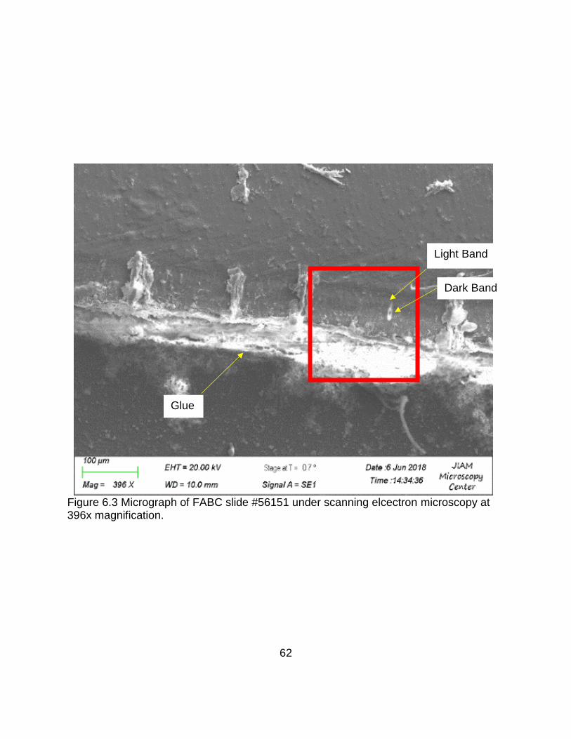
Figure 6.3 Micrograph of FABC slide #56151 under scanning elcectron microscopy at 396x magnification. ................................................................................................ 62
Figure 6.4 Micrograph of FABC slide #56151 under regular light microscopy at 200x magnification, and digitally enhanced with Adobe Photoshop CC Embossing Tool. ............................................................................................................................... 63
Figure 6.5 Micrograph of lacunae on FABC slide #53741 under regular light microscopy and a fluorescent filter, at 200x magnification. ....................................................... 64
Figure 6.6 Micrograph of lacunae on FABC slide #53741 under scanning elcectron microscopy at 382x magnification. ......................................................................... 65
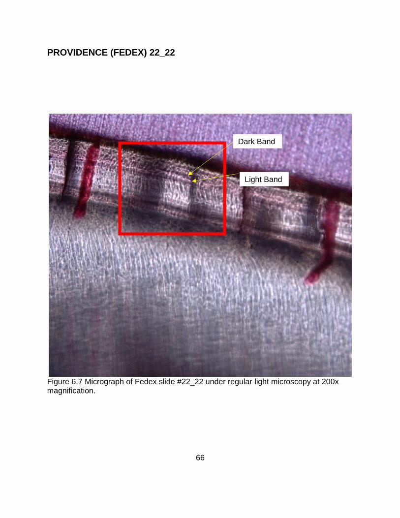
Figure 6.7 Micrograph of Fedex slide #22_22 under regular light microscopy at 200x magnification. ......................................................................................................... 66
Figure 6.8 Micrograph of Fedex slide #22_22 under regular light microscopy and a fluorescent filter, at 200x magnification. ................................................................. 67
Figure 6.9 Micrograph of Fedex slide #22_22 under scanning elcectron microscopy at 115x magnification. ................................................................................................ 68
Figure 6.10 Micrograph of Fedex slide #22_22 under regular light microscopy at 200x magnification, and digitally enhanced with Adobe Photoshop CC Embossing Tool. ............................................................................................................................... 69
Figure 6.11 Micrograph of Fedex slide #22_22B under regular light microscopy at 200x magnification. ......................................................................................................... 70
Figure 6.12 Micrograph of Fedex slide #22_22B under regular light microscopy and a fluorescent filter, at 200x magnification. ................................................................. 71
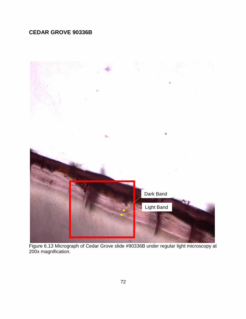
Figure 6.13 Micrograph of Cedar Grove slide #90336B under regular light microscopy at 200x magnification. ................................................................................................ 72
Figure 6.14 Micrograph of Cedar Grove slide #90336B under regular light microscopy and a fluorescent filter, at 200x magnification. ....................................................... 73
Figure 6.15 Micrograph of Cedar Grove slide #90336B under scanning elcectron microscopy at122x magnification. .......................................................................... 74
Figure 6.16 Micrograph of Cedar Grove slide #90336B under regular light microscopy at 200x magnification, and digitally enhanced with Adobe Photoshop CC Embossing Tool. ....................................................................................................................... 75
Figure 6.17 Micrograph of Cedar Grove slide #90336B Part 2 under regular light microscopy at 200x magnification. ......................................................................... 76
Figure 6.18 Micrograph of Cedar Grove slide #90336B Part 2 under regular light microscopy and a fluorescent filter, at 200x magnification. .................................... 77
Figure 6.19 Micrograph of Cedar Grove slide #90336B Part 2 under scanning elcectron microscopy at 209x magnification .......................................................................... 78

ix
Figure 6.20 Micrograph of Cedar Grove slide #90336B Part 2 under regular light microscopy at 200x magnification, and digitally enhanced with Adobe Photoshop CC Embossing Tool. .............................................................................................. 79
Figure 6.21 Micrograph of Cedar Grove slide #90852 under regular light microscopy at 200x magnification. ................................................................................................ 80
Figure 6.22 Micrograph of Cedar Grove slide #90852 under regular light microscopy and a fluorescent filter, at 200x magnification. ....................................................... 81
Figure 6.23 Micrograph of Cedar Grove slide #90852 under scanning elcectron microscopy at 144x magnification. ......................................................................... 82
Figure 6.24 Micrograph of Cedar Grove slide #90852 under regular light microscopy at 200x magnification, and digitally enhanced with Adobe Photoshop CC Embossing Tool. ....................................................................................................................... 83
Figure 6.25 Micrograph of Cedar Grove slide #90852 Part 2 under regular light microscopy at 200x magnification. ......................................................................... 84
Figure 6.26 Micrograph of Cedar Grove slide #90852 Part 2 under regular light microscopy and a fluorescent filter. ........................................................................ 85
Figure 6.27 Micrograph of Cedar Grove slide #90852 Part 2 under scanning elcectron microscopy at 144x magnification. ......................................................................... 86
Figure 6.28 Micrograph of Cedar Grove slide #90852 Part 2 under regular light microscopy at 200x magnification, and digitally enhanced with Adobe Photoshop CC Embossing Tool. .............................................................................................. 87
Figure 6.29 Micrograph of MB 101 under regular light microscopy at 200x magnification. ............................................................................................................................... 88
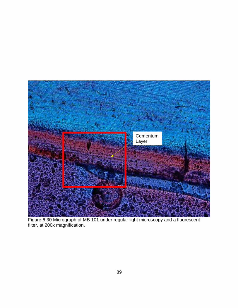
Figure 6.30 Micrograph of MB 101 under regular light microscopy and a fluorescent filter, at 200x magnification. ................................................................................... 89
Figure 6.31 Micrograph of MB 101 under scanning elcectron microscopy at 396x magnification. ......................................................................................................... 90
Figure 6.32 Micrograph of MB101 under regular light microscopy at 200x magnification, and digitally enhanced with Adobe Photoshop CC Embossing Tool. ..................... 91
Figure 6.33 Micrograph of the transverse cut thin section under regular light microscopy at 200x magnification. ............................................................................................ 92
Figure 6.34 Micrograph of the transverse cut thin section under regular light microscopy at 200x magnification and digitally enhanced with Adobe Photoshop CC Embossing Tool. ....................................................................................................................... 92
Figure 6.35 Micrograph of thin section using the Von Kossa method under fluorecent light microscopy at 200x magnification. .................................................................. 93
Figure 6.36 Micrograph of thin section using the Von Kossa method under fluorecent light microscopy at 200x magnification and digitally enhanced with Adobe Photoshop CC Embossing Tool. ............................................................................ 93
Figure 6.37 Micrograph of thin section using the Von Kossa method under fluorecent light microscopy at 200x magnification. .................................................................. 94
Figure 6.38 Micrograph of thin section using the Von Kossa method under fluorecent light microscopy at 200x magnification and digitally enhanced with Adobe Photoshop CC Embossing Tool. ............................................................................ 94

1
CHAPTER ONE THE PROGRESSION OF DENTISTRY AND DENTAL
ANATOMY: CEMENTUM
Introduction
Even though teeth have been with humankind since the beginning of time serving
primarily as instruments of mastication and tools of survival, their exact functionality has
yet to be fully understood. This is made evident as their history is documented
extensively over the centuries; from toothaches being considered a curse from God, to
their healing being conducted by the performance of rituals and offering off as
sacrifices. It is not until 7000 BC that there is a recorded practice of dental procedures
that involved the use of tools, such as bows operating as drills. This record of dentistry
is further enhanced during the Egyptian Dynasty with the Ebers papyrus. It is one of the
most well preserved and extensive texts of the time that notes various ailments of the
oral cavity and their remedies (Hussain and Khan 2014).
The History of Dentistry and Its Progression
Throughout antiquity, advancements were made that established, by today’s
standards, what is considered the basics of dentistry. But it is not until the seventeenth
century that the practice of dentistry is evaluated on an anatomical level, and separated
from medicine into its own distinguished, but intertwined discipline. It is during this era
that anatomist, Adrian Spiegel, recognized the teeth’s fixation within the alveolar bone.
Furthermore, it is also during this time that, Diemberbroek, a Dutchman, noted the

2
replacement of deciduous teeth after an extraction and theorized that permanent teeth
are developed from the roots of the deciduous dentition remaining in the alveoli (Guerini
1909).
It is not until 1678, with the development of the first powerful microscope, does
Antoni Van Leeuwenhoek notate the structure of dentine. Shortly afterwards, Jean
Duverney, a French anatomist, examines fetal jaws, and at the diminishing of the root
cavity with age along with other age-related anomalies (Guerini 1909).
The identification of cementum does not come until the nineteenth century.
Further explained in Yamamoto and Hasegawa (2016),” …cementum was first
demonstrated microscopically by Fraenkel and Raschkow (1835) and Retzius (1836),
and has since become a part of general knowledge in dentistry.”
After this initial focus of dental anatomy and histology, there is a one-hundred-
and-fifty-year gap before the development of a microscope capable of viewing
cementum and its examination. In recognizing the extended periods of scientific
breakthroughs; again, it is not until the passing of another century that functionality and
form is brought to the understanding of cementum. As cementum is studied, it’s
appositional growth is utilized in association with wildlife management.
Throughout antiquity and in animal husbandry, age has been estimated by the
evaluation of teeth. “Knowledge of the ages of individuals is essential to understanding
the rates of growth, onset of sexual maturity, fertility peak, senescent decline and life
span, as well as social behaviours” (Spinage 1973:165-187). As mammals have been
tested in different environments and their cementum analyzed, it was concluded that

3
instances such as,” cold stress is a calcium-consuming process, the lack of available
calcium in newly forming cementum could be responsible for the observed
hypomineralization. The appositional growth characteristics of dental cementum serve
as a record for such life-history events” (Cipriano 2002:21-31). Forensic anthropologists
began using it as a means of identification in the early 1980’s. During growth, sequential
changes assist in the estimation of age, but this metric is only limited to the growth of
subadults. As age increases, so does degenerative properties that increase the difficulty
in using this methodology (Roksandic, Vlak et al. 2009).
By taking these commonalities into consideration, the question of what causes
cementum hyper- and hypo-mineralization, or light and dark banding, in humans begs
attention. In Cipriano 2002, that cementum of captive apes were analyzed. This analysis
displayed hyper-mineralization based on climate and diet. This raises the question to its
reflection within humans.
To analyze this occurrence, historic African American populations were analyzed
to assess the value of cementum for age estimation and the accuracy of total cementum
annulation (TCA), within historic and deprived populations. Additionally, cementum will
be critiqued based on its depiction in microscopy. As mammals display cementum
variation based on dietary restrictions during seasonality; this study analyzes members
of historic African American populations. Over the span of a century which included
social restrictions and inhumane practices such as: slavery (1619-1865), pre-
reconstruction (1865-1867), reconstruction (1867-1877), and the era of Jim Crow Law
1877-1950) in the United States.

4
By analyzing cementum development over this time period, we may become
more versed on the characteristics of cementum and its functionality outside of
anchoring the tooth. Second, a methodology will be developed for best practice
procedures to grind, polish and/or etch historic dental remains. Finally, this research
will provide a methodological roadmap on how to analyze cementum in historic and/or
modern populations that are socially restricted and/or malnourished.
Cementum
Cementum, from its Latin origin, means quarry stone and was first described by
students of Purkinje in 1835. It is a mineralized, avascular tissue that encases the root
of the tooth. Its borders create the cemento-enamel junction (CEJ) and the cemento-
dentinal junction (CDJ) terminating at the root apex. To understand the importance of
cementum, one needs to understand it function. Diagrams of cementum can be found at
the end of this chapter in figure 1.1 showing the basic single rooted tooth anatomy, and
figure 1.2 showing the basic double rooted tooth anatomy.
The anchoring mechanism of teeth to the jaws is called the periodontium. The
word “periodontium” is of Greek origin, meaning around tooth. Specifically, the
periodontium, “anchors the teeth to the bone of the jaws, provide interdental linkage to a
row of teeth, and seals the oral mucosal openings created by the erupting teeth. Thus,
the periodontium comprises four different tissues, e.g., root cementum, alveolar bone
proper, periodontal ligament and the gingiva” Schroeder (1986:12). Thus, cementum
functions as a glue anchoring the periodontal ligament to the tooth’s surface.
5
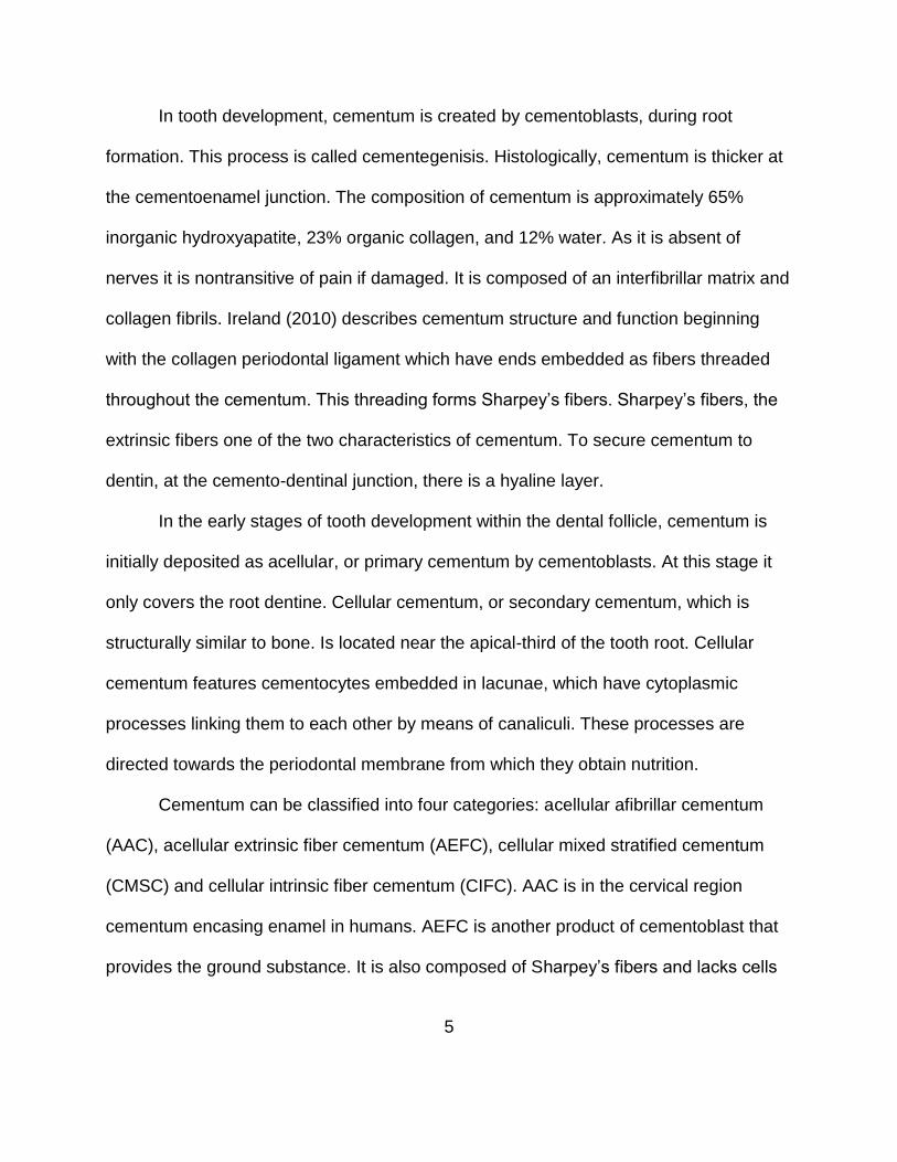
In tooth development, cementum is created by cementoblasts, during root
formation. This process is called cementegenisis. Histologically, cementum is thicker at
the cementoenamel junction. The composition of cementum is approximately 65%
inorganic hydroxyapatite, 23% organic collagen, and 12% water. As it is absent of
nerves it is nontransitive of pain if damaged. It is composed of an interfibrillar matrix and
collagen fibrils. Ireland (2010) describes cementum structure and function beginning
with the collagen periodontal ligament which have ends embedded as fibers threaded
throughout the cementum. This threading forms Sharpey’s fibers. Sharpey’s fibers, the
extrinsic fibers one of the two characteristics of cementum. To secure cementum to
dentin, at the cemento-dentinal junction, there is a hyaline layer.
In the early stages of tooth development within the dental follicle, cementum is
initially deposited as acellular, or primary cementum by cementoblasts. At this stage it
only covers the root dentine. Cellular cementum, or secondary cementum, which is
structurally similar to bone. Is located near the apical-third of the tooth root. Cellular
cementum features cementocytes embedded in lacunae, which have cytoplasmic
processes linking them to each other by means of canaliculi. These processes are
directed towards the periodontal membrane from which they obtain nutrition.
Cementum can be classified into four categories: acellular afibrillar cementum
(AAC), acellular extrinsic fiber cementum (AEFC), cellular mixed stratified cementum
(CMSC) and cellular intrinsic fiber cementum (CIFC). AAC is in the cervical region
cementum encasing enamel in humans. AEFC is another product of cementoblast that
provides the ground substance. It is also composed of Sharpey’s fibers and lacks cells

6
and may come in contact with AAC. CMSC is another product of cementoblasts and is
composed of varied intrinsic and extrinsic fibers. It is most commonly found within the
apical one-third of the tooth-root. CIFC is another product of cementoblast, containing
cells, but not collagenous fibers. It is mainly found filling in the areas of resorbed
lacunae (Schroeder 1986).
After cementegenisis has deposited cementum on the root dentin, it continues
appositionally, throughout life. Unlike the other dental tissues, cementum is dependent
upon the tooth root’s functionally and requirements during eruption and orthodontic
restructuring as a result of movement repositioning of the tooth. Cementum is only
reabsorbed when there is extreme stress and pressure on the root.
Research has linked the appositional deposition of cementum to growth analysis.
The incremental light and dark banding lines created throughout a calendar year, have
provided valuable information into the seasonality of wildlife (Weinand 1997). This
information was used to detect the seasonality of death within wildlife, as well as serve
as an indicator of population longevity. This method has been attempted for age
estimation in humans Charles, Condon et al. (1986)

7
Figure 1.1 Basic Single Rooted Tooth Anatomy. Photo taken by the Human Anatomy Web Site at San Diego Mesa College

8
Figure 1.2 Basic Double Rooted Tooth Anatomy (Leigh 1998).

9
CEMENTUM ANNULATIONS IN WILDLIFE
Throughout the years, archeologists have used the appositional deposition of
cementum, of known human prey, to analyze human behavior. As the deposition of
occurs, it gradually creates an alternating ring-like anatomical structure, or annulations.
By observing this patterning, archeologists can estimate age, in addition to infer
environmental and metabolic changes.
Weinand (1997) studied the incremental cemental annuli of white-tailed deer in
comparison to mammalian wildlife. This incremental growth banding is found in most
vertebrates and invertebrate fauna. Archaeologists are more concerned with its ability to
discern season at death to interpret human ‘hunting’ behavior and understanding death
rates of certain species. This infers cultural understanding of the annual and life-long
foraging habits of the animal kingdom.
Lieberman and coworkers initiated the ground work on cementum in 1969. Their
research indicates that banding is a result of photo-periodicity, climate, and hormonal
cycles. Lieberman (1995), suggests that the light and dark annulations are a result of
variations in relative mineralization and colloagen orientation due to nutritional and
biochemical stress associated with the differences in seasonal food quality. This was
proven by changing the texture of goats’ food and observing the cementum annuli with
scanning electron microscopy. Through this research, he determined that coarser foods
influenced mastication which altered the orientation of the extrinsic collagen Sharpey’s
fibers. This additional biomechanical stress caused the fibers to orient more on the y-
plane, relative to the x-axis of the tooth. In addition to scanning electron microscopy,

10
Lieberman also used microradiographic analysis to detect changes in the mineral
density. This research improves our understanding of how function affects form.

11
AGE ESTIMATION USING TOOTH CEMENTUM ANNULATIONS
In forensic science age estimation is a crucial tool used in the identification of
decomposed human remains, skeletonized or comingled. Once a body becomes
decomposed, visual identification becomes impossible. As a result, forensic scientists
turn to skeletal and dental markers to discover and create methods to help estimation of
age, sex, ancestry and stature. Age estimation is arguably the most challenging (Senn
and Weems 2013).
Reliability and accuracy in age estimation is of high importance, as its use
sometimes has legal implications such as: narrowing the search possibilities of
unknown victims, estimating the age at death, differentiating cluster victims, aiding
immigration services in the processing of undocumented immigrants, awarding of social
benefits, and establishing the legal age of majority amongst other things (Lewis and
Senn 2010).
Established methodology in age estimation is supported by scientific rationale.
There are numerous methods of conducting biological age estimation from skeletal and
dental remains. From bone, these include analysis of the pubic symphysis, sacro-iliac
surface, sternal ends of the ribs, medial clavicle, cranial sutures and hand-wrist bone
development. Furthermore, there are numerous gross and radiographic methods using
teeth. Regardless of the tissue analyzed, as age increases, the accuracy of the
estimation decreases.

12
Of these different markers, pubic symphysis is regarded as the most widely used,
reliable, anthropologic determinant of age estimation in medicolegal cases. “Current
pubic symphyseal aging methods combine morphological characteristics associated
with developmental changes during the period immediately following adolescence with
degenerative changes spanning the latter portion of the age spectrum” (Dudzik and
Langley 2015). It utilizes a scoring system, and therefore, its accuracy is dependent
upon subjectivity of the forensic anthropologist conducting the analysis. As age
increases, the accuracy of age estimation decreases creating an increase in the
potential age interval. Charles and coworkers (1986) describe the best level of accuracy
at approximately plus or minus 5 years, featuring less precision in certain aspects.
The utilization of the teeth are frequently and more prominently used as a means
to estimate age in recent years. As dentistry has evolved, different scientifically derived
methods of age estimation have come into fruition such as: development and eruption
charts, aspartic acid racemization radiocarbon, root length and translucency, and most
noted in this research – root cementum apposition (Lewis and Senn 2010). Figures 3.1
through 3.4 shows the detailed documentation through the Atlas of Human Tooth
Development and Eruption, Descripton of Moorrees’ Stages, Dental Age Assesment
Procedures, and the Adult Dental Age Assessment Technique Chart respectively.

13
Figure 3.1 Atlas of Human Tooth Development and Eruption (Qahtani 2012).

14
Figure 3.2 Descripton of Moorrees’ Stages (Qahtani 2012).

15
Figure 3.3 Dental Age Assessment Procedures (Odontology 2016).

16
Figure 3.4 Adult Dental Age Assessment Technique Chart (Odontology 2018).

17
Root cementum apposition, tooth cementum annulation, or cementum annuli has been
increasingly used by forensic anthropologist for its low error rate. Measurement of
cementum deposition has been used to estimate age. It has been observed
histologically that a years’ time is equal to two deposition layers, indicated by
contrasting light and dark banding. Charles et. al., (1986) reviewed the history of
cementum annulations used on humans in forensic science. The use of cementum
annuli for age estimation can be properly introduced as followed:
Recently, Stott and coworkers (Stott et al., 1982; Naylor et al., 1985) described the application of a method of human age-at-death estimation based on counts of incremental cementum layers or annulations. These layers are assumed to be added yearly. Layering in cementum andor dentin has been identified in virtually every group of mammals, and their use in age determination is routine among wildlife biologists (Grue and Jensen, 1979; Klevezal’ and Kleinenberg, 1967; Morris, 1978; Spinage, 1973; Stallibrass, 1982).
They (Stott and coworkers) described the use of human age-at-death estimation
based on counts of incremental cementum layers or annulations. Additionally, they note
the annual deposition of each layer is seen by cutting thin, ground sections of the
apical-third part of the tooth root.
Because these annulations are a biologically created development pattern, the
use of cementum annulations has become a more accurate estimator of age. Charles
and coworkers describe Stotts’ error values as ranging from 0.5 to 2.0 years once the
average eruption time was added.
However, the common use of cementum annulation is still tentative. Because of
the biological structure and its physiological background as a thin sheath is hard to

18
elucidate, well and in a manner, that is able to be repetitively reproduced (Wittwer‐
Backofen, Gampe et al. 2004).

19
ENSLAVED AFRICAN-AMERICAN BIO-HISTORY
America was first colonized in 1607 by the establishment of Jamestown, Virginia.
In less than two-hundred years civil discourse led to independence. It was not until
1776-1783 that The United States was established as a sovereign nation. In less than
fifty years, civil discourse takes its toll again as represented by the Trans-Atlantic Slave
Trade, emancipation, pre- and post-reconstruction, and the era of Jim Crow. Because of
the economic and social pressures during this era; this chapter will focus on the how
these experiences impacted African Americans.
The effects of the slavery are still present in today’s society, more than four-
hundred years after its fruition and approximately one-hundred fifty years after its
abolishment. Little is known of the diet, health, nutrition, disease, and metabolic demand
that slavery and its social/ethical constructs had on African Americans (Brooks 2005).
The objective of this bio-history chapter is to analyze and review the research done on
the remains of each respective population. This analysis will provide background
information on this population of individuals.
Cedar Grove Baptist Church – Texarkana (AR)
In the 1980s, Cedar Grove Baptist Church cemetery was discovered on the south
bank of the Red River near Texarkana, Arkansas (see figure 4.1). This discovery
exposed 104 unmarked and 9 marked graves. These were exhumed and reburied
nearby. These graves were excavated, removed, analyzed and relocated. The Red
River had previously destroyed numerous graves (see figure 4.2-4.4).

20
Members of the Cedar Grove Baptist Church included slaves and several
generations of their descendants, whose lives conceivably spanned pre-Emancipation
(prior to 1865), Reconstruction (1865-1880), and post-Reconstruction (1880-1927). This
chronology was established through artifacts and clothing in the grave, coffin hardware
and church records detailing the site and burial practices.
The area surrounding Cedar Grove Baptist Church is best described as a
dilapidated community of nutritionally-deprived farmers and sharecroppers Analysis of
this population revealed: a high subadult mortality rate, females outnumbering males 21
to 15, a mean age of 38 years, and active/or healed pathological skeletal lesions
indictive of infections (see figure 4.4).
Periostitis was also found within the subadult population. Periostitis indicates
uterine and congenitally-acquired infections, which are pathognomonic for treponemas
associated with maternal syphilis. Other credible causes for the high mortality rate
includes an area filled with tetanus, diarrhea, worms, fever, and most notably, malaria.
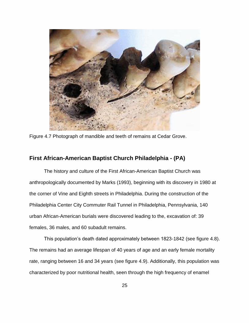
A visual representation of dental remains can be found in figures 4.6 and 4.7.

21
Figure 4.1 Map displaying the area of Cedar Grove and Texarkana, AR.

22
Figure 4.2 Historic burials in the direct impact zone.
Red River
Erosion location and direction

23
Figure 4.3 25-foot drop-off created by erosion that destroyed 9 sets of remains.
Figure 4.4 Illustration of sharecroppers working.

24
Figure 4.5 Photo of remains estimated at +/- 10 years of age at Cedar Grove.
Figure 4.6 Condition of oral dentition found in human remains at Cedar Grove.
25
Figure 4.7 Photograph of mandible and teeth of remains at Cedar Grove.
First African-American Baptist Church Philadelphia - (PA)
The history and culture of the First African-American Baptist Church was
anthropologically documented by Marks (1993), beginning with its discovery in 1980 at
the corner of Vine and Eighth streets in Philadelphia. During the construction of the
Philadelphia Center City Commuter Rail Tunnel in Philadelphia, Pennsylvania, 140
urban African-American burials were discovered leading to the, excavation of: 39
females, 36 males, and 60 subadult remains.
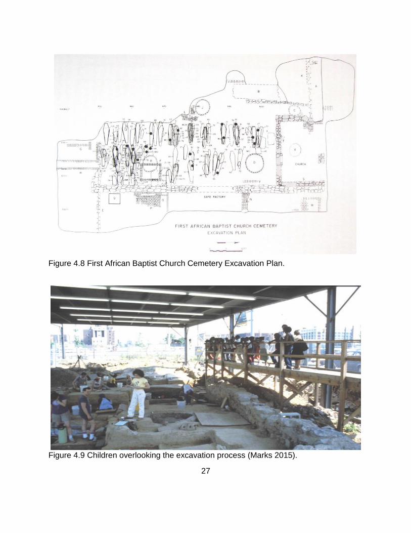
This population’s death dated approximately between 1823-1842 (see figure 4.8).
The remains had an average lifespan of 40 years of age and an early female mortality
rate, ranging between 16 and 34 years (see figure 4.9). Additionally, this population was
characterized by poor nutritional health, seen through the high frequency of enamel

26
hypoplasia and dental caries. Caries was more frequent in males than in females as
was a greater childhood morbidity rate. Additionally, mineral deficient diets were noted
through the findings of pathological lesions such as porotic hyperostosis and cribra
orbitalia.
These statistics reflect the state of life during the second quarter of the
nineteenth century (see figure 4.10). This period was plagued with racially motivated
riots, kidnappings, murders and gang violence. However, skeletal analysis of the
remains did not feature a high frequency of skeletal trauma.
During this challenging period for African-Americans, the church served as a
cushion to daily life providing a financial aid and suppressing poverty (see figure 4.11).
These benefits were most important as African-Americans were often denied the ability
to develop wealth as a result of segregation.
27
Figure 4.8 First African Baptist Church Cemetery Excavation Plan.
Figure 4.9 Children overlooking the excavation process (Marks 2015).

28
Figure 4.10 Photo of remains found with artifact.

29
Figure 4.11 1838 Lithograph of Reverend Henry Simmons, pastor of the First African Baptist Church until 1848.
30
Providence Baptist Church - Memphis (TN)
The history of Providence Baptist Church is greatly provided by Rebecca Wilson,
who also serves as a primary source to the excavation and analysis of the burial site.
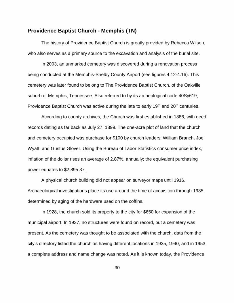
In 2003, an unmarked cemetery was discovered during a renovation process
being conducted at the Memphis-Shelby County Airport (see figures 4.12-4.16). This
cemetery was later found to belong to The Providence Baptist Church, of the Oakville
suburb of Memphis, Tennessee. Also referred to by its archeological code 40Sy619,
Providence Baptist Church was active during the late to early 19th and 20th centuries.
According to county archives, the Church was first established in 1886, with deed
records dating as far back as July 27, 1899. The one-acre plot of land that the church
and cemetery occupied was purchase for $100 by church leaders: William Branch, Joe
Wyatt, and Gustus Glover. Using the Bureau of Labor Statistics consumer price index,
inflation of the dollar rises an average of 2.87%, annually; the equivalent purchasing
power equates to $2,895.37.
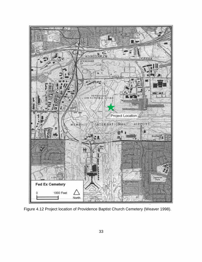
A physical church building did not appear on surveyor maps until 1916.
Archaeological investigations place its use around the time of acquisition through 1935
determined by aging of the hardware used on the coffins.
In 1928, the church sold its property to the city for $650 for expansion of the
municipal airport. In 1937, no structures were found on record, but a cemetery was
present. As the cemetery was thought to be associated with the church, data from the
city’s directory listed the church as having different locations in 1935, 1940, and in 1953
a complete address and name change was noted. As it is known today, the Providence

31
Missionary Baptist Church does not claim lineage to the original Providence Baptist
Church and its cemetery.
During the airport renovation, approximately 20% of the cemetery was impacted,
resulting in the exhumation of 65 burial sites containing 62 individuals. Of these
remains, personal artifacts were recovered included: pieces of newspapers, personal
belongs and tombstones identifying persons and their age(see figures 4.17 through
4.19). Individuals were later matched with obituaries found in dated newspapers.
The condition of the remains varied from poor to excellent (see figure 4.20). The
best preserved remains were those that had been buried underneath some of the
original runway asphalt. As a result of coffin material and drainage issues, remains
outside of the original runway path had the most damage.
Seven years before the establishment of the church, Yellow Fever plagued the
city of Memphis from 1878 and 1879. During this time 25,000 citizens fled the city,
5,000 relocated into camps in the surrounding areas. There were more than 5,000
deaths recorded and the city lost its charter in 1879. The city was not re-incorporated
until 1893.
Wilson details the occupations of African American as the following:

32
Vital statistics for the Memphis area indicate that most of the Black population not involved in agricultural activities were employed in menial and unskilled tasks. By 1920, agriculture was still the predominant employment opportunity for Tennessee Black males with 360,000 individuals; domestics came in second with 180,910; and mining, manufacturing, trade, and transportation completed the top occupations (Coomer 1920). Black female occupations were domestics at 56,000, followed by agriculture at 36,000, and finished out by manufacturing and clerical jobs (Coomer 1920). From 1880 and 1920 there was a shift from agriculture to manufacturing. In 1880, 49.4 percent of the Black population of the Unites States was employed in agriculture, while only 25.6 percent 16 were employed in manufacturing (Coomer 1920). By 1920, only 27.2 percent of Blacks were involved in agriculture with 33 percent employed in manufacturing (Coomer 1920).
33
Figure 4.12 Project location of Providence Baptist Church Cemetery (Weaver 1998).

34
Figure 4.13 Land survey of cemetery site (Weaver 1998).
35
Figure 4.14 Aerial view of airport runway in 1940 (Womack 2003).

36
Figure 4.15 Aerial view of runway in 1938 (Womack 2003).

37
Figure 4.16 Map of proposed runway construction at the Memphis Municipal Airport
(Wilson 2005).

38
Figure 4.17 East view of probable grave sites (Womack 2003).

39
Figure 4.18 Southern view of probable grave sites (Womack 2003).

40
Figure 4.19 Field photograph of Frankie LeFlore's tombstone (Weaver 1998).

41
Figure 4.20 Burial 29, a 10-12 year old sub-adult, exhibiting a lateral curvature of the vertebral column (Wilson 2005). .

42
MATERIALS AND METHODS
Materials
The materials used to conduct this research are divided into two parts: analysis
of African American populations and establishing a methodology to evaluate cementum
(see table 5.1). The primary are dental sources are historic African American, modern
African American, and modern White mandibular canines including five male and five
female thin sections selected from Cedar Grove Baptist Church Cemetery (Texarkana,
AR) and the First African Baptist Church Cemetery (Philadelphia, PA). Just five male
thin sections were selected from Providence Baptist Church Cemetery, Memphis (TN).
All slides were originally collected, produced, and furnished by Marks as a sample from
his doctoral thesis “Dental Enamel Microdefects as Indicators of Childhood Morbidity
Among Historic African Americans,” at the University of Tennessee (1993).
The modern tooth samples were acquired through the Department of
General Dentistry, at the University of Tennessee Medical Center Knoxville (see table
5.2). These teeth were removed as a result of routine dental procedures. To establish a
methodology, these samples were used to establish an adequate thickness and staining
technique for best observation (see figure 5.1).

43
Table 5.1 Population Information.
CEMETARY BURIAL
NUMBER SLIDE
NUMBER ID
NUMBER SEX AGE
TOOTH NUMBER
FABC
13800 138 5329 F 33 22
09900 99 8890 F 44 22
00000 100 1137 F 21 22
11400 114 7320 F 40 22
12300 123 2599 F 15 22
10100 101 5380 M 46 22
03200 32 2695 M 51 27
03300 33 6919 M 31 22
07700 77 6859 M 39 22
11500 115 5615 M 58 27
CEDAR GROVE
00103 91650 F103 F 25 22
00081 90657 F81 F 22 22
00065 94362 F65 F 27 27
00014 93376 F14 F 32 22
00021 94742 F21 F 55 22
00061 96743 M61 M 44.5 22

44
Table 5.1 Continued
CEMETARY BURIAL NUMBER
SLIDE NUMBER
ID NUMBER
SEX AGE TOOTH
NUMBER
CEDAR GROVE
00055 90366 M55 M 17 22
00010 91009 M10 M 27 22
00024 98052 M24 M 37 22
00096 91403 M96 M 47 22
PROVIDENCE
00008 1-2 1 M 50 22
00016 3-4 2 M 40 27
00012 8-9 4 M 40 22
00028 10-11 5 M 35 22
00019 12 6 M 60 27

45
Thin sections of the teeth were fabricated utilizing coper wire, 22 x 30 mm Peel-
A-Way disposable molds by Polysciences Inc. (Warrington, PA), Buehler’s EpoThin™ 2
Epoxy Resin(20-3440-128) and EpoThin™ 2 hardener(20-3432-064), a diamond blade
Buehler Isomet Slow Speed Saw, gel super glue, microscopy slides, and a grinder with
600 C and 1500 Micro Fine 3M paper were used. The slides were labeled using a
diamond tip scribe (see figures 5.2 through 5.4).
Slides were stained with the Hematoxylin and Eosin Stain Kit by TissuePro
Technology. The staining process also included 70% isopropyl alcohol, tap and distilled
water, and one normal solution of hydrochloric acid.
Microscopic evaluation was performed using a Leica DMRX light microscope,
Sony DXC-S500 IEEE1394 Color Digital Camera, Image-Pro Express software, Zeiss
Auriga Crossbeam FIB/scanning electron microscope, and a SPI SPUTTER/Carbon
Coater at the Joint Institute for Advanced Material.
For part two, four single rooted teeth were used, ultimately resulting in a total of 6
slides. The materials used to embed, create thin sections, mount ground, and view
slides were duplicated from part one. Additional materials used in part two included
Xylol Xylene by Klean Strip and a micrometer.

46
Table 5.2 Cementum Methodology Study Slide Demographics.
Figure 5.1 Photograph of embedded teeth used to take transverse thin sections, by Kortney Dewayne Powell.
SLIDE NUMBER INITIAL SLIDE THICKNESS IN
INCHES
INITIAL SLIDE THICKNESS IN
MICRONS
1 .00860 218.44
2 .01450 368.3
3 .01615 410.21
4 .00140 35.56
5 .01360 345.44
6 .07775 1974.85

47
Methods
Sample Collection
The collection of thin sections was first organized, examined, and documented.
From this initial evaluation, sample sizes were established. For each population, 5
female and 5 male thin sections were selected when available. All thin sections
evaluated were permanent maxillary canines of individuals over an evenly distributed
age range. They were then analyzed for quality and noted for repair. To repair the
slides, super glue was added to the epoxy resin thin sections and remounted onto their
slides. Afterwards, the slides were grounded.
For the modern reference samples, three White teeth and one African American
tooth were embedded in an epoxy resin solution which stabilized the tooth for
microscopic examination. This procedure is a modification of the Marks, Rose and
Davenport method (Marks, Rose et al. 1996).
The Embedding Process
First, teeth were wrapped around the cementoenamel junction, in copper wire,
forming a butterfly pattern to serve as a stand. The copper wire and suspended tooth
were the placed in 22 x 30 mm Peel-A-Way disposable molds, by Polysciences Inc.
(Warrington, PA), and secured using superglue. Molds were then filled with the
embedding medium and left at room temperature for twenty-four hours to cure.

48
Figure 5.2 A tooth suspended within a mold using copper transformer wire (Marks, Rose et al. 1996).

49
Figure 5.3 Photograph of teeth being prepared of embedding, wrapped in copper wire and placed in molding trays.
Figure 5.4 Photograph of teeth in molding trays, filled epoxy.

50
Creating Thin Sections
Second, the cured blocks were removed from their molds for in preparation for
thin sectioning. The sectioning involved the use of a diamond blade Buehler Isomet
Slow Speed Saw. Cured blocks were mounted, and the saw positioned to create 15 mm
thick sections, off centering the tooth longitudinally and labiolingually for the first cut.
Calibrated on a scale of 1-10, the saw speed was held at 7.5 with a pressured weight of
70 g. After the first cut, the cured block is cleaned with 70% isopropyl alcohol to remove
the oil that cools the saw blade. Then a slide is attached to the block with superglue, the
saw is positioned back to the center of the tooth, for a second cut. This step is then
repeated to obtain a second copy. Completed slides were then etched with a diamond
engraving pencil for identification of the thin section (see figure 5.5).
The Grinding Process
Third, thin sections went through a grinding process to smooth edges created by
the saw. This was performed with a grinder using 3M 600 and 1500 microfine grinding
paper and water. To grind, an appropriately-sized slide holder must be used. Once the
turntables are spinning, the sandpaper is moistened, and the slide is held gently over
the sandpaper; Maneuvering the slide in a zig-zag motion.

51
Figure 5.5 Photograph of longitudinal thin section being made.
Hematoxylin and Eosin Staining
Fourth, the slides were stained with a hematoxylin and eosin staining kit. This
process includes rehydration in 70% isopropyl alcohol to be for three minutes, then in
distilled water for one minute. They were next stained with the hematoxylin for ten-
minutes and rinsed with tap water. The slides were then etched in hydrochloric acid for
twenty-five seconds and rinsed again in tap water. The slides were next stained with
eosin for two minutes, dehydrated in isopropyl alcohol for three minutes, and allowed to
dry.

52
Light Microscopy
After staining, slides where examined using the Leica DMRX light microscope,
Leica MZ6 dissecting-scope, Leica KL1500 LCD light source, Sony DXC-S500
IEEE1394 Color Digital Camera, and Image-Pro Express software. Viewing on the light
microscope was done using a magnification of 50x-400x, but all results were
documented at 200x. Viewing on the dissecting scope was done using 10x.
Scanning Electron Microscopy
In preparation for SEM, copper tape was added to slides so it is not isolated
when electrons are aimed at the stage. Afterwards, they were taken to the sputter
coater, radiated in argon for twenty seconds, to form a continuous conductive film (see
figure 5.6). Slides were then loaded into the Zeiss Auriga Crossbeam FIB/scanning
electron microscope’s stage chamber, pumped with nitrogen, and then beamed for
evaluation (see figure 5.7).
Creating A Method to Best Analyze Cementum
To establish a best practice in observing cementum, there was multiple
deviations from the methods above. The first four teeth were sectioned within the apical-
third of the root, transversely, opposing the traditional longitudinal cut (see figures 5.8-
5.9). After slides were cut and mounted, they were ground to 5 varying thicknesses
ranging from 37-410 microns. Slides were then analyzed on the light microscope at
varying magnifications, ultimately 200x was the chosen standard of best practice.
The speed of the diamond blade saw was adjusted to level 3 for slide number 5.
After the section was cut and ground, it was etched in the hydrochloric acid for 5
seconds before being observed on the light microscope, at 20x magnification.

53
The speed of the diamond blade saw was adjusted, again, to level 10 for slide
number 6. The slide was also etched for 5 seconds in hydrochloric acid and measured
for slide thickness using the micrometer. After observation, the slide was dehydrated in
alcohol for 2min, and defilmed in xylene for 30 seconds.
The slide was then observed under fluorescent lighting, ultimately at 200x
magnification. After observation, the slide was ground down to a (.04435in) thickness.
In order to establish the proper staining technique to observe cementum, slides
were taken to the University of Tennessee’s College of Veterinary Medicine. There, thin
sections were stained using the Von Kossa method as follows: hydrated to distilled
water, rinsed in Millipore water, place in 5% Silver Nitrate solution for 60 minutes, rinsed
in Millipore water, placed in 5% Sodium Thiosulfate for 3 minutes, rinsed in Millipore
water, counterstained in Nuclear Fast Red for 3 minutes, dehydrated, and then cleared.

54
Figure 5.6 Photograph of thin section being coated in gold by sputter coater.

55
Figure 5.7 Photograph of slides being prepared and positioned within the scanning electron microscope.

56
Once the slide was ground, it was then etched for 10 seconds in the hydrochloric
acid. The slide was ultimately ground down to (.03835 in). Afterwards, the slide was
observed the H&E staining method was followed as described: cleared in xylene for 6
minutes, dehydrated in alcohol for 9 minutes, and then preceded the with the steps
mentioned above.
Figure 5.8 Photograph of embedded tooth being transversely cut.

57
Figure 5.9 Photograph of the apical-third of the root being mounted to a slide and cut.

58
RESULTS AND DISCUSSION
Banding is visibly present in the historic African-American populations as seen in
figures 6.1-6.4, 6.7-6.10, and 6.13-6.28. This banding occurs in reasonably defined
alternating increments. Based on the results, age can be estimated using these
samples. The cementum annuli are made legible by progressively enhancing the
microscopic technology with digital imaging software. This can be best seen within the
Cedar Grove samples, figures 6.13-6.28.
There was not a positive change in sample clarity from the Providence Baptist
Church Cemetery, Figures 6.11 and 6.12. As the fluorescent filter was added to the light
microscope it did not have a positive effect. These two samples lack the potential for
further study. Additionally, the modern African-American sample, figures 6.29 and 6.30
are of poor quality when viewed with the light microscope. Visibility of the annuli was
increased with the use of scanning electron microscopy and further enhanced using
Photoshop.
Figures 6.33 and 6.34, the transversely cut sample, possessed great initial
potential. As all of the other slides underwent staining, it was only treated with xylene.
Photoshop improved its quality; but did not provide the same quality of visualization as
the samples from Cedar Grove.
The poor quality of figures 6.11 and 6.11 could be caused by sample aging,
demarked by the cracks caused by shrinkage within the epoxy. Additionally, the vertical
lines made across the samples, or ‘chatter lines’ were created by the saw in the process

59
of creating the thin sections, this could also cause slide quality to decline. This can be
seen in great detail on transversely cut sample, figures 6.33 and 6.34.

60
FABC 56151
Figure 6.1 Micrograph of FABC slide #56151 under regular light microscopy at 200x magnification.
Light Band
Dark Band

61
Figure 6.2 Micrograph of FABC slide #56151 under regular light microscopy and a fluorescent filter, at 200x magnification.
Light Band
Dark Band
62
Figure 6.3 Micrograph of FABC slide #56151 under scanning elcectron microscopy at 396x magnification.
Light Band
Dark Band
Glue

63
Figure 6.4 Micrograph of FABC slide #56151 under regular light microscopy at 200x magnification, and digitally enhanced with Adobe Photoshop CC Embossing Tool.
Light Band
Dark Band

64
FABC 68591
Figure 6.5 Micrograph of lacunae on FABC slide #53741 under regular light microscopy and a fluorescent filter, at 200x magnification.

65
Figure 6.6 Micrograph of lacunae on FABC slide #53741 under scanning elcectron microscopy at 382x magnification.
66
PROVIDENCE (FEDEX) 22_22
Figure 6.7 Micrograph of Fedex slide #22_22 under regular light microscopy at 200x magnification.
Light Band
Dark Band

67
Figure 6.8 Micrograph of Fedex slide #22_22 under regular light microscopy and a fluorescent filter, at 200x magnification.
Light Band
Dark Band

68
Figure 6.9 Micrograph of Fedex slide #22_22 under scanning elcectron microscopy at 115x magnification.
Light Band
Dark Band

69
Figure 6.10 Micrograph of Fedex slide #22_22 under regular light microscopy at 200x magnification, and digitally enhanced with Adobe Photoshop CC Embossing Tool.
PROVIDENCE (FEDEX) 22_22B
Light Band
Dark Band

70
Figure 6.11 Micrograph of Fedex slide #22_22B under regular light microscopy at 200x magnification.
Cementum layer

71
Figure 6.12 Micrograph of Fedex slide #22_22B under regular light microscopy and a fluorescent filter, at 200x magnification.
Cementum layer
72
CEDAR GROVE 90336B
Figure 6.13 Micrograph of Cedar Grove slide #90336B under regular light microscopy at 200x magnification.
Light Band
Dark Band
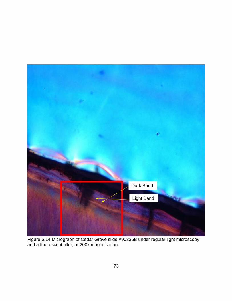
73
Figure 6.14 Micrograph of Cedar Grove slide #90336B under regular light microscopy and a fluorescent filter, at 200x magnification.
Light Band
Dark Band

74
Figure 6.15 Micrograph of Cedar Grove slide #90336B under scanning elcectron microscopy at122x magnification.
Light Band
Dark Band

75
Figure 6.16 Micrograph of Cedar Grove slide #90336B under regular light microscopy at 200x magnification, and digitally enhanced with Adobe Photoshop CC Embossing Tool.
Light Band
Dark Band

76
CEDAR GROVE 90336B PART 2
Figure 6.17 Micrograph of Cedar Grove slide #90336B Part 2 under regular light microscopy at 200x magnification.
Light Band
Dark Band

77
Figure 6.18 Micrograph of Cedar Grove slide #90336B Part 2 under regular light microscopy and a fluorescent filter, at 200x magnification.
Light Band
Dark Band

78
Figure 6.19 Micrograph of Cedar Grove slide #90336B Part 2 under scanning elcectron microscopy at 209x magnification
Light Band
Dark Band

79
Figure 6.20 Micrograph of Cedar Grove slide #90336B Part 2 under regular light microscopy at 200x magnification, and digitally enhanced with Adobe Photoshop CC Embossing Tool.
Light Band
Dark Band

80
CEDAR GROVE 90852
Figure 6.21 Micrograph of Cedar Grove slide #90852 under regular light microscopy at 200x magnification.
Light Band
Dark Band

81
Figure 6.22 Micrograph of Cedar Grove slide #90852 under regular light microscopy and a fluorescent filter, at 200x magnification.
Light Band
Dark Band

82
Figure 6.23 Micrograph of Cedar Grove slide #90852 under scanning elcectron microscopy at 144x magnification.
Light Band
Dark Band

83
Figure 6.24 Micrograph of Cedar Grove slide #90852 under regular light microscopy at 200x magnification, and digitally enhanced with Adobe Photoshop CC Embossing Tool.
Light Band
Dark Band

84
CEDAR GROVE 90852 PART 2
Figure 6.25 Micrograph of Cedar Grove slide #90852 Part 2 under regular light microscopy at 200x magnification.
Light Band
Dark Band

85
Figure 6.26 Micrograph of Cedar Grove slide #90852 Part 2 under regular light microscopy and a fluorescent filter.
Light Band
Dark Band

86
Figure 6.27 Micrograph of Cedar Grove slide #90852 Part 2 under scanning elcectron microscopy at 144x magnification.
Light Band
Dark Band

87
Figure 6.28 Micrograph of Cedar Grove slide #90852 Part 2 under regular light microscopy at 200x magnification, and digitally enhanced with Adobe Photoshop CC Embossing Tool.
Light Band
Dark Band

88
MB101
Figure 6.29 Micrograph of MB 101 under regular light microscopy at 200x magnification.
Cementum Layer
89
Figure 6.30 Micrograph of MB 101 under regular light microscopy and a fluorescent filter, at 200x magnification.
Cementum Layer

90
Figure 6.31 Micrograph of MB 101 under scanning elcectron microscopy at 396x magnification.
Cementum Layer

91
Figure 6.32 Micrograph of MB101 under regular light microscopy at 200x magnification, and digitally enhanced with Adobe Photoshop CC Embossing Tool.
Light Band
Dark Band

92
Figure 6.33 Micrograph of the transverse cut thin section under regular light microscopy at 200x magnification.
Figure 6.34 Micrograph of the transverse cut thin section under regular light microscopy at 200x magnification and digitally enhanced with Adobe Photoshop CC Embossing Tool.
Light Band
Dark Band
Light Band
Dark Band

93
Figure 6.35 Micrograph of thin section using the Von Kossa method under fluorecent light microscopy at 200x magnification.
Figure 6.36 Micrograph of thin section using the Von Kossa method under fluorecent light microscopy at 200x magnification and digitally enhanced with Adobe Photoshop CC Embossing Tool.

94
Figure 6.37 Micrograph of thin section using the Von Kossa method under fluorecent light microscopy at 200x magnification.
Figure 6.38 Micrograph of thin section using the Von Kossa method under fluorecent light microscopy at 200x magnification and digitally enhanced with Adobe Photoshop CC Embossing Tool.

95
CHAPTER SEVEN CONCLUSIONS AND RECOMMENDATIONS
This light and scanning electron microscopic research began as a singular effort
to document structure and measure changes in cementum of several historic African
American burial ground samples dating from approximately 1820-1920. As it progresses
it created a twofold question: can cementum be used to analyze the age of these
individuals and what is the best methodology in achieving this? Figures 6.1-6.38 shows
a progressive timeline to answer these questions.
In observing cementum annuli for the use of age estimation, Charles and
coworkers (1986) is a good reference to gain understanding on the topic, and Harris
and McKee (1990) is a great resource in understanding the specificity needed for proper
analysis. Their research, as a collective, prompted the initial use of hematoxylin and
eosin staining as a means to answer this question. Furthermore, it alluded to the use of
scanning electron microscopy to enhance results. The results presented in Chapter 6,
specifically figures 6.1 through 6.28, triggered the use of the Adobe Photoshop CC
Embossing Tool.
The use of the digital imaging enhancement served as a catalyst to modernize,
create a methodology in observing cementum. Figures 6.35 through 6.38 demonstrate
that the Von Koss method followed by image enhancement is serves as a proper
method to display cementum annulations.
Parting thoughts from this research is the notion of applicability of use. As this
research sought to document the structure of cementum and standardize a

96
methodology in observing cementum annuli; it achieved that goal. During this process
every sample yielded differences that make it difficult to standard a method to yield
replicable results on a consistent basis. Cementum holds the capability to be a viable
tool for age estimation, but this research suggests it is an undependable method of use.
• Cementum is one of the most accessible biological indicators of the human body
with a great amount of potential.
• Though cementum is very accessible and has great potential its findings are not
efficiently duplicable to access it.
• While it is possible to obtain higher quality images of cementum annuli its yield of
duplicability is not high enough to be an adequate means of age estimation.

97
LIST OF REFERENCES

98
Brooks, C. N. B. (2005). Patterns of traumatic injury in historic African and African American populations, Thesis (M.A.) -- University of Tennessee, Knoxville, 2005. For my master's thesis project titled, ''Patterns of Traumatic
Injury in Historic African and African American Populations,'' I examined trauma incidence in American slave and free populations. The objectives of this study were (1) to present frequency and distribution analysis of injuries in each sample, (2) to create cross tabulations to show similarities and differences in each site and compare these results to between, (3) interpret the frequency and distribution of injuries from a cultural aspect, to better understand the violence and physical demands endured by American slaves and freeborn African American. Most of the skeletal samples used in this research have been reinterred. Therefore, this research is based off the observations and interpretations of researchers and data found in published papers. Unfortunately, during the time when most of these remains were examined there existed no universal research method when analyzing skeletal material. Each researcher used his or
her own method for analyzing remains; some being more detailed then others. Because of this, this study contains basic information about each site including: site name, total number of individuals examined in each site, total number of individuals observed with fractures, total number of fractures observed in each site, sex of the individuals, which bone(s) were injured, if the injury occurred ante or peri mortem, which are slave communities and which are free populations. Whenever possible a mechanism such as accident, violent encounter or occupational-related injury, was assigned to each injury. For this study trauma was defined as dislocations, fractures, muscle pulls, blunt force trauma and puncture wounds. All bones were examined. There has been a lot of research attempting to reconstruct historic African American lifeways in anthropology. Most of this research consists of analyzing overall health of the populations studied. This study is important because there is not a lot of research specifically on trauma
analysis of slave and free populations that discuss the physical demands of slavery as well as slave mistreatment. Due in large part to small sample sizes and fragmentary conditions of slave and African American skeletal series available for study, there is no effective means to measure the biological brutality of slavery. This study is intended to evoke interest in trauma studies in historic African and African American populations. As more studies of trauma in African American populations emerge, more comparisons can be made resulting in important questions being answered about the past. Studies of trauma distribution and frequency patterning in African American populations are essential for addressing questions about human adaptation to physical, environmental, and social constraints.

99
Cipriano, A. (2002). "Cold stress in captive great apes recorded in incremental lines of dental cementum." Incremental lines in dental cementum of museum specimens of 11 free-ranging
great apes were compared to the respective structures in 5 captive specimens of known age-at-death, and with many known life-history parameters. While the dental cementum of the free-ranging apes was regularly structured into alternating dark and light bands, 4 out of 5 captive animals showed marked irregularities in terms of hypomineralized bands which could all be dated to the year 1963. Cementum preservation was insufficient in the fifth specimen and did not permit such a differentiation. All 4 captive apes had been kept in a zoo located in the northern hemisphere, where 1963 was characterized by an extremely cold winter. Since cold stress is a calcium-consuming process, the lack of available calcium in newly forming cementum could be responsible for the observed hypomineralization. The appositional growth characteristics of dental cementum serve as a record for such life-history events. Copyright (C) 2002 S. Karger AG, Basel.
Dudzik, B. and N. R. Langley (2015). "Estimating age from the pubic symphysis: A new component-based system." Forensic Science International 257(C): 98-105. •A component based scoring system for the pubic symphysis is introduced.•Age
interval estimates for young adults (≤40 years) are provided.•A flow chart is provided for practitioners to implement.•Method gives accurate age estimates of individuals prior to degenerative changes. The os pubis is one of the most widely used areas of the skeleton for age estimation. Current pubic symphyseal aging methods for adults combine the morphology associated with the developmental changes that occur into the mid-30s with the degenerative changes that span the latter portion of the age spectrum. The most popular methods are phase-based however, the definitions currently used to estimate age intervals may not be adequately defined and/or accurately understood by burgeoning researchers and seasoned practitioners alike. This study identifies patterns of growth and maturation in the pubic symphysis to derive more precise age estimates for individuals under 40 years of age. Emphasis is placed on young adults to provide more informative descriptions of epiphyseal changes associated with the final phases of skeletal maturation before degeneration commences. This study investigated macroscopic changes in forensically relevant modern U.S. samples of known age, sex, and ancestry from the Maricopa County Forensic Science Center in Phoenix, Arizona as well as donated individuals from the William M. Bass Forensic and Donated Collections at the University of Tennessee, Knoxville (n=237). Age-related traits at locations with ontogenetic and biomechanical relevance were broken into components and scored. The components included the pubic tubercle, the superior apex of the face, the ventral and dorsal demifaces, and the ventral and dorsal symphyseal margins. Transition analysis was applied to elucidate the transition ages between the morphological states of each component. The categorical scores and transition analysis ages were

100
subjected to multinomial logistic regression and decision tree analysis to derive accurate age interval estimates. Results of these analyses were used to construct a decision tree-style flow chart for practitioner use. High inter-rater agreement of the individual component traits (linear weighted kappa values ≥0.665 for all traits in the decision tree) indicates that the method offers unambiguous scoring for age-related changes of the pubic symphysis. Validation of the flow chart on a sample of 47 individuals provided by the Montana State Crime Lab yielded 94% accuracy overall, indicating that the method has the potential to deliver precise and accurate age estimates of individuals prior to the onset of advanced degenerative changes. A pubic symphysis that exhibits epiphyseal changes and/or billowing is suitable for this method a pubic symphysis that exhibits degenerative changes (i.e. porosity and/or rim erosion) is not suitable.
Guerini, V. (1909). A history of dentistry from the most ancient times until the end of the eighteenth century, Philadelphia and New York: Lea & Febiger. Hussain, A. and F. Khan (2014). "History of dentistry." Archives of Medicine and Health Sciences 2(1): 106-110. Marks, M. K., et al. (1996). "Technical note: Thin section procedure for enamel histology." American Journal of Physical Anthropology 99(3): 493-498. Roksandic, M., et al. (2009). "Technical note: Applicability of tooth cementum annulation to an archaeological population." American Journal of Physical Anthropology 140(3): 583-588. The use of tooth cementum annulations for age determination has been deemed
promising, exhibiting high correlations with chronological age. Despite its apparent potential, to date, the tooth cementum annulations method has been used rarely for estimating ages in archaeological populations. Here we examine the readability of cementum annulations and the consistency of age estimates using a sample of 116 adults from the Iron Gates Gorge Mesolithic/Neolithic series. Our examination of the method pointed to several sources of error that call into question the use of this method for estimating the chronological ages of archaeologically derived dental samples. The poor performance of the method in our analysis might be explained by taphonomic influences, including the effect of chemical and biological agents on dental microstructures. Am J Phys Anthropol 2009. © 2009 Wiley‐Liss, Inc.
Schroeder, H. E. (1986). The Periodontium, Berlin, Heidelberg : Springer Berlin Heidelberg. In their contribution to the first edition of this Handbook, entitled "The
Teeth," LEHNER and PLENK (1936) discussed the tissues constituting the "perio dontium" rather briefly. In contrast to the detailed paragraphs
101
dealing with, for example, enamel and dentine, the section (about 40 pages and 20 illustra tions, mostly drawings) devoted to periodontal tissues failed to provide a factual review and summary of the contemporary knowledge and latest developments in research on the various components of the periodontium. Instead, much of the text was an attempt to arrive at conclusions from often purely semantic speculations, playing the various schools of thought against each other, provid ing arguments in favor of the authors' views and arguments for the feasibility and probability of accepting or rejecting the often diverse opinions, while the reader was referred to the already existing literature for factual details. Since 1936, however, factual details ofthe structural biology of the periodon tal tissues, i. e. their development, structure, function, and physiology, have been greatly extended and have been internationally accepted. With much less opin ionated belief to cope with, this knowledge has formed the solid foundation upon which diagnosis, prevention, and treatment in the fields of clinical perio dontology, modem orthodontics, and re- and transplantation procedures of teeth have been built.
Spinage, C. A. (1973). "A review of the age determination of mammals by means of teeth, with especial reference to Africa." African Journal of Ecology 11(2): 165-187. A growing requirement exists to determine the ages of the larger African
mammals, concomitant with increasing management. Teeth offer the most practicable means of age determination, but no wholly reliable method, based on teeth, has been demonstrated. Tooth structure, growth and function, are considered, and related to methods of age determination. Methods are divided into those separating groups, and those for determining the chronological ages of groups. The various methods used in these two procedures are evaluated. It is pointed out that studies on the most promising method, cementum line counts, have been neglected in Africa, while methods of determining the ages of large carnivores are virtually uninvestigated, and there is a lack of basic studies describing tooth growth and changes for most large African mammals.
Jane X. Doe was born. She wrote a thesis. She graduated. She followed instructions and wrote her vita in third person and paragraph form (as opposed to a resume or CV), which made her thesis consultant very happy. This page is required.
Brooks, C. N. B. (2005). Patterns of traumatic injury in historic African and African American populations, Thesis (M.A.) -- University of Tennessee, Knoxville, 2005. For my master's thesis project titled, ''Patterns of Traumatic
Injury in Historic African and African American Populations,'' I examined trauma incidence in American slave and free populations. The objectives of this study were (1) to present frequency and distribution analysis of injuries in each sample, (2) to create cross tabulations to show similarities and differences in each site and compare these results to between, (3) interpret the
102
frequency and distribution of injuries from a cultural aspect, to better understand the violence and physical demands endured by American slaves and freeborn African American. Most of the skeletal samples used in this research have been reinterred. Therefore, this research is based off the observations and interpretations of researchers and data found in published papers. Unfortunately, during the time when most of these remains were examined there existed no universal research method when analyzing skeletal material. Each researcher used his or her own method for analyzing remains; some being more detailed then others. Because of this, this study contains basic information about each site including: site name, total number of individuals examined in each site, total number of individuals observed with fractures, total number of fractures observed in each site, sex of the individuals, which bone(s) were injured, if the injury occurred ante or peri mortem, which are slave communities and which are free populations. Whenever possible a mechanism such as accident, violent encounter or occupational-related injury, was assigned to each injury. For this study trauma was defined as dislocations, fractures, muscle pulls, blunt force trauma and puncture wounds. All bones were examined. There has been a lot of research attempting to reconstruct historic African American lifeways in anthropology. Most of this research consists of analyzing overall health of the populations studied. This study is important because there is not a lot of research specifically on trauma analysis of slave and free populations that discuss the physical demands of slavery as well as slave mistreatment. Due in large part to small sample sizes and fragmentary conditions of slave and African American skeletal series available for study, there is no effective means to measure the biological brutality of slavery. This study is intended to evoke interest in trauma studies in historic African and African American populations. As more studies of trauma in African American populations emerge, more comparisons can be made resulting in important questions being answered about the past. Studies of trauma distribution and frequency patterning in African American populations are essential for addressing questions about human adaptation to physical, environmental, and social constraints.
Charles, D. K., et al. (1986). "Cementum annulation and age determination in Homo sapiens . I. Tooth variability and observer error." American Journal of Physical Anthropology 71(3): 311-320. In order to test the feasibility of cementum annulations to estimate age in
humans, observer error and tooth variability in cementum ring counts were evaluated in a sample of 42 mandibular canine and first premolar pairs. Additionally, two sectioning techniques were evaluated. Demineralized thin sections (7 μm) stained with hematoxylin are the preferred technique since their age related variance is greater than 75% for all tooth types examined. In contrast, less than 50% of the total variance was accounted for among individuals when mineralized sections (80 μm) stained with alizarin red were used. Intertooth variability in ring counts of demineralized sections was large

103
between canines and premolars (43%). Premolars provide counts with lower interobserver error and are the preferred tooth. In an expanded sample (N = 51) of demineralized premolars, intraobserver and interobserver error accounted for 2% and 5% of the total variance, respectively. Evaluation of several experimental designs showed that increasing the number of slides per tooth has the greatest effect on reducing variance followed by increasing the number of observers. Increasing the number of observations has little effect. Cementum ring counts are measurable to a highly repeatable extent and provide a level of repeatability greater than that reported for the pubic symphysis and auricular surface aging techniques.
Cipriano, A. (2002). "Cold stress in captive great apes recorded in incremental lines of dental cementum." Incremental lines in dental cementum of museum specimens of 11 free-ranging
great apes were compared to the respective structures in 5 captive specimens of known age-at-death, and with many known life-history parameters. While the dental cementum of the free-ranging apes was regularly structured into alternating dark and light bands, 4 out of 5 captive animals showed marked irregularities in terms of hypomineralized bands which could all be dated to the year 1963. Cementum preservation was insufficient in the fifth specimen and did not permit such a differentiation. All 4 captive apes had been kept in a zoo located in the northern hemisphere, where 1963 was characterized by an extremely cold winter. Since cold stress is a calcium-consuming process, the lack of available calcium in newly forming cementum could be responsible for the observed hypomineralization. The appositional growth characteristics of dental cementum serve as a record for such life-history events. Copyright (C) 2002 S. Karger AG, Basel.
Dudzik, B. and N. R. Langley (2015). "Estimating age from the pubic symphysis: A new component-based system." Forensic Science International 257(C): 98-105. •A component based scoring system for the pubic symphysis is introduced.•Age
interval estimates for young adults (≤40 years) are provided.•A flow chart is provided for practitioners to implement.•Method gives accurate age estimates of individuals prior to degenerative changes. The os pubis is one of the most widely used areas of the skeleton for age estimation. Current pubic symphyseal aging methods for adults combine the morphology associated with the developmental changes that occur into the mid-30s with the degenerative changes that span the latter portion of the age spectrum. The most popular methods are phase-based however, the definitions currently used to estimate age intervals may not be adequately defined and/or accurately understood by burgeoning researchers and seasoned practitioners alike. This study identifies patterns of growth and maturation in the pubic symphysis to derive more precise age estimates for individuals under 40 years of age. Emphasis is placed on young adults to provide more informative descriptions of epiphyseal changes associated with the final
104
phases of skeletal maturation before degeneration commences. This study investigated macroscopic changes in forensically relevant modern U.S. samples of known age, sex, and ancestry from the Maricopa County Forensic Science Center in Phoenix, Arizona as well as donated individuals from the William M. Bass Forensic and Donated Collections at the University of Tennessee, Knoxville (n=237). Age-related traits at locations with ontogenetic and biomechanical relevance were broken into components and scored. The components included the pubic tubercle, the superior apex of the face, the ventral and dorsal demifaces, and the ventral and dorsal symphyseal margins. Transition analysis was applied to elucidate the transition ages between the morphological states of each component. The categorical scores and transition analysis ages were subjected to multinomial logistic regression and decision tree analysis to derive accurate age interval estimates. Results of these analyses were used to construct a decision tree-style flow chart for practitioner use. High inter-rater agreement of the individual component traits (linear weighted kappa values ≥0.665 for all traits in the decision tree) indicates that the method offers unambiguous scoring for age-related changes of the pubic symphysis. Validation of the flow chart on a sample of 47 individuals provided by the Montana State Crime Lab yielded 94% accuracy overall, indicating that the method has the potential to deliver precise and accurate age estimates of individuals prior to the onset of advanced degenerative changes. A pubic symphysis that exhibits epiphyseal changes and/or billowing is suitable for this method
a pubic symphysis that exhibits degenerative changes (i.e. porosity and/or rim erosion) is not suitable.
Guerini, V. (1909). A history of dentistry from the most ancient times until the end of the eighteenth century, Philadelphia and New York: Lea & Febiger. Hussain, A. and F. Khan (2014). "History of dentistry." Archives of Medicine and Health Sciences 2(1): 106-110. Ireland, R. (2010). cementoblast n, Oxford University Press. A cell that takes part in the formation of *cementum. They are formed from the
*dental follicle Leigh, A. T. (1998). SEASONALITY IN ARCHAEOLOGICAL FAUNAL ASSEMBLAGES: APPLICATIONS OF CEMENTUM INCREMENT ANALYSIS TO DEER FROM NORTHERN CALIFORNIA. Anthropology, California State University, Chico. Master of Arts: 148. Lewis, J. M. and D. R. Senn (2010). "Dental age estimation utilizing third molar development: A review of principles, methods, and population studies used in the United States." Forensic Science International 201(1): 79-83.

105
When an individual reaches the age of legal majority, their treatment within the criminal and civil legal systems is changed dramatically in the United States. Forensic odontologists are often asked to assist government agencies in estimating the ages of persons who may or may not have reached that legally important age. The third molars are the only teeth useful as forensic estimators of chronological age in the target age group. This study reviews the principles, methodology, and population data of the most commonly used technique in the United States, the analysis of the third molar development based on modified Demirjian staging. The method analyzes the developing third molar to estimate mean age, age intervals and the empirical probability that an individual has reached the anniversary of her or his eighteenth birthday.
Lieberman, D. E. (1995). "Testing Hypotheses About Recent Human Evolution From Skulls: Integrating Morphology, Function, Development, and Phylogeny." Current Anthropology 36(2): 159-197. Marks, M. (2015). Distinguished Alumni Lecture. Marks, M. K., et al. (1996). "Technical note: Thin section procedure for enamel histology." American Journal of Physical Anthropology 99(3): 493-498. Odontology, A. B. o. F. (2016). Dental Age Assessment Procedures Chart The American Board of Forensic Odontology, Inc. Odontology, A. B. o. F. (2018). Adult DAA Technique Chart 2018, The American Board of Forensic Odontology, Inc. Qahtani, S. J. A. (2012). The London Atlas: developing an atlas of tooth development and testing its quality and performance measures Centre for Oral Growth and Development, nstitute of Dentistry at Barts and The London Scho ol of Medicine and Dentistry. PhD: 209. Roksandic, M., et al. (2009). "Technical note: Applicability of tooth cementum annulation to an archaeological population." American Journal of Physical Anthropology 140(3): 583-588. The use of tooth cementum annulations for age determination has been deemed
promising, exhibiting high correlations with chronological age. Despite its apparent potential, to date, the tooth cementum annulations method has been used rarely for estimating ages in archaeological populations. Here we examine the readability of cementum annulations and the consistency of age estimates using a sample of 116 adults from the Iron Gates Gorge Mesolithic/Neolithic series. Our examination of the method pointed to several sources of error that call into question the use of this method for estimating the chronological ages of

106
archaeologically derived dental samples. The poor performance of the method in our analysis might be explained by taphonomic influences, including the effect of chemical and biological agents on dental microstructures. Am J Phys Anthropol 2009. © 2009 Wiley‐Liss, Inc.
Schroeder, H. E. (1986). The Periodontium, Berlin, Heidelberg : Springer Berlin Heidelberg. In their contribution to the first edition of this Handbook, entitled "The
Teeth," LEHNER and PLENK (1936) discussed the tissues constituting the "perio dontium" rather briefly. In contrast to the detailed paragraphs dealing with, for example, enamel and dentine, the section (about 40 pages and 20 illustra tions, mostly drawings) devoted to periodontal tissues failed to provide a factual review and summary of the contemporary knowledge and latest developments in research on the various components of the periodontium. Instead, much of the text was an attempt to arrive at conclusions from often purely semantic speculations, playing the various schools of thought against each other, provid ing arguments in favor of the authors' views and arguments for the feasibility and probability of accepting or rejecting the often diverse opinions, while the reader was referred to the already existing literature for factual details. Since 1936, however, factual details ofthe structural biology of the periodon tal tissues, i. e. their development, structure, function, and physiology, have been greatly extended and have been internationally accepted. With much less opin ionated belief to cope with, this knowledge has formed the solid foundation upon which diagnosis, prevention, and treatment in the fields of clinical perio dontology, modem orthodontics, and re- and transplantation procedures of teeth have been built.
Senn, D. R. and R. A. Weems (2013). Manual of forensic odontology. Boca Raton, FL, Boca Raton, FL : Taylor & Francis. Spinage, C. A. (1973). "A review of the age determination of mammals by means of teeth, with especial reference to Africa." African Journal of Ecology 11(2): 165-187. A growing requirement exists to determine the ages of the larger African
mammals, concomitant with increasing management. Teeth offer the most practicable means of age determination, but no wholly reliable method, based on teeth, has been demonstrated. Tooth structure, growth and function, are considered, and related to methods of age determination. Methods are divided into those separating groups, and those for determining the chronological ages of groups. The various methods used in these two procedures are evaluated. It is pointed out that studies on the most promising method, cementum line counts, have been neglected in Africa, while methods of determining the ages of large carnivores are virtually uninvestigated, and there is a lack of basic studies describing tooth growth and changes for most large African mammals.

107
Weaver, G. (1998). Archaelogical Investigation of an Abandoned Cemetery at the Memphis International Airport, Memphis, Shelby County, Tennessee: 32. Weinand, D. C. (1997). Increment Studies of White-Tailed Deer ( Odocoileus Virginanus) From Coastal Georgia. Graduate School, University of Georgia. Master of Science: 112. Wilson, R. J. (2005). The health status of early 20th century Blacks from Providence Baptist Church Cemetery (40SY619) in Shelby County, Tennessee, Thesis (M.A.) -- University of Tennessee, Knoxville, 2005. Paleopathological investigations of health are an important component in the
construction of a population's history. Such studies make possible analyses regarding Black health in the late 19th and early 20th centuries, a time period where the availability of relevant and objective literature is limited. Also, these investigations permit a comparison between similar populations to determine the extent to which the demographic, social, economic, and political conditions of this time period affect a specific population. This study compares the Providence Baptist Church cemetery in Shelby County, Tennessee to two contemporary historic Black cemeteries in order to address an urban versus rural dichotomy suggested by Davidson et al. (2002). The Cedar Grove cemetery, located in rural Arkansas, and the late-period Freedman's cemetery, located in Dallas, Texas, were used for the comparison. Comparisons of the skeletal and dental indicators of stress across these samples, using data from the
Western Hemisphere Database (Steckel et al. 2002), help place the Providence Baptist Church skeletal series in relation to the other sites. A series of pair-wise chi-tests was employed to determine significant differences at the 95 percent confidence level, between the populations for the frequency and severity of each skeletal indicator of stress. The Providence Baptist Church cemetery demonstrates a high incidence of degenerative joint disease, moderate infectious lesions, few dietary or metabolic disorders, and little trauma. When compared to the other populations, the pathology frequencies indicate a population that is not clearly associated with either the rural or urban condition as defined by Davidson et al. (2002). Significant variation among the three sites was observed with each stress indicator examined. The Providence population demonstrated a relationship with the Cedar Grove Cemetery for osteoarthritis, and with Freedman's Cemetery for indicators of metabolic stress. The
results for trauma and infection were inconclusive, but a possible relationship between Cedar Grove and Providence for trauma and between Freedman's and Providence for infection was noted. These pathological conditions indicate a rural community that is augmenting its health status by taking advantage of the resources at its disposal. This suggests that other factors, such as a direct church affiliation, an association with Masonic organizations, or the proximity to the Memphis metropolitan area affected the population's health.

108
Wittwer‐Backofen, U., et al. (2004). "Tooth cementum annulation for age estimation: Results from a large known‐age validation study." American Journal of Physical Anthropology 123(2): 119-129. Recent research indicates that tooth‐cementum annulations (TCA) may be used
more reliably than other morphological or histological traits of the adult skeleton to estimate age. Until now, however, confidence intervals for age estimated by this method have not been available for paleodemographic and forensic applications. The present study addresses this problem. Based on a large known‐age sample, age estimates by TCA were conducted in a blind study involving 363 teeth. Tooth‐root cross sections were made using a refined preparation technique. Improved digital graphic procedures and enhancement strategies were used to produce digital images with a specially adapted software package. This resulted in high concordance between the TCA age estimates and chronological age. Assessment of the method's accuracy, as expressed by 95% confidence intervals, showed that error bounds for age estimates do not exceed 2.5 years. Sex differences, intraindividual correlations, and the effects of periodontal disease were studied. None of these indicators had a quantitative effect on the number of TCA bands when the proposed methodological standard was followed. We conclude that the TCA technique is a reliable method for estimating a subject's age from cementum annulations. Am J Phys Anthropol 2003. © 2003 Wiley‐Liss, Inc.
Womack, R. (2003). MSCAA v. Federal Express Corporation and All Interested Parties, Chancery Court of Shelby County, Tennessee No. CH-03-0454-1. C. C. o. S. C. T. f. t. T. J. D. a. Memphis.

109
VITA
Kortney DeWayne Powell was born in Memphis, TN, to Carol Pollard Powell. He
is the younger brother to Kenneth Powell Junior. He attended Alton Elementary,
Downtown Elementary, Bellevue Middle, and continued to Central High School in
Memphis, Tennessee. After graduation, he relocated to Knoxville, TN to attend the
University of Tennessee. During his time at the university he blossomed in leadership
working in the division of Enrollment Management, Academic Affairs - ultimately serving
as the Senior Student Adviser to the Provost and Senior Vice Chancellor. Outside of his
academic and professional career, Kortney served as the Executive Director of Catering
and Auction Coordinator for VolThon - the UT extension of the Children’s Miracle
Network Dance Marathon program. He obtained a Bachelor of Science degree from the
University of Tennessee, Knoxville in August 2017 in Food Science and Technology,
and subsequently began his graduate studies in Comparative and Experimental
Medicine, concentrating in Forensic Odontology. During this time, he was able to work
with notable members in the forensic community such as: Bill Bass, Murray Marks, Jim
Lewis and Thomas David. While pursuing this degree Kortney worked as a Graduate
Student Assistant in the Office of Research and Engagement, Office of Community
Engagement and Outreach. In this role, Kortney was able to help reinvigorate the
university engagement portfolio, a critical part in the university’s classification as a
highly engaged public land-grant institution by the Carnegie Foundation.


![Adv in Cementum Devt[1]](https://static.fdocuments.net/doc/165x107/55cf99ce550346d0339f453c/adv-in-cementum-devt1.jpg)