Cell The smallest unit of life that can perform all life processes.

PRE-READINGPRE-READING
Cells: The Basic Units of Life
About the
Harmful bacteria may invade your body and make you sick. But wait—your white blood cells come to the rescue! In this image, a white blood cell (the large, yellowish cell) reaches out its pseudopod to destroy bacteria (the purple cells). The red discs are red blood cells.
Key-Term Fold Before you read the chapter, create the FoldNote entitled “Key-
Term Fold” described in the Study Skills section of the Appendix. Write a key term from the chapter on each tab of the key-term fold. Under each tab, write the definition of the key term.
3
58 Chapter 3
SECTION 1 The Diversity of Cells . . . 60
SECTION 2 Eukaryotic Cells . . . . . . . 68
SECTION 3 The Organization of Living Things . . . . . . . . . 76
Chapter Lab . . . . . . . . . . . . . . . . . . . . . 80
Chapter Review . . . . . . . . . . . . . . . . . . 82
Standardized Test Preparation . . . . . 84
Science in Action . . . . . . . . . . . . . . . . 86
Copyright © by Holt, Rinehart and Winston. All rights reserved.
START-UPSTART-UP
Cells: The Basic Units of Life
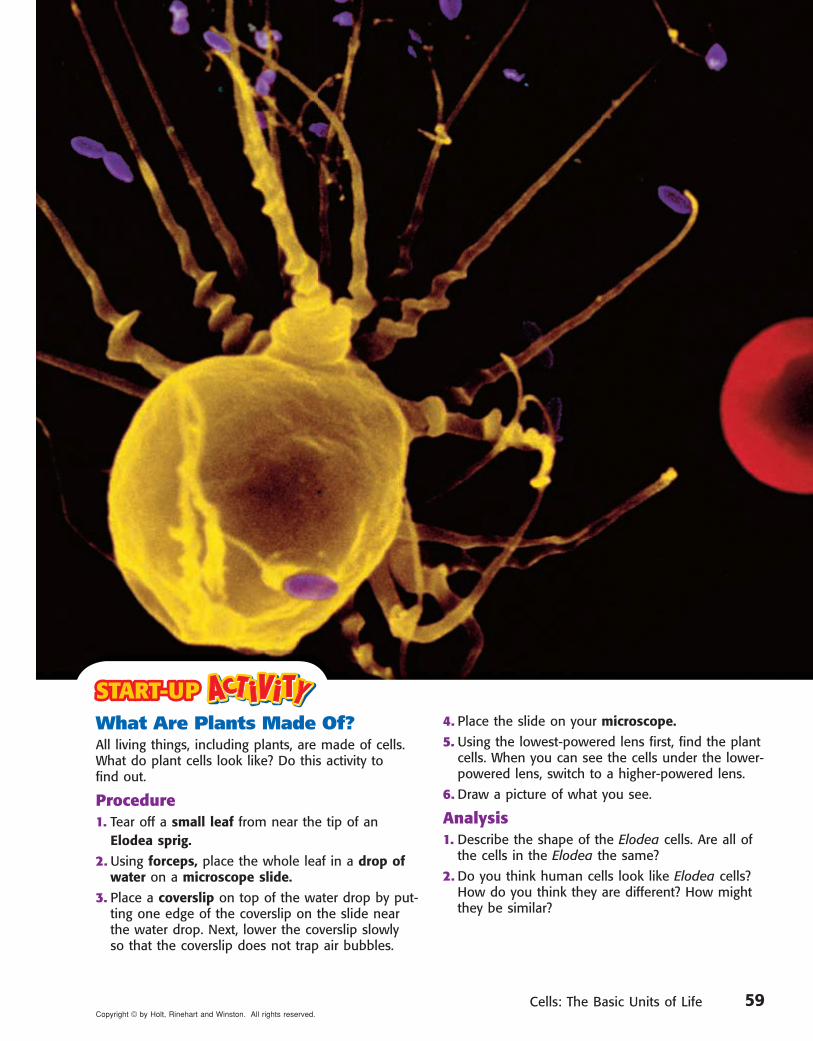
What Are Plants Made Of?All living things, including plants, are made of cells. What do plant cells look like? Do this activity to find out.
Procedure1. Tear off a small leaf from near the tip of an Elodea sprig.
2. Using forceps, place the whole leaf in a drop of water on a microscope slide.
3. Place a coverslip on top of the water drop by put-ting one edge of the coverslip on the slide near the water drop. Next, lower the coverslip slowly so that the coverslip does not trap air bubbles.
4. Place the slide on your microscope.
5. Using the lowest-powered lens first, find the plant cells. When you can see the cells under the lower-powered lens, switch to a higher-powered lens.
6. Draw a picture of what you see.
Analysis1. Describe the shape of the Elodea cells. Are all of
the cells in the Elodea the same?
2. Do you think human cells look like Elodea cells? How do you think they are different? How might they be similar?
59Copyright © by Holt, Rinehart and Winston. All rights reserved.

READING WARM-UPObjectives •• State the parts of the cell theory. •• Explain why cells are so small. •• Describe the parts of a cell. •• Describe how eubacteria are differ-
ent from archaebacteria. •• Explain the difference between pro-
karyotic cells and eukaryotic cells.
Terms to Learncell nucleuscell membrane prokaryoteorganelle eukaryote
Reading Organizer As you read this section, create an outline of the section. Use the headings from the section in your outline.
READING STRATEGY
1 The Diversity of CellsMost cells are so small they can’t be seen by the naked eye. So how did scientists fi nd cells? By accident, that’s how! The fi rst person to see cells wasn’t even looking for them.
All living things are made of tiny structures called cells. A cell cell is the smallest unit that can perform all the processes necessary for life. Because of their size, cells weren’t discovered until microscopes were invented in the mid-1600s.
Cells and the Cell TheoryRobert Hooke was the first person to describe cells. In 1665, he built a microscope to look at tiny objects. One day, he looked at a thin slice of cork. Cork is found in the bark of cork trees. The cork looked like it was made of little boxes. Hooke named these boxes cells, which means “little rooms” in Latin. Hooke’s cells were really the outer layers of dead cork cells. Hooke’s microscope and his drawing of the cork cells are shown in Figure 1.
Hooke also looked at thin slices of living plants. He saw that they too were made of cells. Some cells were even filled with “juice.” The “juicy” cells were living cells.
Hooke also looked at feathers, fish scales, and the eyes of houseflies. But he spent most of his time looking at plants and fungi. The cells of plants and fungi have cell walls. This makes them easy to see. Animal cells do not have cell walls. This absence of cell walls makes it harder to see the outline of animal cells. Because Hooke couldn’t see their cells, he thought that animals weren’t made of cells.
Figure 1 Hooke discovered cells using this microscope. Hooke’s drawing of cork cells is shown to the right of his microscope.
60 Chapter 3 Cells: The Basic Units of LifeCopyright © by Holt, Rinehart and Winston. All rights reserved.
Section 1 The Diversity of Cells
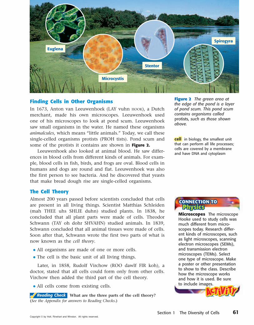
Finding Cells in Other OrganismsIn 1673, Anton van Leeuwenhoek (LAY vuhn HOOK), a Dutch merchant, made his own microscopes. Leeuwenhoek used one of his microscopes to look at pond scum. Leeuwenhoek saw small organisms in the water. He named these organisms animalcules, which means “little animals.” Today, we call these single-celled organisms protists (PROH tists). Pond scum and some of the protists it contains are shown in Figure 2.
Leeuwenhoek also looked at animal blood. He saw differ-ences in blood cells from different kinds of animals. For exam-ple, blood cells in fish, birds, and frogs are oval. Blood cells in humans and dogs are round and flat. Leeuwenhoek was also the first person to see bacteria. And he discovered that yeasts that make bread dough rise are single-celled organisms.
The Cell TheoryAlmost 200 years passed before scientists concluded that cells are present in all living things. Scientist Matthias Schleiden (mah THEE uhs SHLIE duhn) studied plants. In 1838, he concluded that all plant parts were made of cells. Theodor Schwann (TAY oh dohr SHVAHN) studied animals. In 1839, Schwann concluded that all animal tissues were made of cells. Soon after that, Schwann wrote the first two parts of what is now known as the cell theory.
• All organisms are made of one or more cells.
• The cell is the basic unit of all living things.
Later, in 1858, Rudolf Virchow (ROO dawlf FIR koh), a doctor, stated that all cells could form only from other cells. Virchow then added the third part of the cell theory.
• All cells come from existing cells.
✓✓Reading Check What are the three parts of the cell theory? (See the Appendix for answers to Reading Checks.)
Figure 2 The green area at the edge of the pond is a layer of pond scum. This pond scum contains organisms called protists, such as those shown above.
Microcystis
Stentor
Spirogyra
Euglena
cellcell in biology, the smallest unit that can perform all life processes; cells are covered by a membrane and have DNA and cytoplasm
Microscopes The microscope Hooke used to study cells was much different from micro-scopes today. Research differ-ent kinds of microscopes, such as light microscopes, scanning electron microscopes (SEMs), and transmission electron microscopes (TEMs). Select one type of microscope. Make a poster or other presentation to show to the class. Describe how the microscope works and how it is used. Be sure to include images.
61Copyright © by Holt, Rinehart and Winston. All rights reserved.
Cell SizeMost cells are too small to be seen without a microscope. It would take 50 human cells to cover the dot on this letter i.
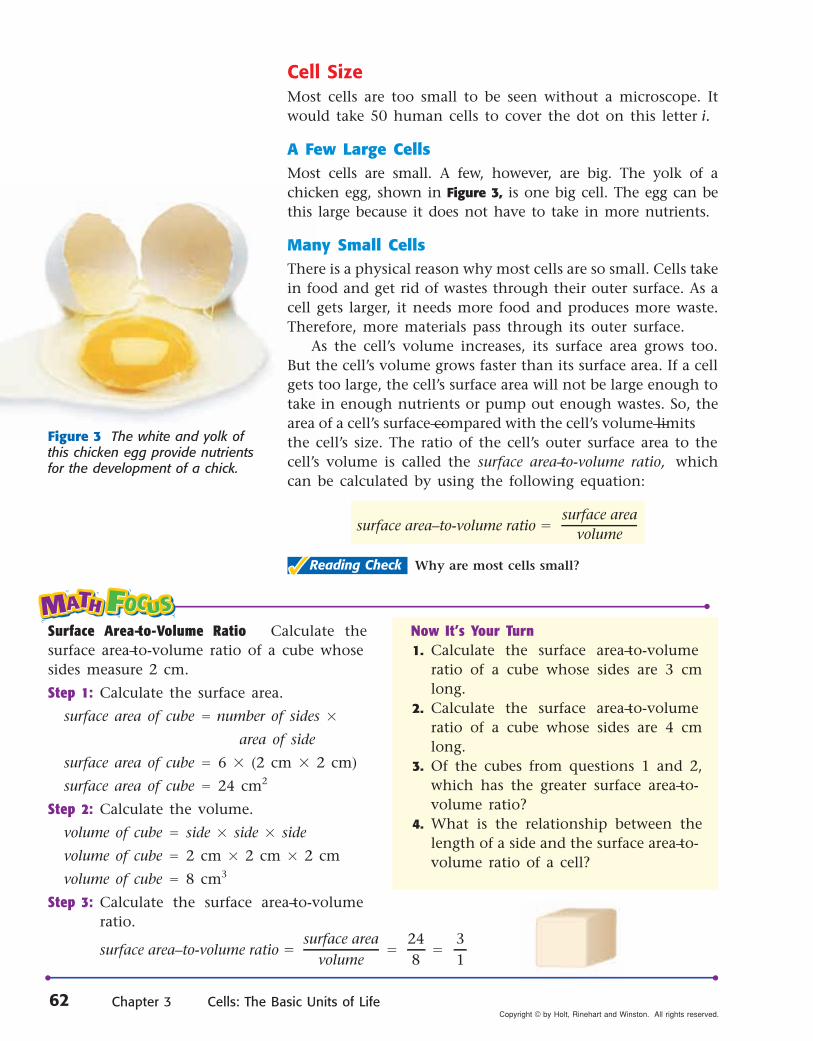
A Few Large CellsMost cells are small. A few, however, are big. The yolk of a chicken egg, shown in Figure 3, is one big cell. The egg can be this large because it does not have to take in more nutrients.
Many Small CellsThere is a physical reason why most cells are so small. Cells take in food and get rid of wastes through their outer surface. As a cell gets larger, it needs more food and produces more waste. Therefore, more materials pass through its outer surface.
As the cell’s volume increases, its surface area grows too. But the cell’s volume grows faster than its surface area. If a cell gets too large, the cell’s surface area will not be large enough to take in enough nutrients or pump out enough wastes. So, the area of a cell’s surface—compared with the cell’s volume—limits the cell’s size. The ratio of the cell’s outer surface area to the cell’s volume is called the surface area–to-volume ratio, which can be calculated by using the following equation:
��Reading Check Why are most cells small?
Figure 3 The white and yolk of this chicken egg provide nutrients for the development of a chick.
Surface Area–to-Volume Ratio Calculate the surface area–to-volume ratio of a cube whose sides measure 2 cm.
Step 1: Calculate the surface area.
surface area of cube � number of sides �
area of side
surface area of cube � 6 � (2 cm � 2 cm)
surface area of cube � 24 cm2
Step 2: Calculate the volume.
volume of cube � side � side � side
volume of cube � 2 cm � 2 cm � 2 cm
volume of cube � 8 cm3
Step 3: Calculate the surface area–to-volume ratio.
Now It’s Your Turn1. Calculate the surface area–to-volume
ratio of a cube whose sides are 3 cm long.
2. Calculate the surface area–to-volume ratio of a cube whose sides are 4 cm long.
3. Of the cubes from questions 1 and 2, which has the greater surface area–to-volume ratio?
4. What is the relationship between the length of a side and the surface area–to-volume ratio of a cell?
surface area– to-volume ratio �surface area
volume248
�31
�
surface area– to-volume ratio �surface area
volume
62 Chapter 3 Cells: The Basic Units of LifeCopyright © by Holt, Rinehart and Winston. All rights reserved.

DNAE. coli bacterium
Parts of a CellCells come in many shapes and sizes. Cells have many different functions. But all cells have the following parts in common.
The Cell Membrane and CytoplasmAll cells are surrounded by a cell membrane. The cell membranecell membrane is a protective layer that covers the cell’s surface and acts as a barrier. It separates the cell’s contents from its environment. The cell membrane also controls materials going into and out of the cell. Inside the cell is a fluid. This fluid and almost all of its contents are called the cytoplasm (SIET oh PLAZ uhm).
OrganellesCells have organelles that carry out various life processes. OrganellesOrganelles are structures that perform specific functions within the cell. Different types of cells have different organelles. Most organelles are surrounded by membranes. For example, the algal cell in Figure 4 has membrane-bound organelles. Some organelles float in the cytoplasm. Other organelles are attached to membranes or other organelles.
��Reading Check What are organelles?
Genetic MaterialAll cells contain DNA (deoxyribonucleic acid) at some point in their life. DNA is the genetic material that carries information needed to make new cells and new organisms. DNA is passed on from parent cells to new cells and controls the activities of a cell. Figure 5 shows the DNA of a bacterium.
In some cells, the DNA is enclosed inside an organelle called the nucleus. nucleus. For example, your cells have a nucleus. In contrast, bacterial cells do not have a nucleus.
In humans, mature red blood cells lose their DNA. Red blood cells are made inside bones. When red blood cells are first made, they have a nucleus with DNA. But before they enter the bloodstream, red blood cells lose their nucleus and DNA. They survive with no new instructions from their DNA.
Figure 4 This green alga has organelles. The organelles and the fluid surrounding them make up the cytoplasm.
Figure 5 This photo shows an Escherichia coli bacterium. The bacterium’s cell membrane has been treated so that the cell’s DNA is released.
Cellmembrane
cell membranecell membrane a phospholipid layer that covers a cell’s surface; acts as a barrier between the inside of a cell and the cell’s environment
organelleorganelle one of the small bodies in a cell’s cytoplasm that are special-ized to perform a specific function
nucleusnucleus in a eukaryotic cell, a membrane-bound organelle that contains the cell’s DNA and that has a role in processes such as growth, metabolism, and reproduction
Organelles
DNA
63Copyright © by Holt, Rinehart and Winston. All rights reserved.

Flagellum
Cell wall
Cell membrane
DNA
Two Kinds of CellsAll cells have cell membranes, organelles, cytoplasm, and DNA in common. But there are two basic types of cells— cells without a nucleus and cells with a nucleus. Cells with no nucleus are prokaryotic (proh KAR ee AHT ik) cells. Cells that have a nucleus are eukaryotic (yoo KAR ee AHT ik) cells. Prokaryotic cells are further classified into two groups: eubacteria (yoo bak TIR ee uh) and archaebacteria (AHR kee bak TIR ee uh).
Prokaryotes: Eubacteria and ArchaebacteriaEubacteria and archaebacteria are prokaryotes (pro KAR ee OHTS). ProkaryotesProkaryotes are single-celled organisms that do not have a nucleus or membrane-bound organelles.
EubacteriaThe most common prokaryotes are eubacteria (or just bacteria). Bacteria are the world’s smallest cells. These tiny organisms live almost everywhere. Bacteria do not have a nucleus, but they do have DNA. A bacteria’s DNA is a long, circular molecule, shaped sort of like a rubber band. Bacteria have no membrane-covered organelles. But they do have ribosomes. Ribosomes are tiny, round organelles made of protein and other material.
Bacteria also have a strong, weblike exterior cell wall. This wall helps the cell retain its shape. A bacterium’s cell mem-brane is just inside the cell wall. Together, the cell wall and cell membrane allow materials into and out of the cell.
Some bacteria live in the soil and water. Others live in, or on, other organisms. For example, you have bacteria living on your skin and teeth. You also have bacteria living in your digestive system. These bacteria help the process of digestion. A typical bacterial cell is shown in Figure 6.
prokaryote prokaryote an organism that con-sists of a single cell that does not have a nucleus
Figure 6 This diagram shows the DNA, cell membrane, and cell wall of a eubacterial cell. The flagellum helps the bacterium move.
Bacteria in Your Lunch?Most of the time, you don’t want bacteria in your food. Many bacteria make toxins that will make you sick. However, some foods— such as yogurt— are supposed to have bacteria in them! The bacteria in these foods are not dangerous.
In yogurt, masses of rod-shaped bacteria feed on the sugar (lactose) in milk. The bacteria convert the sugar into lactic acid. Lactic acid causes milk to thicken. This thickened milk makes yogurt.1. Using a cotton swab, put
a small dot of yogurt on a microscope slide.
2. Add a drop of water. Use the cotton swab to stir.
3. Add a coverslip.4. Use a microscope to
examine the slide. Draw what you observe.
64 Chapter 3 Cells: The Basic Units of LifeCopyright © by Holt, Rinehart and Winston. All rights reserved.
Section 1 The Diversity of Cells
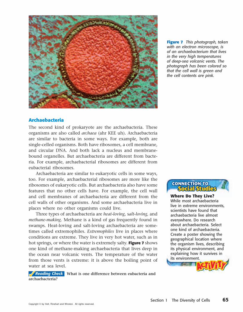
ArchaebacteriaThe second kind of prokaryote are the archaebacteria. These organisms are also called archaea (ahr KEE uh). Archaebacteria are similar to bacteria in some ways. For example, both are single-celled organisms. Both have ribosomes, a cell membrane, and circular DNA. And both lack a nucleus and membrane-bound organelles. But archaebacteria are different from bacte-ria. For example, archaebacterial ribosomes are different from eubacterial ribosomes.
Archaebacteria are similar to eukaryotic cells in some ways, too. For example, archaebacterial ribosomes are more like the ribosomes of eukaryotic cells. But archaebacteria also have some features that no other cells have. For example, the cell wall and cell membranes of archaebacteria are different from the cell walls of other organisms. And some archaebacteria live in places where no other organisms could live.
Three types of archaebacteria are heat-loving, salt-loving, and methane-making. Methane is a kind of gas frequently found in swamps. Heat-loving and salt-loving archaebacteria are some-times called extremophiles. Extremophiles live in places where conditions are extreme. They live in very hot water, such as in hot springs, or where the water is extremely salty. Figure 7 showsone kind of methane-making archaebacteria that lives deep in the ocean near volcanic vents. The temperature of the water from those vents is extreme: it is above the boiling point of water at sea level.
✓✓Reading Check What is one difference between eubacteria and archaebacteria?
Figure 7 This photograph, taken with an electron microscope, is of an archaebacterium that lives in the very high temperatures of deep-sea volcanic vents. The photograph has been colored so that the cell wall is green and the cell contents are pink.
Where Do They Live?While most archaebacteria live in extreme environments, scientists have found that archaebacteria live almost everywhere. Do research about archaebacteria. Select one kind of archaebacteria. Create a poster showing the geographical location where the organism lives, describing its physical environment, and explaining how it survives in its environment.
65Copyright © by Holt, Rinehart and Winston. All rights reserved.

Organelles Nucleus
Eukaryotic Cells and EukaryotesEukaryotic cells are the largest cells. Most eukaryotic cells are still microscopic, but they are about 10 times larger than most bacterial cells. A typical eukaryotic cell is shown in Figure 8.
Unlike bacteria and archaebacteria, eukaryotic cells have a nucleus. The nucleus is one kind of membrane-bound organelle. A cell’s nucleus holds the cell’s DNA. Eukaryotic cells have other membrane-bound organelles as well. Organelles are like the dif-ferent organs in your body. Each kind of organelle has a specific job in the cell. Together, organelles, such as the ones shown in Figure 8, perform all the processes necessary for life.
All living things that are not bacteria or archaebacteria are made of one or more eukaryotic cells. Organisms made of eukaryotic cells are called eukaryotes.eukaryotes. Many eukaryotes are multicellular. Multicellular means “many cells.” Multicellular organisms are usually larger than single-cell organisms. So, most organisms you see with your naked eye are eukaryotes. There are many types of eukaryotes. Animals, including humans, are eukaryotes. So are plants. Some protists, such as amoebas, are single-celled eukaryotes. Other protists, including some types of green algae, are multicellular eukaryotes. Fungi are organisms such as mushrooms or yeasts. Mushrooms are multicellular eukaryotes. Yeasts are single-celled eukaryotes.
✓✓Reading Check How are eukaryotes different from prokaryotes?
Nucleus
Organelles in a Typical Eukaryotic CellFigure 8
eukaryoteeukaryote an organism made up of cells that have a nucleus enclosed by a membrane; eukaryotes include animals, plants, and fungi, but not archaebacteria or eubacteria
Golgi complex
Mitochondrion
Lysosome
Endoplasmicreticulum
Cell membrane
Ribosome
For another activity related to this chapter, go to go.hrw.com and type in the keyword HL5CELW.
66 Chapter 3 Cells: The Basic Units of LifeCopyright © by Holt, Rinehart and Winston. All rights reserved.Copyright © by Holt, Rinehart and Winston. All rights reserved.
Developed and maintained by theNational Science Teachers Association
For a variety of links related to this chapter, go to www.scilinks.org
SummarySummary
Flagellum
Cell wall
Cell membrane
A
Review
Using Key Terms
1. In your own words, write a definition for the term organelle.
2. Use the following terms in the same sentence: prokaryotic, nucleus, and eukaryotic.
Understanding Key Ideas
3. Cell size is limited by the
a. thickness of the cell wall.b. size of the cell’s nucleus.c. cell’s surface area– to-volume ratio.d. amount of cytoplasm in the cell.
4. What are the three parts of the cell theory?
5. Name three structures that every cell has.
6. Give two ways in which archaebacteria are different from bacteria.
Critical Thinking
7. Applying Concepts You have discovered a new single-celled organism. It has a cell wall, ribosomes, and long, circular DNA. Is it a eukaryote or a prokaryote cell? Explain.
8. Identifying Relationships One of your stu-dents brings you a cell about the size of the period at the end of this sentence. It is a single cell, but it also forms chains. What character-istics would this cell have if the organism is a eukaryote? If it is a prokaryote? What would you look for first?
Interpreting Graphics
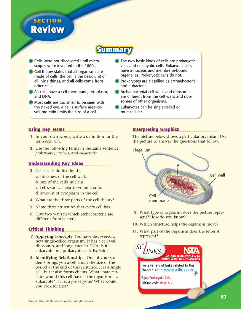
The picture below shows a particular organism. Use the picture to answer the questions that follow.
9. What type of organism does the picture repre-sent? How do you know?
10. Which structure helps the organism move?
11. What part of the organism does the letter A represent?
•• Cells were not discovered until micro-scopes were invented in the 1600s.
•• Cell theory states that all organisms are made of cells, the cell is the basic unit of all living things, and all cells come from other cells.
•• All cells have a cell membrane, cytoplasm, and DNA.
•• Most cells are too small to be seen with the naked eye. A cell’s surface area– to-volume ratio limits the size of a cell.
•• The two basic kinds of cells are prokaryotic cells and eukaryotic cells. Eukaryotic cells have a nucleus and membrane-bound organelles. Prokaryotic cells do not.
•• Prokaryotes are classified as archaebacteria and eubacteria.
•• Archaebacterial cell walls and ribosomes are different from the cell walls and ribo-somes of other organisms.
•• Eukaryotes can be single-celled or multicellular.
Topic: Prokaryotic CellsSciLinks code: HSM1225
67Copyright © by Holt, Rinehart and Winston. All rights reserved.
READING WARM-UP
2 Eukaryotic CellsMost eukaryotic cells are small. For a long time after cells were discovered, scientists could not see what was going on inside cells. They did not know how complex cells are.
Now, scientists know a lot about eukaryotic cells. These cells have many parts that work together and keep the cell alive.
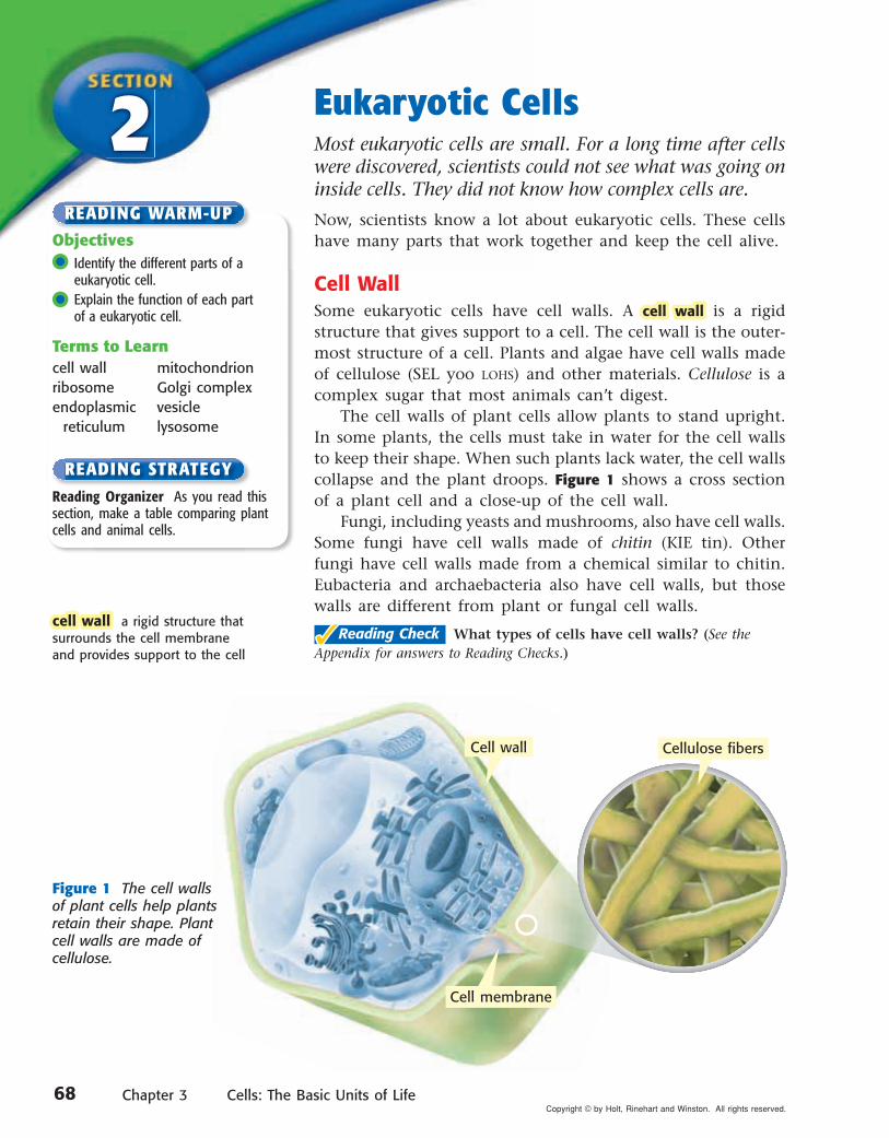
Cell WallSome eukaryotic cells have cell walls. A cell wallcell wall is a rigid structure that gives support to a cell. The cell wall is the outer-most structure of a cell. Plants and algae have cell walls made of cellulose (SEL yoo LOHS) and other materials. Cellulose is a complex sugar that most animals can’t digest.
The cell walls of plant cells allow plants to stand upright. In some plants, the cells must take in water for the cell walls to keep their shape. When such plants lack water, the cell walls collapse and the plant droops. Figure 1 shows a cross section of a plant cell and a close-up of the cell wall.
Fungi, including yeasts and mushrooms, also have cell walls. Some fungi have cell walls made of chitin (KIE tin). Other fungi have cell walls made from a chemical similar to chitin. Eubacteria and archaebacteria also have cell walls, but those walls are different from plant or fungal cell walls.
��Reading Check What types of cells have cell walls? (See the
Appendix for answers to Reading Checks.)
Objectives •• Identify the different parts of a
eukaryotic cell. •• Explain the function of each part
of a eukaryotic cell.
Terms to Learncell wall mitochondrionribosome Golgi complexendoplasmic vesicle reticulum lysosome
Reading Organizer As you read this section, make a table comparing plant cells and animal cells.
READING STRATEGY
Figure 1 The cell walls of plant cells help plants retain their shape. Plant cell walls are made of cellulose.
cell wall cell wall a rigid structure that surrounds the cell membrane and provides support to the cell
Cell wall Cellulose fibers
Cell membrane
68 Chapter 3 Cells: The Basic Units of LifeCopyright © by Holt, Rinehart and Winston. All rights reserved.
Section 2 Eukaryotic Cells
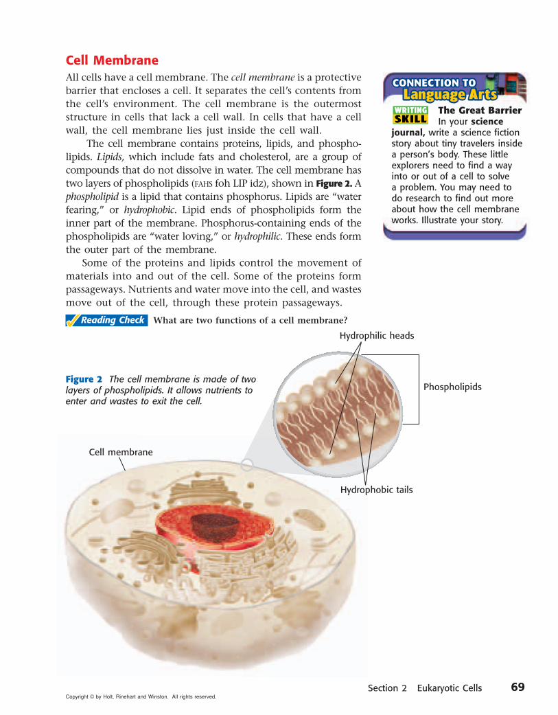
Cell MembraneAll cells have a cell membrane. The cell membrane is a protective barrier that encloses a cell. It separates the cell’s contents from the cell’s environment. The cell membrane is the outermost structure in cells that lack a cell wall. In cells that have a cell wall, the cell membrane lies just inside the cell wall.
The cell membrane contains proteins, lipids, and phospho-lipids. Lipids, which include fats and cholesterol, are a group of compounds that do not dissolve in water. The cell membrane has two layers of phospholipids (FAHS foh LIP idz), shown in Figure 2. Aphospholipid is a lipid that contains phosphorus. Lipids are “water fearing,” or hydrophobic. Lipid ends of phospholipids form the inner part of the membrane. Phosphorus-containing ends of the phospholipids are “water loving,” or hydrophilic. These ends form the outer part of the membrane.
Some of the proteins and lipids control the movement of materials into and out of the cell. Some of the proteins form passageways. Nutrients and water move into the cell, and wastes move out of the cell, through these protein passageways.
✓✓Reading Check What are two functions of a cell membrane?
Cell membrane
Figure 2 The cell membrane is made of two layers of phospholipids. It allows nutrients to enter and wastes to exit the cell.
The Great BarrierIn your science
journal, write a science fiction story about tiny trav elers inside a person’s body. These little explorers need to find a way into or out of a cell to solve a problem. You may need to do research to find out more about how the cell membrane works. Illustrate your story.
WRITINGSKILL
Hydrophilic heads
Hydrophobic tails
Phospholipids
69Copyright © by Holt, Rinehart and Winston. All rights reserved.
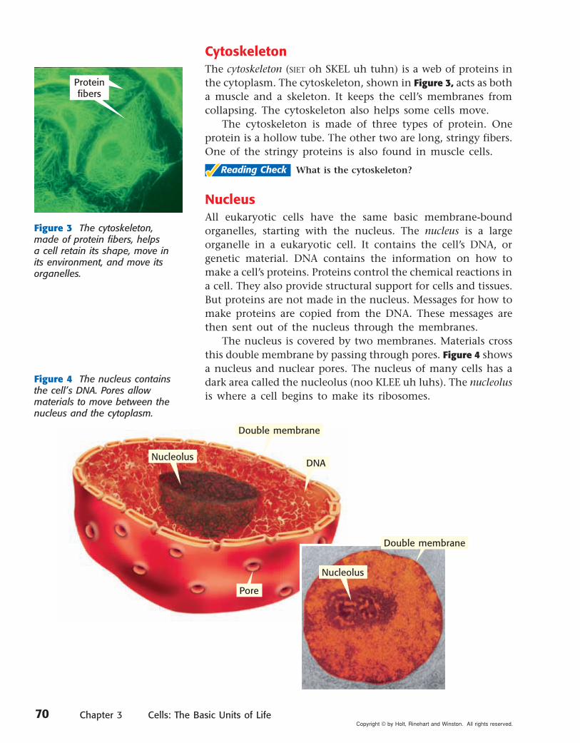
CytoskeletonThe cytoskeleton (SIET oh SKEL uh tuhn) is a web of proteins in the cytoplasm. The cytoskeleton, shown in Figure 3, acts as both a muscle and a skeleton. It keeps the cell’s membranes from collapsing. The cytoskeleton also helps some cells move.
The cytoskeleton is made of three types of protein. One pro tein is a hollow tube. The other two are long, stringy fibers. One of the stringy proteins is also found in muscle cells.
��Reading Check What is the cytoskeleton?
NucleusAll eukaryotic cells have the same basic membrane-bound organelles, starting with the nucleus. The nucleus is a large organelle in a eukaryotic cell. It contains the cell’s DNA, or genetic material. DNA contains the information on how to make a cell’s proteins. Proteins control the chemical reactions in a cell. They also provide structural support for cells and tissues. But proteins are not made in the nucleus. Messages for how to make proteins are copied from the DNA. These messages are then sent out of the nucleus through the membranes.
The nucleus is covered by two membranes. Materials cross this double membrane by passing through pores. Figure 4 shows a nucleus and nuclear pores. The nucleus of many cells has a dark area called the nucleolus (noo KLEE uh luhs). The nucleolus is where a cell begins to make its ribosomes.
Figure 4 The nucleus contains the cell’s DNA. Pores allow materials to move between the nucleus and the cytoplasm.
Figure 3 The cytoskeleton, made of protein fibers, helps a cell retain its shape, move in its environment, and move its organelles.
Protein fibers
Double membrane
DNA
Pore
Nucleolus
Double membrane
Nucleolus
70 Chapter 3 Cells: The Basic Units of LifeCopyright © by Holt, Rinehart and Winston. All rights reserved.
Section 2 Eukaryotic Cells
Rough ER
Smooth ER
RibosomesOrganelles that make proteins are called ribosomes.ribosomes. Ribosomes are the smallest of all organelles. And there are more ribosomes in a cell than there are any other organelles. Some ribosomes float freely in the cytoplasm. Others are attached to membranes or the cytoskeleton. Unlike most organelles, ribosomes are not covered by a membrane.
Proteins are made within the ribosomes. Proteins are made of amino acids. An amino acid is any one of about 20 differ-ent organic molecules that are used to make proteins. All cells need proteins to live. All cells have ribosomes.
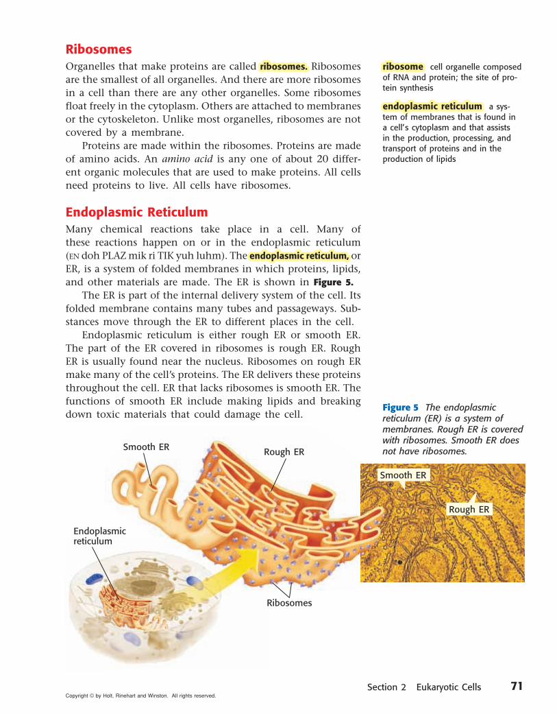
Endoplasmic ReticulumMany chemical reactions take place in a cell. Many of these reactions happen on or in the endoplasmic reticulum (EN doh PLAZ mik ri TIK yuh luhm). The endoplasmic reticulum, endoplasmic reticulum, or ER, is a system of folded membranes in which proteins, lipids, and other materials are made. The ER is shown in Figure 5.
The ER is part of the internal delivery system of the cell. Its folded membrane contains many tubes and passageways. Sub-stances move through the ER to different places in the cell.
Endoplasmic reticulum is either rough ER or smooth ER. The part of the ER covered in ribosomes is rough ER. Rough ER is usually found near the nucleus. Ribosomes on rough ER make many of the cell’s proteins. The ER delivers these proteins throughout the cell. ER that lacks ribosomes is smooth ER. The functions of smooth ER include making lipids and breaking down toxic materials that could damage the cell.
Endoplasmic reticulum
Figure 5 The endoplasmic reticulum (ER) is a system of membranes. Rough ER is covered with ribosomes. Smooth ER does not have ribosomes.
Ribosomes
ribosome ribosome cell organelle composed of RNA and protein; the site of pro-tein synthesis
endoplasmic reticulum endoplasmic reticulum a sys-tem of membranes that is found in a cell’s cytoplasm and that assists in the production, processing, and transport of proteins and in the production of lipids
Smooth ER Rough ER
71Copyright © by Holt, Rinehart and Winston. All rights reserved.

MitochondriaA mitochondrion (MIET oh KAHN dree uhn) is the main power source of a cell. A mitochondrionmitochondrion is the organelle in which sugar is broken down to produce energy. Mito-chondria are covered by two membranes, as shown in Figure 6. Energy released by mitochondria is stored in a substance called ATP (adenosine triphosphate). The cell then uses ATP to do work. ATP can be made at several places in a cell. But most of a cell’s ATP is made in the inner membrane of the cell’s mitochondria.
Most eukaryotic cells have mitochondria. Mito-chondria are the size of some bacteria. Like bacteria, mitochondria have their own DNA, and mitochondria can divide within a cell.
��Reading Check Where is most of a cell’s ATP made?
ChloroplastsAnimal cells cannot make their own food. Plants and algae are different. They have chloroplasts (KLAWR uh PLASTS) in some of their cells. Chloroplasts are organelles in plant and algae cells in which photosynthesis takes place. Like mitochondria, chloroplasts have two mem-branes and their own DNA. A chloroplast is shown in Figure 7. Photosynthesis is the process by which plants and algae use sunlight, carbon dioxide, and water to make sugar and oxygen.
Chloroplasts are green because they contain chloro-phyll, a green pigment. Chloro phyll is found inside the inner membrane of a chloroplast. Chloro phyll traps the energy of sunlight, which is used to make sugar. The sugar produced by photosynthesis is then used by mito-chondria to make ATP.
Figure 6 Mitochondria break down sugar and make ATP. ATP is produced on the inner membrane.
Figure 7 Chloroplasts harness and use the energy of the sun to make sugar. A green pigment— chlorophyll—traps the sun’s energy.
Outer membrane
Inner membrane
Outer membrane
Inner membrane
mitochondrionmitochondrion in eukaryotic cells, the cell organelle that is surrounded by two membranes and that is the site of cellular respiration
Inner membrane
Outer membrane
Outer membrane
Inner membrane
72 Chapter 3 Cells: The Basic Units of LifeCopyright © by Holt, Rinehart and Winston. All rights reserved.
Section 2 Eukaryotic Cells
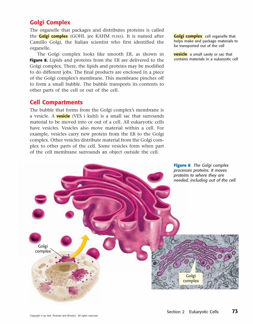
Golgi ComplexThe organelle that packages and distributes proteins is called the Golgi complexGolgi complex (GOHL jee KAHM PLEKS). It is named after Camillo Golgi, the Italian scientist who first identified the organelle.
The Golgi complex looks like smooth ER, as shown in Figure 8. Lipids and proteins from the ER are delivered to the Golgi complex. There, the lipids and proteins may be modified to do different jobs. The final products are enclosed in a piece of the Golgi complex’s membrane. This membrane pinches off to form a small bubble. The bubble transports its contents to other parts of the cell or out of the cell.
Cell CompartmentsThe bubble that forms from the Golgi complex’s membrane is a vesicle. A vesiclevesicle (VES i kuhl) is a small sac that surrounds material to be moved into or out of a cell. All eukaryotic cells have vesicles. Vesicles also move material within a cell. For example, vesicles carry new protein from the ER to the Golgi complex. Other vesicles distribute material from the Golgi com-plex to other parts of the cell. Some vesicles form when part of the cell membrane surrounds an object outside the cell.
Golgi complexGolgi complex cell organelle that helps make and package materials to be transported out of the cell
vesiclevesicle a small cavity or sac that contains materials in a eukaryotic cell
Figure 8 The Golgi complex processes proteins. It moves proteins to where they are needed, including out of the cell.
Golgicomplex
Golgicomplex
73Copyright © by Holt, Rinehart and Winston. All rights reserved.

Cellular DigestionLysosomes (LIE suh SOHMZ) are vesicles that are respon-sible for digestion inside a cell. LysosomesLysosomes are organelles that contain digestive enzymes. They destroy worn-out or damaged organelles, get rid of waste materials, and protect the cell from foreign invaders. Lysosomes, which come in a wide variety of sizes and shapes, are shown in Figure 9.
Lysosomes are found mainly in animal cells. When eukaryotic cells engulf particles, they enclose the particles in vesicles. Lysosomes bump into these vesicles and pour enzymes into them. These enzymes digest the particles in the vesicles.
��Reading Check Why are lysosomes important?
VacuolesA vacuole (VAK yoo OHL) is a large vesicle. In plant and fungal cells, some vacuoles act like large lysosomes. They store digestive enzymes and aid in digestion within the cell. Other vacuoles in plant cells store water and other liquids. Vacuoles that are full of water, such as the one in Figure 9, help support the cell. Some plants wilt when their vacuoles lose water. Table 1 shows some organelles and their functions.
Figure 9 Lysosomes digest materials inside a cell. In plant and fungal cells, vacuoles often perform the same function.
Lysosome
Vacuole
lysosomelysosome a cell organelle that con-tains digestive enzymes
Nucleusthe organelle that contains the cell’s DNA and is the control center of the cell
Chloroplastthe organelle that uses the energy of sunlight to make food
Ribosomethe organelle in which amino acids are hooked together to make proteins
Endoplasmic reticulumthe organelle that makes lipids, breaks down drugs and other substances, and packages pro-teins for Golgi complex
Mitochondriathe organelle that breaks down food molecules to make ATP
Lysosomethe organelle that digests food particles, wastes, cell parts, and foreign invaders
Vacuolethe organelle that stores water and other materials
Golgi complexthe organelle that processes and transports proteins and other materials out of cell
Table 1 Organelles and Their Functions
74 Chapter 3 Cells: The Basic Units of LifeCopyright © by Holt, Rinehart and Winston. All rights reserved.Copyright © by Holt, Rinehart and Winston. All rights reserved.
Developed and maintained by theNational Science Teachers Association
For a variety of links related to this chapter, go to www.scilinks.org
SummarySummary
cb
a
Review
•• Eukaryotic cells have organelles that per-form functions that help cells remain alive.
•• All cells have a cell membrane. Some cells have a cell wall. Some cells have a cytoskeleton.
•• The nucleus of a eukaryotic cell contains the cell’s genetic material, DNA.
•• Ribosomes are the organelles that make proteins. Ribosomes are not covered by a membrane.
•• The endoplasmic reticulum (ER) and the Golgi complex make and process proteins before the proteins are transported to other parts of the cell or out of the cell.
•• Mitochondria and chloroplasts are energy-producing organelles.
•• Lysosomes are organelles responsible for digestion within a cell. In plant cells, organ-elles called vacuoles store cell materials and sometimes act like large lysosomes.
Using Key Terms
1. In your own words, write a definition for each of the following terms: ribosome, lysosome, and cell wall.
Understanding Key Ideas
2. Which of the following are found mainly in animal cells?
a. mitochondriab. lysosomesc. ribosomesd. Golgi complexes
3. What is the function of a Golgi complex? What is the function of the endoplasmic reticulum?
Critical Thinking
4. Making Comparisons Describe three ways in which plant cells differ from animal cells.
5. Applying Concepts Every cell needs ribo-somes. Explain why.
6. Predicting Consequences A certain virus attacks the mitochondria in cells. What would happen to a cell if all of its mitochondria were destroyed?
7. Expressing Opinions Do you think that hav-ing chloroplasts gives plant cells an advantage over animal cells? Support your opinion.
Interpreting Graphics
Use the diagram below to answer the questions that follow.
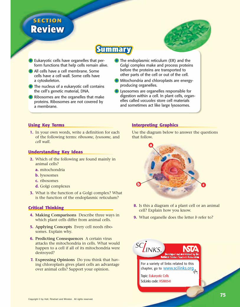
8. Is this a diagram of a plant cell or an animal cell? Explain how you know.
9. What organelle does the letter b refer to?
Topic: Eukaryotic CellsSciLinks code: HSM0541
75Copyright © by Holt, Rinehart and Winston. All rights reserved.

READING WARM-UP
The Organization of Living ThingsIn some ways, organisms are like machines. Some machines have just one part. But most machines have many parts. Some organisms exist as a single cell. Other organisms have many—even trillions—of cells.
Most cells are smaller than the period that ends this sentence. Yet, every cell in every organism performs all the processes of life. So, are there any advantages to having many cells?
The Benefits of Being MulticellularYou are a multicellular organism. This means that you are made of many cells. Multicellular organisms grow by making more small cells, not by making their cells larger. For example, an elephant is bigger than you are, but its cells are about the same size as yours. An elephant just has more cells than you do. Some benefits of being multicellular are the following:
• Larger Size Many multicellular organisms are small. But they are usually larger than single-celled organisms. Larger organisms are prey for fewer predators. Larger predators can eat a wider variety of prey.
• Longer Life The life span of a multicellular organism is not limited to the life span of any single cell.
Objectives •• List three advantages of being
multicellular. •• Describe the four levels of organi-
zation in living things. •• Explain the relationship between the
structure and function of a part of an organism.
Terms to Learntissue organismorgan structureorgan system function
Paired Summarizing Read this section silently. In pairs, take turns summariz-ing the material. Stop to discuss ideas that seem confusing.
READING STRATEGY
Figure 1 This photomicrograph shows a small part of one heart muscle cell. The green line surrounds one of many mitochondria, the powerhouses of the cell. The pink areas are muscle filaments.
• Specialization Each type of cell has a particular job. Spe-cialization makes the organism more efficient. For example, the cardiac muscle cell in Figure 1 is a specialized muscle cell. Heart muscle cells contract and make the heart pump blood.
��Reading Check List three advantages of being multi cellular.
(See the Appendix for answers to Reading Checks.)
3
Chapter 3 76Copyright © by Holt, Rinehart and Winston. All rights reserved.

Section 3 The Organization of Living Things
Cells Working TogetherA tissue tissue is a group of cells that work together to perform a specific job. The material around and between the cells is also part of the tissue. The cardiac muscle tissue, shown in Figure 2, is made of many cardiac muscle cells. Cardiac muscle tissue is just one type of tissue in a heart.
Animals have four basic types of tissues: nerve tissue, mus-cle tissue, connective tissue, and protective tissue. In contrast, plants have three types of tissues: transport tissue, protective tissue, and ground tissue. Transport tissue moves water and nutrients through a plant. Protective tissue covers the plant. It helps the plant retain water and protects the plant against damage. Photosynthesis takes place in ground tissue.
Tissues Working TogetherA structure that is made up of two or more tissues working together to perform a specific function is called an organ.organ. For example, your heart is an organ. It is made mostly of cardiac muscle tissue. But your heart also has nerve tissue and tissues of the blood vessels that all work together to make your heart the powerful pump that it is.
Another organ is your stomach. It also has several kinds of tissue. In the stomach, muscle tissue makes food move in and through the stomach. Special tissues make chemicals that help digest your food. Connective tissue holds the stomach together, and nervous tissue carries messages back and forth between the stomach and the brain. Other organs include the intestines, brain, and lungs.
Plants also have different kinds of tissues that work together as organs. A leaf is a plant organ that contains tissue that traps light energy to make food. Other examples of plant organs are stems and roots.
��Reading Check What is an organ?
tissuetissue a group of similar cells that perform a common function
organorgan a collection of tissues that carry out a specialized function of the body
Figure 2 This photomicrograph shows cardiac muscle tissue. Cardiac muscle tissue is made up of many cardiac cells.
A Pet ProtistImagine that you have a tiny box-shaped protist for a pet. To care for your pet protist properly, you have to fig-ure out how much to feed it. The dimensions of your protist are roughly 25 µm � 20 µm � 2 µm. If seven food particles per second can enter through each square micrometer of surface area, how many particles can your protist eat in 1 min?
77Copyright © by Holt, Rinehart and Winston. All rights reserved.

Organs Working TogetherA group of organs working together to perform a particular function is called an organ system.organ system. Each organ system has a specific job to do in the body.
For example, the digestive system is made up of several organs, including the stomach and intestines. The digestive system’s job is to break down food into small particles. Other parts of the body then use these small particles as fuel. In turn, the digestive system depends on the respiratory and cardiovas-cular systems for oxygen. The cardiovascular system, shown in Figure 3, includes organs and tissues such as the heart and blood vessels. Plants also have organ systems. They include leaf systems, root systems, and stem systems.
��Reading Check List the levels of organization in living things.
OrganismsAnything that can perform life processes by itself is an organism.organism. An organism made of a single cell is called a unicellular organism. Bacteria, most protists, and some kinds of fungi are unicellular. Although some of these organisms live in colonies, they are still unicellular. They are unicellular organisms living together, and all of the cells in the colony are the same. Each cell must carry out all life processes in order for that cell to survive. In contrast, even the simplest multicellular organism has specialized cells that depend on each other for the organ-ism to survive.
organ system organ system a group of organs that work together to perform body functions
organism organism a living thing; anything that can carry out life processes independently
structure structure the arrangement of parts in an organism
functionfunction the special, normal, or proper activity of an organ or part
Cells form tissues.
Tissues form organs.
Organs form organ systems.
And organ systems form organisms such as you!
Tissue Organ Organ systemCell
Levels of Organization in the Cardiovascular SystemFigure 3
78 Chapter 3 Cells: The Basic Units of LifeCopyright © by Holt, Rinehart and Winston. All rights reserved.Copyright © by Holt, Rinehart and Winston. All rights reserved.
For a variety of links related to this chapter, go to www.scilinks.org
Developed and maintained by theNational Science Teachers Association
SummarySummary
Review
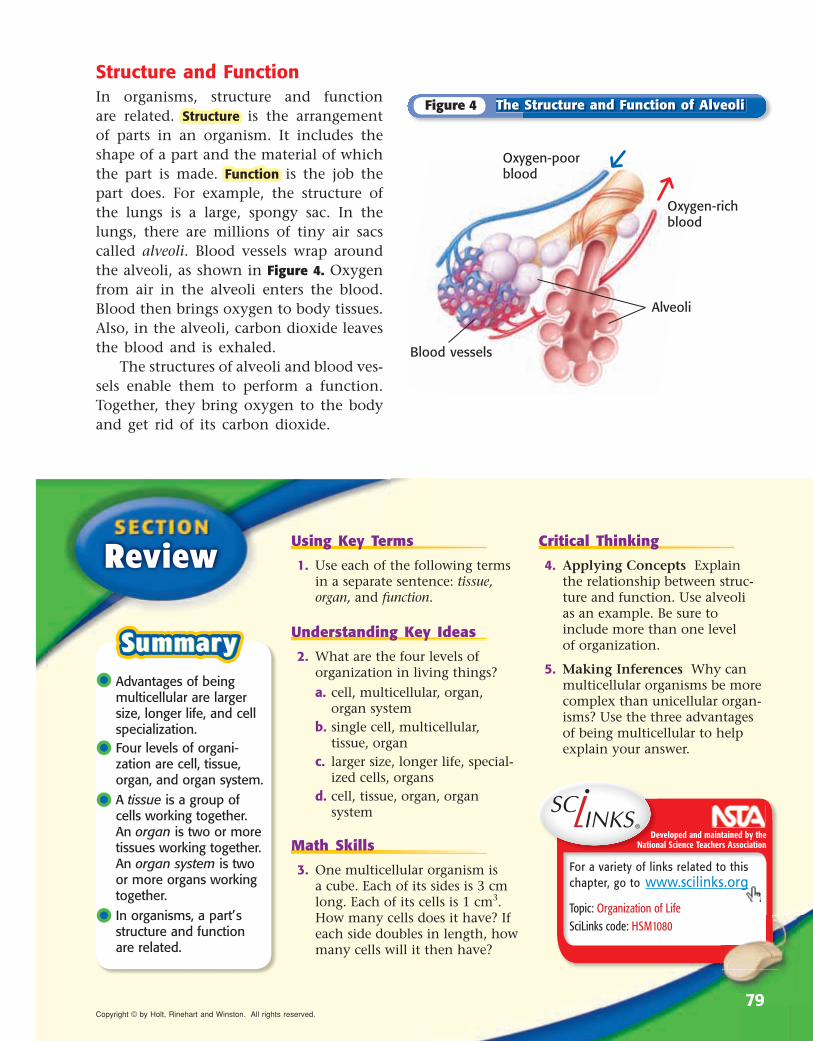
Structure and FunctionIn organisms, structure and function are related. StructureStructure is the arrangement of parts in an organism. It includes the shape of a part and the material of which the part is made. Function Function is the job the part does. For example, the structure of the lungs is a large, spongy sac. In the lungs, there are millions of tiny air sacs called alveoli. Blood vessels wrap around the alveoli, as shown in Figure 4. Oxygen from air in the alveoli enters the blood. Blood then brings oxygen to body tissues. Also, in the alveoli, carbon dioxide leaves the blood and is exhaled.
The structures of alveoli and blood ves-sels enable them to perform a function. Together, they bring oxygen to the body and get rid of its carbon dioxide.
Blood vessels
Alveoli
Oxygen-poor blood
Oxygen-rich blood
•• Advantages of being multicellular are larger size, longer life, and cell specialization.
•• Four levels of organi-zation are cell, tissue, organ, and organ system.
•• A tissue is a group of cells working together. An organ is two or more tissues working together. An organ system is two or more organs working together.
•• In organisms, a part’s structure and function are related.
Using Key Terms
1. Use each of the following terms in a separate sentence: tissue, organ, and function.
Understanding Key Ideas
2. What are the four levels of or ganization in living things?
a. cell, multicellular, organ, organ system
b. single cell, multicellular, tissue, organ
c. larger size, longer life, special-ized cells, organs
d. cell, tissue, organ, organ system
Math Skills
3. One multicellular organism is a cube. Each of its sides is 3 cm long. Each of its cells is 1 cm3. How many cells does it have? If each side doubles in length, how many cells will it then have?
Critical Thinking
4. Applying Concepts Explain the relationship between struc-ture and function. Use alveoli as an example. Be sure to include more than one level of organization.
5. Making Inferences Why can multicellular organisms be more complex than unicellular organ-isms? Use the three advantages of being multicellular to help explain your answer.
Topic: Organization of Life SciLinks code: HSM1080
The Structure and Function of AlveoliFigure 4
79Copyright © by Holt, Rinehart and Winston. All rights reserved.
Model-Making Lab
Explore why a single-celled organism cannot grow to the size of an elephant.
Create a model of a cell to illustrate the concept of surface area– to-volume ratio.
• calculator (optional)
• cubic cell patterns
• heavy paper or poster board
• sand, fine
• scale or balance
• scissors
• tape, transparent
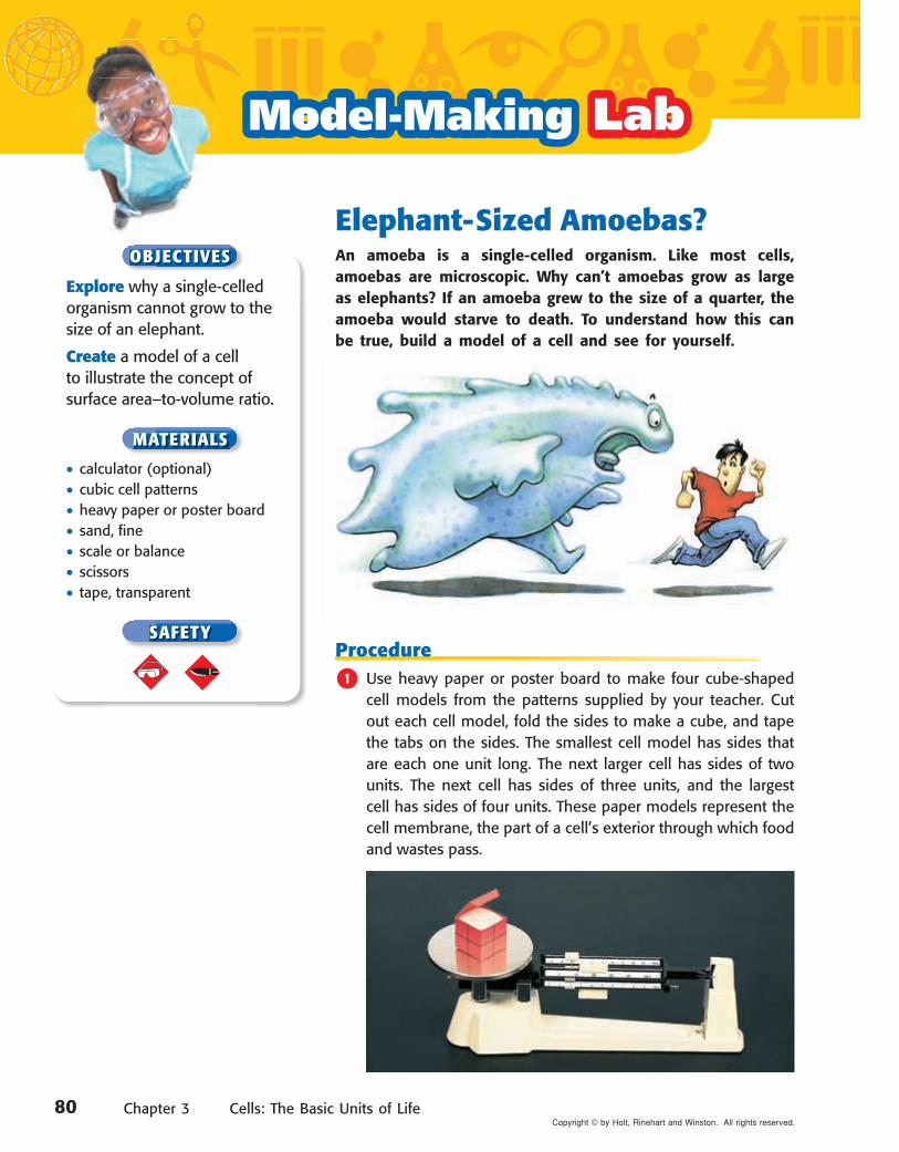
Elephant-Sized Amoebas?An amoeba is a single-celled organism. Like most cells, amoebas are microscopic. Why can’t amoebas grow as large as elephants? If an amoeba grew to the size of a quarter, the amoeba would starve to death. To understand how this can be true, build a model of a cell and see for yourself.
Procedure
1 Use heavy paper or poster board to make four cube-shaped cell models from the patterns supplied by your teacher. Cut out each cell model, fold the sides to make a cube, and tape the tabs on the sides. The smallest cell model has sides that are each one unit long. The next larger cell has sides of two units. The next cell has sides of three units, and the largest cell has sides of four units. These paper models represent the cell membrane, the part of a cell’s exterior through which food and wastes pass.
OBJECTIVES
MATERIALS
SAFETY
80 Chapter 3 Cells: The Basic Units of LifeCopyright © by Holt, Rinehart and Winston. All rights reserved.
Chapter Lab
2 Copy the data table shown above. Use each formula to calculate the data about your cell models. Record your calculations in the table. Calculations for the smallest cell have been done for you.
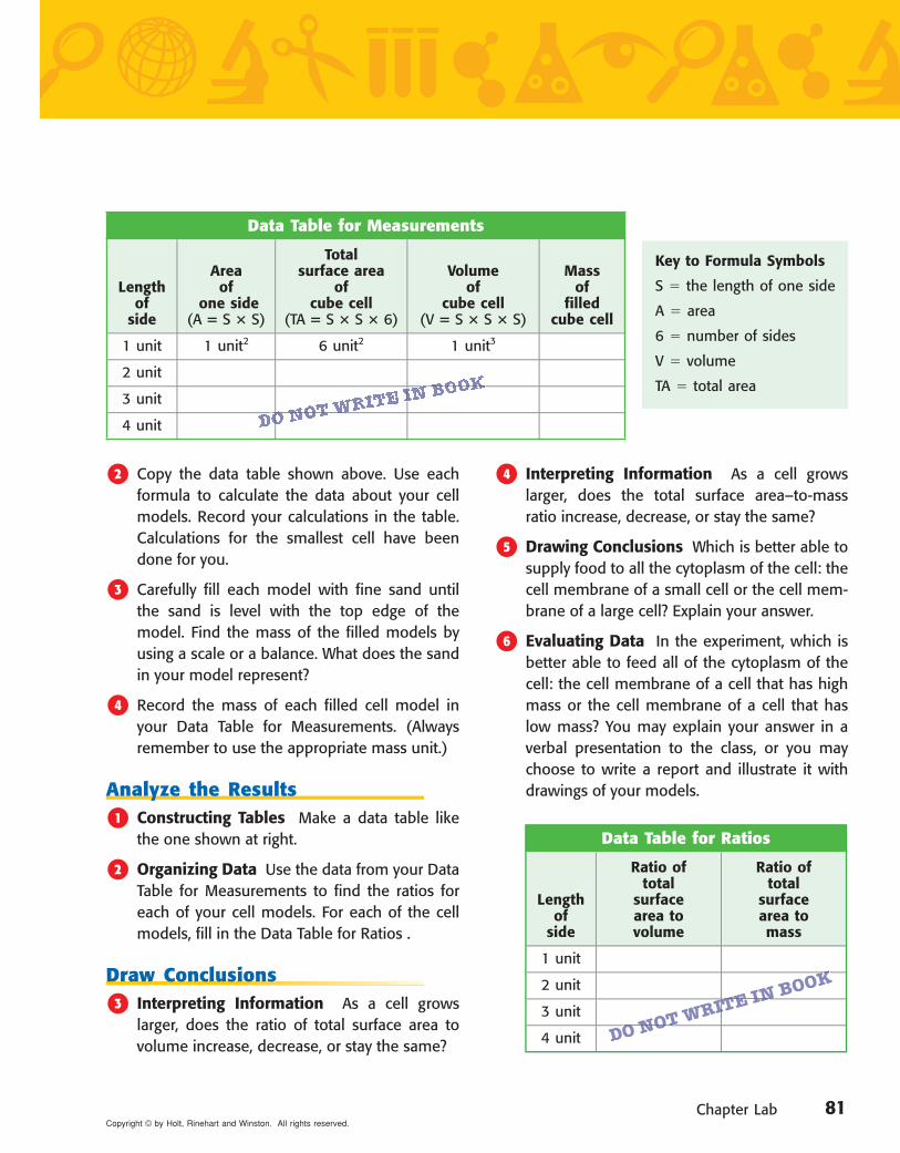
3 Carefully fill each model with fine sand until the sand is level with the top edge of the model. Find the mass of the filled models by using a scale or a balance. What does the sand in your model represent?
4 Record the mass of each filled cell model in your Data Table for Measurements. (Always remember to use the appropriate mass unit.)
Analyze the Results
1 Constructing Tables Make a data table like the one shown at right.
2 Organizing Data Use the data from your Data Table for Measurements to find the ratios for each of your cell models. For each of the cell models, fill in the Data Table for Ratios .
Draw Conclusions
3 Interpreting Information As a cell grows larger, does the ratio of total surface area to volume increase, decrease, or stay the same?
4 Interpreting Information As a cell grows larger, does the total surface area– to-mass ratio increase, decrease, or stay the same?
5 Drawing Conclusions Which is better able to supply food to all the cytoplasm of the cell: the cell membrane of a small cell or the cell mem-brane of a large cell? Explain your answer.
6 Evaluating Data In the experiment, which is better able to feed all of the cytoplasm of the cell: the cell membrane of a cell that has high mass or the cell membrane of a cell that has low mass? You may explain your answer in a verbal presentation to the class, or you may choose to write a report and illustrate it with drawings of your models.
Data Table for Measurements
Length of
side
Area of
one side(A � S � S)
Total surface area
of cube cell
(TA � S � S � 6)
Volume of
cube cell(V � S � S � S)
Mass of
filled cube cell
1 unit 1 unit2 6 unit2 1 unit3
2 unit
3 unit
4 unit
Key to Formula Symbols
S � the length of one side
A � area
6 � number of sides
V � volume
TA � total area
Data Table for Ratios
Length of
side
Ratio of total
surface area to volume
Ratio of total
surface area to mass
1 unit
2 unit
3 unit
4 unit
DO NOT WRITE IN BOOK
DO NOT WRITE IN BOOK
DO NOT WRITE IN BOOK
O NOT WRITE IN BOOK
81Copyright © by Holt, Rinehart and Winston. All rights reserved.

Complete each of the following sen-tences by choosing the correct term from the word bank.
cell organcell membrane prokaryoteorganelles eukaryotecell wall tissuestructure function
1 A(n) is the most basic unit of all living things.
2The job that an organ does is the of that organ.
3 Ribosomes and mitochondria are types of .
4 A(n) is an organism whose cells have a nucleus.
5 A group of cells working together to perform a specifi c function is a(n) .
6 Only plant cells have a(n) .
Multiple Choice
7 Which of the following best describes an organ?
a. a group of cells that work together to perform a specifi c job
b. a group of tissues that belong to dif-ferent systems
c. a group of tissues that work together to perform a specifi c job
d. a body structure, such as muscles or lungs
8 The benefi ts of being multicellular include
a. small size, long life, and cell specialization.
b. generalized cells, longer life, and ability to prey on small animals.
c. larger size, more enemies, and spe-cialized cells.
d. longer life, larger size, and special-ized cells.
9 In eukaryotic cells, which organelle contains the DNA?
a. nucleus c. smooth ERb. Golgi complex d. vacuole
0Which of the following statements is part of the cell theory?
a. All cells suddenly appear by themselves.
b. All cells come from other cells.c. All organisms are multicellular.d. All cells have identical parts.
qThe surface area–to-volume ratio of a cell limits
a. the number of organelles that the cell has.
b. the size of the cell.c. where the cell lives.d. the types of nutrients that a cell
needs.
w Two types of organisms whose cells do not have a nucleus are
a. prokaryotes and eukaryotes.b. plants and animals.c. eubacteria and archaebacteria.d. single-celled and multicellular
organisms.
USING KEY TERMS
UNDERSTANDING KEY IDEAS
82 Chapter 3 Cells: The Basic Units of LifeCopyright © by Holt, Rinehart and Winston. All rights reserved.

Chapter Review
Short Answer
eExplain why most cells are small.
r Describe the four levels of organization in living things.
t What is the difference between the structure of an organ and the function of the organ?
y Name two functions of a cell membrane.
u What are the structure and function of the cytoskeleton in a cell?
i Concept Mapping Use the following terms to create a concept map: cells, organisms, Golgi complex, organ systems, organs, nucleus, organelle, and tissues.
o Making Comparisons Compare and contrast the functions of the endoplas-mic reticulum and the Golgi complex.
p Identifying Relationships Explain how the structure and function of an organism’s parts are related. Give an example.
a Evaluating Hypotheses One of your classmates states a hypothesis that all organisms must have organ systems. Is your classmate’s hypothesis valid? Explain your answer.
s Predicting Consequences What would happen if all of the ribosomes in your cells disappeared?
d Expressing Opinions Scientists think that millions of years ago the surface of the Earth was very hot and that the atmosphere contained a lot of methane. In your opinion, which type of organism, a eubacterium or an archaebacterium, is the older form of life? Explain your reasoning.
Use the diagram below to answer the questions that follow.
f What is the name of the structure identifi ed by the letter a?
g Which letter identifi es the structure that digests food particles and foreign invaders?
h Which letter identifi es the structure that makes proteins, lipids, and other materials and that contains tubes and passageways that enable substances to move to different places in the cell?
CRITICAL THINKING
INTERPRETING GRAPHICS
c
b
a
83Copyright © by Holt, Rinehart and Winston. All rights reserved.

READINGRead each of the passages below. Then, answer the questions that follow each passage.
Passage 1 Exploring caves can be dangerous but can also lead to interesting discoveries. For exam-ple, deep in the darkness of Cueva de Villa Luz, a cave in Mexico, are slippery formations called snottites. They were named snottites because they look just like a two-year-old’s runny nose. If you use an electron microscope to look at them, you see that snottites are bacteria; thick, sticky fluids; and small amounts of minerals produced by the bacteria. As tiny as they are, these bacteria can build up snottite structures that may eventually turn into rock. Formations in other caves look like hardened snottites. The bacteria in snottites are acidophiles. Acidophiles live in environments that are highly acidic. Snottite bacteria produce sulfuric acid and live in an environment that is similar to the inside of a car battery.
1. Which statement best describes snottites?
A Snottites are bacteria that live in car batteries.
B Snottites are rock formations found in caves.
C Snottites were named for a cave in Mexico.D Snottites are made of bacteria, sticky fl uids,
and minerals.
2. Based on this passage, which conclusion about snottites is most likely to be correct?
F Snottites are found in caves everywhere.G Snottite bacteria do not need sunlight.H You could grow snottites in a greenhouse.I Snottites create other bacteria in caves.
3. What is the main idea of this passage?
A Acidophiles are unusual organisms.B Snottites are strange formations.C Exploring caves is dangerous.D Snottites are large, slippery bacteria.
Passage 2 The world’s smallest mammal may be a bat about the size of a jelly bean. The scientific name for this tiny animal, which was unknown until 1974, is Craseonycteris thonglong-yai. It is so small that it is sometimes called the bumblebee bat. Another name for this animal is the hog-nosed bat. Hog-nosed bats were given their name because one of their distinctive features is a piglike muzzle. Hog-nosed bats differ from other bats in another way: they do not have a tail. But, like other bats, hog-nosed bats do eat insects that they catch in mid-air. Scientists think that the bats eat small insects that live on the leaves at the tops of trees. Hog-nosed bats live deep in limestone caves and have been found in only one country, Thailand.
1. According to the passage, which statement about hog-nosed bats is most accurate?
A They are the world’s smallest animal.B They are about the size of a bumblebee.C They eat leaves at the tops of trees.D They live in hives near caves in Thailand.
2. Which of the following statements describes distinctive features of hog-nosed bats?
F The bats are very small and eat leaves.G The bats live in caves and have a tail.H The bats live in Thailand and are birds.I The bats have a piglike muzzle and no tail.
3. From the information in this passage, which conclusion is most likely to be correct?
A Hog-nosed bats are similar to other bats.B Hog-nosed bats are probably rare.C Hog-nosed bats can sting like a bumblebee.D Hog-nosed bats probably eat fruit.
84 Chapter 3 Cells: The Basic Units of LifeCopyright © by Holt, Rinehart and Winston. All rights reserved.
Stand
ardized
Test Prep
aration
3
3 3
Cell 2B
Cell 1
A
Standardized Test Preparation
1. What is the name of the organelle labeled A in Cell 1?
A endoplasmic reticulumB mitochondrionC vacuoleD nucleus
2. What type of cell is Cell 1?
F a bacterial cellG a plant cellH an animal cellI a prokaryotic cell
3. What is the name and function of the organelle labeled B in Cell 2?
A The organelle is a vacuole, and it stores water and other materials.
B The organelle is the nucleus, and it contains the DNA.
C The organelle is the cell wall, and it gives shape to the cell.
D The organelle is a ribosome, where proteins are put together.
4. What type of cell is Cell 2? How do you know?
F prokaryotic; because it does not have a nucleus
G eukaryotic; because it does not have a nucleus
H prokaryotic; because it has a nucleusI eukaryotic; because it has a nucleus
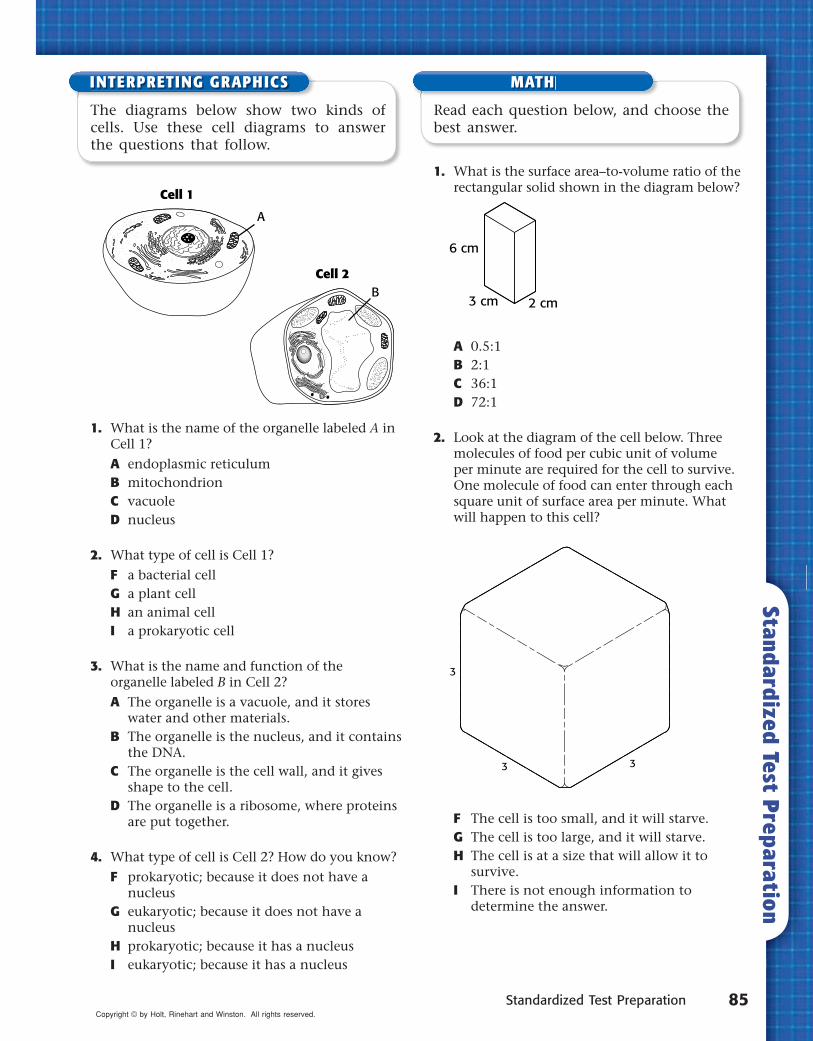
1. What is the surface area–to-volume ratio of the rectangular solid shown in the diagram below?
A 0.5:1B 2:1C 36:1D 72:1
2. Look at the diagram of the cell below. Three molecules of food per cubic unit of volume per minute are required for the cell to survive. One molecule of food can enter through each square unit of surface area per minute. What will happen to this cell?
F The cell is too small, and it will starve.G The cell is too large, and it will starve.H The cell is at a size that will allow it to
survive.I There is not enough information to
determine the answer.
The diagrams below show two kinds of cells. Use these cell diagrams to answer the questions that follow.
Read each question below, and choose the best answer.
6 cm
3 cm 2 cm
INTERPRETING GRAPHICS MATH
85Copyright © by Holt, Rinehart and Winston. All rights reserved.
Language ArtsLanguage ArtsImagine that you are a doc-tor who treats diseases such as
Parkinson’s disease. Design and create a pamphlet or brochure that you could use to explain what stem cells are. Include in your pamphlet a description of how stem cells might be used to treat one of your patients who has Parkinson’s disease. Be sure to include information about Parkinson’s disease.
WRITINGSKILL
Social StudiesSocial StudiesChoose one of the four types of extremophiles. Do some research about the organism you have chosen and make a poster showing what you learned about it, including where it can be found, under what conditions it lives, how it survives, and how it is used.
Weird ScienceExtremophilesAre there organisms on Earth that can give scientists clues about possible life elsewhere? Yes, there are! These organisms are called extremophiles, and they live where the environ ment is extreme. For example, some extremophiles live in the hot volcanic thermal vents deep in the ocean. Other extremophiles live in the extreme cold of Antarctica. But these organisms do not live only in extreme environments. Research shows that extremophiles may be abun-dant in plankton in the ocean. And not all extremophiles are archaebacteria; some extremophiles are eubacteria.
Scientific DiscoveriesDiscovery of the Stem CellWhat do Parkinson’s disease, diabetes, aplas-tic anemia, and Alzheimer’s disease have in common? All of these diseases are diseases for which stem cells may provide treatment or a cure. Stem cells are unspecialized cells from which all other kinds of cells can grow. And research on stem cells has been going on almost since microscopes were invented. But scientists have been able to culture, or grow, stem cells in laboratories for only about the last 20 years. Research during these 20 years has shown scientists that stem cells can be useful in treating—and possibly curing—a variety of diseases.
Sciencein Action
86 Chapter 3 Cells: The Basic Units of LifeCopyright © by Holt, Rinehart and Winston. All rights reserved.
Science in Action
MathMathAn average bacterium is about 0.000002 m long. A pencil point is about 0.001 m wide. Approximately how many bacteria would fit on a pencil point?
To learn more about these Science in Action topics, visit go.hrw.com and type in the keyword HL5CELF.
Check out Current Science® articles related to this chapter by visiting go.hrw.com. Just type in the keyword HL5CS03.
People in SciencePeople in Science
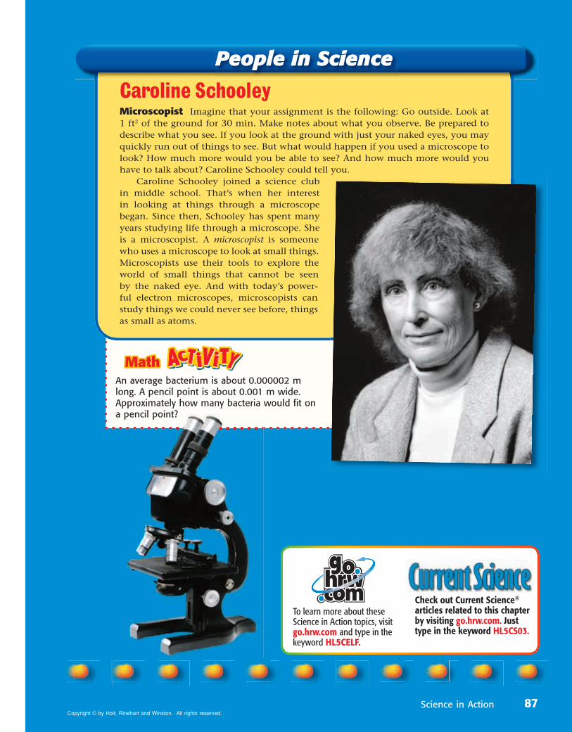
Caroline SchooleyMicroscopist Imagine that your assignment is the following: Go outside. Look at 1 ft2 of the ground for 30 min. Make notes about what you observe. Be prepared to describe what you see. If you look at the ground with just your naked eyes, you may quickly run out of things to see. But what would happen if you used a microscope to look? How much more would you be able to see? And how much more would you have to talk about? Caroline Schooley could tell you.
Caroline Schooley joined a science club in middle school. That’s when her interest in looking at things through a microscope began. Since then, Schooley has spent many years studying life through a microscope. She is a microscopist. A microscopist is someone who uses a microscope to look at small things. Microscopists use their tools to explore the world of small things that cannot be seen by the naked eye. And with today’s power-ful electron microscopes, microscopists can study things we could never see before, things as small as atoms.
87Copyright © by Holt, Rinehart and Winston. All rights reserved.