
Cell. Vol. 50, 453-463, July 31, 1987, Copyright 0 1987 by ......phase endocytosis were decreased by...
11
Cell. Vol. 50, 453-463, July 31, 1987, Copyright 0 1987 by Cell Press Inhibition of Endocytosis by Anti-Clathrin Antibodies Stephen J. Doxsey,’ Frances M. Brodsky,t Gregory S. Blank,* and Ari Helenius’ *Yale University School of Medicine Department of Cell Biology New Haven, Connecticut 06510 tuniversity of San Francisco Department of Pharmacy San Francisco, California 94143 t Becton-Dickinson Monoclonal Center 2375 Garcia Avenue Mountain View, California 94309 Summary We examined the function of clathrin, a cytoplasmic protein associated with coated pits and vesicles, by in- troducing monoclonal antibodies into living cells and determining their effects on membrane transport. When anti-clathrin heavy chain antibodies were used, the following effects were observed: clathrin became aggregated in the cytoplasm, the number of coated pits on the plasma membrane was reduced, and ad- sorbtive endocytosis of Semliki Forest virus and fluid- phase endocytosis were decreased by 40%-500/b. No change in transport of newly synthesized influenza hemagglutinin to the plasma membrane was observed. The results indicated that clathrin in CV-1 cells is in- volved in fluid-phase uptake and receptor-mediated endocytosis, but not in constitutive transport within the secretory pathway. Introduction Since their discovery, clathrin-coated pits and vesicles have been widely viewed as important organelles in the selective transport of macromolecules between mem- brane compartments. Coated pits on the cell surface are thought to concentrate receptors and receptor-ligand complexes at sites of internalization, and to participate in the formation of clathrin-coated endocytic vesicles (for reviews see Goldstein et al., 1979; Pearse and Bretcher, 1981). It has been suggested that clathrin-coated mem- branes and vesicles found in association with the Golgi complex may be involved in transport of plasma mem- brane glycoproteins, secretory proteins, and lysosomal enzymes (see Pearse and Bretcher, 1981; Griffiths and Si- mons, 1986). Clathrin-coated vesicles have also been in- voked in membrane retrieval from granules and the plasma membrane in secretory cells, in the transcytotic transport across epithelial cells, and in transporting mem- brane to localized areas of the cell surface growth (see Sil- verstein et al., 1977; Pearse and &etcher, 1981; Farquhar, 1985; Griffiths and Simons, 1986). In contrast with these views, which place clathrin at the center of membrane transport and molecular sorting, a number of reports have suggested that clathrin may not be a crucial factor in exocytic and endocytic transport. Wehland et al. (1981) reported that microinjection of affinity-purified anti-clathrin antibodies into the cytoplasm of tissue culture cells had no effect on the receptor- mediated endocytosis of a*-macroglobulin (a ligand that is concentrated in and internalized by coated pits). Payne and Schekman (1985) demonstrated that yeast cells can survive without clathrin heavy chains and that secretion of invertase in these cells is virtually normal. The role of clathrin-coated membranes in the trans-golgi network as sites of departure for viral membrane glycoproteins bound for the plasma membrane has also been questioned in re- cent immunocytochemical studies (Wehland et al., 1982; Griffiths et al., 1985; Orci et al., 1986). Thus, while detailed structural analysis of coated vesi- cles and clathrin rapidly progresses (see Pearse and Crowther, 1987) their functional roles still remain unclear. In this study we have examined the function of clathrin using a newly developed bulk delivery technique that al- lows the introduction of anti-clathrin antibodies into the cytoplasm of living cells. We employed well-characterized monoclonal antibodies with known inhibitory action on clathrin assembly in vitro (Blank and Brodsky, 1986). Our results showed that antibodies to clathrin heavy chains (the 180 kd subunits of the clathrin triskelions; Pearse and Crowther, 1987) impaired fluid-phase uptake and adsorb- tive endocytosis of Semliki Forest virus, but did not affect the constitutive transport of newly synthesized influenza hemagglutinin (HA) from the endoplasmic reticulum to the plasma membrane. Results Anti-Clathrin Antibodies Four monoclonal anti-clathrin antibodies of isotype IgG, and their Fab fragments were used. They were raised in mice against purified human brain clathrin, and have been extensively characterized using immunoprecipita- tion, immunoblotting, radioimmunoassays and functional in vitro assays (Brodsky, 1985a; Blank and Brodsky, 1986). Three of the antibodies (X19, X35, and X22) bind to clathrin heavy chains. They react with both free and polymerized clathrin triskelions, which form protein baskets (Pearse and Crowther, 1987). Antibodies X19 and X35 compete for binding to adjacent sites, whereas X22 appears to react with an independent site. All three cause extensive aggre- gation of clathrin baskets in vitro. In addition, X19, X35, and their Fab fragments perturb the assembly of baskets from triskelions; they induce the formation of large mats of aggregated clathrin and other abnormal clathrin struc- tures (Blank and Brodsky, 1986). Another antibody, X16, recognizes one of the clathrin light chain subunits (LCa). It does so preferentially when LCa is dissociated from the heavy chain. The Xl6 antibody causes little aggregation of preformed clathrin baskets in vitro. A mouse monoclo-
Transcript of Cell. Vol. 50, 453-463, July 31, 1987, Copyright 0 1987 by ......phase endocytosis were decreased by...

Cell. Vol. 50, 453-463, July 31, 1987, Copyright 0 1987 by Cell Press
Inhibition of Endocytosis by Anti-Clathrin Antibodies
Stephen J. Doxsey,’ Frances M. Brodsky,t Gregory S. Blank,* and Ari Helenius’ *Yale University School of Medicine Department of Cell Biology New Haven, Connecticut 06510 tuniversity of San Francisco Department of Pharmacy San Francisco, California 94143 t Becton-Dickinson Monoclonal Center 2375 Garcia Avenue Mountain View, California 94309
Summary
We examined the function of clathrin, a cytoplasmic protein associated with coated pits and vesicles, by in- troducing monoclonal antibodies into living cells and determining their effects on membrane transport. When anti-clathrin heavy chain antibodies were used, the following effects were observed: clathrin became aggregated in the cytoplasm, the number of coated pits on the plasma membrane was reduced, and ad- sorbtive endocytosis of Semliki Forest virus and fluid- phase endocytosis were decreased by 40%-500/b. No change in transport of newly synthesized influenza hemagglutinin to the plasma membrane was observed. The results indicated that clathrin in CV-1 cells is in- volved in fluid-phase uptake and receptor-mediated endocytosis, but not in constitutive transport within the secretory pathway.
Introduction
Since their discovery, clathrin-coated pits and vesicles have been widely viewed as important organelles in the selective transport of macromolecules between mem- brane compartments. Coated pits on the cell surface are thought to concentrate receptors and receptor-ligand complexes at sites of internalization, and to participate in the formation of clathrin-coated endocytic vesicles (for reviews see Goldstein et al., 1979; Pearse and Bretcher, 1981). It has been suggested that clathrin-coated mem- branes and vesicles found in association with the Golgi complex may be involved in transport of plasma mem- brane glycoproteins, secretory proteins, and lysosomal enzymes (see Pearse and Bretcher, 1981; Griffiths and Si- mons, 1986). Clathrin-coated vesicles have also been in- voked in membrane retrieval from granules and the plasma membrane in secretory cells, in the transcytotic transport across epithelial cells, and in transporting mem- brane to localized areas of the cell surface growth (see Sil- verstein et al., 1977; Pearse and &etcher, 1981; Farquhar, 1985; Griffiths and Simons, 1986).
In contrast with these views, which place clathrin at the center of membrane transport and molecular sorting, a
number of reports have suggested that clathrin may not be a crucial factor in exocytic and endocytic transport. Wehland et al. (1981) reported that microinjection of affinity-purified anti-clathrin antibodies into the cytoplasm of tissue culture cells had no effect on the receptor- mediated endocytosis of a*-macroglobulin (a ligand that is concentrated in and internalized by coated pits). Payne and Schekman (1985) demonstrated that yeast cells can survive without clathrin heavy chains and that secretion of invertase in these cells is virtually normal. The role of clathrin-coated membranes in the trans-golgi network as sites of departure for viral membrane glycoproteins bound for the plasma membrane has also been questioned in re- cent immunocytochemical studies (Wehland et al., 1982; Griffiths et al., 1985; Orci et al., 1986).
Thus, while detailed structural analysis of coated vesi- cles and clathrin rapidly progresses (see Pearse and Crowther, 1987) their functional roles still remain unclear. In this study we have examined the function of clathrin using a newly developed bulk delivery technique that al- lows the introduction of anti-clathrin antibodies into the cytoplasm of living cells. We employed well-characterized monoclonal antibodies with known inhibitory action on clathrin assembly in vitro (Blank and Brodsky, 1986). Our results showed that antibodies to clathrin heavy chains (the 180 kd subunits of the clathrin triskelions; Pearse and Crowther, 1987) impaired fluid-phase uptake and adsorb- tive endocytosis of Semliki Forest virus, but did not affect the constitutive transport of newly synthesized influenza hemagglutinin (HA) from the endoplasmic reticulum to the plasma membrane.
Results
Anti-Clathrin Antibodies Four monoclonal anti-clathrin antibodies of isotype IgG, and their Fab fragments were used. They were raised in mice against purified human brain clathrin, and have been extensively characterized using immunoprecipita- tion, immunoblotting, radioimmunoassays and functional in vitro assays (Brodsky, 1985a; Blank and Brodsky, 1986). Three of the antibodies (X19, X35, and X22) bind to clathrin heavy chains. They react with both free and polymerized clathrin triskelions, which form protein baskets (Pearse and Crowther, 1987). Antibodies X19 and X35 compete for binding to adjacent sites, whereas X22 appears to react with an independent site. All three cause extensive aggre- gation of clathrin baskets in vitro. In addition, X19, X35, and their Fab fragments perturb the assembly of baskets from triskelions; they induce the formation of large mats of aggregated clathrin and other abnormal clathrin struc- tures (Blank and Brodsky, 1986). Another antibody, X16, recognizes one of the clathrin light chain subunits (LCa). It does so preferentially when LCa is dissociated from the heavy chain. The Xl6 antibody causes little aggregation of preformed clathrin baskets in vitro. A mouse monoclo-

Cell 454
Figure 1. lmmunofluorescence Localization of Clathrin and Delivered Antibodies
(a) shows the distribution of control IgG in CV-1 cells after delivery as visualized by immunofluorescence. One hour after delivery, cells were fixed, permeabilized, and stained using FITC-goat anti-mouse IgG. The cytoplasm of most cells contains delivered antibody, but the phase-contrast image (b) reveals some cells (see arrow heads in b) that have not received detectable IgG. Unfused erythrocytes remaining on the ceil surface appear as fluorescent patches, (a and b, bar = 10 urn). (c) shows the distribution of clathrin heavy chains in CV-1 cells. Fixed and permeabilized cells were stained using X19 anti-heavy chain antibody followed by FITC-goat anti-mouse. (d) shows the distribution of X19 1 hr after delivery into CV-1 cells. The delivered antibody was visualized as in (a) (c and d, bar = 7.5 urn)
nal IgG (anti-lghdb; Oi et al., 1978), which has the same isotype as the test antibodies but does not react with any proteins in CV-1 cells, was used as a control.
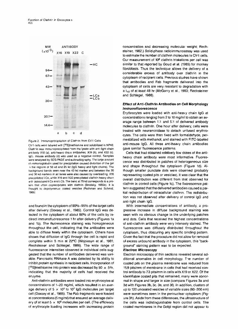
To test the cross-reactivity of the antibodies with CV-1 cells, immunofluorescence and immunoprecipitation were performed. All three anti-heavy chain antibodies gave in- tense staining (Figure lc). The pattern was similar to that previously observed for clathrin in a variety of other cell types (Anderson et al., 1978; Kartenbeck et al., 1981) i.e., discrete punctate fluorescence associated with the cell surface and concentrated fluorescence in the Golgi region. lmmunoprecipitation from [%]methionine-labeled lysates confirmed that these antibodies reacted with CV-1 cell clathrin triskelions (Figure 2, lanes b and c). No detectable fluorescence was observed with X16, the anti-light chain antibody, but in agreement with its specificity in other cell types (Brodsky, 1985a and 1985b), it immunoprecipitated a small amount of LCa without associated heavy chains (Figure 2a). The control antibody did not react with clathrin (Figure 2, lane d). As the effects of the Xl6 were virtually indistinguishable from that of the control IgG with respect
to staining and precipitation of clathrin triskelions, it served, in effect, as a second control antibody.
Method for Bulk Delivery Antibodies were introduced into the cytoplasm of living cells using a bulk delivery technique recently developed by Doxsey et al. (1985). As recipients we used CV-1 cells that expressed influenza virus hemagglutinin (HA) on their surfaces following infection with an SV40 vector con- taining the HA gene (Gething and Sambrook, 1981). Erythrocytes, loaded with antibodies by hypotonic lysis (Rechsteiner, 1982), were allowed to attach to the cell sur- face HA. The HA-mediated fusion between the mem- branes was induced by a brief drop in the pH of the medium. The resulting delivery was efficient, reproduc- ible, and virtually nondeleterious to the target ceils. Since entire cultures of cells could be processed simultane- ously, the method was well suited for biochemical studies.
The efficiency of delivery was routinely monitored using three test molecules: horseradish peroxidase (HRP) was easily visualized by cytochemistry and light microscopy,
Function of Clathrin I” Endocytosis 455
MW ANTIBODY (x163) Xl6 X19 X22 C
94-
67- - .-.
-HC
- LCa -LCb
-*I ‘._
20.1-
14.4- make
a b c d
Figure 2 lmmunoprecipitatron of Clathrin from CV-1 Cells
CV-1 cells were labeled with [%]methionine and solubilized in NP40. Clathrin was rmmunoprecipitated from the lysate with anti-light chain anhbody Xl6 (a), anti-heavy chain antibodies, X19 (b), and X22 (C). IgG, mouse antibody (d) was used as a negative control. Samples were analyzed by SDS-PAGE and autoradiography. The large amount of rmmunoglobulin used for precipitation caused distortion of the gel rn the regions of 50 kd and 25 kd (IgG heavy and light chains). The background bands seen near the 43 kd marker and between the 20 and 30 kd markers in all lanes were also caused by overloading. Xl6 precipitated LCa, while X19 and X22 precipitated clathrin heavy char” with associated LCa and LCb. The band at 70 kd corresponds to a pro- tein that often coprecrpitates with clathrin (Brodsky, 1985b). It is thought to depolymerize coated vesicles (Rothman and Schmid, 1986)
and found in the cytoplasm of 90%-95% of the target cells after delivery (Doxsey et al., 1985). Control IgG was de- tected in the cytoplasm of about 80% of the cells by in- direct immunofluorescence 1 hr after delivery (Figures la and lb). The fluorescence staining was homogeneous throughout the cell, indicating that the antibodies were able to diffuse freely within the cytoplasm. Others have shown that diffusion of IgG through the cell is rapid and complete within 5 min at 22% (Wojciesyn et al., 1981; Rechsteiner and Schlegel, 1986). The wide range of fluorescence intensities observed in individual cells sug- gested that the number of antibodies delivered was vari- able. Pancreatic RNAase A was detected by its ability to inhibit protein synthesis in recipient cells. Incorporation of [35S]methionine into protein was decreased by 90 f 5%, indicating that the majority of cells had received the enzyme.
Anti-clathrin antibodies were loaded into erythrocytes at concentrations of l-20 mglml, which resulted in an aver- age delivery of 5 x lo5 to 10’ IgG molecules per target cell (Doxsey et al., 1985). The Fab fragments were loaded at concentrations (5 mg/ml) that ensured an average deliv- ery of at least 5 x 10” molecules per cell. (The efficiency of erythrocyte loading increases with increasing protein
concentration and decreasing molecular weight; Rech- Steiner, 1982.) Solid-phase radioimmunoassay was used to estimate the number of clathrin molecules in CV-1 cells, Our measurement of lo6 clathrin triskelions per cell was similar to that reported by Goud et al. (1985) for monkey fibroblasts. Thus the technique allows the delivery of a considerable excess of antibody over sclathrin in the cytoplasm of recipient cells. Previous studies have shown that antibodies and Fab fragments delivered into the cytoplasm of cells are very resistant to degradation with a tr/z of at least 48 hr (McGarry et al., 1983; Rechsteiner and Schlegel, 1986).
Effect of Anti-Clathrin Antibodies on Cell Morphology /mmunofluorescence Erythrocytes were loaded with anti-heavy chain IgG at concentrations ranging from 2 to 10 mglml to obtain an av- erage range between 1:l and 5:l of delivered antibody molecules to clathrin. One hour after delivery, cells were treated with neuraminidase to detach unfused erythro- cytes. The cells were then fixed with formaldehyde, per- meabilized with methanol, and stained with FITC-labeled anti-mouse IgG. All three anti-heavy chain antibodies gave similar fluorescence patterns.
Cells that had obtained relatively low doses of the anti- heavy chain antibody were most informative. Fluores- cence was distributed in patches of heterogeneous size and shape throughout the cytoplasm (Figure Id). Al- though smaller punctate dots were observed (probably representing coated pits or vesicles), it was clear that the overall distribution was different from that observed for clathrin in control cells (Figure lc). The fluorescence pat- tern suggested that the delivered antibodies caused a par- tial redistribution of intracellular clathrin. The redistribu- tion was not observed after delivery of control IgG and anti-light chain IgG.
With intermediate concentrations of antibody, a pro- gressive increase in diffuse background staining was seen with no obvious change in the underlying patches and dots. Cells that received the highest concentrations of anti-clathrin antibody were very intensely stained. The fluorescence was diffusely distributed throughout the cytoplasm, thus obscuring any specific binding pattern. Given the fact that the procedure did not allow for removal of excess unbound antibody in the cytoplasm, this “back- ground” staining pattern was to be expected. Electron Microscopy Electron microscopy of thin sections revealed several ad- ditional anomalies in cell morphology. Tlhe number of coated pits on the plasma membrane was reduced from 15.9 pits/mm of membrane in cells that had received con- trol antibody to 7.3 pits/mm in cells with X19 or X22. Of the identifiable coated pits that remained, many were abnor- mal in shape and larger in size (compare Figures 3a and 3d with Figures 3b, 3c, 3e, and 3f). In addition, clusters of up to 120 uncoated vesicles of variable sizes (60-300 nm) were sometimes seen in the perinuclear cytoplasm (Fig- ure 3h). Aside from these differences, the ultrastructure of the cells was indistinguishable from control cells. The coated membranes in the Golgi region did not appear to

Cell 456
be affected, although the Golgi complexes in CV-1 cells are relatively small and difficult to analyze quantitatively.
Figure 3. Cell Ultrastructure after Anti-Clathrin Antibody Delivery
Cells containing X19 (b, c, e, f. h) or control IgG (a, d, g) were examined by electron microscopy in the presence and absence of SFV. Of the few coated pits present in the X19 containing cells, many were larger and less invaginated (b, c, e, f) than in control cells (a, d). In addition, clusters containing several hundred noncoated vesicles of variable size (60 to 300 nm in di- ameter) were occasionally found in the cyto- plasm (h). In (e) and (f), X19-containing cells were incubated with SFV for 60 min and pro- cessed for electron microscopy. SFV was found in association with the misshapen coated pits at the surface (e, f). Several viruses were often found in the same pit (e), a situation rarely ob- served in control cells(d). Vacuoles containing intracellular viruses were frequently found in control cells (g), while only half as many were found in X19-containing cells. (a-f, bar = 90 nm; g, bar = 200 nm; h, bar = 300 nm).
Effect of Anti-Clathrin Antibodies on Receptor-Mediated Endocytosis Uptake of Semliki Forest Virus (SFV) To determine whether anti-clathrin antibodies affected en- docytosis of extracellular ligands, the uptake of SFV was examined by morphological and biochemical methods. This well-characterized Toga (a) virus has been shown to be internalized via coated pits and coated vesicles in a va- riety of cell types (Helenius et al., 1980; Keilian and Helenius, 1986). It is delivered to endosomes and finally to secondary lysosomes. The acidic pH in endosomes in- duces a conformational change in the spike glycoproteins and triggers membrane fusion and nucleocapsid penetra- tion of about 50% of incoming viruses (see Keilian and Helenius, 1986).
One hour after delivery of anti-clathrin antibodies or control antibodies, SFV was added to the extracellular medium. The cells were incubated for 60-120 min and processed for electron microscopy. In cells containing control antibodies (or no antibodies), virus particles were found attached to the cell surface, in coated invaginations at the plasma membrane (Figure 3d), and in intracellular vacuoles in the peripheral and perinuclear areas of the cell (Figure 39). In cells that had received anti-heavy chain antibodies (X19), virus particles were seen in similar loca- tions but the fraction of viruses in intracellular vacuoles was only half that found in control cells or cells containing anti-light chain antibodies(Xl6; Table 1). Some virus parti- cles could be seen within the abnormally shaped coated
Table 1. Inhibition of SFV Uptake by Anti-Clathrin Antibodies
Number of Viruses Total Number of Cell Antibody per Cell Profile Profiles Examined
Control 31 85 Xl6 28 91 x19 14 176
One hour after delivery of control IgG, anti-light chain IgG (X16) or anti- heavy chain IgG (X19) cells were incubated with SFV for 80 min. Cells were then processed for electron microscopy and the average num- ber of intracellular virus particles per cellular profile was determined as described in Experimental Procedures.
pits described earlier. Unlike the control cells (Figure 3d), these structures frequently contained several viruses (Fig- ure 3e). Considerable variation in the apparent inhibition of internalization was observed between individual cells: while a few cells appeared normal in virus uptake, others contained no internalized viruses. This heterogeneity was consistent with variation in the amount of antibody re- ceived by individual target cells (see above)
Transport of SFV to Lysosomes Endocytic transport of SFV to lysosomes was next quanti- tated by monitoring the degradation of internalized [W]- methionine-labeled virus (Marsh and Helenius, 1980; Marsh et al., 1983). Labeled SFV was added to the cells and aliquots of the extracellular medium were taken at var- ious times of incubation and analyzed for the presence of trichloroacetic acid (TCA)-soluble radioactivity. In CV-1 cells that had been fused with mock-loaded erythrocytes, soluble [35S]methionine counts (representing products of

Function of Clathrin in Endocytosis 457
@?- 0 E E e
s! D 2 51
2 I-
A. IgG i /’ B. Fab
15 -a Xl6 + x19 + Control
10 -
n ^
Figure 4. Inhibition of SFV Degradation by Anti-Clathrin Antibodies
Lysosomal degradation of [35S]methionine- labeled SFV was determined by measuring TCA-soluble radioactivity(Marsh and Helenius, 1980). One hour after delivery of about 5 x 1Oa IgG molecules or Fab fragments per cell, labeled SFV was added. Aliquots removed at hourly intervals were assayed for TCA-soluble radioactivity. (A) Cells containing X16, X19, or control antibodies. (B) Cells containing Fab fragments of X16, X19, or control IgG. Values represent means and standard deviations of four independent experiments. Similar results
Hours after SFV addition
viral protein degradation), were detected 30-45 min after the addition of virus and increased linearly thereafter. Controls included cells incubated with virus either at 4% (to inhibit endocytosis) or at 37% in the presence of 10 uM monensin (to elevate lysosomal pH). Degradation in both cases was insignificant (Marsh et al., 1982; 10/o-5% of ex- perimental values). Thus uptake of SFV in CV-1 cells was similar to that observed in other cell types (Marsh and Helenius, 1980) except that the lag time was somewhat longer and the efficiency of degradation lower. The dif- ference can be explained by the relatively low endocytic activity of CV-1 cells and by diminished access of virus particles to the CV-1 cell surface in the presence of ob- structing erythrocytes.
The TCA-soluble radioactivity released from cells con- taining anti-heavy chain antibody (X19, X22, and X35) was found to be consistently lower than that released from cells that had received the same amount of control anti- body or Xl6 (Figure 4A). The difference was observed when degradation was first detectable and persisted throughout the experiment. Results with all three heavy chain antibodies were identical and highly reproducible from one experiment to the next. The maximal inhibition observed was between 40% and 55%.
Figure 5 shows SFV degradation at different levels of anti-heavy chain antibody (X19). Inhibition was detectable when an average of 5 x lo4 anti-heavy chain antibodies were delivered per cell. Maximal inhibition was reached with red cells loaded at IgG concentrations of 5 mglml. This concentration corresponded to an average delivery of 2 x lo6 IgG molecules per cell, a value roughly equivalent to the average number of clathrin molecules in CV-1 cells. No further reduction in SFV degradation was observed at higher concentrations.
Several attempts were made to increase the inhibitory effect of the anti-heavy chain antibodies on SFV endocy- tosis beyond the observed 400/o-55%. Since X19 and X35 recognize epitopes distinct from X22, mixtures of X22 and X19 (and X35 and X19) were delivered. Even when loaded at combined concentrations of 30 mglml, the antibodies failed to increase inhibition beyond 50%, suggesting that their effects were not additive. In an attempt to increase the inhibitory effect by cross-linking immune complexes in the cytoplasm, anti-clathrin heavy chain IgG and anti-
were obtained with IgG and Fab fragments of the other two anti-heavy chain antibodies (X22 and X35)
4-
I ,......I , ,..,..I . . . . ..J ,.A.u 103 104 105 106
Number of X19 per cell
Figure 5. Inhibition of SFV Degradation of Different Antibody Doses
Erythrocytes loaded with different concentrations of X19 were mixed with erythrocytes containing control antibodies so as to achieve deliv- ery of a constant number of IgG molecules (about 5 x 10s) but a vari- able number of X19 antibodies. The number of antibodies delivered was estimated on the basis of IgG concentrations during erythrocyte loading and previous quantitative determination (Doxsey et al., 1985). Degradation of labeled SFV was determined as in Figure 4. Maximal inhibition in this experiment was 45%.
mouse IgG were delivered simultaneously. The two anti- bodies were loaded into separate erythrocyte populations that were mixed before addition to cells at ratios of 1:l to 1:5. No additional effect on the rate of SFV degradation was observed. The degree of inhibition also remained un- changed when the time of SFV addition to antibody- containing cells was varied between 15 and 240 min after delivery. This indicated that the maximal effect of the anti- bodies was expressed relatively early after delivery, and that it persisted for several hours. In cells that had re- ceived anti-heavy chain Fab fragments, the amount of TCA-soluble [35S]methionine radioactivity released into the medium was 25%-300/o less than in cells containing control or anti-light chain Fab fragments (Figure 48).
These results demonstrated that the endocytic uptake or intracellular transport of SFV to the lysosomal compart-

Cdl 456
Fab
25
Figure 6. Effect of Anti-Clathrin Antibodies on Replication of SFV
One hour after delivery of antibodies or Fab fragments, SFV was added to CV-1 cells. Two and one-half hours later, the amount of [%l]uri- dine incorporated into viral RNA was deter- mined. Values represent the mean values and standard deviations derived from four indepen- dent determinations. The incorporation of ra- dioactivity in cells containing control antibody (used as 100% to normalize the values) was highly reproducible between experiments (4681 + 400 cpm).
Control Xl6 x19 X22 Control Xl6 x19 x22
ment was inhibited by the presence of anti-clathrin heavy chain antibodies in the cytoplasm. Since only 80%-85% of cells actually received antibody, one can estimate that SFV degradation was decreased by an average 60% in the population of cells that contained antibody. Maximal inhibition was observed when the number of delivered antibodies was roughly equivalent to or higher than the number of clathrin molecules. The observation that Fab fragments displayed inhibitory activity suggested, in agree- ment with previous in vitro results (Blank and Brodsky, 1986), that the mechanism of perturbation in clathrin ac- tivity was not due to cross-linking alone. 7iansport of SFV to Endosomes To determine whether the block in SFV degradation reflected a block in the transport of SFV between the plasma membrane and endosomes, we examined the penetration of the viral genome into the cytoplasm. Previ- ous studies have shown that SFV delivers its RNA into the cytoplasm for replication by fusing the viral envelope with that of the limiting membrane of the endosome (Marsh et al., 1983; Helenius, 1984). Penetration can be determined by measuring the amount of [3H]uridine incorporated into viral RNA and provides an indirect assay for the efficiency of virus transport into endosomes. The results in Figure 6 show that 13H]uridine incorporation into viral RNA was decreased by 41%-550/o in the presence of anti-heavy chain IgG and 260/o-30% in the presence of Fab frag- ments. This indicated that the inhibitory action of the anti- bodies was exerted at a step before the endosome and suggests that clathrin was needed to transport viruses from the cell surface to endosomes.
Effect of Anti-Clathrin Antibodies on Fluid-Phase Endocytosis Our previous studies have indicated that a large propor- tion of the fluid uptake observed in tissue culture cells oc- curs by coated vesicles (Marsh and Helenius, 1980). It was therefore of interest to determine whether fluid-phase up- take was affected by the anti-clathrin antibodies. We used lucifer yellow as fluid-phase marker and quantitated its internalization spectrophotometrically (Swanson et al.,
1985). As shown in Figure 7, uptake of the marker was, on average, 46% lower in cells containing X19 antibody than in Cells containing control antibody. This result indicated that inhibition was not restricted to adsorbtive or receptor- mediated endocytosis, but also affected pinocytic activity of the cells.
The maximum inhibitory effect on fluid-phase uptake (Figure 7) was observed earlier than SFV degradation (Figure 4). The difference is explained by the lag time re- quired for fusion of virus-containing endocytic vesicles with lysosomes (about 45 min; Keilian and Helenius, 1986) and the time needed for degradation of SFV in lysosomes (Marsh and Helenius, 1980).
Effect of Anti-Clathrin Antibodies on Exocytic Transport The effect of anti-clathrin heavy chain antibodies on trans- port in the exocytic pathway was monitored by the appear- ance of HAO, the uncleaved precursor of the influenza hemagglutinin, on the cell surface. Intracellular transport of HA0 in CV-1 cells has been studied in some detail (Geth- ing and Sambrook, 1981; Copeland et al., 1986). The newly synthesized HA0 follows the normal consti!utive pathway from the endoplasmic reticulum via the Golgi complex to the plasma membrane. The efficiency of trans- port is easily determined by exposing pulse-labeled cells to trypsin at 4OC. The surface HA0 is thereby converted into mature HA, and the two subunits (HA1 and HA2) are easily distinguished from HA0 after SDS-PAGE and fluo- rography (Matlin and Simons, 1983).
Figure 8 shows the results obtained 1 hr after delivery of anti-light chain antibody (Figures 8a and 8b), control an- tibody (Figure 8c), or anti-heavy chain antibodies (Figures 8d and 8e). It is apparent that the fraction of HA1 and HA2, proteolytically generated from HAO, is similar in all sam- ples. Densitometric quantitation (Figure 8) showed that in each case 51 + 5% of the labeled HA0 was on the cell surface and was accessible for cleavage after a 45 min chase. Similar results were obtained when the transport assay was performed at earlier or later times after delivery (Table 2).

Function of Clathrln in Endocytosis 459
0 0 30 60 90 120
Minutes after LY uptake
Figure 7. Effect of Anti-Clathrin Antibodies on Fluid-Phase Endocy- tosis
One hour after delivery of X19 or control antibody (5 x lOa per cell), cells were incubated with lucifer yellow for various periods of time. The Internalized dye was quantttated and expressed as the amount of Iu- cifer yellow per mg of cellular protein. Background fluorescence in cell lysates devoid of lucifer yellow (<I%), were subtracted from experi- mental values. Values represent the mean of six determinations.
This result indicated that the presence of anti-clathrin antibodies in the cytoplasmic compartment had no detect- able effect on the transport of HA0 from the endoplasmic reticulum to the cell surface. When the experiment was repeated using Fab fragments of the anti-clathrin heavy chain antibodies, the results were the same (Table 2). We concluded that, while important in the receptor-mediated uptake of SFV from the cell surface and fluid-phase en- docytosis, clathrin did not play a crucial role in the exocytic transport of newly synthesized HA0 from the endoplasmic reticulum to the cell surface.
TRYPSIN - ++++
(0 a ANTIBODY ‘i ; 0
a fl 2:
HA0
HA1
Discussion
Role of Clathrin in Endocytosis The main finding in this study was that anti-clathrin heavy chain antibodies, when delivered into the cytoplasm of liv- ing cells, inhibited receptor-mediated and fluid-phase en- docytosis. They failed to affect constitutive exocytic trans- port of HA0 from the endoplasmic reticulum to the plasma membrane. The block in endocytosis appeared to take place at a step prior to delivery to endosomes, suggesting that it occurred at the level of the plasma membrane. Coated pits at the plasma membrane were found to be re- duced in number and altered in form. These results pro- vide biochemical evidence for a role of clathrin in the early stages of receptor-mediated and fluid-phase endocytosis.
Although our data showed that clathrin is necessary for endocytosis, the molecular mechanism of inhibition is not completely clear. The antibody-induced loss of coated pits on the plasma membrane and the apparent deformation of pit structure were consistent with inhibition of coat as-
HA2
abcde
% HA0 cleaved: 52 47 56 48
Figure 8. Effect of Anti-Clathrin Antibodies on HA0 Transport in the Secretory Pathway
One hour after delivery of various antibodies, ceils were subjected to a 5 min [%]methionine pulse followed by a 45 min chase. Cells were then treated with trypsin at 4OC to cleave cell surface HA0 into mature HA, which was resolved as two polypeptides. HAI and HA2, after SDS- PAGE and fluorography. Lane a, anti-light chain IgG (X16), cells were not trypsinized. Lane b, anti-light chain IgG (X16), cells were tryp- sinized. Lane c, control antibody, cells were trypsinized. Lane d, anti- heavy chain IgG (X19); cells were trypsinized. Lane e, anti-heavy chain IgG (X22); cells were trypsinized. Densitometry of gel bands showed that the amount of HA0 accessible to trypsin was 51% f 5% in all samples. Similar results were obtained in four inclependent experi- ments, and in three experiments where Fab fragments were used in- stead of intact IgGs.
Table 2. Exocytic Transport of HA0 at Different Times after Antibody Delivery
W
Antibody
Control Xl6 x19 x22
Control
% HA0 Cleaved -.
Minutes after Delivery 30 60 120
55 52 77 57 47 81 56 56 70 61 48 76
- 68
Fab Xl6 63 x19 - 69 x22 65
Transport of HA0 to the plasma membrane was measured essentially as described in Figure 8. At various times after delivery of antibodies or Fab fragments (30, 60, 120 min), cells were pulse-labeled and chased (see Experimental Procedures). The percentage of HA0 that reached the cell surface was determined by cleavage with trypsin. (X16, anti-light cham IgG; X19, X22, anti-heavy chain IgG)

Cell 460
sembly and/or clathrin recycling. The fact that monovalent Fab fragments caused detectable inhibition indicated that the effect could not be exclusively attributed to cross- linking of clathrin and clathrin-coated structures. Our in vitro studies have shown that Fab fragments of X19 and X35 induce the formation of amorphous clumps of tri- skelions and novel geometric forms resembling incom- plete baskets (Blank and Brodsky, 1986). It is likely that, in the presence of Fab fragments, the assembly of triskel- ions into normal coated vesicles within cells is similarly impaired.
Why Only 50% Inhibition? It is somewhat puzzling that receptor-mediated and fluid- phase endocytosis could not be inhibited by more than about 50% in spite of various efforts to increase the effec- tiveness of the delivered reagents. The partial inhibition we observe may be for several reasons. The first reason is that only a fraction of the cells received sufficient anti- body. While the average number of antibodies delivered per cell exceeded that of clathrin, there were large differ- ences in the efficiency of delivery to individual cells. About 15% of the cells did not receive any detectable IgG and de- livery to the rest was variable. The variability was due to uneven loading of erythrocytes and to differences in the number of erythrocytes that fused with target cells (Doxsey et al., 1985). As a result, there was always a fraction of cells (15% or above) that did not contain a critical, inhibitory dose of antibody. Our studies have indicated that three IgG molecules or nine Fab fragments are required per triskelion to induce the maximum morphological effects on clathrin assembly in vitro (Blank and Brodsky, 1986). Such molar ratios were probably achieved in only a frac- tion of the recipient cells. The second reason for incom- plete inhibition could be that antibodies to clathrin were in- capable of completely inhibiting clathrin function in the cytoplasm of the living cell. Other systems in which the ef- fects of monoclonal antibodies on cellular function have been studied include inhibition of lymphocyte recognition and neural cell adhesion (Sanchez-Madrid et al., 1982; Keilhauer et al., 1985). The inhibitory effects ranged from 200/o-70%. When anti-fodrin antibodies were delivered into the cytoplasm of chromaffin cells, maximum inhibition was 55% (Perrin et al., 1987). The inhibition that we have observed is thus well within the range observed by others. The apparent inefficiency of a variety of antibodies in blocking the functions of their antigens in the living cell may be due to competition with other molecular and cellu- lar interactions. The third reason, an increase in clathrin biosynthesis, seems unlikely because we observe a fairly constant level of inhibition over the 4 hr time period stud- ied (Figure 4) even after delivery of 5- to lo-fold excess of anti-clathrin antibody over clathrin. Finally, a fraction of viruses and fluid-phase material may have entered CV-1 cells by a pathway independent of clathrin. Clathrin-inde- pendent endocytosis of viruses, receptor-bound ligands, and fluid-phase material have been proposed in several studies (see Mellman et al., 1986), but the contribution of these pathways to overall endocytosis in the cell remains unclear.
The partial inhibition of endocytosis observed in this study may explain the apparent discrepancy between our results and those of Wehland et al. (1981), who found no effect of anti-clathrin antibodies on receptor-mediated en- docytosis. The qualitative immunofluorescence assay used by Wehland and co-workers to determine uptake of a*-macroglobulin may not have been sensitive enough to detect a partial inhibitory effect. The antiserum-derived antibodies used in their study were primarily directed against light chains and did not induce redistribution of native clathrin in the cells. This is consistent with our studies using the light chain antibody X16, which had no apparent effect on endocytosis in our assays.
Role of Clathrin in the Secretory Pathway The fact that synthesis and transport of newly synthesized HA0 from the endoplasmic reticulum to the plasma mem- brane was unaffected in this study is in keeping with find- ings from other groups. Payne and Schekman (1985) re- cently showed that deletion of the gene for the clathrin heavy chain in yeast had little effect on secretion of newly synthesized invertase from the cells. The yeast cells were able to survive, although they grew more slowly than wild- type cells. Griffiths et al. (1985) and others (Wehland et al., 1982; Orci et al., 1986) using immunolabeling tech- niques, have shown that a constitutively transported viral membrane glycoprotein (G protein) is not localized to clathrin-coated membranes in the Golgi complex. Con- trary to earlier beliefs, it is increasingly apparent that the clathrin-coated vesicles in the Golgi region may not be in- volved in the constitutive pathway to the cell surface.
This contention is supported by our results on HA0 transport. No difference in the rate of HA0 transport could be detected in cells containing anti-clathrin antibodies and control antibodies. The functional role of clathrin and clathrin-coated vesicles in the Golgi complex therefore re- mains an open question. It has been suggested that they may be involved in shuttling of membrane between the trans Golgi network and endosomes and lysosomes (Schulze-Lohoff et al., 1985; also see Griffiths and Si- mons, 1986, and Farquhar, 1985). Studies are in progress to determine whether the transport of lysosomal mem- brane proteins and hydrolases is compromised by cyto- plasmic delivery of anti-clathrin heavy chain antibodies.
Experimental Procedures
Cells and Viruses African green monkey kidney cells (CV-1 cells) were grown I” Dul- becco’s modified essential medium with 10% fetal calf serum (com- plete DMEM) and plated in 35 mm wells (7 x lo4 cells/well) or on 12 mm coverslips (2 x 10“) for experiments (Doxsey et al., 1985). HA was expressed in the cells by infection with an SV40 vector containing a cDNA coding for HA (SVEHAS; Gething and Sambrook, 1981; Doxsey et al., 1985). Unlabeled and [%]methionine-labeled SFV was propa- gated in baby hamster kidney (SHK-21) cells and purified as descrrbed (Marsh and Helenius, 1980).
Anti-Clathrin Antibodies and Fab Fragments Four mouse monoclonaf anti-clathrin antibodies (IgG, isotype) were used. They were raised against human brain clathrin and have been characterized in detail elsewhere (Brodsky, 1985a; Blank and Brodsky. 1966). All four are of the IgG, isotype X19, X22, X35 recognize

Funcbon of Clathrin in Endocytosis 461
clathrin heavy chains and Xl6 binds to the LCa light chain preferen- tlally when it is not associated with heavy chain. The antibody 11-6.3 (Oi et al., 1978), which recognizes the mouse immunoglobulin allotype Ighdb, was used as a control antibody since it does not react with CV-1 cells and is an IgG,. Antibodies were purified from ascites fluid (Parham et al., 1982) and Fab fragments were prepared as previously described (Parham, 1986). Antibodies and Fab fragments were con- centrated to 2-20 mg/ml using a Centricon concentrating device or a stirred cell concentrator (Amicon) and dialysed into 10 mM Tris before loading into erythrocytes (Doxsey et al., 1985).
Bulk Delivery of Macromolecules into Cells The protocol for introducing macromolecules into living cells was as previously described (Doxsey et al., 1985). Briefly, the precursor HA0 molecule was expressed on the surface of CV-1 cells by infection with the SV40 vector (see above). Approximately 44 hr later, cells were tryp sinized to produce the fusion-active HA molecule. Erythrocytes were loaded by hypotonic lysis (Aechsteiner, 1982) with antibodies (I-20 mg/ml), Fab fragments (5 mg/ml), RNAase A (20 mglml, boiled 15 min before use), horseradish peroxidase (HRP, 40 mglml), or buffer alone (mock-loaded). Unless otherwise specified, antibodies were loaded at IO-20 mglml. The red cells were allowed to bind to target cells and the pH of the medium was lowered to 4.8 for 2 min to trigger fusion between erythrocyte and cell membranes and hence the delivery of the macro- molecules into the cytoplasm. Most assays described in these studies were performed 1 hr after delivery.
Morphological Techniques Distribution of clathrin in CV-I cells was determined by indirect immu- nofluorescence as follows. Cells on 12 mm coverslips were fixed in 3% formaldehyde in phosphate-buffered saline (PBS) for 15 min and per- meabilized in methanol at 22OC. Cells were incubated with anti-clathrin antibodies (20-50 bglml), then with FITC goat anti-mouse IgG and mounted as described (Doxsey et al., 1985). To assess the efficiency of delivery, HRP and IgG were introduced into target ceils and localized bydiaminobenzidinecytochemistryand immunofluorescence, respec- tively (Doxsey et al., 1985).
To examine coated pits at the plasma membrane, cells were processed for transmission electron microscopy 1 hr after delivery of the anti-clathrin antibodies as described (Doxsey et al., 1985). Thin sections were cut perpendicular to the cell monolayer and examined in a Philips EM300 electron microscope. The number of coated pits on the surface of cells containing control antibodies or anti-heavy chain antibodies was determined as previously described (Anderson et al., 1978; Larkin et al., 1983). In each case, approximately IO mm of plasma membrane was surveyed from two separate experiments. To study the uptake of SFV in cells containing anti-clathrin antibodies, cells were in- cubated with virus (20-40 fig protein/32 mm plate) for 1 hr at 37oC, then processed for electron microscopy. The number of intracellular viruses was counted in at least 75 cellular profiles from two separate experi- ments. Only cells sectioned through the center of the nucleus were examined.
SFV Degradation Assay SFV degradation in lysosomes was determined as described (Marsh and Helenius, 1980; Marsh et al., 1983). One hour after antibody deliv- ery, cells (on 35 mm dishes) were washed in RPMI-1640 with 0.2% bo- vine serum albumin (BSA) and 10 mM 4-(2-hydroxyethel)-1-piperazine ethane sulphonic acid (HEPES) [pH 6.8](R-medium). Cells were then incubated with [35S]methionine-labeled SFV (2 x10⁶ cpm/dish, with 0.1-0.25 pg cold SFV/well) in 1.2 ml of R-medium at 37oC in a nonCOp incubator. At the times indicated, 0.3 ml of the medium was collected, made 10% with TCA, and kept on ice for 1 hr. Tubes were spun for 5 min in a microfuge (Brinkmann Instrument Co., Westbury, NY), super- natants were added to 5 ml scintillation fluid (Optifluor), and radioactiv- ity counted using a Beckman LS 7500 counter.
SFV Replication Assay Replication of viral RNA was assayed essentially as described previ- ously for BHK cells (Helenius et al., 1982). One hour after delivery of anbbodies, cells on 12 mm coverslips were washed with 1 ml of R-medium. The solution was replaced with 0.25 ml of medium contain- ing SFV (moi of 200) and cells were incubated in a non-COP incubator
at 3PC for 2.5 hr. Actinomycin Cl (3 pglml) m R-medium was added to cells to inhibit cellular RNA synthesis. Thirty minutes later, cells were incubated in the same medium containing 3-5 PCi [3H]uridine/well for 2 hr. Coverslips were washed extensively with ice-cold PBS containing IO mM uridine and TCA-precipitable material was counted. Back- ground values, represented by cells incubated in virus-free medium or cells incubated with virus in the presence of monensin to inhibit penetration of the viral genome into the cytoplasm (Marsh et al., 1982) were IO%-20% of experimental values. An moi of 200 was used in these studies since it gave maximal 13H]uridine incorporation in stan- dard curves (not shown). Higher moi’s did not resull in higher uridine incorporation. All determinations, including standard curves and virus- free backgrounds, were performed in quadruplicate.
Fluid-Phase Uptake Fluid-phase uptake was measured usmg lucifer yellow (LY; Swanson et al., 1985). One hour after antibody delivery, cells in 32 mm dishes were incubated with LY (1 mg/ml in complete DMEM). At various times, cells were washed five times with PBS at 4OC, then lysed in 0.05% Nonidet P40 (NP40) for 30 min at 4OC. Background values represented by cells that were unexposed to LY were subtracted. Fluorescence was measured using a Perkin-Elmer LS-5 fluorescence spectrophotome- ter. (Excitation wavelength = 430 nm, emitted wavelength = 540 nm).
Tryptic Cleavage of Cell Surface HA0 The appearance of pulse-labeled HA0 on the cell surface was detected by proteolytic cleavage (Matlin and Simon?,, 1983; Copeland et al., 1986). One hour after delivery, cells grown In 35 mm dishes were washed in 1 ml of methionine-free medium for 10 min at 37oC. Cells were pulse-labeled with [s5S]methiomne (100-150 PCi in 0.6 ml/dish) for 5 min in methionine-free medium then ‘<chased” in DMEM contain- ing IO mM methionine at 37oC for various periods of time. The chase period for most experiments was 45 min, although longer times were also used (Table 2; for the 120 min time point the chase time was 65 min, for the Fab’s the chase was 55 min). After the chase period, cells
were washed with PBS, placed on ice, and treated with ice-cold trypsin (100 pglml, tosylamidephenylethylchloromethyl ketone (TPCK)- treated) for 30 min at 4°C to cleave HA0 into two disulfide-linked pep- tides, HA1 and HA2. Soybean trypsin inhibitor was added to a final concentration of 500 Kg/ml, and IO min later cells were washed twice in PBS then lysed in NP40 (0.5%) in PBS with 2 rnM EDTA. 1 mM phenylmethylsulfonyl fluoride and 0.1 U/ml aprotinin for 15 min at 4OC. Lysates were used for immunoprecipitation immediately or frozen and stored at -20°C.
lmmunoprecipitation of Clathrin, HAO, and HA lmmunoprecipitation of clathrin from [?S]methionlne-labeled (4 hr) CV-I cell lysates was done using preformed immune complexes as pre- viously described (van Agthoven et al., 1981; Brodsky, 1985b). For im- munoprecipitation of HA, 1 VI of polyclonal anti-serum against in- fluenza virus (Jap strain, C. Copeland, Yale University) was added to 200 ~1 of cell lysate and incubated for 1 hr at 4OC. Goat anti-rabbit IgG (IO Kg) was added and 1 hr later, lysates were incubated with 100 PI of a 10% suspension of fixed Staphylococcus aureus at 4OC with con- tinuous agitation for 1 hr. The immunoprecipitated material was washed in high-salt and detergent buffers as described (Mellman et al., 1980). Pellets were resuspended in sample buffer (200 mM Tris [pH 6.81, 3% SDS, 10% glycerol, 20 mM dithiothreitol, and 0.004% bromophenol blue) and boiled for 3-5 min. The S. aureus was pelleted and samples were subjected to SDS-PAGE (Laemmli, 1970). Fluorog- raphy and densitometry of gel bands were performed as described (Copeland et al., 1986).
Solid-Phase Radioimmunoassay To determine the number of clathrin molecules in CV-1 cells, solid- phase radioimmunoassays were performed (Brodsky and Parham, 1983). Monoclonal antibody X22 (at 100 nglml) was preincubated with dilutions of CV-1 cell lysates (IO7 cells solubilized in 1 ml of IO mM Tris [pH 8.01, 150 mM NaCI, 1 mM EDTA, 1 mM EGTA), then tested for bind- ing to purified clathrin applied to a polyvinyl chloride plate. Binding was detected using [‘251]enti-immunoglobulin. The degree of inhibition of X22 binding by lysate was correlated with a standard csurve of inhibition by purified clathrin.

Cell 462
Other Methods [%]methionine labeling of cells after delivery of RNAase was as for HA0 (above). Cells were then washed, lysed. and TCA precipitates were either subjected to SDS-PAGE, fluorography and densitometry, or counted directly.
Reagents FITC-conjugated goat anti-mouse IgG and goat anti-rabbit were pur- chased from TAG0 (Burlingame, CA); fixed S. aureus was obtained from Zymed (S. San Francisco, CA); TPCK-trypsin, protease inhibitors, DAB, HRP (neuraminidase (type V), pancreatic RNAase A and lucifer yellow were from Sigma (St. Louis, MO); [35S]methionine (sp. act. 800 Cilmmol) was from Amersham (Arlington Heights, IL); [sH)uridine (sp. act. 27 Cilmmol) was from DuPont, New England Nuclear (Boston, MA); Optifluor was from Packard Instrument Co. (Downers Grove, IL); monensin and actinomycin D were from Calbiochem-Behring Corp. (La Jolla, CA).
Acknowledgments
We wish to thank Ira Mellman for advice and stimulating discussion, Sandy Schmid and Susan Froshauer for critical reading of the manu- script, Ann Curley-Whitehouse for photographic assistance, Helena Hoover-Liddy for tissue culture work, L. Kelley and 8. Abrams for tech- nical assistance, and Barbara Longobardi for secretarial help. This work was supported by the National Institutes of Health (GM 38346 and GM 07223).
The costs of publication of this article were defrayed in part by the payment of page charges. This article must therefore be hereby marked ‘advertisement" in accordance with 18 U.S.C. Section 1734 solely to indicate this fact.
Received May 20, 1987; revised June 5, 1967.
References
Anderson, R. G., Goldstein, J. L., and Brown, M. S. (1976). Localization of low density lipoprotein receptors on plasma membrane of normal human fibroblasts and their absence in cells from a familial hyper- cholesterolemia homozygote. Proc. Natl. Acad. Sci. USA 73, 2434- 2438.
Anderson, R. G. W., Vasile, E., Mello, N. J., Brown, M. S., and Gold- stein, J. L. (1978). lmmunocytochemical visualization of coated pits and vesicles in human fibroblasts: relation to low density lipoprotein recep- tor distribution. Cell 15, 919-933.
Blank, G. S., and Brodsky, F. M. (1986). Site-specific disruption of clathrin assembly produces novel structures. EMBO J. 5, 2087-2095.
Brodsky, F. M. (1985a). Clathrin structure characterized with monoclo- nal antibodies. I. Analysis of multiple antigenic sites. J. Cell Biol. 101, 2047-2054.
Brodsky, F. M. (1985b). Clathrin structure characterized with monoclo- nal antibodies. II. Identification of in viva forms of clathrin. J. Cell Biol. 101, 2055-2062.
Brodsky, F. M., and Parham, P. (1983). Polymorphism in clathrin light chains from different tissues, J. Mol. Biol. 167, 197-204.
Copeland, C. S., Doms, R. W., Bolzau, E. M., Webster, R. G., and Helenius, A. (1986). Assembly of Influenza hemagglutinin trimers and its role in intracellular transport, J. Cell Biol. 103, 1179-1199.
Doxsey, S. J., Sambrook, J., Helenius, A., and White, J. (1985). An effi- cient method for introducing macromolecules into living cells. J. Cell Biol. 101, 19-27.
Farquhar, M. G. (1965). Progress in unraveling pathways of Golgi traf- fic, Ann. Rev. Cell Biol. 1, 447-468.
Gething, M. J., and Sambrook, J. (1981). Cell surface expression of In- fluenza haemagglutinin from a cloned DNA copy of the RNA gene. Na- ture 293, 620-625.
Goldstein, J. L., Anderson, R. A., and Brown, M. (1979). Coated pits, coated vesicles and receptor-mediated endocytosis. Nature 279, 679-685.
Goud, B., Huet, C., and Louvard, D. (1985). Assembled and unassem-
bled pools of clathrin: a quantitative study using an enzyme immuno- assay. J. Cell Biol. 100, 521-527.
Griffiths, G., and Simons, K. (1986). The bans Golgi network: sorting at the exit site of the Golgi complex. Science 234, 438-443.
Griffiths, G., Pfeiffer, S., Simons, K., and Matlin, K. (1985). Exit of newly synthesized membrane proteins from the trans cisterna of the Golgi complex to the plasma membrane. J. Cell Biol. 101, 949-964.
Helenius, A. (1984). Semliki Forest virus penetration from endosomes: a morphological study. Biol. Cell 51, 181-186.
Helenius, A., Kartenbeck, J., Simons, K., and Fries, E. (1980). On the uptake of Semliki Forest virus into BHK-21 cells. J. Cell Biol. 84, 404-420.
Helenius. A., Marsh, M., and White, J. (1982). Inhibition of Semliki For- est virus penetration by lysosomotropic weak bases. J. Gen. Virol. 58, 47-61.
Kartenbeck, J., Schmid, E., Muller, H., and Franke, W. W. (1981). Im- munological identification and localization of clathrin and coated vesi- cles in cultured cells and in tissues. Exp. Cell Res. 133, 191-211.
Keilhauer, G.. Faissner, A., and Schachner, M. (1985). Differential inhr- bition of neurone-neurone. neurone-astrocyte and astrocyte-astro- cyte adhesion by Ll, L2 and N-CAM antibodies. Nature 316, 728-730.
Kielian, M., and Helenius, A. (1986). Entry of alphaviruses. In The Togaviridae and Flaviviridae, S. Schlesinger and M. J. Schlesinger, eds. (New York: Plenum), pp. 91-119.
Laemmli, U. K. (1970). Cleavage of structural proteins during the as- sembly of the head of the bacteriophage T4. Nature 27; 680-685.
Larkin, J. M., Brown, M. S., Goldstein, J. L., and Anderson, R. G. W. (1983). Depletion of intracellular potassium arrests coated pit formation and receptor-mediated endocytosis in fibroblasts. Cell 33, 273-285.
Marsh, M., and Helenius, A. (1980). Adsorptive endocytosis of Semliki Forest virus. J. Mol. Biol. 142, 439-454.
Marsh, M., Wellsteed, J., Kern, Ii., Harms, E., and Helenius, A. (1962). Monensin inhibits Semliki Forest virus penetration into culure cells. Proc. Natl. Acad. Sci. USA 79, 5297-5301.
Marsh, M., Bolzau, E., and Helenius, A. (1983). Penetration of Semliki Forest virus from acidic prelysosomal vacuoles. Cell 32, 931-940.
Matlin, K. S., and Simons, K. (1983). Reduced temperature prevents transfer of a membrane glycoprotein to the cell surface but does not prevent terminal glycosylation. Cell 34, 233-243.
McGarry, T., Hough, R., Rogers, S., and Rechsteiner, M. (1983). Intra- cellular distribution and degradation of immunoglobulin G and immu- noglobulin G fragments injected into HeLa cells. J. Cell Biol. 96, 338-346.
Mellman, I. S.. Steinman, R. M., Unkless, J. C., and Cohn, Z. A. (1980). Selective iodination and polypeptide composition of pinocytic vesicles. J. Cell Biol. 86, 712-722.
Mellman, I. S., Fuchs, R., and Helenius, A. (1986). Acidification of the endocytic and exocytic pathways. Ann. Rev. Biochem. 55, 663-700.
Oi, V., Jones, P P., Goding, J. W., Herzenberg, L. A., and Herzenberg, L. A. (1978). Properties of monoclonal antibodies to mouse lg antigens, H-2 and la antigens. Curr. Top. Microbial. Immunol. 87, 115-129.
Orci, L., Glick, B. S., and Rothman, J. E. (1986). A new type of coated vesicular carrier that appears not to contain clathrin: its possible role in protein transport within the Golgi stack. Cell 46, 171-184.
Parham, P (1986). Preparation and purification of active fragments from mouse monoclonal antibodies. In Handbook of Experimental Im- munology, Volume IV, D. M. Weir, C. Blackwell, L. A. Herzenberg, and L. A. Herzenberg, eds. (Oxford: Blackwell Scientific), pp. 14.1-14.23.
Parham, P., Androlewicz. M. J., Brodsky, F. M., Holmes, N. J., and Ways, J. !? (1982). Monoclonal antibodies: purification, fragmentation, and application to structural and functional studies of Class I MHC antr- gens. J. Immunol. Meth. 53, 133-173.
Payne, G. S., and Schekman, R. (1985). A test of clathrin function in protein secretion and cell growth. Science 230, 1009-1014.
Pearse, 8. M. F., and Bretcher, M. S. (1981). Membrane recycling by coated vesicles. Ann. Rev. Biochem. 50. 85-101.
Pearse, B. M. F., and Crowther, R. A. (1987). Structure and assembly of coated vesicles. Ann. Rev. Biophys. Biophys. Chem. 16, 49-68.

Function of Clathrrn in Endocytosrs 463
Perrrn, D., Langley, 0. K., and Aunis, D. (1987). Anti-a-fodrin inhibits secretron from permeabtlrzed chromaffin cells. Nature 3.26, 498-501.
Rechsteiner, M. C. (1982). Transfer of macromolecules using erythro- cyte ghosts. In Techniques in Somatic Cell Genetics, J. Shay, ed. (New York. Plenum), pp. 385-398
Rechsteiner, M. C., and Schlegel, R. A. (1986). Mrcroinjection and Or- ganelle Transplantation Techmques, J. E. Celis, A. Graessmann, and A Loyter, eds. (New York: Academic Press), pp. 89-116.
Rothman, J. E.. and Schmid, S. L. (1986). Enzymatic recycling of clathrrn from coated vesicles. Cell 46, 5-9.
Sanchez-Madnd, F., Krensky, A. M., Ware, C. F., Robbins, E., Strominger, J. L., Burakoff, S. J., and Springer, T. A. (1982). Three dis- trnct antrgens associated wrth human T-lymphocyte-mediated cytoly- SIS’ LFA-1, LFA-2, and LFA-3. Proc. Natl. Acad. Sci. USA 79, 7489.
Schulze-Lohoff, E., Hasilrk. A., and von Figura, K. (1985). Cathepsin D precursors in clathrin-coated organelles from human fibroblasts. J. Cell Biol. 707, 824-829.
Silverstein, S. C., Stemman, Ft. M., and Cohn, 2. A. (1977). Endocyto- SIS. Ann. Rev. Biochem. 46, 669-722.
Swanson, J. A., Yirinec, B. D., and Silverstein, S. C. (1985). Phorbol es- ters and horseradish peroxidase stimulate pinocytosis and redirect flow of pinocytosed fluid through macrophages. J. Cell Biol. 100, 851-859.
van Agthoven, A., Terhorst, C., Reinherz, E., and Schlossman, S. (1981). Characterization of T cell surface glycoproteins Ti and T3 pres- ent on all human peripheral T lymphocytes and functionally mature thymocytes. Eur. J. Immunol. 17, 18-21.
Wehland, J., Willingham, M C., Dickson, R., and Pastan, I. (1981). Mtcroinjection of anticlathrin antibodies into fibroblasts does not inter- fere with the receptor-mediated endocytosis of a,-macroglobulin. Cell 25, 105-119.
Wehland, J., Wrllingham, M. C., Gallo, M. G., and Pastan. I. (1982). The morphologic pathway of exocytosis of the vesicular stomatitis virus G protein in cultured fibroblasts. Cell 28, 831-841.
Wojciesyn, J. W., Schlegel, R. A., Wu, E-S., and Jacobson, K. A. (1981). Diffusion of injected macromolecules within the cytoplasm of Irving cells. Proc. Natl Acad Sci. USA 78, 4407-4410.


![Intracellular Trafficking Network of Protein Nanocapsules: Endocytosis… · 2016-09-13 · endocytosis, recycling endocytosis and exocytosis pathways [22]. Rab5 and Rab7 have been](https://static.fdocuments.net/doc/165x107/5f34351cd6125f288673d8b5/intracellular-trafficking-network-of-protein-nanocapsules-endocytosis-2016-09-13.jpg)