Cells The smallest unit of life. Discovering the Cell 1665 - Robert Hooke.
Upload
phunghuongCategory
view
219download
0
Cell Structure and Function
I. The Discovery of the Cell
A. Robert Hooke - 16651. The word "cell" was first used in late 1665 by Robert Hooke. He looked at thin slices of cork
(plant cells) under the microscope.2. Cork seemed to be made of thousands of tiny, empty chambers.3. Hooke called these chambers “cells” because they reminded him of the tiny rooms in which he
lived in the monastery.4. Today we know that cells are not empty chambers, but contain much living matter.
B. Anton van Leeuwenhoek – Late 1600’s1. Leeuwenhoek made many simple microscopes to observe things in nature that interested him.2. He discovered the hidden world of microorganisms in a drop of water. He called them “little
beasties”.3. He was the first to see and describe microorganisms under the microscope.
C. Matthias Schleiden - 18381. German botanist2. Schleiden said that all plants tissues are composed of cells.
D. Theodore Schwann - 18391. Zoologist2. Schwann said that all animals are composed of cells.
E. Virchow1. In 1858, Rudolph Virchow said that cells could only arise from preexisting cells.
F. The Cell Theory1. All living things are composed of cells.2. Cells are the basic units of structure and function in living things.3. New cells are produced from existing cells.
II. Energy Requirements of Living Organisms
A. Living organisms need a constant supply of energy to maintain themselves and to grow and reproduce.
B. Ways Organisms Get Energy
1. Heterotrophsa) Heterotrophs are consumers.b) Heterotrophs cannot make their own food. They must get it from outside sources.c) Examples: All animals and fungi are heterotrophs.
1

2. Autotrophsa) Autotrophs are producers.b) Autotrophs can make their own food and are not dependent on outside sources for their food.c) Examples include: All green plants, some protists, and some bacteria.
III. All cells must be able to perform the following processes:
1. Ingestion: Taking in food and water.
2. Digestion: Breaking down food into small molecules that can be used by the cell.
3. Cyclosis: Movement of materials inside a cell.
4. Respiration: Burning food for energy; the release of energy from food.
5. Biosynthesis: Using food to form new cell parts; as in growth and repair.
6. Excretion: The removal of liquid waste.
7. Egestion: Removal of solid waste.
8. Movement
9. Reproduction: Either sexual or asexual.
10. Irritability: Responding to a stimulus.
11. Secretions: Something made in one place but used in another place.
IV. Structures of Animal Cells
A. Organelles: 1. Organelles are the specialized structures found within a cell.2. Each organelle has a specific job or function.
B. A cell is divided into 2 parts:1. Nucleus – The control center of the cell.2. Cytoplasm – The portion of the cell outside of the nucleus.
2

V. The Animal CellA. Let’s start by learning the basic structures found in an animal cell.
1. Cell Membrane2. Cytoplasm3. Nucleus4. Nuclear Membrane5. Nucleoplasm6. Nucleolus7. Chromosomes8. Vacuole9. Ribosomes10. Rough Endoplasmic Reticulum11. Golgi Apparatus12. Lysosome13. Mitochondria14. Centrioles15. Smooth Endoplasmic Reticulum
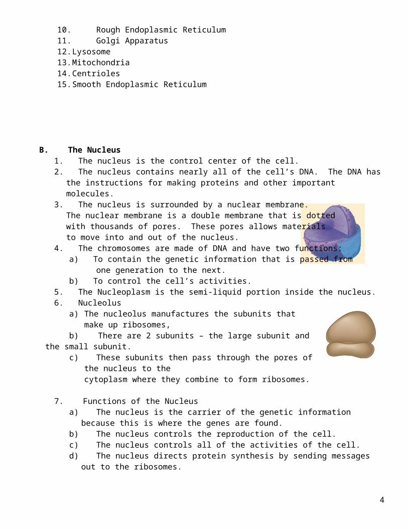
B. The Nucleus1. The nucleus is the control center of the cell. 2. The nucleus contains nearly all of the cell’s DNA. The DNA has the instructions for making
proteins and other important molecules.3. The nucleus is surrounded by a nuclear membrane.
The nuclear membrane is a double membrane that is dotted with thousands of pores. These pores allows materials to move into and out of the nucleus.
4. The chromosomes are made of DNA and have two functions: a) To contain the genetic information that is passed from one generation to the next.
b) To control the cell’s activities.5. The Nucleoplasm is the semi-liquid portion inside the nucleus.6. Nucleolus
a) The nucleolus manufactures the subunits that make up ribosomes,b) There are 2 subunits – the large subunit and the small subunit.c) These subunits then pass through the pores of the nucleus to the
cytoplasm where they combine to form ribosomes.
7. Functions of the Nucleusa) The nucleus is the carrier of the genetic information because this is where the genes are found. b) The nucleus controls the reproduction of the cell.c) The nucleus controls all of the activities of the cell.d) The nucleus directs protein synthesis by sending messages out to the ribosomes.
3
C. Ribosomes1. Ribosomes may be found free floating in the cytoplasm, or they may be found attached to the
endoplasmic reticulum. 2. Ribosomes are the most numerous of the cell’s organelles.3. Ribosomes are the site of protein synthesis. All proteins of the cell are made by the
ribosomes.
D. Endoplasmic Reticulum1. The internal membrane system of a cell is known as the
endoplasmic reticulum.2. This system of membranes is so extensive throughout
the cell that it accounts for more than half the total membrane in a cell.
3. It connects the cell membrane to the nuclear membrane.4. The smooth endoplasmic reticulum has no ribosomes. The function of the smooth
endoplasmic reticulum is to make lipids that will be used in the cell membrane.5. The rough endoplasmic reticulum has ribosomes attached to it. This type of endoplasmic
reticulum is involved in the making of proteins. Newly made proteins leave the ribosome and are inserted into spaces of the endoplasmic reticulum where they are modified and shaped into a functioning protein.
E. Golgi Apparatus1. Proteins that were produced in the rough endoplasmic reticulum now
move to the Golgi apparatus.2. The Golgi apparatus appears as a stack of loosely connected membranes.3. The function of the Golgi is to modify, sort and package the proteins
that have arrived from the endoplasmic reticulum.4. These proteins will either be stored inside the cell or be secreted to the
outside of the cell.5. The finishing touches are put on proteins here before they are
shipped off to their final destinations.
F. Lysosomes1. Lysosomes are filled with very strong digestive enzymes.2. One function is the digestion of carbohydrates, proteins, and lipids into small molecules that
can be used by the rest of the cell. They recycle the cell's own organic materials, breaking them down into their building blocks, and returning them to the cytoplasm to be used again.
3. Lysosomes are responsible for destroying old organelles that can no longer carry out their function.
4. Lysosomes help to “clean up” or destroy any debris that might build up inside the cell.5. Lysosomes are surrounded by a thick membrane, because the cell would be destroyed if the
enzymes were released.
G. Vacuoles1. A vacuole is a storage area inside a cell.2. A vacuole may store water, salts, proteins, and carbohydrates.
4

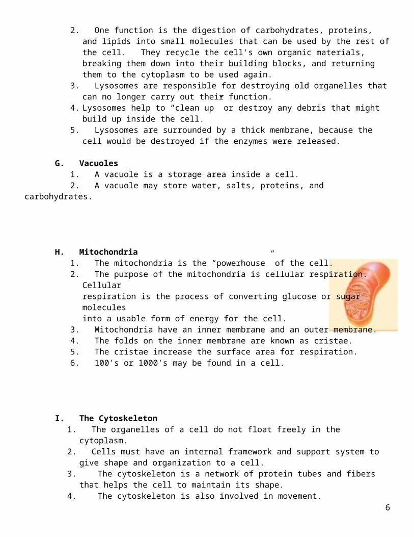
H. Mitochondria1. The mitochondria is the “powerhouse” of the cell.2. The purpose of the mitochondria is cellular respiration. Cellular
respiration is the process of converting glucose or sugar moleculesinto a usable form of energy for the cell.
3. Mitochondria have an inner membrane and an outer membrane.4. The folds on the inner membrane are known as cristae.5. The cristae increase the surface area for respiration.6. 100's or 1000's may be found in a cell.
I. The Cytoskeleton1. The organelles of a cell do not float freely in the cytoplasm.2. Cells must have an internal framework and support system to give shape and organization to a
cell.3. The cytoskeleton is a network of protein tubes and fibers that helps the cell to maintain its
shape.4. The cytoskeleton is also involved in movement.5. Two of the types of fibers found in the cytoskeleton are microfilaments and microtubules.
6. Microfilaments are solid, threadlike, protein structures. Microfilaments form extensive frameworks inside the cell to give support to the cell. They help to bear mechanical stress.
7. Microfilaments also help cells to move. They can assemble and disassemble rapidly causing movement.
8. Microtubules are hollow structures. Their functions include:a) Cell shape.b) The separation of chromosomes during cell division.c) The formation of cilia and flagella.
J. The Cell Membrane1. Also called the plasma membrane.2. Maintains the shape of a cell.3. Separates one animal cell from the next.4. Regulates the passage of materials into and out of the cell.5. Made mostly of lipids and proteins.
5
VI. The Plant Cell
A. A plant cell has many of the same partsfound inside an animal cell, but there are afew organelles that are only found inplant cells.
1 – Golgi apparatus2 – Mitochondria3 – Central vacuole4 – Chloroplasts5 – Ribosomes6 – Endoplasmic reticulum7 – Nucleus8 – Cytoplasm9 – Cell wall10 – Cell membrane
B. Differences Between Plant and Animal Cells1. Structures never found in plant cells: lysosomes, centrioles, flagella (except for sperm cells).2. Structures never found in animal cells: plastids (chloroplast), central vacuole, cell wall.
C. Large, Central Vacuole1. A central vacuole is a very large vacuole found in mature plant cells.2. When filled with water, it creates turgor pressure to give strength and support to the cell. This
allows the plant to support heavy structures such as flowers and leaves.3. It can also serve as a storage area for organic compounds
D. Plastids1. There are three types of plastids found in plant cells
a) Chloroplastsb) Chromaplastsc) Leukoplasts
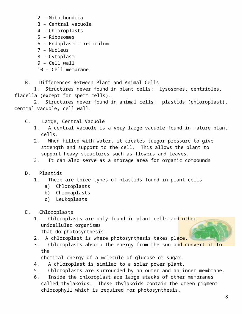
E. Chloroplasts1. Chloroplasts are only found in plant cells and other unicellular organisms
that do photosynthesis.2. A chloroplast is where photosynthesis takes place.3. Chloroplasts absorb the energy from the sun and convert it to the
chemical energy of a molecule of glucose or sugar.4. A chloroplast is similar to a solar power plant.5. Chloroplasts are surrounded by an outer and an inner membrane. 6. Inside the chloroplast are large stacks of other membranes called thylakoids. These thylakoids
contain the green pigment chlorophyll which is required for photosynthesis.
F. Chromoplasts1. “Chromo” means color.2. Chromoplasts contain pigments of all colors except green.3. Chromoplasts give fruits and flowers their colors.
6

G. Leukoplasts1. Leukoplasts have no color.2. This is an area of starch storage inside a cell.
H. The Cell Wall1. The cell wall is a supporting structure found in the cells of plants and fungi.2. The main function of the cell wall is to provide support and protection for the cell.3. The cell wall is composed mostly of cellulose, a tough carbohydrate fiber.
VII. The Diversity of Cellular Life (Levels of Cellular Organization)
A. Unicellular Organisms1. A single-celled organism.2. Examples: bacteria, yeast, the ameba.
B. Colonial Organisms1. Unicellular organisms that live together in groups.2. The cells have no relationship to each other.3. There is no specialization or differentiation.
C. Multicellular Organisms1. A multicellular organism is a group of cells that live and work together in one organism.2. There is differentiation and cell specialization.3. Advantage of having cell specialization: A cell that only has to do one function can be much
more efficient at that one job.4. Disadvantage of cell specialization: The cells are dependent upon one another. If one group of
cells fails to do its job, the other cells will perish.
D. Levels of Organization
1. Cell Specialization: A cell that becomes specialized for just one function.2. Tissue: A group of similar cells all performing a similar activity.3. Organ: A group of several tissues functioning as a unit, performing the same function.4. Organs work together to form systems.5. Various systems work together to form a multicellular organism.
VIII. Prokaryotic and Eukaryotic Cells
A. All cells have two characteristics in common:1. They are surrounded by a barrier called a cell membrane.2. They contain DNA.3. All cells fall into two broad groups, depending on whether or not they contain a nucleus.
a) Prokaryotic cellsb) Eukaryotic cells
7
B. Prokaryotic Cells1. Prokaryotic cells lack a nucleus and membrane-bound organelles.2. Prokaryotic cells have genetic material (DNA) that is not contained inside a nucleus. No
membrane separates this from the rest of the cell.3. Prokaryotic cells are generally smaller and simpler than eukaryotic cells.4. Prokaryotic cells have a cell wall.5. Prokaryotic cells have cell membranes and ribosomes.6. Bacteria are prokaryotic cells.
C. Eukaryotic Cells1. Eukaryotic cells are generally larger and much more complex than prokaryotic cells. 2. Eukaryotic cells have a true nucleus and membrane-bound organelles.3. Eukaryotic cells contain a nucleus which is kept separate from the rest of the cell.4. Plants, animals, protists, fungi all have eukaryotic cells.
IX. How Do Materials Enter and Leave A Cell? (The Cell Membrane)
A. Structure of the Cell Membrane
1. Parts of the membrane1 – Cell membrane2 – Proteins3 – Lipid bilayer4 – Carbohydrates5 – Transport protein
2. The cell membrane regulates what enters and what leavesthe cell. It also provides protection and support to the cell.
3. The membrane consists of a lipid bilayer (double layer) in which proteins are embedded. The lipid bilayer gives the membrane a flexible structure that forms a strong barrier between the inside and the outside of the cell.
4. Many of the proteins form channels and pumps to help move materials across the membrane.
5. The carbohydrates serve as identification markers to help individual cells to identify one another.
B. Homeostasis1. Homeostasis is a balance that organisms maintain through self-regulating adjustments.2. It requires self-regulation of materials coming into the cell and going out of the cell.3. The cell is an open system: it requires the constant inflow of matter and energy and the constant
out flow of waste.
C. Permeability of the Membrane1. The cell membrane is called a selectively permeable membrane or a semipermeable membrane.2. It has the ability to let one substance pass through more readily than others; some materials are
not allowed to enter at all.3. It can control the speed at which molecules are allowed to enter.
8

D. The Concentration Gradient 1. In the absence of other forces, materials will tend to
move from an area of high concentration to an area of lowerconcentration.
2. On the drawing to the right, label the area of higher concentration.Label the area of lower concentration.Draw an arrow on the drawing showing the direction of movementfor this solute.
3. Describe what is happening in the drawing below.
A. B. C.
A) There is a higher concentration of solute molecules on one side of the membrane.
B) The solute molecules move from the side of higher concentration to the side of lower concentration. This movement will continue until the concentration is equal on both sides of the membrane.
C) Equilibrium has been reached; the concentration is equal on both sides of the membrane. There will still be movement in both directions, but the concentrations will remain equal.
E. Types of Passive Transport1. Passive transport means that no energy is being used to move molecules across the
membrane.
2. Diffusiona) Diffusion is the spreading out of molecules from a region of high concentration to a region of
low concentration.
3. Osmosisa) Osmosis is the movement of water across a membrane from a region of high concentration to
a region of low concentrationb) Refers to the movement of water only.
4. There are three types of water solutions:a) Isotonicb) Hypertonicc) Hypotonic
9

Type of Solution Animal Cell Plant Cell
Isotonic
The amount of water is the same on the inside and the outside of the cell. Water will still flow back and forth across the membrane, but at the same rate in both directions.
Water in Water out Water in Water out
Hypertonic
If a cell is placed in a hypertonic solution, there is more water on the inside of the cell than on the outside of the cell. There is a net movement of water out of the cell.
Plasmolysis: Too much water moves out and the cell collapses.
Water moves out
Plasmolysis has occurred.
Water moves out
Plasmolysis has occurred.
Hypotonic
If a cell is placed in a hypotonic solution, there is more water on the outside of the cell than on the inside of the cell. There is a net movement of water into the cell.
Cytolysis: Too much water moves in and the cell membrane bursts because of the water pressure.
Water enters cell
Cytolysis has occurred.
Water enters cell
In cells with a cell wall, cytolysis is not likely to occur. The central vacuole of a plant cell will become extremely full of water. Turgor pressure will increase. This helps to give structure and support to a plant cell.
10

Let’s Practice!!
Label the drawing as we work through this.
1. The bag contains a 20% salt solution.
2. The water surrounding the bag is pure (100%) water.
3. What is the concentration of water inside the bag? (80%)
4. Is the bag hypotonic or hypertonic to the water on the outside? (hypertonic)
5. Is the water on the outside hypertonic or hypotonic to the bag? (hypotonic)
6. In which direction will water move? (in)
7. In which direction will salt move? (out)
8. What process might occur if too much water moves into the bag? (cytolysis)
9. The movement of the salt and the water will continue until??? (both sides are equal)
10. After equilibrium has been reached, what will happen to the movement of these molecules? (Movement
will continue in both directions, but the equilibrium will be maintained.)
11. Water always moves from an area of high concentration to an area of low concentration. In other words,
water moves from the hypotonic side to the hypertonic side.
Label the drawing as we work through this.
1. The bag contains a 40% sugar solution.
2. The water solution surrounding the bag contains a 40% sugar solution.
3. What is the concentration of water inside the bag? (60%)
4. What is the concentration of water on the outside of the bag? (60%)
5. What type of solutions are these? (isotonic)
6. In which direction will water move? (In both directions)
11

Label the drawing as we work through this.
1. The bag contains a 5% salt solution.
2. The water surrounding the bag contains a 25% salt solution.
3. What is the concentration of water inside the bag? (95%)
4. What is the concentration of water outside the bag? (75%)
5. Is the bag hypotonic or hypertonic to the water on the outside? (hypotonic)
6. Is the water on the outside hypertonic or hypotonic to the bag? (hypertonic)
7. In which direction will water move? (out)
8. In which direction will salt move? (in)
9. What process might occur if too much water leaves the bag? (plasmolysis)
10. The movement of the salt and the water will continue until??? (both sides are equal)
11. After equilibrium has been reached, what will happen to the movement of these molecules? (Movement
will continue in both directions, but the equilibrium will be maintained.)
12. Water always moves from an area of high concentration to an area of low concentration. In other words,
water moves from the hypotonic side to the hypertonic side.
F. Facilitated Diffusion1. Polar molecules (water, glucose) have difficulty crossing through
the lipid bilayer of the membrane. 2. Transport proteins help these molecules to pass through the
membrane more easily.3. Polar molecules cross directly through the protein without
coming into contact with the lipid bilayer.4. This is known as facilitated diffusion because these proteins
“facilitate or help” the diffusion of these molecules across the membrane.
5. Facilitated diffusion is considered passive transport because the solute is moving down its concentration gradient. Facilitated diffusion speeds the passage of a solute by providing a passage through the membrane. It does not alter the direction of transport.
G. Types of Active Transport
1. Materials must sometimes move against the concentration gradient. The cell must often move materials from an area of low concentration to an area of higher concentration.
2. This is called active transport, and the cell must expend energy to accomplish it.
3. If small molecules and ions need to be moved across the membrane against the concentration gradient, it will require the use of protein pumps
12

that are embedded in the membrane. This use of protein pumpsrequires much energy.
4. Large molecules may have to be transported by a movement of the cell membrane.
Endocytosis is the process of taking material into the cell by means of infoldings, or pockets, of the cell membrane. The pocket that results breaks loose from the cell membrane and forms a vacuole within the cytoplasm. Large molecules and clumps of food are taken up in this way.
This requires much energy.
Two type of endocytosis are phagocytosis and pinocytosis.
Phagocytosis is the engulfing of large food particles.
Pinocytosis is “cellular drinking”. The cell surrounds and engulfs droplets of extracellular fluid. It is not the fluid that is needed, but the molecules dissolved in the droplets.
Exocytosis is the release of large materials from the cell. A vacuole fuses with the cell membrane, forcing the contents out of the cell.
13