Cell Host & Microbe Article - COnnecting REpositories · Cell Host & Microbe Article ... E. coli,...
13
Cell Host & Microbe Article An Iron-Containing Dodecameric Heptosyltransferase Family Modifies Bacterial Autotransporters in Pathogenesis Qiuhe Lu, 1,8 Qing Yao, 1,8 Yue Xu, 1,2,8 Lin Li, 1 Shan Li, 1 Yanhua Liu, 3 Wenqing Gao, 1 Miao Niu, 1 Michal Sharon, 4 Gili Ben-Nissan, 4 Alla Zamyatina, 5 Xiaoyun Liu, 3 She Chen, 1 and Feng Shao 1,6,7, * 1 National Institute of Biological Sciences, Beijing 102206, China 2 College of Biological Sciences, China Agricultural University, Beijing 100094, China 3 Institute of Analytical Chemistry & Synthetic and Functional Biomolecules Center, College of Chemistry and Molecular Engineering, Peking University, Beijing 100871, China 4 Department of Biological Chemistry, The Weizmann Institute of Science, Rehovot 76100, Israel 5 Department of Chemistry, University of Natural Resources and Life Sciences, A-1190 Vienna, Austria 6 National Laboratory of Biomacromolecules, Institute of Biophysics, Chinese Academy of Sciences, Beijing 100101, China 7 National Institute of Biological Sciences, Beijing, Collaborative Innovation Center for Cancer Medicine, Beijing 102206, China 8 Co-first author *Correspondence: [email protected] http://dx.doi.org/10.1016/j.chom.2014.08.008 SUMMARY Autotransporters deliver virulence factors to the bacterial surface by translocating an effector pas- senger domain through a membrane-anchored bar- rel structure. Although passenger domains are diverse, those found in enteric bacteria autotrans- porters, including AIDA-I in diffusely adhering Es- cherichia coli (DAEC) and TibA in enterotoxigenic E. coli, are commonly glycosylated. We show that AIDA-I is heptosylated within the bacterial cyto- plasm by autotransporter adhesin heptosyltransfer- ase (AAH) and its paralogue AAH2. AIDA-I heptosyla- tion determines DAEC adhesion to host cells. AAH/ AAH2 define a bacterial autotransporter heptosyl- transferase (BAHT) family that contains ferric ion and adopts a dodecamer assembly. Structural ana- lyses of the heptosylated TibA passenger domain reveal 35 heptose conjugates forming patterned and solenoid-like arrays on the surface of a b helix. Additionally, CARC, the AIDA-like autotransporter from Citrobacter rodentium, is essential for coloni- zation in mice and requires heptosylation by its cognate BAHT. Our study establishes a bacterial glycosylation system that regulates virulence and is essential for pathogenesis. INTRODUCTION A common mechanism in bacterial pathogenesis is to deliver various virulence factors onto bacterial surface by the auto- transporter secretion pathway (also known as type V secretion) (Dautin and Bernstein, 2007; Leyton et al., 2012). Since the IgA protease autotransporter was discovered in Neisseria gonor- rheae (Pohlner et al., 1987), the number of autotransporters iden- tified has expanded rapidly. All autotransporters share a similar modular structure consisting of a signal peptide, an N-terminal functional passenger domain, and a C-terminal b barrel domain that mediates translocation of the passenger domain across bac- terial outer membrane. Owing to the difference in the passenger domain, diverse virulence functions, including cell adhesion and invasion, aggregation and biofilm formation, proteolytic diges- tion of host proteins, host cell vacuolation, and formation of bac- terial actin tail, have been recorded with autotransporters from different bacterial pathogens (Benz and Schmidt, 2011; Hender- son et al., 2004; Lazar Adler et al., 2011; Wells et al., 2007). A subfamily of autotransporters, including AIDA-I from DAEC 2787 (Benz and Schmidt, 1989), TibA from enterotoxigenic E. coli (ETEC) H10407 (Elsinghorst and Weitz, 1994), and Ag43 from pathogenic E. coli species (van der Woude and Henderson, 2008), are long known to be glycosylated in their passenger domains (Benz and Schmidt, 2001; Lindenthal and Elsinghorst, 1999; Sherlock et al., 2006). The prototypical AIDA-I is proposed to mediate bacterial autoaggregation and adhesion to host cells (Benz and Schmidt, 1989; Charbonneau and Mourez, 2007; Sherlock et al., 2004). AIDA-I is widely present (Wells et al., 2010), particularly in porcine diarrheagenic E. coli, and has been a promising vaccine candidate (Ravi et al., 2007; Zhao et al., 2009). Glycosylation of AIDA-I and related autotransport- ers represents an example of protein glycosylation in bacteria (Benz and Schmidt, 2002) despite an undefined functional signif- icance. Studies performed in the heterologous system (Benz and Schmidt, 2001; Charbonneau et al., 2012; Moormann et al., 2002) indicate the involvement of AAH in DAEC and its ETEC homolog TibC in AIDA-I and TibA glycosylation, respectively. However, AAH and TibC share little sequence homology to known glycosyltransferases, and it is not clear whether AAH/ TibC harbor a glycosyltransferase activity from a biochemical perspective. Thus, how AIDA-I and related autotransporters are glycosylated and the functional significance of this modifica- tion in infection are not established. Cell Host & Microbe 16, 351–363, September 10, 2014 ª2014 Elsevier Inc. 351
Transcript of Cell Host & Microbe Article - COnnecting REpositories · Cell Host & Microbe Article ... E. coli,...

Cell Host & Microbe
Article
An Iron-Containing DodecamericHeptosyltransferase Family ModifiesBacterial Autotransporters in PathogenesisQiuhe Lu,1,8 Qing Yao,1,8 Yue Xu,1,2,8 Lin Li,1 Shan Li,1 Yanhua Liu,3 Wenqing Gao,1 Miao Niu,1 Michal Sharon,4
Gili Ben-Nissan,4 Alla Zamyatina,5 Xiaoyun Liu,3 She Chen,1 and Feng Shao1,6,7,*1National Institute of Biological Sciences, Beijing 102206, China2College of Biological Sciences, China Agricultural University, Beijing 100094, China3Institute of Analytical Chemistry & Synthetic and Functional Biomolecules Center, College of Chemistry and Molecular Engineering,
Peking University, Beijing 100871, China4Department of Biological Chemistry, The Weizmann Institute of Science, Rehovot 76100, Israel5Department of Chemistry, University of Natural Resources and Life Sciences, A-1190 Vienna, Austria6National Laboratory of Biomacromolecules, Institute of Biophysics, Chinese Academy of Sciences, Beijing 100101, China7National Institute of Biological Sciences, Beijing, Collaborative Innovation Center for Cancer Medicine, Beijing 102206, China8Co-first author
*Correspondence: [email protected]://dx.doi.org/10.1016/j.chom.2014.08.008
SUMMARY
Autotransporters deliver virulence factors to thebacterial surface by translocating an effector pas-senger domain through a membrane-anchored bar-rel structure. Although passenger domains arediverse, those found in enteric bacteria autotrans-porters, including AIDA-I in diffusely adhering Es-cherichia coli (DAEC) and TibA in enterotoxigenicE. coli, are commonly glycosylated. We show thatAIDA-I is heptosylated within the bacterial cyto-plasm by autotransporter adhesin heptosyltransfer-ase (AAH) and its paralogue AAH2. AIDA-I heptosyla-tion determines DAEC adhesion to host cells. AAH/AAH2 define a bacterial autotransporter heptosyl-transferase (BAHT) family that contains ferric ionand adopts a dodecamer assembly. Structural ana-lyses of the heptosylated TibA passenger domainreveal 35 heptose conjugates forming patternedand solenoid-like arrays on the surface of a b helix.Additionally, CARC, the AIDA-like autotransporterfrom Citrobacter rodentium, is essential for coloni-zation in mice and requires heptosylation by itscognate BAHT. Our study establishes a bacterialglycosylation system that regulates virulence and isessential for pathogenesis.
INTRODUCTION
A common mechanism in bacterial pathogenesis is to deliver
various virulence factors onto bacterial surface by the auto-
transporter secretion pathway (also known as type V secretion)
(Dautin and Bernstein, 2007; Leyton et al., 2012). Since the IgA
protease autotransporter was discovered in Neisseria gonor-
Cell Host & M
rheae (Pohlner et al., 1987), the number of autotransporters iden-
tified has expanded rapidly. All autotransporters share a similar
modular structure consisting of a signal peptide, an N-terminal
functional passenger domain, and a C-terminal b barrel domain
thatmediates translocation of thepassenger domain across bac-
terial outer membrane. Owing to the difference in the passenger
domain, diverse virulence functions, including cell adhesion and
invasion, aggregation and biofilm formation, proteolytic diges-
tion of host proteins, host cell vacuolation, and formation of bac-
terial actin tail, have been recorded with autotransporters from
different bacterial pathogens (Benz and Schmidt, 2011; Hender-
son et al., 2004; Lazar Adler et al., 2011; Wells et al., 2007).
A subfamily of autotransporters, including AIDA-I from DAEC
2787 (Benz and Schmidt, 1989), TibA from enterotoxigenic
E. coli (ETEC) H10407 (Elsinghorst and Weitz, 1994), and Ag43
from pathogenic E. coli species (van der Woude and Henderson,
2008), are long known to be glycosylated in their passenger
domains (Benz and Schmidt, 2001; Lindenthal and Elsinghorst,
1999; Sherlock et al., 2006). The prototypical AIDA-I is proposed
to mediate bacterial autoaggregation and adhesion to host cells
(Benz and Schmidt, 1989; Charbonneau and Mourez, 2007;
Sherlock et al., 2004). AIDA-I is widely present (Wells et al.,
2010), particularly in porcine diarrheagenic E. coli, and has
been a promising vaccine candidate (Ravi et al., 2007; Zhao
et al., 2009). Glycosylation of AIDA-I and related autotransport-
ers represents an example of protein glycosylation in bacteria
(Benz and Schmidt, 2002) despite an undefined functional signif-
icance. Studies performed in the heterologous system (Benz and
Schmidt, 2001; Charbonneau et al., 2012; Moormann et al.,
2002) indicate the involvement of AAH in DAEC and its ETEC
homolog TibC in AIDA-I and TibA glycosylation, respectively.
However, AAH and TibC share little sequence homology to
known glycosyltransferases, and it is not clear whether AAH/
TibC harbor a glycosyltransferase activity from a biochemical
perspective. Thus, how AIDA-I and related autotransporters
are glycosylated and the functional significance of this modifica-
tion in infection are not established.
icrobe 16, 351–363, September 10, 2014 ª2014 Elsevier Inc. 351

Figure 1. Glycosylation of AIDA-I Mediates DAEC Adhesion to Host Cells and Requires AAH and AAH2
(A and B) Reconstitution of AAH-induced AIDA-I glycosylation and bacterial adhesion to HeLa cells. E. coli BL21 (DE3) was transformed with a plasmid harboring
AIDA-I alone (pAIDA-I) or the aah-aidA locus (pAAH�AIDA-I). In (A), EGFP was additionally expressed to label the bacteria. Infected cells and the bacteria were
visualized by differential interference contrast (DIC) microscopy and the green fluorescence, respectively. The average numbers of adherent bacteria were
expressed as the mean number of bacteria adhering to one cell (the total number of recovered bacteria/the number of seeded cell in each well) ± SD of four
technical repeats. Scale bar, 20 mm. In (B), bacterial lysates were subjected to anti-AIDA-I and anti-AAH immunoblotting; AIDA-I glycosylation was detected with
Amersham ECL glycoprotein detection system.
(C) Genetic analyses of the aah-aidA locus in conferring DAEC adhesion. HeLa cells were infected with wild-type (WT) or indicated DAEC deletion strains ex-
pressing EGFP. The Daah/DaidA double deletion strain was complemented with a plasmid expressing AAH (pAAH) or AIDA-I (pAIDA-I) or both (pAAH�AIDA-I).
Data are presented similarly as in (A).
(D) Effects of aah deletion on endogenous AIDA-I glycosylation. Lysates of DAEC (WT orDaah) or BL21 (DE3) cells expressing AIDA-I alone (NC) were subjected to
anti-AIDA-I and anti-AAH immunoblotting.
(E) Gain-of-function screen of the cryptic AIDA-I glycosyltransferase. Genomic library prepared from DAEC Daah/DaidA was introduced into E. coli Top10
expressing Flag-AIDA-I. Results of representative clones are shown. Lysates of Top10 cells harboring the aah-aidA locus and aidA alone serve as the positive (PC)
and negative (NC) controls, respectively.
(F–H) Functional analyses of AAH2 in supporting AIDA-I glycosylation and bacterial adhesion to host cells. In (F and G), aah/aah2 double deletion (D/D)
DAEC strain was complemented with a plasmid expressing AAH or AAH2. In (H), AIDA-I (pAIDA-I) was expressed alone or together with AAH2 (pAAH2)
in E. coli Top10. Adhesion assay in (G and H) was performed and data were presented similarly as in (A). AIDA-I glycosylation in (F) was assayed similarly
as in (B).
See also Figure S1.
Cell Host & Microbe
BAHT Modification of Bacterial Autotransporters
In this study, we provide genetic evidences that heptosylation
of AIDA-I mediates DAEC adhesion to host cells, which requires
AAH as well as AAH-like AAH2. In vitro biochemical analyses
demonstrate that AAH and AAH2 are bone fide heptosyltrans-
ferases exclusively modifying serine residues in AIDA-I. Crystal
structure of heptosylated TibA passenger domain identifies 35
heptose moieties forming patterned and solenoid-like arrays
on the surface of a b helix structure. AAH and TibC define a large
bacterial autotransporter heptosyltransferase (BAHT) family. We
further identify an autotransporter required for C. rodentium
colonization in infected mice, which requires hyperheptosylation
by its cognate BAHT, demonstrating the importance of the BAHT
family in pathogenesis.
352 Cell Host & Microbe 16, 351–363, September 10, 2014 ª2014 El
RESULTS AND DISCUSSION
Identification of AAH2 Required for AIDA-I Glycosylationbut Not for DAEC Adhesion to Host CellsConsistent with previous studies (Benz and Schmidt, 2001;
Charbonneau et al., 2012; Moormann et al., 2002), ectopic
expression of the aah-aidA locus, but not aidA alone, in E. coli
BL21 resulted in a tight adhesion of the bacteria to HeLa cells
(Figure 1A) and concurrent AIDA-I glycosylation (Figure 1B). To
investigate the physiological significance of these phenomena,
we performed genetic analyses of the aidA locus. Wild-type
DAEC robustly adhered to HeLa cells, and deletion of the aah-
aidA locus completely abolished such property (Figure 1C).
sevier Inc.

Cell Host & Microbe
BAHT Modification of Bacterial Autotransporters
Notably, deletion of aidA or its upstream aah alone also disrupted
the adhesion (Figure 1C). Complementation analyses confirmed
that aidA and aahwere both required for DAEC adhesion to HeLa
cells (Figure 1C).
Contradictory to previous understandings (Benz and Schmidt,
2001; Moormann et al., 2002), deletion of aah only reduced but
did not completely abolish AIDA-I glycosylation (Figure 1D).
This intriguing observation suggested two possibilities. AAH
determines AIDA-I-mediated DAEC adhesion independently of
AIDA-I glycosylation; alternatively, another cryptic glycosyltrans-
ferase accounts for the remaining AIDA-I glycosylation in DAEC
Daah, but it is not sufficient to confer bacterial adhesion to host
cells. Given that nonglycosylated AIDA-I could not confer adhe-
sion of E. coli BL21 to HeLa cells (Figures 1A and 1B), we fol-
lowed up with the latter hypothesis and attempted to identify
the cryptic glycosyltransferase. The genome of DAEC 2787
has not been sequenced; a genomic library with an average
insert size of 5–10 kb was prepared from the aah/aidA double
deletion strain. The library plasmids were introduced into
E. coli Top10 expressing Flag-AIDA-I. Individual recombinant
was then screened for clones capable of inducing AIDA-I glyco-
sylation. Among�1,000 recombinants examined, clone 14 could
induce AIDA-I glycosylation (Figure 1E). Sequencing of clone 14
identified a gene encoding a protein of �75% homology to AAH
(Figure S1A available online), which was named as aah2. Impor-
tantly, when the plasmid-born aah and chromosome-encoded
aah2 were both deleted, AIDA-I glycosylation in DAEC was
completely abolished (Figure 1F). Anti-AAH antibody that could
crossreact with overexpressed AAH2 (Figure 1F) only detected
endogenous AAH, but not AAH2 (Figure 1D). The messenger
RNA level of AAH2 was also much lower than that of AAH (data
not shown). Thus, the low expression of endogenous AAH2 con-
tributes to AIDA-I glycosylation but plays a negligible role in bac-
terial adhesion to host cells. Notably, AAH2 overexpression
could restore AIDA-I glycosylation in DAEC DaahDaah2 (Figures
1F and S1B) and confer AIDA-I-mediated adhesion to HeLa cells
in both DAEC DaahDaah2 (Figure 1G) and E. coli Top10 (Fig-
ure 1H). Thus, AAH2 and AAH harbor a similar biochemical func-
tion, and both of them are important for AIDA-I glycosylation. It is
also a possibility that AAH2might be involved in heptosylation of
other bacterial proteins in addition to AIDA-I.
AAH/AAH2 Induce AIDA-I Hyperheptosylation on SerineResidues in the Passenger DomainResults from above analyses and those in previous studies indi-
cate that AAH2 and possibly also AAH may harbor a glycosyl-
transferase activity on AIDA-I. To identify an AIDA-I fragment
that allows for testing the hypothesis in a defined in vitro system,
extensive truncation analyses of AIDA-I passenger domain (res-
idues 50–846) were performed (Figure S2A); each fragment,
fusedC-terminal to glutathione S-transferase (GST) andN-termi-
nal to a Flag tag, was expressed in E. coli in the presence or
absence of AAH. Multiple AIDA-I fragments exhibited a slow
migration on the SDS-PAGE gel due to the presumed glycosyla-
tion modification (Figure S2B). The smallest one, containing res-
idues 531–611 (AIDA-I531-611), was further analyzed (Figure 2A).
Following removal of the GST, AIDA-I531-611-Flag was subjected
to mass spectrometry analysis to unambiguously define the
sugar moiety and the exact chemical linkage. Matrix-assisted
Cell Host & M
laser desorption/ionization time-of-flight mass spectrometry
(MALDI-TOF-MS) gave a mass of 9,867 Da that matched the
theoretic mass of AIDA-I531-611-Flag (9,866 Da) (Figure 2B).
Notably, coexpression of either AAH or AAH2 resulted in a
mass increase of 1,536 Da. The sample was further digested
with trypsin and analyzed by liquid chromatography-tandem
mass spectrometry (LC-MS/MS). Full sequence coverage
was obtained, with the following peptides detected in the full-
scan MS analysis: GPLGSGRPM-531NQEGR535 (peptide 1),
536QYVYSGATATSTVGNNEGR554 (peptide 2), 555EYVLSG-
GITDGTVLNSGGLQAVSSGGK581 (peptide 3), 582ASATVINEG
GAQFVYDGGQVTGTNIK607 (peptide 4), and 608NGGT611-
DYKDDDDK (peptide 5). In AAH/AAH2-modified samples, Pep-
tides 1 and 4 showed a homogenous mass increase of 192 Da;
peptides 2 and 3 were predominantly modified with 384 Da
(23 192 Da) and 768 Da (43 192 Da), respectively, and peptide
5, containing no serine/threonine residues, was not modified.
The 192 Da mass increase corresponded exactly to a heptose
modification. Accordingly, the 1,536 Da mass increase on
AAH/AAH2-modified AIDA-I531-611 suggested eight heptosyla-
tion modifications.
Classical collision-induced dissociation (CID) mass spec-
trometry was not successful in identifying the heptosylation
sites in AIDA-I531-611-Flag due to the neutral loss during peptide
fragmentation. To circumvent this problem, electron transfer
dissociation (ETD) mass spectrometry that can better preserve
the labile sugar moiety was employed, which identified hepto-
sylation modifications on the two serines in peptide 2 (Fig-
ure 2C). No threonine residues in this peptide were heptosy-
lated. All the eight serines in AIDA-I531-611, but not any other
residues, were modified according to mass spectra of other
four peptides, which agreed with the total mass increase of
1,536 Da (8 3 192 Da) on AIDA-I531-611 (Figure 2B). Thus,
AAH/AAH2-induced AIDA-I heptosylation is exclusively on
serine residues, a unique phenomenon in the conventional
O-linked glycan modification.
AAH/AAH2 Induce AIDA-I Heptosylation in BacterialCytoplasm using ADP-Heptose from the LPSSynthesis PathwayTo investigate whether AAH/AAH2 and the heptosylation play a
role in AIDA-I transport across the bacterial membrane, we first
examined the subcellular localization of AAH and AIDA-I. As
expected, endogenous AIDA-I was accessible to recognition
by anti-AIDA-I antibody in nonpermeabilized DAEC cells (Fig-
ure S2C). Fractionation of DAEC lysates confirmed the mem-
brane localization of AIDA-I and also revealed the cytoplasmic
localization of AAH (Figure S2D). As a control, OmpA, DnaK,
and Bla were enriched in the membrane, cytoplasm, and peri-
plasm fractions, respectively. This suggests that AAHs-cata-
lyzed AIDA-I heptosylation occurs within bacterial cytoplasm.
Deletion of both aah and aah2 did not affect the membrane tar-
geting of AIDA-I (Figure S2C); AIDA-I remained enriched in the
membrane fraction in the presence of a nonfunctional AAH
D104A mutant in reconstituted BL21 cells (Figure S2E). Thus,
AAH-catalyzed AIDA-I glycosylation is not required for surface
presentation of AIDA-I. Consistent with this notion, AIDA-I is
sufficient for surface display of other proteins in aah-deficient
bacteria (Jose and Meyer, 2007).
icrobe 16, 351–363, September 10, 2014 ª2014 Elsevier Inc. 353
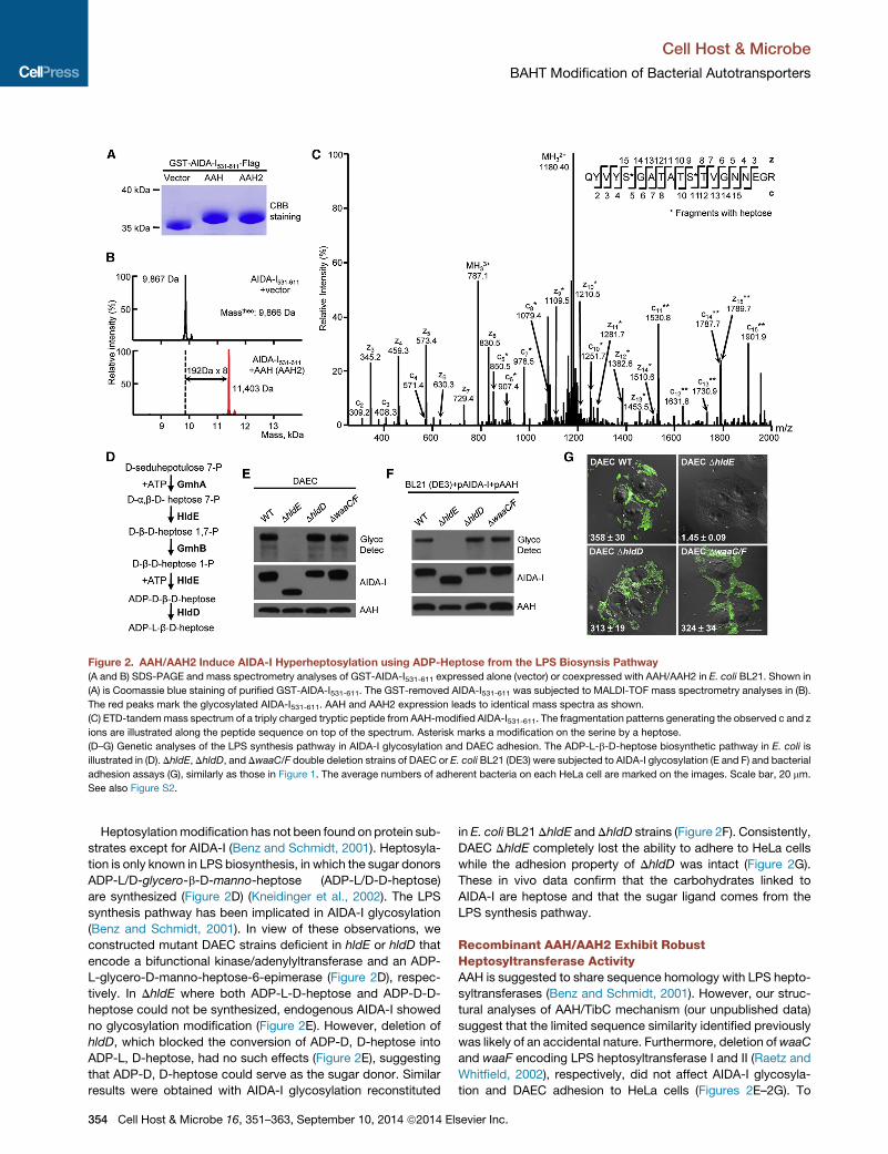
Figure 2. AAH/AAH2 Induce AIDA-I Hyperheptosylation using ADP-Heptose from the LPS Biosynsis Pathway
(A and B) SDS-PAGE and mass spectrometry analyses of GST-AIDA-I531-611 expressed alone (vector) or coexpressed with AAH/AAH2 in E. coli BL21. Shown in
(A) is Coomassie blue staining of purified GST-AIDA-I531-611. The GST-removed AIDA-I531-611 was subjected to MALDI-TOF mass spectrometry analyses in (B).
The red peaks mark the glycosylated AIDA-I531-611. AAH and AAH2 expression leads to identical mass spectra as shown.
(C) ETD-tandemmass spectrum of a triply charged tryptic peptide from AAH-modified AIDA-I531-611. The fragmentation patterns generating the observed c and z
ions are illustrated along the peptide sequence on top of the spectrum. Asterisk marks a modification on the serine by a heptose.
(D–G) Genetic analyses of the LPS synthesis pathway in AIDA-I glycosylation and DAEC adhesion. The ADP-L-b-D-heptose biosynthetic pathway in E. coli is
illustrated in (D). DhldE, DhldD, and DwaaC/F double deletion strains of DAEC or E. coli BL21 (DE3) were subjected to AIDA-I glycosylation (E and F) and bacterial
adhesion assays (G), similarly as those in Figure 1. The average numbers of adherent bacteria on each HeLa cell are marked on the images. Scale bar, 20 mm.
See also Figure S2.
Cell Host & Microbe
BAHT Modification of Bacterial Autotransporters
Heptosylationmodification has not been found on protein sub-
strates except for AIDA-I (Benz and Schmidt, 2001). Heptosyla-
tion is only known in LPS biosynthesis, in which the sugar donors
ADP-L/D-glycero-b-D-manno-heptose (ADP-L/D-D-heptose)
are synthesized (Figure 2D) (Kneidinger et al., 2002). The LPS
synthesis pathway has been implicated in AIDA-I glycosylation
(Benz and Schmidt, 2001). In view of these observations, we
constructed mutant DAEC strains deficient in hldE or hldD that
encode a bifunctional kinase/adenylyltransferase and an ADP-
L-glycero-D-manno-heptose-6-epimerase (Figure 2D), respec-
tively. In DhldE where both ADP-L-D-heptose and ADP-D-D-
heptose could not be synthesized, endogenous AIDA-I showed
no glycosylation modification (Figure 2E). However, deletion of
hldD, which blocked the conversion of ADP-D, D-heptose into
ADP-L, D-heptose, had no such effects (Figure 2E), suggesting
that ADP-D, D-heptose could serve as the sugar donor. Similar
results were obtained with AIDA-I glycosylation reconstituted
354 Cell Host & Microbe 16, 351–363, September 10, 2014 ª2014 El
in E. coli BL21 DhldE and DhldD strains (Figure 2F). Consistently,
DAEC DhldE completely lost the ability to adhere to HeLa cells
while the adhesion property of DhldD was intact (Figure 2G).
These in vivo data confirm that the carbohydrates linked to
AIDA-I are heptose and that the sugar ligand comes from the
LPS synthesis pathway.
Recombinant AAH/AAH2 Exhibit RobustHeptosyltransferase ActivityAAH is suggested to share sequence homology with LPS hepto-
syltransferases (Benz and Schmidt, 2001). However, our struc-
tural analyses of AAH/TibC mechanism (our unpublished data)
suggest that the limited sequence similarity identified previously
was likely of an accidental nature. Furthermore, deletion ofwaaC
and waaF encoding LPS heptosyltransferase I and II (Raetz and
Whitfield, 2002), respectively, did not affect AIDA-I glycosyla-
tion and DAEC adhesion to HeLa cells (Figures 2E–2G). To
sevier Inc.
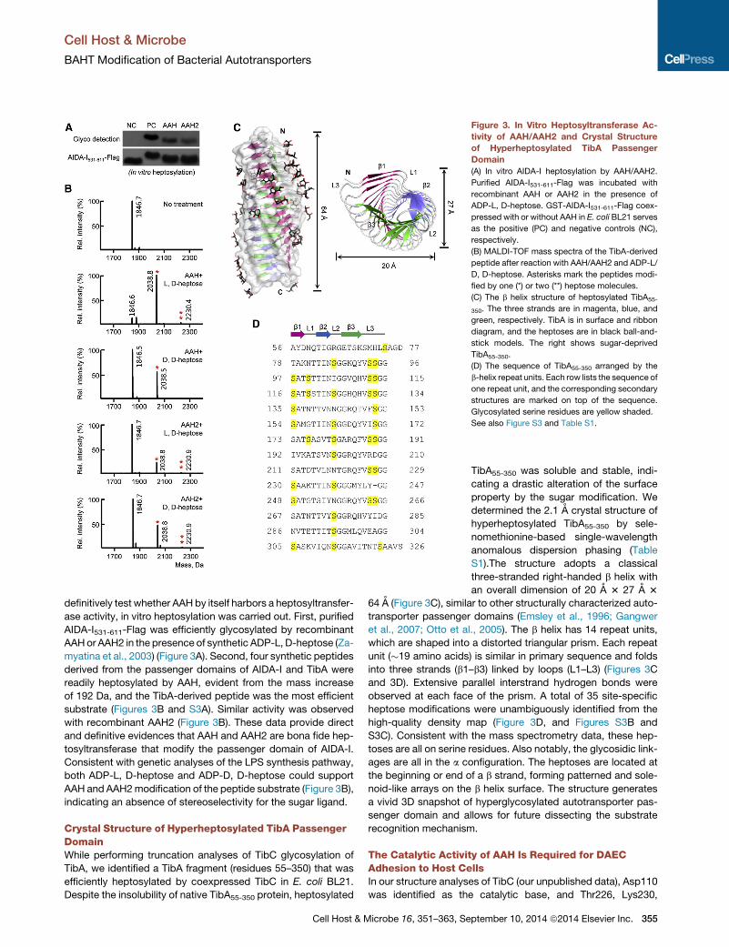
Figure 3. In Vitro Heptosyltransferase Ac-
tivity of AAH/AAH2 and Crystal Structure
of Hyperheptosylated TibA Passenger
Domain
(A) In vitro AIDA-I heptosylation by AAH/AAH2.
Purified AIDA-I531-611-Flag was incubated with
recombinant AAH or AAH2 in the presence of
ADP-L, D-heptose. GST-AIDA-I531-611-Flag coex-
pressed with or without AAH in E. coli BL21 serves
as the positive (PC) and negative controls (NC),
respectively.
(B) MALDI-TOF mass spectra of the TibA-derived
peptide after reaction with AAH/AAH2 and ADP-L/
D, D-heptose. Asterisks mark the peptides modi-
fied by one (*) or two (**) heptose molecules.
(C) The b helix structure of heptosylated TibA55-
350. The three strands are in magenta, blue, and
green, respectively. TibA is in surface and ribbon
diagram, and the heptoses are in black ball-and-
stick models. The right shows sugar-deprived
TibA55-350.
(D) The sequence of TibA55-350 arranged by the
b-helix repeat units. Each row lists the sequence of
one repeat unit, and the corresponding secondary
structures are marked on top of the sequence.
Glycosylated serine residues are yellow shaded.
See also Figure S3 and Table S1.
Cell Host & Microbe
BAHT Modification of Bacterial Autotransporters
definitively test whether AAH by itself harbors a heptosyltransfer-
ase activity, in vitro heptosylation was carried out. First, purified
AIDA-I531-611-Flag was efficiently glycosylated by recombinant
AAHor AAH2 in the presence of synthetic ADP-L, D-heptose (Za-
myatina et al., 2003) (Figure 3A). Second, four synthetic peptides
derived from the passenger domains of AIDA-I and TibA were
readily heptosylated by AAH, evident from the mass increase
of 192 Da, and the TibA-derived peptide was the most efficient
substrate (Figures 3B and S3A). Similar activity was observed
with recombinant AAH2 (Figure 3B). These data provide direct
and definitive evidences that AAH and AAH2 are bona fide hep-
tosyltransferase that modify the passenger domain of AIDA-I.
Consistent with genetic analyses of the LPS synthesis pathway,
both ADP-L, D-heptose and ADP-D, D-heptose could support
AAH and AAH2modification of the peptide substrate (Figure 3B),
indicating an absence of stereoselectivity for the sugar ligand.
Crystal Structure of Hyperheptosylated TibA PassengerDomainWhile performing truncation analyses of TibC glycosylation of
TibA, we identified a TibA fragment (residues 55–350) that was
efficiently heptosylated by coexpressed TibC in E. coli BL21.
Despite the insolubility of native TibA55-350 protein, heptosylated
Cell Host & Microbe 16, 351–363, Se
TibA55-350 was soluble and stable, indi-
cating a drastic alteration of the surface
property by the sugar modification. We
determined the 2.1 A crystal structure of
hyperheptosylated TibA55-350 by sele-
nomethionine-based single-wavelength
anomalous dispersion phasing (Table
S1).The structure adopts a classical
three-stranded right-handed b helix with
an overall dimension of 20 A 3 27 A 3
64 A (Figure 3C), similar to other structurally characterized auto-
transporter passenger domains (Emsley et al., 1996; Gangwer
et al., 2007; Otto et al., 2005). The b helix has 14 repeat units,
which are shaped into a distorted triangular prism. Each repeat
unit (�19 amino acids) is similar in primary sequence and folds
into three strands (b1–b3) linked by loops (L1–L3) (Figures 3C
and 3D). Extensive parallel interstrand hydrogen bonds were
observed at each face of the prism. A total of 35 site-specific
heptose modifications were unambiguously identified from the
high-quality density map (Figure 3D, and Figures S3B and
S3C). Consistent with the mass spectrometry data, these hep-
toses are all on serine residues. Also notably, the glycosidic link-
ages are all in the a configuration. The heptoses are located at
the beginning or end of a b strand, forming patterned and sole-
noid-like arrays on the b helix surface. The structure generates
a vivid 3D snapshot of hyperglycosylated autotransporter pas-
senger domain and allows for future dissecting the substrate
recognition mechanism.
The Catalytic Activity of AAH Is Required for DAECAdhesion to Host CellsIn our structure analyses of TibC (our unpublished data), Asp110
was identified as the catalytic base, and Thr226, Lys230,
ptember 10, 2014 ª2014 Elsevier Inc. 355
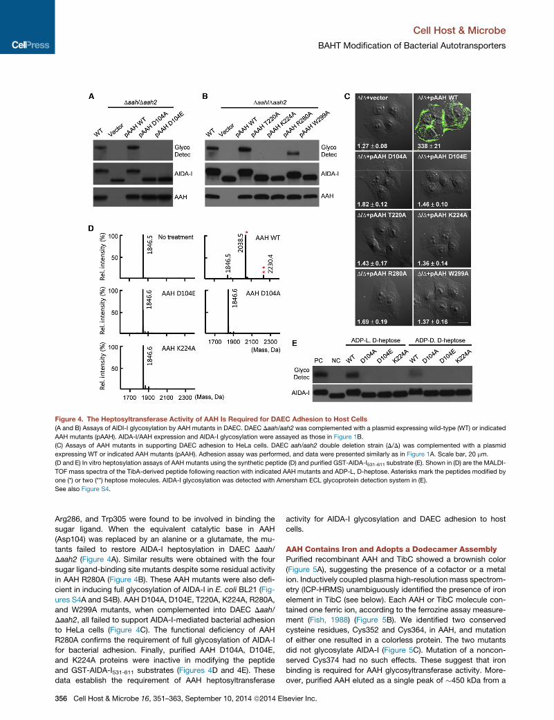
Figure 4. The Heptosyltransferase Activity of AAH Is Required for DAEC Adhesion to Host Cells
(A and B) Assays of AIDI-I glycosylation by AAH mutants in DAEC. DAEC Daah/aah2 was complemented with a plasmid expressing wild-type (WT) or indicated
AAH mutants (pAAH). AIDA-I/AAH expression and AIDA-I glycosylation were assayed as those in Figure 1B.
(C) Assays of AAH mutants in supporting DAEC adhesion to HeLa cells. DAEC aah/aah2 double deletion strain (D/D) was complemented with a plasmid
expressing WT or indicated AAH mutants (pAAH). Adhesion assay was performed, and data were presented similarly as in Figure 1A. Scale bar, 20 mm.
(D and E) In vitro heptosylation assays of AAH mutants using the synthetic peptide (D) and purified GST-AIDA-I531-611 substrate (E). Shown in (D) are the MALDI-
TOF mass spectra of the TibA-derived peptide following reaction with indicated AAH mutants and ADP-L, D-heptose. Asterisks mark the peptides modified by
one (*) or two (**) heptose molecules. AIDA-I glycosylation was detected with Amersham ECL glycoprotein detection system in (E).
See also Figure S4.
Cell Host & Microbe
BAHT Modification of Bacterial Autotransporters
Arg286, and Trp305 were found to be involved in binding the
sugar ligand. When the equivalent catalytic base in AAH
(Asp104) was replaced by an alanine or a glutamate, the mu-
tants failed to restore AIDA-I heptosylation in DAEC Daah/
Daah2 (Figure 4A). Similar results were obtained with the four
sugar ligand-binding site mutants despite some residual activity
in AAH R280A (Figure 4B). These AAH mutants were also defi-
cient in inducing full glycosylation of AIDA-I in E. coli BL21 (Fig-
ures S4A and S4B). AAH D104A, D104E, T220A, K224A, R280A,
and W299A mutants, when complemented into DAEC Daah/
Daah2, all failed to support AIDA-I-mediated bacterial adhesion
to HeLa cells (Figure 4C). The functional deficiency of AAH
R280A confirms the requirement of full glycosylation of AIDA-I
for bacterial adhesion. Finally, purified AAH D104A, D104E,
and K224A proteins were inactive in modifying the peptide
and GST-AIDA-I531-611 substrates (Figures 4D and 4E). These
data establish the requirement of AAH heptosyltransferase
356 Cell Host & Microbe 16, 351–363, September 10, 2014 ª2014 El
activity for AIDA-I glycosylation and DAEC adhesion to host
cells.
AAH Contains Iron and Adopts a Dodecamer AssemblyPurified recombinant AAH and TibC showed a brownish color
(Figure 5A), suggesting the presence of a cofactor or a metal
ion. Inductively coupled plasma high-resolution mass spectrom-
etry (ICP-HRMS) unambiguously identified the presence of iron
element in TibC (see below). Each AAH or TibC molecule con-
tained one ferric ion, according to the ferrozine assay measure-
ment (Fish, 1988) (Figure 5B). We identified two conserved
cysteine residues, Cys352 and Cys364, in AAH, and mutation
of either one resulted in a colorless protein. The two mutants
did not glycosylate AIDA-I (Figure 5C). Mutation of a noncon-
served Cys374 had no such effects. These suggest that iron
binding is required for AAH glycosyltransferase activity. More-
over, purified AAH eluted as a single peak of �450 kDa from a
sevier Inc.
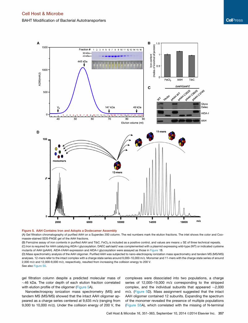
Figure 5. AAH Contains Iron and Adopts a Dodecamer Assembly
(A) Gel filtration chromatography of purified AAH on a Superdex 200 column. The red numbers mark the elution fractions. The inlet shows the color and Coo-
massie-stained SDS-PAGE gel of the AAH fractions.
(B) Ferrozine assay of iron contents in purified AAH and TibC. FeCl3 is included as a positive control, and values are means ± SE of three technical repeats.
(C) Iron is required for AAH catalyzing AIDA-I glycosylation. DAEC aah/aah2 was complemented with a plasmid expressing wild-type (WT) or indicated cysteine
mutants of AAH (pAAH). AIDA-I/AAH expression and AIDA-I glycosylation were assayed as those in Figure 1B.
(D) Mass spectrometry analysis of the AAH oligomer. Purified AAH was subjected to nano-electrospray ionization mass spectrometry and tandem MS (MS/MS)
analyses. 12-mers refer to the intact complex with a charge state series around 9,000–10,000 m/z. Monomer and 11-mers with the charge state series of around
2,000 m/z and 12,000-9,000 m/z, respectively, resulted from increasing the collision energy to 200 V.
See also Figure S5.
Cell Host & Microbe
BAHT Modification of Bacterial Autotransporters
gel filtration column despite a predicted molecular mass of
�46 kDa. The color depth of each elution fraction correlated
with elution profile of the oligomer (Figure 5A).
Nanoelectrospray ionization mass spectrometry (MS) and
tandem MS (MS/MS) showed that the intact AAH oligomer ap-
peared as a charge series centered at 9,635 m/z (ranging from
9,000 to 10,000 m/z). Under the collision energy of 200 V, the
Cell Host & M
complexes were dissociated into two populations, a charge
series of 12,000–19,000 m/z corresponding to the stripped
complex, and the individual subunits that appeared �2,000
m/z. (Figure 5D). Mass assignment suggested that the intact
AAH oligomer contained 12 subunits. Expanding the spectrum
of the monomer revealed the presence of multiple populations
(Figure S5A), which correlated with the missing of N-terminal
icrobe 16, 351–363, September 10, 2014 ª2014 Elsevier Inc. 357
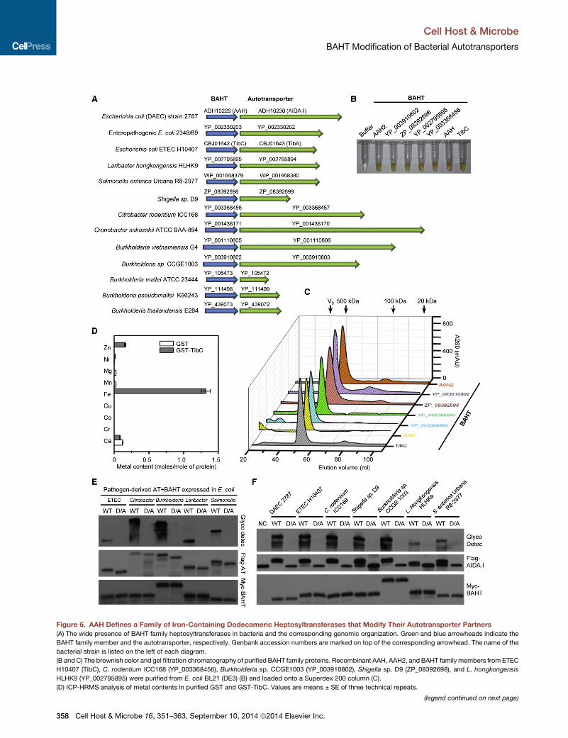
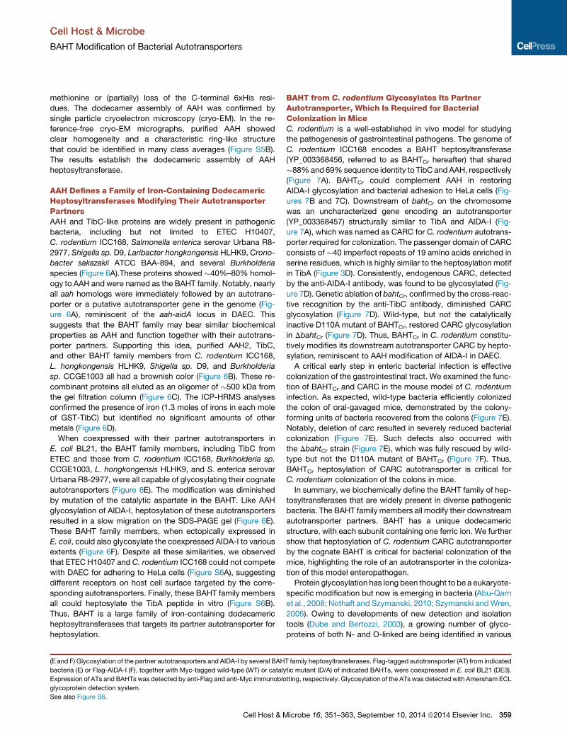
Figure 6. AAH Defines a Family of Iron-Containing Dodecameric Heptosyltransferases that Modify Their Autotransporter Partners
(A) The wide presence of BAHT family heptosyltransferases in bacteria and the corresponding genomic organization. Green and blue arrowheads indicate the
BAHT family member and the autotransporter, respectively. Genbank accession numbers are marked on top of the corresponding arrowhead. The name of the
bacterial strain is listed on the left of each diagram.
(B andC) The brownish color and gel filtration chromatography of purified BAHT family proteins. Recombinant AAH, AAH2, and BAHT family members from ETEC
H10407 (TibC), C. rodentium ICC168 (YP_003368456), Burkholderia sp. CCGE1003 (YP_003910802), Shigella sp. D9 (ZP_08392698), and L. hongkongensis
HLHK9 (YP_002795895) were purified from E. coli BL21 (DE3) (B) and loaded onto a Superdex 200 column (C).
(D) ICP-HRMS analysis of metal contents in purified GST and GST-TibC. Values are means ± SE of three technical repeats.
(legend continued on next page)
Cell Host & Microbe
BAHT Modification of Bacterial Autotransporters
358 Cell Host & Microbe 16, 351–363, September 10, 2014 ª2014 Elsevier Inc.
Cell Host & Microbe
BAHT Modification of Bacterial Autotransporters
methionine or (partially) loss of the C-terminal 6xHis resi-
dues. The dodecamer assembly of AAH was confirmed by
single particle cryoelectron microscopy (cryo-EM). In the re-
ference-free cryo-EM micrographs, purified AAH showed
clear homogeneity and a characteristic ring-like structure
that could be identified in many class averages (Figure S5B).
The results establish the dodecameric assembly of AAH
heptosyltransferase.
AAH Defines a Family of Iron-Containing DodecamericHeptosyltransferases Modifying Their AutotransporterPartnersAAH and TibC-like proteins are widely present in pathogenic
bacteria, including but not limited to ETEC H10407,
C. rodentium ICC168, Salmonella enterica serovar Urbana R8-
2977, Shigella sp. D9, Laribacter hongkongensis HLHK9, Crono-
bacter sakazakii ATCC BAA-894, and several Burkholderia
species (Figure 6A).These proteins showed �40%–80% homol-
ogy to AAH and were named as the BAHT family. Notably, nearly
all aah homologs were immediately followed by an autotrans-
porter or a putative autotransporter gene in the genome (Fig-
ure 6A), reminiscent of the aah-aidA locus in DAEC. This
suggests that the BAHT family may bear similar biochemical
properties as AAH and function together with their autotrans-
porter partners. Supporting this idea, purified AAH2, TibC,
and other BAHT family members from C. rodentium ICC168,
L. hongkongensis HLHK9, Shigella sp. D9, and Burkholderia
sp. CCGE1003 all had a brownish color (Figure 6B). These re-
combinant proteins all eluted as an oligomer of �500 kDa from
the gel filtration column (Figure 6C). The ICP-HRMS analyses
confirmed the presence of iron (1.3 moles of irons in each mole
of GST-TibC) but identified no significant amounts of other
metals (Figure 6D).
When coexpressed with their partner autotransporters in
E. coli BL21, the BAHT family members, including TibC from
ETEC and those from C. rodentium ICC168, Burkholderia sp.
CCGE1003, L. hongkongensis HLHK9, and S. enterica serovar
Urbana R8-2977, were all capable of glycosylating their cognate
autotransporters (Figure 6E). The modification was diminished
by mutation of the catalytic aspartate in the BAHT. Like AAH
glycosylation of AIDA-I, heptosylation of these autotransporters
resulted in a slow migration on the SDS-PAGE gel (Figure 6E).
These BAHT family members, when ectopically expressed in
E. coli, could also glycosylate the coexpressed AIDA-I to various
extents (Figure 6F). Despite all these similarities, we observed
that ETEC H10407 and C. rodentium ICC168 could not compete
with DAEC for adhering to HeLa cells (Figure S6A), suggesting
different receptors on host cell surface targeted by the corre-
sponding autotransporters. Finally, these BAHT family members
all could heptosylate the TibA peptide in vitro (Figure S6B).
Thus, BAHT is a large family of iron-containing dodecameric
heptosyltransferases that targets its partner autotransporter for
heptosylation.
(E and F) Glycosylation of the partner autotransporters and AIDA-I by several BAH
bacteria (E) or Flag-AIDA-I (F), together with Myc-tagged wild-type (WT) or cataly
Expression of ATs and BAHTs was detected by anti-Flag and anti-Myc immunoblo
glycoprotein detection system.
See also Figure S6.
Cell Host & M
BAHT from C. rodentium Glycosylates Its PartnerAutotransporter, Which Is Required for BacterialColonization in MiceC. rodentium is a well-established in vivo model for studying
the pathogenesis of gastrointestinal pathogens. The genome of
C. rodentium ICC168 encodes a BAHT heptosyltransferase
(YP_003368456, referred to as BAHTCr hereafter) that shared
�88%and 69%sequence identity to TibC and AAH, respectively
(Figure 7A). BAHTCr could complement AAH in restoring
AIDA-I glycosylation and bacterial adhesion to HeLa cells (Fig-
ures 7B and 7C). Downstream of bahtCr on the chromosome
was an uncharacterized gene encoding an autotransporter
(YP_003368457) structurally similar to TibA and AIDA-I (Fig-
ure 7A), which was named as CARC for C. rodentium autotrans-
porter required for colonization. The passenger domain of CARC
consists of �40 imperfect repeats of 19 amino acids enriched in
serine residues, which is highly similar to the heptosylation motif
in TibA (Figure 3D). Consistently, endogenous CARC, detected
by the anti-AIDA-I antibody, was found to be glycosylated (Fig-
ure 7D). Genetic ablation of bahtCr, confirmed by the cross-reac-
tive recognition by the anti-TibC antibody, diminished CARC
glycosylation (Figure 7D). Wild-type, but not the catalytically
inactive D110A mutant of BAHTCr, restored CARC glycosylation
in DbahtCr (Figure 7D). Thus, BAHTCr in C. rodentium constitu-
tively modifies its downstream autotransporter CARC by hepto-
sylation, reminiscent to AAH modification of AIDA-I in DAEC.
A critical early step in enteric bacterial infection is effective
colonization of the gastrointestinal tract. We examined the func-
tion of BAHTCr and CARC in the mouse model of C. rodentium
infection. As expected, wild-type bacteria efficiently colonized
the colon of oral-gavaged mice, demonstrated by the colony-
forming units of bacteria recovered from the colons (Figure 7E).
Notably, deletion of carc resulted in severely reduced bacterial
colonization (Figure 7E). Such defects also occurred with
the DbahtCr strain (Figure 7E), which was fully rescued by wild-
type but not the D110A mutant of BAHTCr (Figure 7F). Thus,
BAHTCr heptosylation of CARC autotransporter is critical for
C. rodentium colonization of the colons in mice.
In summary, we biochemically define the BAHT family of hep-
tosyltransferases that are widely present in diverse pathogenic
bacteria. The BAHT family members all modify their downstream
autotransporter partners. BAHT has a unique dodecameric
structure, with each subunit containing one ferric ion. We further
show that heptosylation of C. rodentium CARC autotransporter
by the cognate BAHT is critical for bacterial colonization of the
mice, highlighting the role of an autotransporter in the coloniza-
tion of this model enteropathogen.
Protein glycosylation has long been thought to be a eukaryote-
specific modification but now is emerging in bacteria (Abu-Qarn
et al., 2008; Nothaft and Szymanski, 2010; Szymanski andWren,
2005). Owing to developments of new detection and isolation
tools (Dube and Bertozzi, 2003), a growing number of glyco-
proteins of both N- and O-linked are being identified in various
T family heptosyltransferases. Flag-tagged autotransporter (AT) from indicated
tic mutant (D/A) of indicated BAHTs, were coexpressed in E. coli BL21 (DE3).
tting, respectively. Glycosylation of the ATs was detected with Amersham ECL
icrobe 16, 351–363, September 10, 2014 ª2014 Elsevier Inc. 359
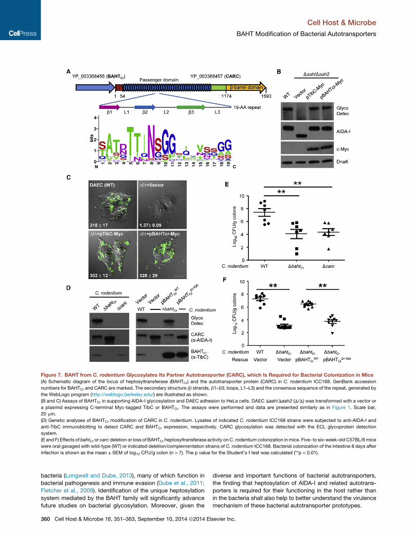
Figure 7. BAHT from C. rodentium Glycosylates Its Partner Autotransporter (CARC), which Is Required for Bacterial Colonization in Mice
(A) Schematic diagram of the locus of heptosyltransferase (BAHTCr) and the autotransporter protein (CARC) in C. rodentium ICC168. GenBank accession
numbers for BAHTCr and CARC are marked. The secondary structure (b strands, b1–b3; loops, L1–L3) and the consensus sequence of the repeat, generated by
the WebLogo program (http://weblogo.berkeley.edu/) are illustrated as shown.
(B and C) Assays of BAHTCr in supporting AIDA-I glycosylation and DAEC adhesion to HeLa cells. DAEC Daah/Daah2 (D/D) was transformed with a vector or
a plasmid expressing C-terminal Myc-tagged TibC or BAHTCr. The assays were performed and data are presented similarly as in Figure 1. Scale bar,
20 mm.
(D) Genetic analyses of BAHTCr modification of CARC in C. rodentium. Lysates of indicated C. rodentium ICC168 strains were subjected to anti-AIDA-I and
anti-TibC immunoblotting to detect CARC and BAHTCr expression, respectively. CARC glycosylation was detected with the ECL glycoprotein detection
system.
(E and F) Effects of bahtCr or carc deletion or loss of BAHTCr heptosyltransferase activity onC. rodentium colonization inmice. Five- to six-week-old C57BL/6mice
were oral gavaged with wild-type (WT) or indicated deletion/complementation strains of C. rodentium ICC168. Bacterial colonization of the intestine 8 days after
infection is shown as the mean ± SEM of log10 CFU/g colon (n > 7). The p value for the Student’s t test was calculated (**p < 0.01).
Cell Host & Microbe
BAHT Modification of Bacterial Autotransporters
bacteria (Longwell and Dube, 2013), many of which function in
bacterial pathogenesis and immune evasion (Dube et al., 2011;
Fletcher et al., 2009). Identification of the unique heptosylation
system mediated by the BAHT family will significantly advance
future studies on bacterial glycosylation. Moreover, given the
360 Cell Host & Microbe 16, 351–363, September 10, 2014 ª2014 El
diverse and important functions of bacterial autotransporters,
the finding that heptosylation of AIDA-I and related autotrans-
porters is required for their functioning in the host rather than
in the bacteria shall also help to better understand the virulence
mechanism of these bacterial autotransporter prototypes.
sevier Inc.

Cell Host & Microbe
BAHT Modification of Bacterial Autotransporters
EXPERIMENTAL PROCEDURES
Bacteria, Plasmids, Antibodies, and Reagents
DAEC 2787 and C. rodentium ICC168 were provided by Dr. John M. Fair-
brother (The Universite de Montreal, Canada) and Dr. Youcun Qian (Institute
of Health Sciences, Chinese Academy of Sciences, China), respectively. Other
E. coli strains used include Top10 for molecular cloning, BL21 (DE3) for protein
expression and glycosylation reconstitution, and SM10 (l pir) for plasmid con-
jugal transfer. Bacterial deletion mutants were generated by homologous
recombination as described previously (Li et al., 2013). For complementation
of DAECmutants, DNAs for AAH/AAH2, together with the respective promoter,
were inserted into the pBluescript II SK (+) vector; the coding sequence of
AIDA-I (fused to the promoter of aah) or the entire aah-aidA locus (including
the promoter) was constructed into the EZ-T vector. For complementation in
C. rodentium ICC168, DNAs for BAHTCr (together with its natural promoter)
and the CARC autotransporter (fused to BAHTCr promoter) were amplified
by PCR and introduced into pACYC184. To assay AAH modification of
AIDA-I, Flag-tagged AIDA-I fragments and AAH were cloned into pGEX-6P-2
and pACYCDuet-1, respectively. For coexpression of BAHT with its partner
autotransporter (AT), the coding sequences of BAHTs and ATs from different
pathogens were introduced into pET21a and pACYCDuet-1, respectively. To
detect protein expression, a Myc tag was fused N terminal to the BAHT, and
a Flag tag was inserted between the signal peptide and the passenger domain
of the AT. Point mutations were generated by the QuikChange Site-Directed
Mutagenesis Kit (Stratagene). All the plasmids were verified by DNA
sequencing.
All bacterial strains were grown in Luria Bertani (LB) medium at 37�C. Con-centrations of the antibiotics used are 100 mg/mL for ampicillin and strepto-
mycin, 30 mg/mL for kanamycin, 34 mg/mL for chloramphenicol, 25 mg/mL
for tetracycline, and 50 mg/mL for nalidixic acid. Anti-Flag (M2) was from
Sigma; mouse anti-HA (16B12) and anti-c-Myc monoclonal antibodies
(9E10) were from Covance; anti-DnaK (clone 8E2/2) was purchased from
Enzo Life Sciences. Purified AAH-His6, TibC-His6, and GST-AIDA-I50-846were used to immunize rabbits for generating polyclonal antisera. Synthetic
peptides were made by Scilight Biotechnology LLC. ADP-L/D-glycero-b-D-
manno-heptose were synthesized previously (Zamyatina et al., 2003). All other
chemical reagents used were from Sigma unless noted.
Cell Culture and Bacterial Adhesion Assay
HeLa cells (American Type Culture Collection, ATCC) were cultured in Dulbec-
co’s modified Eagle’s medium (Thermo Scientific) supplemented with 10%
fetal bovine serum (Invitrogen) at 37�C in a 5%CO2 incubator. Tomonitor bac-
terial adhesion, cells were seeded onto glass coverslips, and 5 ml of overnight
bacteria was added onto cells without centrifugation. Following 2 hr incubation
(37�C in 5%CO2), infected cells were washed to remove nonadherent bacteria
and fixed with 4% paraformaldehyde. The microscopy images shown are
representative of those on the entire coverslip. To quantify the adherence,
HeLa cells seeded in 24-well plates (1.2 3 105 cells/well) were used, and in-
fected cells were lysed (0.1%Triton X-100 in PBS) to liberate the adherent bac-
teria. The lysates were plated on LB agar with specific antibiotics to enumerate
the adherent bacteria. The results were expressed as themean number of bac-
teria adhering to one cell (the total number of recovered bacteria/the number of
seeded cell in each well) ± SD of four technical repeats. All experiments were
independently repeated more than three times.
Immunoblotting and Glycoprotein Detection
Of 100 ml total lysates from 1 ml of bacteria culture, 1/10 was separated on
SDS-PAGE gels for immunoblotting analysis and/or glycoprotein detection.
Expression of AIDA-I and AAH was detected by the anti-AIDA-I and anti-
AAH sera, respectively. Expression of BAHTCr and the CARC autotransporter
in C. rodentium ICC168 was monitored by anti-TibC and anti-AIDA-I immuno-
blotting, respectively. Glycosylation was detected with the ECL glycoprotein
detection kit (GE Healthcare).
Subcellular Fractionation of Bacterial Lysates
DAEC lysates was separated into the periplasm, cytoplasm, and membrane
fractions as described (Maurer et al., 1997). OmpA and b-lactamase (Bla)
were expressed as the markers for outer membrane and periplamic localiza-
Cell Host & M
tion, respectively. The periplasmwas prepared by cold osmotic shock. Briefly,
the bacteria were suspended in 50 mM Tris-HCl (pH 7.6), 20% sucrose (w/v),
and 1 mM ETDA and incubated for 10 min at 4�C. The centrifuged pellets were
resuspended in ice-cold 5 mM MgSO4 and incubated for another 10 min at
4�C. Following additional centrifugation, the supernatant was recovered as
the periplasm fraction. The resulting pellet containing the spheroplasts was re-
suspended in ice-cold PBS containing a protease-inhibitor cocktail (Roche
Molecular Biochemicals) and lysed by sonication. Intact spheroplasts and
large debris were removed by centrifugation (12,000 g, 10 min). The superna-
tant was centrifuged again (100,000 g, 60 min). The final supernatant was
recovered as the cytosol, and the pellet, suspended in 1% Sarkosyl (N-Laur-
oylsarcosine sodium salt) in PBS, was the membrane fraction (both inner
and outer membranes).
Gain-of-Function Screen of aah2 in DAEC 2787
Genomic DNA from DAEC Daah/DaidA double mutant was prepared by using
TIANamp bacteria DNA Kit (TIANGENBiotech, China). Of genomic DNA, 10 mg
was digested with Sau3A1, and 5–10 kb fragments were purified from agarose
gels and ligated into BamHI-digested pUC19 vector. The resulting plasmids
were introduced into E. coli Top10 harboring pME6032-Flag-AIDA-I. The re-
combinants were cultured overnight with 1 mM isopropyl-b-D-thiogalactopyr-
anoside (IPTG) to induce AIDA-I expression and then lysed for immunoblotting
and glycosylation analysis.
Purification of Recombinant Proteins
E. coli BL21 (DE3) strains harboring the expression plasmids were grown in LB
medium with appropriate antibiotics. Protein expression was induced by addi-
tion of 0.5 mM IPTG for 12 hr at 22�C after OD600 reached 0.8. Purification of
His-tagged proteins was performed by using Ni-NTA agarose (GE Healthcare).
GST-AIDA-I50-846 was expressed in the inclusion body. The inclusion bodies
were solubilized in a buffer containing 6 M urea, 20 mM Tris-HCl (pH 7.6),
and 20 mM NaCl; GST-AIDA-I50-846 was purified by the glutathione sepharose
resins (GE Healthcare). Refolding of GST-AIDA-I50-846 was performed by dial-
ysis against a buffer containing 20 mM Tris-HCl (pH 7.6) and 20 mM NaCl with
a linear urea gradient from 6 to 0 M. GST, GST-AIDA-I531-611-Flag, and GST-
TibC were also purified by the glutathione Sepharose resins. When needed,
GST was removed from the fusion protein by PreScission protease digestion.
Protein concentrations were estimated by Coomassie blue staining of SDS-
PAGE gels using the BSA standard.
Mass Spectrometry Analyses
To examine the AAH oligomer, nanoelectrospray ionization mass spectrom-
etry (MS) and tandem MS (MS/MS) were performed on a QSTAR ELITE instru-
ment. Briefly, 20 ml of the AAH complex (100 mM) was buffer exchanged into
200 mM ammonium acetate using the biospin columns (Bio-Rad). A total of
2 ml of the sample was loaded into a gold-coated nano-ESI capillary (home-
made from borosilicate glass tubes). The conditions within the mass spec-
trometer were adjusted to preserve noncovalent interactions. The following
parameters were used: capillary voltage �1.18 KV, declustering potential
�150 V, focusing potential�200 V, declustering potential 2–15 V, collision en-
ergy �20 V. For tandem MS experiments, the peak centered at the m/z of 9,
635 was selected in the quadrupole and the collision energy was elevated to
200 V. Argon was used as a collision gas at the maximum pressure. Spectra
are shown with minimal smoothing and without background subtraction. The
mass spectrometry protocol used for determining AAH heptosylation of
AIDA-I or the peptide substrate was similar to that previously described for
analyzing other modifications (Li et al., 2013).
Metal Contents Determination and In Vitro Heptosyltransferase
Assay
Metal contents of purified GST and GST-TibC were determined by using a Fin-
nigan Element ICP-HRMS as described previously (Wiley et al., 2007). The iron
contents of AAH-His6 and TibC-His6 were measured under reducing condi-
tions by using the classical Ferrozine assay (Fish, 1988). For in vitro heptosy-
lation, 2 mg of AAH or other purified BAHT proteins were reacted with 10 mg of
AIDA-I or TibA-derived synthetic peptides for 4 hr at 30�C in a 20 ml reaction
containing 10 mM Tris-HCl (pH 7.5), 50 mM NaCl, 10 mM MgCl2, 1 mM DTT,
and 0.5 mM ADP-L, D-heptose, or ADP-D, D-heptose. The reaction mixtures
icrobe 16, 351–363, September 10, 2014 ª2014 Elsevier Inc. 361

Cell Host & Microbe
BAHT Modification of Bacterial Autotransporters
were analyzed by mass spectrometry. To assay protein substrates, 0.5 mg of
GST-AIDA-I531-611-Flag, 0.5 mg of AAH, and 0.1 mM sugar ligand were used.
The reaction mixtures were subjected to immunoblotting and glycoprotein
detection analyses.
Crystallization, Data Collection, and Structure Determination
The tibA fragment encoding residues 55–350 was coexpressed with wild-type
tibC. The resulting glycosylated TibA55-350 was purified by using the Ni-NTA
resin (Transgen Biotech) followed by removal of the SUMO. The protein was
further purified by gel filtration chromatography and concentrated to 30 mg/
mL in 10 mM Tris-HCl (pH 7.6) and 150 mM NaCl for crystallization. Crystals
of native and SeMet TibA55-350 were grown in 20% (w/v) PEG 8000, 100 mM
magnesium acetate, and 100 mM Tris-HCl (pH 8.0) in 1 week. The TibA55-350
crystals were cryoprotected with Parabar 10312 (Hampton Research) coat
before flash-freezing in liquid nitrogen. The diffraction data were collected at
BL-17U of Shanghai Synchrotron Radiation Facility (SSRF) at 100 K and pro-
cessed with the HKL 2000 suite. The structure was solved by single-wave-
length anomalous dispersion using the AutoSol-AutoBuild in PHENIX. The
model was manually adjusted and completed in Coot. The final mode was
refined in Refmac. Structural pictures were generated in Pymol (http://www.
pymol.org/).
Mice Infection and C. rodentium Colonization Assays
Wild-type C. rodentium ICC168 and indicated derivatives were prepared by
overnight shaking at 37�C. Five- to six-week-old male C57BL/6 mice were
orally inoculated using a gavage needle with 200 ml bacterial suspension in
PBS (�2 3 109 CFU). The number of viable bacteria used as the inoculum
was determined by retrospective plating onto LB agar containing appropriate
antibiotics. Eight days after inoculation, colons were removed aseptically,
weighed, and homogenized in PBS. Homogenates were serially diluted and
plated on MacConkey agar with proper antibiotics to determine CFU counts.
C. rodentium colonies (pink with white rings) were counted after overnight
incubation. Colonization data were analyzed using Student’s t test in the
software GraphPad Prism. p values <0.05 were considered significant. Inde-
pendent experiments were performed using at least seven mice per group.
All animal experiments were conducted following the Ministry of Health na-
tional guidelines for housing and care of laboratory animals and performed
in accordance with institutional regulations after review and approval by the
Institutional Animal Care and Use Committee at National Institute of Biological
Sciences.
ACCESSION NUMBERS
Structural data for hyperheptosylated TibA55-350 are deposited in the Protein
Data Bank (PDB) under the accession number 4Q1Q.
SUPPLEMENTAL INFORMATION
Supplemental Information includes six figures and one table and can be found
with this article at http://dx.doi.org/10.1016/j.chom.2014.08.008.
AUTHOR CONTRIBUTIONS
F.S. conceived of and coordinated the study; Q.L. designedmost of the exper-
iments; Q.L., Q.Y., and Y.X. worked together and performed all the biochem-
ical and cell culture experiments; L.L., Y.L., X.L., and S.C. performed mass
spectrometry analyses of the heptosylation modification; S.L. and W.G.
performed the mouse infection assay; M.S. and G.B.-N. performed mass
spectrometry analysis of the AAH oligomer; M.N. and A.Z. contributed new
reagents and analytic tools; Q.L. and F.S. analyzed the data and wrote the
manuscript.
ACKNOWLEDGMENTS
We thank J. Fairbrother for providing DAEC 2787, J. Fleckenstein for ETEC
H10407, E. Martinez for Burkholderia sp. CCGE1003 genomic DNA, Y. Qian
for C. rodentium ICC168 strain, Y. Lu for L. hongkongensis HLHK9, and S.
362 Cell Host & Microbe 16, 351–363, September 10, 2014 ª2014 El
Roof for S. enterica serovar Urbana R8-2977. We thank T. Huston for ICP-
HRMS analysis, F. Song and P. Zhu for cyro-EM analyses, the NIBS antibody
facility for generating the antibodies, and the NIBS Biological Resource Center
for gene synthesis. We also thank M. Shi for preparing the artwork, and mem-
bers of the Shao laboratory for helpful discussions and technical assistance.
The research was supported in part by an International Early Career Scientist
grant from the Howard Hughes Medical Institute and by the Beijing Scholar
Program to F.S. This work was also supported by the National Basic Research
Program of China 973 Programs (2012CB518700), the Strategic Priority
Research Program of the Chinese Academy of Sciences (XDB08020202),
and the China National Science Foundation Program for Distinguished Young
Scholars (31225002) to F.S.
Received: March 20, 2014
Revised: July 6, 2014
Accepted: August 1, 2014
Published: September 10, 2014
REFERENCES
Abu-Qarn, M., Eichler, J., and Sharon, N. (2008). Not just for Eukarya anymore:
protein glycosylation in Bacteria and Archaea. Curr. Opin. Struct. Biol. 18,
544–550.
Benz, I., and Schmidt, M.A. (1989). Cloning and expression of an adhesin
(AIDA-I) involved in diffuse adherence of enteropathogenic Escherichia coli.
Infect. Immun. 57, 1506–1511.
Benz, I., and Schmidt, M.A. (2001). Glycosylation with heptose residues medi-
ated by the aah gene product is essential for adherence of the AIDA-I adhesin.
Mol. Microbiol. 40, 1403–1413.
Benz, I., and Schmidt, M.A. (2002). Never say never again: protein glycosyla-
tion in pathogenic bacteria. Mol. Microbiol. 45, 267–276.
Benz, I., and Schmidt, M.A. (2011). Structures and functions of autotransporter
proteins in microbial pathogens. Int. J. Med. Microbiol. 301, 461–468.
Charbonneau, M.E., and Mourez, M. (2007). Functional organization of the
autotransporter adhesin involved in diffuse adherence. J. Bacteriol. 189,
9020–9029.
Charbonneau, M.E., Cote, J.P., Haurat, M.F., Reiz, B., Crepin, S., Berthiaume,
F., Dozois, C.M., Feldman, M.F., and Mourez, M. (2012). A structural motif is
the recognition site for a new family of bacterial protein O-glycosyltrans-
ferases. Mol. Microbiol. 83, 894–907.
Dautin, N., and Bernstein, H.D. (2007). Protein secretion in gram-negative bac-
teria via the autotransporter pathway. Annu. Rev. Microbiol. 61, 89–112.
Dube, D.H., and Bertozzi, C.R. (2003). Metabolic oligosaccharide engineering
as a tool for glycobiology. Curr. Opin. Chem. Biol. 7, 616–625.
Dube, D.H., Champasa, K., and Wang, B. (2011). Chemical tools to discover
and target bacterial glycoproteins. Chem. Commun. (Camb.) 47, 87–101.
Elsinghorst, E.A., and Weitz, J.A. (1994). Epithelial cell invasion and adher-
ence directed by the enterotoxigenic Escherichia coli tib locus is associated
with a 104-kilodalton outer membrane protein. Infect. Immun. 62, 3463–
3471.
Emsley, P., Charles, I.G., Fairweather, N.F., and Isaacs, N.W. (1996). Structure
of Bordetella pertussis virulence factor P.69 pertactin. Nature 381, 90–92.
Fish, W.W. (1988). Rapid colorimetric micromethod for the quantitation of
complexed iron in biological samples. Methods Enzymol. 158, 357–364.
Fletcher, C.M., Coyne, M.J., Villa, O.F., Chatzidaki-Livanis, M., and Comstock,
L.E. (2009). A general O-glycosylation system important to the physiology of a
major human intestinal symbiont. Cell 137, 321–331.
Gangwer, K.A., Mushrush, D.J., Stauff, D.L., Spiller, B., McClain, M.S., Cover,
T.L., and Lacy, D.B. (2007). Crystal structure of the Helicobacter pylori vacuo-
lating toxin p55 domain. Proc. Natl. Acad. Sci. USA 104, 16293–16298.
Henderson, I.R., Navarro-Garcia, F., Desvaux, M., Fernandez, R.C., and
Ala’Aldeen, D. (2004). Type V protein secretion pathway: the autotransporter
story. Microbiol. Mol. Biol. Rev. 68, 692–744.
sevier Inc.
Cell Host & Microbe
BAHT Modification of Bacterial Autotransporters
Jose, J., and Meyer, T.F. (2007). The autodisplay story, from discovery to
biotechnical and biomedical applications. Microbiol. Mol. Biol. Rev. 71,
600–619.
Kneidinger, B., Marolda, C., Graninger, M., Zamyatina, A., McArthur, F.,
Kosma, P., Valvano, M.A., and Messner, P. (2002). Biosynthesis pathway of
ADP-L-glycero-beta-D-manno-heptose in Escherichia coli. J. Bacteriol. 184,
363–369.
Lazar Adler, N.R., Stevens, J.M., Stevens, M.P., and Galyov, E.E. (2011).
Autotransporters and Their Role in the Virulence of Burkholderia pseudomallei
and Burkholderia mallei. Front. Microbiol. 2, 151.
Leyton, D.L., Rossiter, A.E., and Henderson, I.R. (2012). From self sufficiency
to dependence: mechanisms and factors important for autotransporter
biogenesis. Nat. Rev. Microbiol. 10, 213–225.
Li, S., Zhang, L., Yao, Q., Li, L., Dong, N., Rong, J., Gao, W., Ding, X., Sun, L.,
Chen, X., et al. (2013). Pathogen blocks host death receptor signalling by argi-
nine GlcNAcylation of death domains. Nature 501, 242–246.
Lindenthal, C., and Elsinghorst, E.A. (1999). Identification of a glycoprotein
produced by enterotoxigenic Escherichia coli. Infect. Immun. 67, 4084–4091.
Longwell, S.A., and Dube, D.H. (2013). Deciphering the bacterial glycocode:
recent advances in bacterial glycoproteomics. Curr. Opin. Chem. Biol. 17,
41–48.
Maurer, J., Jose, J., and Meyer, T.F. (1997). Autodisplay: one-component sys-
tem for efficient surface display and release of soluble recombinant proteins
from Escherichia coli. J. Bacteriol. 179, 794–804.
Moormann, C., Benz, I., and Schmidt, M.A. (2002). Functional substitution of
the TibC protein of enterotoxigenic Escherichia coli strains for the autotrans-
porter adhesin heptosyltransferase of the AIDA system. Infect. Immun. 70,
2264–2270.
Nothaft, H., and Szymanski, C.M. (2010). Protein glycosylation in bacteria:
sweeter than ever. Nat. Rev. Microbiol. 8, 765–778.
Otto, B.R., Sijbrandi, R., Luirink, J., Oudega, B., Heddle, J.G., Mizutani, K.,
Park, S.Y., and Tame, J.R. (2005). Crystal structure of hemoglobin protease,
a heme binding autotransporter protein from pathogenic Escherichia coli.
J. Biol. Chem. 280, 17339–17345.
Cell Host & M
Pohlner, J., Halter, R., Beyreuther, K., and Meyer, T.F. (1987). Gene structure
and extracellular secretion of Neisseria gonorrhoeae IgA protease. Nature 325,
458–462.
Raetz, C.R., and Whitfield, C. (2002). Lipopolysaccharide endotoxins. Annu.
Rev. Biochem. 71, 635–700.
Ravi, M., Ngeleka, M., Kim, S.H., Gyles, C., Berthiaume, F., Mourez, M.,
Middleton, D., and Simko, E. (2007). Contribution of AIDA-I to the pathoge-
nicity of a porcine diarrheagenic Escherichia coli and to intestinal colonization
through biofilm formation in pigs. Vet. Microbiol. 120, 308–319.
Sherlock, O., Schembri, M.A., Reisner, A., and Klemm, P. (2004). Novel roles
for the AIDA adhesin from diarrheagenic Escherichia coli: cell aggregation
and biofilm formation. J. Bacteriol. 186, 8058–8065.
Sherlock, O., Dobrindt, U., Jensen, J.B., Munk Vejborg, R., and Klemm, P.
(2006). Glycosylation of the self-recognizing Escherichia coli Ag43 autotrans-
porter protein. J. Bacteriol. 188, 1798–1807.
Szymanski, C.M., and Wren, B.W. (2005). Protein glycosylation in bacterial
mucosal pathogens. Nat. Rev. Microbiol. 3, 225–237.
van der Woude, M.W., and Henderson, I.R. (2008). Regulation and function of
Ag43 (flu). Annu. Rev. Microbiol. 62, 153–169.
Wells, T.J., Tree, J.J., Ulett, G.C., and Schembri, M.A. (2007). Autotransporter
proteins: novel targets at the bacterial cell surface. FEMS Microbiol. Lett. 274,
163–172.
Wells, T.J., Totsika, M., and Schembri, M.A. (2010). Autotransporters of
Escherichia coli: a sequence-based characterization. Microbiology 156,
2459–2469.
Wiley, S.E., Murphy, A.N., Ross, S.A., van der Geer, P., and Dixon, J.E. (2007).
MitoNEET is an iron-containing outer mitochondrial membrane protein that
regulates oxidative capacity. Proc. Natl. Acad. Sci. USA 104, 5318–5323.
Zamyatina, A., Gronow, S., Puchberger, M., Graziani, A., Hofinger, A., and
Kosma, P. (2003). Efficient chemical synthesis of both anomers of ADP L-glyc-
ero- and D-glycero-D-manno-heptopyranose. Carbohydr. Res. 338, 2571–
2589.
Zhao, L., Chen, X., Xu, X., Song, G., and Liu, X. (2009). Analysis of the AIDA-I
gene sequence and prevalence in Escherichia coli isolates frompigs with post-
weaning diarrhoea and oedema disease. Vet. J. 180, 124–129.
icrobe 16, 351–363, September 10, 2014 ª2014 Elsevier Inc. 363