CASE SCENARIO 1 - Emory Universityweb1.sph.emory.edu/GCCS/NAACCR_WEBINARS/20140109/GIST NET... ·...
14
Please review the Q&A prior to completing. CASE SCENARIO 1 Patient is 79 year-old white male with gastric lesion found on routine CT done for history of prostate cancer. 3/4/13 CT Abdomen/Pelvis: Significant enlargement of the mass at the gastric fundus. It now measures 2.4 cm in diameter compared with 1.7 cm on the prior study 2/20/13. Despite its smooth morphology, this is clearly a neoplastic lesion. Gastrointestinal stromal tumor or leiomyoma could both have this appearance but the enlargement is worrisome and resection is recommended. There are no signs of metastatic disease. 3/21/13 EGD: The stomach was entered and insufflated with air. All the curvatures were carefully examined including the retroflexed view of the fundus where we identified the presence of a bulging through the mucosa at approximately 5 cm from the cardia probably towards the lesser curvature of the mucosa with no breakthrough and no evidence of ulceration. The biopsy forceps were then introduced and as we made contact with the mucosa, the tumor appeared to be firm, likely consistent with a leiomyoma. 3/21/13 Proximal gastric lesion biopsy: Fragments of gastric mucosa without significant morphologic abnormality. 9/10/13 EGD/EUS INDICATION FOR THE PROCEDURES: Gastric fundic subepithelial mass, which has grown in size from 2 cm to around 3 cm in size. We are doing a fine-needle aspiration examination on this mass as well as trying to get an idea about the size and growth of this mass. PROCEDURE FINDINGS Esophagogastroduodenoscopy portion of the examination: Esophagus in proximal, mid, and distal portion was carefully examined. There is esophagitis noticed at the GE junction, which was located at around 40 to 41 cm from incisors. There is irregular Z-line with the possible short segment Barrett's esophagus. Biopsies are taken to exclude the Barrett's esophagus. A small to moderate hiatal hernia was seen. Gastric lumen was examined, which appears within normal limits in the body and antrum; however, in the retroflexed position a subepithelial mass is seen. This mass is subepithelial with normal overlying mucosa and measures at least 2 cm in transverse diameter. The rest of the gastric examination was unremarkable. Pylorus was patent. Duodenal bulb as well as up to the second portion of duodenum appears normal. Endoscopic ultrasound examination: The Pentax radial EUS scope was advanced from the patient's mouth into the gastric fundic station. Immediately noted upon entering into the gastric fundus there is a large hypoechoic heterogeneous mass with some cystic changes and significant vascularity arising from the muscularis propria layer as clearly shown on image 3 and image 4. The "footplate" is clearly visible on image 5 indicating arising of this mass from muscularis propria layer. Elastography as shown on image 6 and image 8 clearly shows the mass is predominantly stiff or hard in its elasticity. Mass is very close to spleen as shown on image 12, but is not invading or involving spleen. It does appear to involve more on the left upper quadrant away from the left lobe of the liver and more towards the spleen. Celiac axis and superior mesenteric axis appeared within normal limits. Pancreatic parenchyma
Transcript of CASE SCENARIO 1 - Emory Universityweb1.sph.emory.edu/GCCS/NAACCR_WEBINARS/20140109/GIST NET... ·...

Please review the Q&A prior to completing.
CASE SCENARIO 1
Patient is 79 year-old white male with gastric lesion found on routine CT done for history of prostate
cancer.
3/4/13 CT Abdomen/Pelvis: Significant enlargement of the mass at the gastric fundus. It now measures
2.4 cm in diameter compared with 1.7 cm on the prior study 2/20/13. Despite its smooth morphology,
this is clearly a neoplastic lesion. Gastrointestinal stromal tumor or leiomyoma could both have this
appearance but the enlargement is worrisome and resection is recommended. There are no signs of
metastatic disease.
3/21/13 EGD: The stomach was entered and insufflated with air. All the curvatures were carefully
examined including the retroflexed view of the fundus where we identified the presence of a bulging
through the mucosa at approximately 5 cm from the cardia probably towards the lesser curvature of the
mucosa with no breakthrough and no evidence of ulceration. The biopsy forceps were then introduced
and as we made contact with the mucosa, the tumor appeared to be firm, likely consistent with a
leiomyoma.
3/21/13 Proximal gastric lesion biopsy: Fragments of gastric mucosa without significant morphologic
abnormality.
9/10/13 EGD/EUS
INDICATION FOR THE PROCEDURES: Gastric fundic subepithelial mass, which has grown in size from 2
cm to around 3 cm in size. We are doing a fine-needle aspiration examination on this mass as well as
trying to get an idea about the size and growth of this mass.
PROCEDURE FINDINGS Esophagogastroduodenoscopy portion of the examination: Esophagus in proximal, mid, and distal portion was carefully examined. There is esophagitis noticed at the GE junction, which was located at around 40 to 41 cm from incisors. There is irregular Z-line with the possible short segment Barrett's esophagus. Biopsies are taken to exclude the Barrett's esophagus. A small to moderate hiatal hernia was seen. Gastric lumen was examined, which appears within normal limits in the body and antrum; however, in the retroflexed position a subepithelial mass is seen. This mass is subepithelial with normal overlying mucosa and measures at least 2 cm in transverse diameter. The rest of the gastric examination was unremarkable. Pylorus was patent. Duodenal bulb as well as up to the second portion of duodenum appears normal. Endoscopic ultrasound examination: The Pentax radial EUS scope was advanced from the patient's mouth into the gastric fundic station. Immediately noted upon entering into the gastric fundus there is a large hypoechoic heterogeneous mass with some cystic changes and significant vascularity arising from the muscularis propria layer as clearly shown on image 3 and image 4. The "footplate" is clearly visible on image 5 indicating arising of this mass from muscularis propria layer. Elastography as shown on image 6 and image 8 clearly shows the mass is predominantly stiff or hard in its elasticity. Mass is very close to spleen as shown on image 12, but is not invading or involving spleen. It does appear to involve more on the left upper quadrant away from the left lobe of the liver and more towards the spleen. Celiac axis and superior mesenteric axis appeared within normal limits. Pancreatic parenchyma
examined from the neck all the way to the tail of pancreas appears within normal limits. Pancreatic duct is only about 2.5 to 3 mm in diameter. The rest of the upper GI EUS examination was unremarkable. I proceeded with the withdrawal of radial EUS scope and introduced the linear scope. Mass was re-measured and was noticed to be a 4 cm x 37.2 mm in diameter. This has clearly grown compared to previous measurements when the mass was in 3 cm range. Avascular plane was found using Doppler ultrasonography. Spindle-cell was yielded within first pass confirming the notion that this is a spindle-cell neoplasm in order to obtain additional staining material. A second pass with 19-gauge flex needle was carried out and this pass was reserved for special stains for CD 117 smooth muscle actin and other stains to further delineate the etiology of this mass. At this point, scope was withdrawn and a GIF scope was reintroduced, all the secretions suctioned from the fundus and the mass was once again visualized carefully to ensure there is no ongoing bleeding. At this point, scope was withdrawn. 9/10/13 Gastric fundus FNA: Fragments consistent with a gastrointestinal stromal tumor.
10/28/13 Laparoscopic partial gastrectomy
DESCRIPTION OF PROCEDURE: The patient was taken to the operating room, prepped and draped in the
usual customary fashion for laparoscopic partial gastrectomy. The patient was placed on reverse
Trendelenburg position, and then attention was turned into 4 more port sites created in the upper
abdomen and the lateral-most port was used to retract the omentum laterally, as the middle port was
used to retract the stomach and endoscopic LigaSure was used to take down the greater curvature to
mobilize the fundus completely. Care was taken not to hurt the integrity of the mass. Once the mass was
totally mobilized with the fundus, an Ethicon Echelon blue load staple was fired at the base of the mass
at the fundus, where SeamGuard was used. Two staples of 45 mm were fired and the specimen was
placed in an EndoCatch bag and pulled out of the mid port site as it was enlarged. Once this was
accomplished, this port site was closed with a figure-of-eight #1 PDS suture by using an EndoClose. Then
attention was turned to the stomach and also the abdomen was somewhat surveyed with the scope.
There were no other issues within the stomach.
10/28/13 Partial gastrectomy
Microscopic Description: Sections show features of a gastric stromal tumor characterized by moderate
cellularity with a proliferation of spindle cells arranged in short fascicles. Some of the spindled tumor
cells exhibit cytoplasmic vacuoles. Immunohistochemical stains show that the tumor cells are strongly
positive for CD34 and CD117 (KIT) but are negative for smooth muscle actin and S100. A mitotic rate of
up to 8 mitoses per 50 high power fields is demonstrated. This mitotic rate, in conjunction with the size
of the tumor (5 cm.), places this into the moderate risk category for gastric stromal tumors according to
the criteria of Miettinen et al (Semin Diagn Pathol 23:70-83, 2006).
Final Diagnosis: STOMACH, FUNDUS, PARTIAL GASTRECTOMY: GASTRIC STROMAL TUMOR WITH FEATURES SUGGESTING MODERATE RISK FOR RECURRENCE (SEE MICROSCOPIC DESCRIPTION); RESECTION MARGINS ARE CLEAR. Patient started on Gleevec 12/17/13 for at least 36 months

`Case Scenario Worksheet 1
Primary Site C16.1 Fundus, Stomach NOT REPORTABLE
Morphology 8936/1 GIST, NOS
Stage/ Prognostic Factors CS Tumor Size 050 CS SSF 9 988
CS Extension 999 CS SSF 10 988
CS Tumor Size/Ext Eval 3 CS SSF 11 988
CS Lymph Nodes 000 CS SSF 12 988
CS Lymph Nodes Eval 0 CS SSF 13 988
Regional Nodes Positive 98 CS SSF 14 988
Regional Nodes Examined 00 CS SSF 15 988
CS Mets at Dx 00 CS SSF 16 988
CS Mets Eval 0 CS SSF 17 988
CS SSF 1 988 CS SSF 18 988
CS SSF 2 988 CS SSF 19 988
CS SSF 3 988 CS SSF 20 988
CS SSF 4 988 CS SSF 21 988
CS SSF 5 988 CS SSF 22 988
CS SSF 6 Mitotic Rate 080 CS SSF 23 988
CS SSF 7 KIT 010 CS SSF 24 988
CS SSF 8 988 CS SSF 25 988
Summary Stage 9 Derived AJCC TNM Stage (indicate c or p in the space before the
T, N, or M)
pT2 cN0 cM0
Stage II
Clinical AJCC TNM Stage T2 N0 M0
Stage 99
Pathologic AJCC TNM Stage T2 N0 M
Mitotic Rate High
Stage 2
Treatment Diagnostic Staging Procedure 02
Surgery Codes Radiation Codes
Surgical Procedure of Primary Site 30 Radiation Treatment Volume 00
Scope of Regional Lymph Node
Surgery
0 Regional Treatment Modality 00
Surgical Procedure/ Other Site 0 Regional Dose 00000
Systemic Therapy Codes Boost Treatment Modality 00
Chemotherapy 02 Boost Dose 00000
Hormone Therapy 00 Number of Treatments to Volume 000
Immunotherapy 00 Reason No Radiation 1
Hematologic Transplant/Endocrine
Procedure
00 Radiation/Surgery Sequence 0
Systemic/Surgery Sequence 3

CASE SCENARIO 2 9/10/13 Open partial gastrectomy and splenectomy INDICATIONS: This is a 61-year-old male who developed a hernia after a fem-fem bypass and had a CT scan performed, which showed a heterogeneously enhancing mass of approximately 8 cm in size, located in the left part of the abdomen. An MRI confirmed it was most likely related to the stomach with infiltration of the wall in the region of the gastric fundus and antrum. He had approximately a 50 pound weight loss, but this was intentional secondary to increased activity and exercise. All risks, benefits and alternatives to the procedure were discussed with the patient. DESCRIPTION OF PROCEDURE: After appropriate consent was obtained, the patient was taken back to the operating suite, placed on the table in the supine position. At the time of induction of the anesthesia, the abdomen was prepped and draped in a standard sterile fashion. A previous midline incision was identified in the lower part of the abdomen. This midline incision was followed starting about the umbilicus all the way up to approximately 1 fingerbreadth beneath the xiphoid process. Dissection was carried into the subcutaneous tissue using electrocautery. The abdomen was entered sharply and the incision was extended cephalad and caudad. A Bookwalter retractor was placed. The stomach and the transverse colon were identified. The lesser sac was entered. The short gastrics were taken down using a LigaSure device all the way around to the GE junction. The stomach was rotated medially and the tumor appeared to be extending off the posterior wall of the stomach, extending into the retroperitoneum in fairly close proximity to the splenic vein and artery as well as the spleen. It was encroaching upon the mass, making it difficult for adequate visualization of the tumor as well as its blood supply. The decision was made that taking the spleen would allow better visualization and easier mobilization of the mass. The splenic vein and artery were identified in the lesser sac and were taken down with clips and ties. The spleen was rotated medially and its posterior attachments were all taken down using Metzenbaum scissors. The remaining portion of the hilum was taken with the LigaSure. The spleen was sent off to pathology. The mesentery between the liver and the lesser curvature of the stomach was entered using electrocautery, and a Penrose drain was placed around the proximal GE junction and used for retraction. The surrounding attachments of the blood supply and the desmoplastic reaction around the mass were taken down with the LigaSure device and the mass in the greater curvature of the stomach was rotated up into the wound. The 2-0 silk traction sutures were placed proximal and distal circumferentially around the mass. A GIA-60 was fired across the greater curvature of the stomach ensuring that the NG tube was in proper position. The GIA stapler was fired x3 and the specimen was sent off to pathology. The staple line was inverted with a running 3-0 Prolene stitch and the 2-0 silk ties were removed. Again, the NG tube was palpated and felt to be freely mobile within the stomach and in good position. At this point, a 10-French flat Blake drain was placed in the left upper quadrant, brought out through the anterior abdominal wall and sutured into place with a 3-0 nylon suture. A small incisional hernia was identified with preperitoneal fat within it. The preperitoneal fat was removed and the hernia sac was opened. The abdomen was then copiously irrigated with 3 L of sterile saline. The pancreas was inspected and found no evidence of injury to the pancreas. It did not appear to have the staple line fired across the tail of the pancreas. The fascia was then approximated with #1 looped PDS sutures x2, incorporating the incisional hernia as well. The subcutaneous tissues were irrigated with sterile saline. The skin was closed with staples. All needle and instruments were correct at the end of the case.

Pathology: Final Diagnosis
A. SPLEEN, SPLENECTOMY (WEIGHT=280 GRAMS): RED PULP CONGESTION. NO EVIDENCE OF
MALIGNANCY.
B. STOMACH, GREATER CURVATURE, PARTIAL GASTRECTOMY: MALIGNANT GASTROINTESTINAL
STROMAL TUMOR, 6.5 CM. IN MAXIMUM DIMENSION, FREE FROM ALL SPECIMEN MARGINS.
TUMOR CELLS SHOW STRONG EXPRESSION OF c-kit (CD117).
C. OMENTUM, OMENTECTOMY: NO EVIDENCE OF MALIGNANCY.
Diagnostic Comments: This gastrointestinal stromal tumor is considered malignant based on its size of 6.5 cm. and its mitotic count of 12 mitoses per 50 high power fields. 10/3/13 Patient started Gleevec.
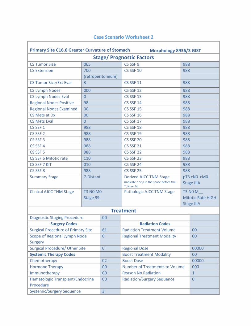
Case Scenario Worksheet 2
Primary Site C16.6 Greater Curvature of Stomach Morphology 8936/3 GIST
Stage/ Prognostic Factors CS Tumor Size 065 CS SSF 9 988
CS Extension 700
(retroperitoneum)
CS SSF 10 988
CS Tumor Size/Ext Eval 3 CS SSF 11 988
CS Lymph Nodes 000 CS SSF 12 988
CS Lymph Nodes Eval 0 CS SSF 13 988
Regional Nodes Positive 98 CS SSF 14 988
Regional Nodes Examined 00 CS SSF 15 988
CS Mets at Dx 00 CS SSF 16 988
CS Mets Eval 0 CS SSF 17 988
CS SSF 1 988 CS SSF 18 988
CS SSF 2 988 CS SSF 19 988
CS SSF 3 988 CS SSF 20 988
CS SSF 4 988 CS SSF 21 988
CS SSF 5 988 CS SSF 22 988
CS SSF 6 Mitotic rate 110 CS SSF 23 988
CS SSF 7 KIT 010 CS SSF 24 988
CS SSF 8 988 CS SSF 25 988
Summary Stage 7-Distant Derived AJCC TNM Stage (indicate c or p in the space before the
T, N, or M)
pT3 cN0 cM0
Stage IIIA
Clinical AJCC TNM Stage T3 N0 M0
Stage 99
Pathologic AJCC TNM Stage T3 N0 M__
Mitotic Rate HIGH
Stage IIIA
Treatment Diagnostic Staging Procedure 00
Surgery Codes Radiation Codes
Surgical Procedure of Primary Site 61 Radiation Treatment Volume 00
Scope of Regional Lymph Node
Surgery
0 Regional Treatment Modality 00
Surgical Procedure/ Other Site 0 Regional Dose 00000
Systemic Therapy Codes Boost Treatment Modality 00
Chemotherapy 02 Boost Dose 00000
Hormone Therapy 00 Number of Treatments to Volume 000
Immunotherapy 00 Reason No Radiation 1
Hematologic Transplant/Endocrine
Procedure
00 Radiation/Surgery Sequence 0
Systemic/Surgery Sequence 3

CASE SCENARIO 3 6/26/13 CT abdomen/pelvis: Large heterogeneous mass in lower right abdomen and pelvis, measuring 17 cm x 9 cm. 6/28/13 Exploratory laparotomy, small bowel resection BRIEF PREOPERATIVE HISTORY: This is a 67-year-old white male who has no significant medical problems. He was diagnosed on a CAT scan with a right pelvic mass. He came into the hospital with abdominal pain. On investigation, this was found as the etiology. He underwent surgery today. PROCEDURE NOTE: The patient was taken to the operating room, underwent general anesthesia, and was prepped and draped in the usual customary fashion for exploratory laparotomy. A midline laparotomy was made. The abdomen was entered without any problems. There was blood inside the abdomen, and this was suctioned. The mass was easily separated from the surrounding tissues, and it was attached to the small bowel, at the distal jejunum level, and was about 20 x 20 cm in size. Also, there were multiple mesenteric feet. One of the feet was sent out to pathology, and the frozen section came back as spindle-cell neoplasm; there was no discreet diagnosis. Small bowel resection was performed. A half-staple, hand-sewn anastomosis was made, about 20 cm of bowel, with the attached segment to the tumor taken out. Care was given to get the resection done at one of the tumor-free sites of the bowel. The 80-GIA stapler was used for resection, to staple parts of the anastomosis, and the holes the staple ends were placed through were closed with 2 layers of 3-0 silk. The abdomen was irrigated with copious amounts of fluid. Hemostasis was achieved. After the lap count, the abdomen was closed with #1 looped Maxon, in a running fashion. At the end of the procedure, all the sponge, instrument, and needle counts were complete. PATHOLOGY
1. Peritoneal nodule excision: Metastatic GI stromal tumor. 2. Small intestine resection of an abdominal mass: Malignant GIST tumor involving mesentery and
small bowel wall with multiple metastatic tumor nodules involving entire small bowel and mesentery; negative margins.
7/18/13 Patient started Gleevec post-operatively.

Case Scenario Worksheet 3
Primary Site C17.1 Jejunum Morphology 8936/3 Malignant GIST
Stage/ Prognostic Factors CS Tumor Size 200 CS SSF 9 988
CS Extension 450 CS SSF 10 988
CS Tumor Size/Ext Eval 3 CS SSF 11 988
CS Lymph Nodes 000 CS SSF 12 988
CS Lymph Nodes Eval 0 CS SSF 13 988
Regional Nodes Positive 98 CS SSF 14 988
Regional Nodes Examined 00 CS SSF 15 988
CS Mets at Dx 40 CS SSF 16 988
CS Mets Eval 3 CS SSF 17 988
CS SSF 1 988 CS SSF 18 988
CS SSF 2 988 CS SSF 19 988
CS SSF 3 988 CS SSF 20 988
CS SSF 4 988 CS SSF 21 988
CS SSF 5 988 CS SSF 22 988
CS SSF 6 999 CS SSF 23 988
CS SSF 7 999 CS SSF 24 988
CS SSF 8 988 CS SSF 25 988
Summary Stage Distant Derived AJCC TNM Stage (indicate
c or p in the space before the T, N, or M) pT4 cN0 pM1
Stage IV
Clinical AJCC TNM Stage T4 N0 M0
Stage 99
Pathologic AJCC TNM Stage T4 N0 M1
Mitotic Rate Unk
Stage IV
Treatment Diagnostic Staging Procedure 00
Surgery Codes Radiation Codes
Surgical Procedure of Primary Site 40 Radiation Treatment Volume 00
Scope of Regional Lymph Node
Surgery
0 Regional Treatment Modality 00
Surgical Procedure/ Other Site 0 Regional Dose 00000
Systemic Therapy Codes Boost Treatment Modality 00
Chemotherapy 02 Boost Dose 00000
Hormone Therapy 00 Number of Treatments to Volume 000
Immunotherapy 00 Reason No Radiation 1
Hematologic Transplant/Endocrine
Procedure
00 Radiation/Surgery Sequence 0
Systemic/Surgery Sequence 3

CASE SCENARIO 4 4/26/13 CT Abdomen/Pelvis Indication: Abdominal pain and vomiting IMPRESSION:
1. Small bowel obstruction with location of obstruction in the right lower quadrant with uninvolved terminal ileum. An associated 1 cm soft tissue mass is seen, possibly representing a carcinoid tumor or adenocarcinoma of the small bowel, and inflammatory changes in the adjacent mesentery are noted. A volvulus is not excluded.
2. Marked constipation within the colon. 3. Stable hepatic cysts.
4/29/13
1. DIAGNOSTIC LAPAROSCOPY. 2. EXPLORATORY LAPAROTOMY. 3. SMALL BOWEL RESECTION. 4. SIDE-TO-SIDE STAPLED ANASTOMOSIS.
INDICATIONS: A 68-year-old white female presented with symptoms of a bowel obstruction. She underwent a CT scan, which showed a high grade small bowel obstruction with some calcified lymph nodes in the small bowel mesentery and an abrupt transition point with some thickening of the adjacent bowel wall suspicious for neoplasia. This was her second episode of a bowel obstruction. We discussed with the patient that she needed exploration with possibility of having a bowel resection if we did find a malignancy. DESCRIPTION OF PROCEDURE: Patient was placed on the table in the supine position. Once appropriate anesthesia was ensured, the patient's abdomen was prepped and draped in standard sterile fashion. Initially, we chose the left upper quadrant as the entry site. We made a 5 mm stab incision; a Veress needle was inserted, and the abdomen was inflated. Once appropriate insufflation was verified, a 5 mm trocar was placed. A 30 degrees 5 mm camera was inserted in the abdomen, and the abdomen was inspected with no evidence of intra-abdominal injury during the insufflation process. At this point, we then placed a subxiphoid 5 mm trocar as well as a left-sided inferior 5 mm trocar all those under direct vision. We then ran the bowel from ligament of Treitz to the terminal ileum; there were three areas of what appeared to be desmoplastic reaction with some hypervascularity involving the small bowel with extension into the adjacent mesenteric fat consistent with possible carcinoid. We decided at this point that doing an intracorporeal dissection may have been limited in the ability to adequately assess and safely resect the adjacent mesenteric lymph node packet. Therefore we chose a midline incision. We made a 7 cm infraumbilical incision initially, going through a prior surgical scar in this location. The Gelport wound protector device was inserted. We dissected down through the subcutaneous tissues through the fascia and into the peritoneum. We then eviscerated the small bowel. We identified the area in question. There were three separate areas of suspicion, all within an approximately 30 cm are of mid small bowel, and as had been seen laparoscopically. We marked the proximal and distal ends for our resection, fired GIA staplers across the distal and proximal bowel, and then we scored the mesentery. We attempted to do a full resection of the adjacent mesentery using the LigaSure device. After delivering the specimen, we palpated the base of the area of resected mesentery, and noted there appeared to be some bulky lymph nodes in the base of the mesentery. We felt this area required removal out of concern for residual disease, and therefore we took another 3 cm to 4 cm of small bowel

mesentery with the LigaSure device, extending more proximally in the mesentery than the original excision. Despite the LigaSure use, a larger caliber proximal mesenteric vessel bled after this tissue was excised, including all the grossly palpably thickened tissue in the more proximal mesentery. We then extended our incision to allow better exposure of this deeper mesenteric area and held digital pressure for control while the incision was extended. Using a 3-0 Prolene, we did a mattress suture around the vessel to control it and was hemostatic at that point. We then examined the bowel. The distal small bowel appeared somewhat dusky and had less appreciable Doppler signals. Therefore we took another approximately 25 cm of small bowel to ensure we had excellent blood supply. We did use the intraoperative ultrasound to confirm excellent arterial supply to the edges of the resected area of small bowel. Proximal small bowel appeared viable without any evidence of compromise. In total we have resected approximately 55 cm of small bowel that was approximately 90 cm from the ileocecal bowel. We then performed a side-to-side stapled anastomosis using a GIA 60 stapler. A TA-60 stapler was used to complete the anastomosis. We closed the mesenteric defect with a 3-0 silk suture. We used 3-0 silk crotch stitches to ensure no tension on her anastomosis. We then placed the small bowel back into the abdomen and irrigated. We then closed the fascia with #1 non-looped PDS x2. We irrigated the subcutaneous tissues and stapled the skin. We stapled our three 5 mm port sites for skin closure. All needle and instruments were correct at the case. Sterile dressings were applied. Final Diagnosis
1. SMALL BOWEL AND ADJACENT MESENTERY, RESECTION: WELL-DIFFERENTIATED NEUROENDOCRINE CARCINOMA (CARCINOID TUMOR) FORMING FOUR MUCOSAL AND BOWEL WALL TUMORS RANGING IN SIZE UP TO 1.0 CM. TUMORS INVADE THROUGH MUSCULAR WALL TO INVOLVE ADJACENT ADIPOSE TISSUE. FOCAL AREAS HIGHLY SUGGESTIVE OF ANGIOLYMPHATIC INVASION ARE NOTED. TUMOR IS MICROSCOPICALLY PRESENT AT THE SAMPLED RADIAL MARGIN. MUCOSAL MARGINS ARE NEGATIVE FOR NEOPLASM.
2. MULTIPLE (7 OF 18) LYMPH NODES ARE POSITIVE FOR METASTATIC TUMOR. Diagnostic Comments: Noted is a well-differentiated neuroendocrine carcinoma with metastasis to multiple lymph nodes. It forms four masses along the bowel wall. One of these masses is predominantly located in the muscularis propria. Focal areas suspicious for angiolymphatic invasion are present. Perineural invasion is also identified in this case. No further treatment recommended.

Case Scenario Worksheet 4
Primary Site C17.9 Small bowel Morphology 8246/3 Well diff neuroendocrine carcinoma
Stage/ Prognostic Factors CS Tumor Size 010 CS SSF 9 988
CS Extension 465 CS SSF 10 988
CS Tumor Size/Ext Eval 3 CS SSF 11 CgA 999
CS Lymph Nodes 300 CS SSF 12 5-HIAA 999
CS Lymph Nodes Eval 3 CS SSF 13 988
Regional Nodes Positive 07 CS SSF 14 988
Regional Nodes Examined 18 CS SSF 15 988
CS Mets at Dx 00 CS SSF 16 988
CS Mets Eval 0 CS SSF 17 988
CS SSF 1 988 CS SSF 18 988
CS SSF 2 988 CS SSF 19 988
CS SSF 3 988 CS SSF 20 988
CS SSF 4 988 CS SSF 21 988
CS SSF 5 988 CS SSF 22 988
CS SSF 6 988 CS SSF 23 988
CS SSF 7 988 CS SSF 24 988
CS SSF 8 988 CS SSF 25 988
Summary Stage 4-Reg DE & LN Derived AJCC TNM Stage (indicate c or p in the space before the T,
N, or M)
pT3 pN1 cM0
Stage IIIB
Clinical AJCC TNM Stage TX N0 M0
Stage IIB
Pathologic AJCC TNM Stage T3m N1 M
Stage IIIB
Treatment Diagnostic Staging Procedure 00
Surgery Codes Radiation Codes
Surgical Procedure of Primary Site 40 Radiation Treatment Volume 00
Scope of Regional Lymph Node
Surgery
5 Regional Treatment Modality 00
Surgical Procedure/ Other Site 0 Regional Dose 00000
Systemic Therapy Codes Boost Treatment Modality 00
Chemotherapy 00 Boost Dose 00000
Hormone Therapy 00 Number of Treatments to Volume 000
Immunotherapy 00 Reason No Radiation 1
Hematologic Transplant/Endocrine
Procedure
00 Radiation/Surgery Sequence 0
Systemic/Surgery Sequence 0

CASE SCENARIO 5
3/28/13 CT Abdomen/Pelvis
Clinical history: 66-year-old white female with given diagnosis of pelvic mass. Recent unintentional 15
pound weight loss. Additional given history of depression, anxiety, gastroesophageal reflux and
hyperlipidemia.
Comparison study: CT abdomen and pelvis 3/20 Impression 1. Large partially calcified soft tissue mass at the root of the mesentery most consistent in appearance
with large carcinoid hazy inflammatory stranding throughout the mesentery surrounding the appendix and right colon suspicious for peritoneal metastatic infiltration.
2. The mesenteric arteries and veins form swirled star-like pattern around the carcinoid at the root of the mesentery and the proximal SMV and distal SMA end in the mass with engorged surrounding vessels.
3. Concomitant appendicitis without evidence of appendiceal rupture. 4. Partially calcified gallbladder wall versus partially calcified gallstones. No findings of cholecystitis;
mild common bile duct dilatation. 5. 2 separate sub-centimeter simple cysts in the right lobe of the liver. 6. 3 large simple cysts in the right ovary. Left-sided fundal leiomyoma. 7. Colonic ileus without bowel obstruction
3/29/13 Serum Chromogranin 200 (range <93)
3/29/13 5 HIAA 5.2 mg (range 10.0 or less)
4/4/13 OPERATIONS PERFORMED
1. EXPLORATORY LAPAROTOMY. 2. Appendectomy. 3. Cholecystectomy. 4. Small bowel resection. 5. Biopsy of retroperitoneal mass.
Indications: 66-year-old woman, who presented with abdominal pain. CT scan showed a mildly thickened appendix, a large retroperitoneal mass obstructing several mesenteric blood vessels, and a large ovarian cystic mass as well as a portion of gallbladder. Final Diagnosis
A. OMENTUM, PARTIAL RESECTION: FIBROUS ADHESIONS WITH ADHESED PORTION OF ATROPHIC OVARIAN TISSUE. NO EVIDENCE OF MALIGNANCY.
B. OVARY, RIGHT, OOPHORECTOMY: BENIGN SEROUS CYSTADENOFIBROMA. C. PORTION OF LEFT OVARY, EXCISION: BENIGN SEROUS CYSTADENOMA. FALLOPIAN TUBE
WITHOUT HISTOLOGIC ABNORMALITY. D. FALLOPIAN TUBE, RIGHT, SALPINGECTOMY: NO HISTOLOGIC ABNORMALITY. E. GALLBLADDER, CHOLECYSTECTOMY: CHRONIC FIBROSING CHOLECYSTITIS ("PORCELAIN
GALLBLADDER") WITH DYSTROPHIC CALCIFICATIONS, CHOLESTEROL CLEFTS AND CHOLELITHIASIS. NO EVIDENCE OF MALIGNANCY.
F. SOFT TISSUE, MESENTERY, BIOPSY: DENSE FIBROSIS.
G. SMALL INTESTINE WITH ATTACHED MESENTERY, RESECTION: WELL-DIFFERENTIATED NEUROENDOCRINE CARCINOMA, MULTIFOCAL, INVOLVING THE SMALL BOWEL WALL/MESENTERY (2.5 X 2.3 X 1.0 CM) WITH A SEPARATE TUMOR NODULE INVOLVING THE SMALL BOWEL WALL (2.0 X 0.9 X 0.6 CM). THE TUMOR CELLS ARE POSITIVE FOR CHROMOGRANIN AND SYNAPTOPHYSIN BY IMMUNOHISTOCHEMISTRY. KI-67 LABELING INDEX: < 1% OF TUMOR CELLS POSITIVE. TUMOR CELL NECROSIS IS NOT PRESENT. TUMOR INVOLVES THE FULL THICKNESS OF THE BOWEL WALL AND INVOLVES MESENTERIC SOFT TISSUE. VASCULAR INVASION IS PRESENT. EXTENSIVE PERINEURAL INVASION BY TUMOR IS PRESENT. METASTATIC NEUROENDOCRINE CARCINOMA IS PRESENT IN 3 OF 22 REGIONAL MESENTERIC LYMPH NODES. TUMOR IS PRESENT AT THE RADIAL MESENTERIC SOFT TISSUE RESECTION MARGIN; SMALL BOWEL MUCOSAL MARGINS ARE CLEAR. PATHOLOGIC STAGE: AJCC stage pT3 N1.
H. MESENTERIC BIOPSY #2: EXTENSIVE INVOLVEMENT BY INVASIVE WELL-DIFFERENTIATED NEUROENDOCRINE CARCINOMA WITH PERINEURAL INVASION.
I. SMALL BOWEL NODULE, EXCISION: WELL-DIFFERENTIATED NEUROENDOCRINE CARCINOMA MEASURING 0.5 CM IN SIZE.
J. APPENDIX, APPENDECTOMY: NO HISTOLOGIC ABNORMALITY.
4/10/13 NM Octreoscan
Findings: There are 2 foci of abnormal In-111 pentetreotide accumulation visualized in the mid upper abdomen. These correspond to soft tissue densities visualized on CT and are consistent with somatostatin receptor positive tumor. No additional sites of abnormal indium-111 pentetreotide activity are visualized elsewhere. There is expected In-111 pentetreotide activity in the spleen, kidneys, bladder, and liver. Impression 1. Two Foci of abnormal In-111 pentetreotide accumulation visualized in the mid upper abdomen.
These correspond to soft tissue densities visualized on CT and are consistent with somatostatin receptor positive tumor.
2. No additional sites of abnormal indium-111 pentetreotide activity are visualized elsewhere.
Patient started on Octreotide LAR 4/18/13

Case Scenario Worksheet 5
Primary Site C17.9 Small bowel Morphology 8246/3 Well diff neuroendocrine carcinoma
Stage/ Prognostic Factors CS Tumor Size 025 CS SSF 9 988
CS Extension 465 CS SSF 10 988
CS Tumor Size/Ext Eval 3 CS SSF 11 200
CS Lymph Nodes 300 CS SSF 12 5-HIAA 005
CS Lymph Nodes Eval 3 CS SSF 13 988
Regional Nodes Positive 03 CS SSF 14 988
Regional Nodes Examined 22 CS SSF 15 988
CS Mets at Dx 00 CS SSF 16 988
CS Mets Eval 0 CS SSF 17 988
CS SSF 1 988 CS SSF 18 988
CS SSF 2 988 CS SSF 19 988
CS SSF 3 988 CS SSF 20 988
CS SSF 4 988 CS SSF 21 988
CS SSF 5 988 CS SSF 22 988
CS SSF 6 988 CS SSF 23 988
CS SSF 7 988 CS SSF 24 988
CS SSF 8 988 CS SSF 25 988
Summary Stage 4-DE & RL Derived AJCC TNM Stage (indicate c or p in the space before the T,
N, or M)
pT3 pN1 cM0
Stage IIIB
Clinical AJCC TNM Stage T4 N0 M0
Stage IIIA
Pathologic AJCC TNM Stage T3m N1 M
Stage IIIB
Treatment Diagnostic Staging Procedure 00
Surgery Codes Radiation Codes
Surgical Procedure of Primary Site 40 Radiation Treatment Volume 00
Scope of Regional Lymph Node
Surgery
5 Regional Treatment Modality 00
Surgical Procedure/ Other Site 0 Regional Dose 00000
Systemic Therapy Codes Boost Treatment Modality 00
Chemotherapy 00 Boost Dose 00000
Hormone Therapy 00 Number of Treatments to Volume 000
Immunotherapy 00 Reason No Radiation 1
Hematologic Transplant/Endocrine
Procedure
00 Radiation/Surgery Sequence 0
Systemic/Surgery Sequence 0