Case Report Traumatic Pseudoaneurysm of the Internal...
5
Case Report Traumatic Pseudoaneurysm of the Internal Maxillary Artery: A Rare Life-Threatening Hemorrhage as a Complication of Maxillofacial Fractures E. Nastro Siniscalchi, 1 L. Catalfamo, 1 A. Pitrone, 1 R. Papa, 1 F. Famà, 2 G. Lo Giudice, 1 G. Cervino, 1 M. Cicciu, 1 and F. S. De Ponte 1 1 Department of Clinical and Experimental Medicine Odontoiatric and Biomorfological Images, University of Messina, Messina, Italy 2 Department of Human Pathology, University of Messina, Messina, Italy Correspondence should be addressed to M. Cicciu; [email protected] Received 1 September 2016; Accepted 9 November 2016 Academic Editor: Mark E. Shaffrey Copyright © 2016 E. Nastro Siniscalchi et al. is is an open access article distributed under the Creative Commons Attribution License, which permits unrestricted use, distribution, and reproduction in any medium, provided the original work is properly cited. Pseudoaneurysm of the internal maxillary artery due to a traumatic event is a rare condition. Pseudoaneurysms are usually directly produced by arteries break with extravasation of blood. e compressed perivascular tissue forms the wall of aneurysmal sac. en, this sac gradually expands and can be damaged. It is rare to see pseudoaneurysms of IMA. ey are usually associated with fracture of the neck of the mandible. To the best of our knowledge the pseudoaneurysm of the internal maxillary artery related to maxillofacial trauma is an event extremely rare in the literature and if not quickly managed can lead to the patient’s death. is case underlines how the close cooperation between surgeons and radiologists results in a quick diagnosis and management of such pathological events. 1. Introduction Pseudoaneurysm (PA) is a rare life-threatening complication that consists of an incomplete tear of the vessel causing a blood flow into the surrounding tissues. If the inelasticity of the surrounding tissues allows a compressive effect, bleeding can be counterbalanced by this compressive action, leading to a formation of a hematoma [1–3]. Pseudoaneurysm has been reported as a consequence of mandibular fractures in the treatment of sagittal split ramus osteotomy, Le Fort I osteotomy, temporomandibular joint surgery, distraction osteogenesis, and trauma [1–6]. e internal maxillary artery (IMA) is the last terminal branch of the carotid artery. Because of its deep lie, hemor- rhage can not be easily managed by digital pressure [7]. Treatment of such complication can be achieved by interventional radiology through a selective embolization of the vessel, which allows a well-acknowledged management with excellent outcomes. A case of early traumatic pseu- doaneurysm of IMA as a consequence of maxillary and mandibular trauma and the treatment with endovascular embolization prior to open reduction and fixation of fractures is reported. 2. Case Report A 16-year-old Asian male was brought to the Maxillo- facial Department of University Hospital of Messina. He was previously admitted at the Emergency Department of another hospital aſter a motorbike accident and immediately transferred to our Unit of Maxillofacial Trauma with a diagnosis of Le Fort III fracture and mandibular fracture associated with active bleeding of the right buccal mucosa. He has the pulse of 72/min and blood pressure (BP) of 112/70mmHg with a normal FAST exam and presented 9,2gr/dL of hemoglobin (Hb). e CT scan showed a mandibular right parasymphyseal fracture associated with complex maxillary fractures in a Le Fort III pattern (Figure 1). No condylar fractures were detected. Upon the admission to Hindawi Publishing Corporation Case Reports in Medicine Volume 2016, Article ID 9168429, 4 pages http://dx.doi.org/10.1155/2016/9168429
Transcript of Case Report Traumatic Pseudoaneurysm of the Internal...

Case ReportTraumatic Pseudoaneurysm of the InternalMaxillary Artery: A Rare Life-Threatening Hemorrhageas a Complication of Maxillofacial Fractures
E. Nastro Siniscalchi,1 L. Catalfamo,1 A. Pitrone,1 R. Papa,1 F. Famà,2
G. Lo Giudice,1 G. Cervino,1 M. Cicciu,1 and F. S. De Ponte1
1Department of Clinical and Experimental Medicine Odontoiatric and Biomorfological Images, University of Messina, Messina, Italy2Department of Human Pathology, University of Messina, Messina, Italy
Correspondence should be addressed to M. Cicciu; [email protected]
Received 1 September 2016; Accepted 9 November 2016
Academic Editor: Mark E. Shaffrey
Copyright © 2016 E. Nastro Siniscalchi et al. This is an open access article distributed under the Creative Commons AttributionLicense, which permits unrestricted use, distribution, and reproduction in any medium, provided the original work is properlycited.
Pseudoaneurysm of the internal maxillary artery due to a traumatic event is a rare condition. Pseudoaneurysms are usually directlyproduced by arteries break with extravasation of blood. The compressed perivascular tissue forms the wall of aneurysmal sac.Then, this sac gradually expands and can be damaged. It is rare to see pseudoaneurysms of IMA. They are usually associated withfracture of the neck of the mandible. To the best of our knowledge the pseudoaneurysm of the internal maxillary artery relatedto maxillofacial trauma is an event extremely rare in the literature and if not quickly managed can lead to the patient’s death. Thiscase underlines how the close cooperation between surgeons and radiologists results in a quick diagnosis and management of suchpathological events.
1. Introduction
Pseudoaneurysm (PA) is a rare life-threatening complicationthat consists of an incomplete tear of the vessel causing ablood flow into the surrounding tissues. If the inelasticity ofthe surrounding tissues allows a compressive effect, bleedingcan be counterbalanced by this compressive action, leadingto a formation of a hematoma [1–3].
Pseudoaneurysm has been reported as a consequence ofmandibular fractures in the treatment of sagittal split ramusosteotomy, Le Fort I osteotomy, temporomandibular jointsurgery, distraction osteogenesis, and trauma [1–6].
The internal maxillary artery (IMA) is the last terminalbranch of the carotid artery. Because of its deep lie, hemor-rhage can not be easily managed by digital pressure [7].
Treatment of such complication can be achieved byinterventional radiology through a selective embolization ofthe vessel, which allows a well-acknowledged managementwith excellent outcomes. A case of early traumatic pseu-doaneurysm of IMA as a consequence of maxillary and
mandibular trauma and the treatment with endovascularembolization prior to open reduction and fixation of fracturesis reported.
2. Case Report
A 16-year-old Asian male was brought to the Maxillo-facial Department of University Hospital of Messina. Hewas previously admitted at the Emergency Department ofanother hospital after a motorbike accident and immediatelytransferred to our Unit of Maxillofacial Trauma with adiagnosis of Le Fort III fracture and mandibular fractureassociated with active bleeding of the right buccal mucosa.He has the pulse of 72/min and blood pressure (BP) of112/70mmHg with a normal FAST exam and presented9,2 gr/dL of hemoglobin (Hb). The CT scan showed amandibular right parasymphyseal fracture associated withcomplexmaxillary fractures in a Le Fort III pattern (Figure 1).No condylar fractures were detected. Upon the admission to
Hindawi Publishing CorporationCase Reports in MedicineVolume 2016, Article ID 9168429, 4 pageshttp://dx.doi.org/10.1155/2016/9168429
2 Case Reports in Medicine
Figure 1: Three-dimensional reconstruction of the skull underlinesthe multiple facial fractures.
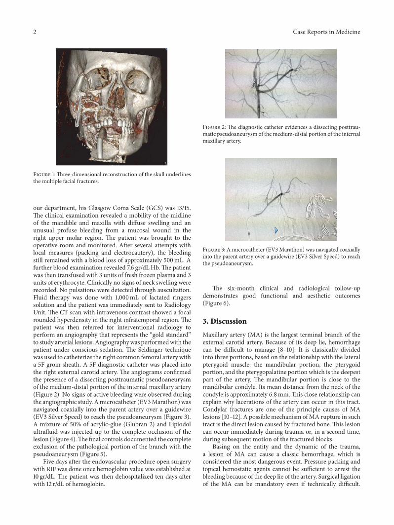
our department, his Glasgow Coma Scale (GCS) was 13/15.The clinical examination revealed a mobility of the midlineof the mandible and maxilla with diffuse swelling and anunusual profuse bleeding from a mucosal wound in theright upper molar region. The patient was brought to theoperative room and monitored. After several attempts withlocal measures (packing and electrocautery), the bleedingstill remained with a blood loss of approximately 500mL. Afurther blood examination revealed 7,6 gr/dLHb.The patientwas then transfused with 3 units of fresh frozen plasma and 3units of erythrocyte. Clinically no signs of neck swelling wererecorded. No pulsations were detected through auscultation.Fluid therapy was done with 1,000mL of lactated ringerssolution and the patient was immediately sent to RadiologyUnit. The CT scan with intravenous contrast showed a focalrounded hyperdensity in the right infratemporal region. Thepatient was then referred for interventional radiology toperform an angiography that represents the “gold standard”to study arterial lesions. Angiographywas performedwith thepatient under conscious sedation. The Seldinger techniquewas used to catheterize the right common femoral artery witha 5F groin sheath. A 5F diagnostic catheter was placed intothe right external carotid artery. The angiograms confirmedthe presence of a dissecting posttraumatic pseudoaneurysmof the medium-distal portion of the internal maxillary artery(Figure 2). No signs of active bleeding were observed duringthe angiographic study. Amicrocatheter (EV3Marathon)wasnavigated coaxially into the parent artery over a guidewire(EV3 Silver Speed) to reach the pseudoaneurysm (Figure 3).A mixture of 50% of acrylic-glue (Glubran 2) and Lipiodolultrafluid was injected up to the complete occlusion of thelesion (Figure 4).Thefinal controls documented the completeexclusion of the pathological portion of the branch with thepseudoaneurysm (Figure 5).
Five days after the endovascular procedure open surgerywith RIF was done once hemoglobin value was established at10 gr/dL. The patient was then dehospitalized ten days afterwith 12 r/dL of hemoglobin.
Figure 2: The diagnostic catheter evidences a dissecting posttrau-matic pseudoaneurysm of themedium-distal portion of the internalmaxillary artery.
Figure 3: Amicrocatheter (EV3Marathon) was navigated coaxiallyinto the parent artery over a guidewire (EV3 Silver Speed) to reachthe pseudoaneurysm.
The six-month clinical and radiological follow-updemonstrates good functional and aesthetic outcomes(Figure 6).
3. Discussion
Maxillary artery (MA) is the largest terminal branch of theexternal carotid artery. Because of its deep lie, hemorrhagecan be difficult to manage [8–10]. It is classically dividedinto three portions, based on the relationship with the lateralpterygoid muscle: the mandibular portion, the pterygoidportion, and the pterygopalatine portion which is the deepestpart of the artery. The mandibular portion is close to themandibular condyle. Its mean distance from the neck of thecondyle is approximately 6.8mm.This close relationship canexplain why lacerations of the artery can occur in this tract.Condylar fractures are one of the principle causes of MAlesions [10–12]. A possible mechanism ofMA rupture in suchtract is the direct lesion caused by fractured bone.This lesioncan occur immediately during trauma or, in a second time,during subsequent motion of the fractured blocks.
Basing on the entity and the dynamic of the trauma,a lesion of MA can cause a classic hemorrhage, which isconsidered the most dangerous event. Pressure packing andtopical hemostatic agents cannot be sufficient to arrest thebleeding because of the deep lie of the artery. Surgical ligationof the MA can be mandatory even if technically difficult.

Case Reports in Medicine 3
Figure 4: Amixture of 50% of acrylic-glue (Glubran 2) and Lipiodolultrafluid was injected up to the complete occlusion of the lesion.
Figure 5: The complete exclusion of the pathological portion of thebranch with the pseudoaneurysm is documented.
Figure 6: Postsurgical 3D CT scan.
Ligation of external carotid artery can be considered as anextreme maneuver.
Pseudoaneurysm, or false aneurysm, is an uncommonconsequence of arterial damage, resulting froman incompletedisruption of the arterial wall causing an expanding lesionbetween the artery and the surrounding tissues [11–14]. Insuch cases, the hematoma of the surrounding tissues counter-balances the arterial pressure, causing the hemorrhage, com-pressing and stabilizing the bleeding. This “natural package”limits the bleeding and if the tear is small lets the plateletsform the clot and stabilize the bleeding with a consequentresolution of the hematoma. If the tear is bigger and plateletsare not able to arrest the bleeding, a pseudoaneurysm canform. The PA is influenced by three factors: (1) the extentof the tera; (2) the elasticity of the surrounding tissues; and(3) the arterial blood flow [15]. Vascular tear depends on thedimension of the fractured bones, related to the artery. Inthe reported case, the maxillary bone fractured in a Le FortIII pattern could have caused the damage. In our experience,the early lesion of the artery could be formed with a suddenshift of the artery in a “concussion way.”This mechanism canexplain those cases in which the entity of fractures seemsnot to be enough to cause a direct lesion of the artery.The inelasticity of the surrounding tissue of the MA can letthe hematoma form a PA, above all in those tracts morecompressed by dense connective tissues.
The diagnosis of PA, although rare, has to be suspected inevery case of posttraumatic and postsurgical severe swellingof the face. Common etiology of PA of the IMA includesblunt and penetrating traumas, orthognathic surgery, neckdissection, surgical removal of impacted third molars, andradiotherapy [11, 14]. Contrast-enhanced CT and catheterangiography are the gold standard in diagnosing PA of theIMA [16].
Some cases of PA are reported to be spontaneouslyresolved. In other cases, complications like delayed hem-orrhage, expansion, neuralgia, pain, and ischemia of thedistal districts have been described [14, 16]. Because of theunpredictable course of the PA a treatment is requested oncethe correct diagnosis is made.
PA treatment includes various surgical and endovascularoptions. Surgical resection is not always possible due toaccessing difficultly of deep-lying lesions [8, 11]. Besides,surgery increases the risk of damage to nerves and itcould cause cosmetic defects as facial scars. Catheter-basedembolization is a safe, quick, and effective technique andit avoids the morbidity of an extensive surgical exposure.Endovascular approach involves either the use of materialsto occlude vessel lumen or the placement of a stent (coveredor not) across the PA base [15]. The best treatment for PAoriginating from IMA is the occlusion of the affected artery,by transarterial embolic agents, distally at the level of themiddle meningeal artery origin. In these cases the collateralcirculation allows vessel sacrifice [16]. Numerous agents havebeen used for the embolization therapy such as metallic coils,polyvinyl alcohol particles, n-butyl cyanoacrylate (NBCA),polymers (Onyx, SQUID), and absorbable sponge gel [9, 11,13]. Metallic coils are permanent embolic agent, with fibersattached or not. Coils are deposited into the vessel lumen

4 Case Reports in Medicine
proximal to the PA to arrest the flow; the positive chargesof the titanium attract the negative charges of blood com-ponents, causing a thrombotic reaction to occlude the vessel.Fibers attached to the coils increase the thrombotic effect.Thecoils choice is fundamental because suitable size and lengthensure adequate thrombosis and flow arrest, preventing theocclusion of normal vessels. NBCA, Onyx, and SQUID pen-etrate deeper into the vessels and theymay go into the venoussystem. Takeshita et al. analyse some series on traumatic IMAPA treatedwith endovascular therapy [14].They reported thatthemost common embolization agents used aremetallic coils(40%), particles (28%), and NBCA (24%).They reported thatNBCA was the most appropriate embolic material for the PAbecause embolization is completed more quickly comparedto other agents, the primary hemostasis rate is higher, andthe recurrent hemorrhage rate is lower [14–16]. Howeveroperators should be familiar with the use of NBCA becausethe reflux of polymerized glue around the microcatheter mayadhere to its tip, increasing the risk of nontarget embolizationor catheter retention. Parent artery occlusion with metalliccoils is considered an effective procedure. A disadvantageof such procedure is the risk of recurrent hemorrhage dueto retrograde filling of the PA through indirect collateralcirculation. The filling of PA sac with coils may rupturethe aneurysm wall, causing the migration of coils outsideof the target lesion. Acute complications of endovasculartreatment include distal thromboembolic events (occlusionof the central retinal artery, ischemic stroke due to potentialanastomosis between the IMA and the ophthalmic artery)and local tissue infarct. Other reported complications areperforations, glued vein, microcatheter fracture, and vesseldissection or branch occlusion [13–16]. Moreover, to reducethe rate of complications, as blindness, facial palsy, and othercranial nerve palsies, it ismandatory to know the anastomosisbetween the external carotid artery and the internal carotidartery [15, 16].
Competing Interests
Authors declare no conflict of interests.
References
[1] M. Bozkurt, E. Kapi, P. Karakol, and E. Yorgancilar, “Sud-den rupture of the internal maxillary artery causing pseu-doaneurysm (mandibular part) secondary to subcondylarmandible fracture,” Journal of Craniofacial Surgery, vol. 20, no.5, pp. 1430–1432, 2009.
[2] S. Chakrabarty, S. K. Majumdar, A. Ghatak, and A. Bansal,“Management of pseudoaneurysm of internal maxillary arteryresulting from trauma,” Journal of Maxillofacial and OralSurgery, vol. 14, no. S1, pp. 203–208, 2015.
[3] X. Fan and Q. Mao, “Life-threatening oral haemorrhage of apseudoaneurysm after raising of a fractured zygoma,” BritishJournal of Oral andMaxillofacial Surgery, vol. 40, no. 6, pp. 508–509, 2002.
[4] G. Gerbino, F. Roccia, M. Grosso, and D. Regge, “Pseudoa-neurysm of the internal maxillary artery and Frey’s syndrome
after blunt facial trauma,” Journal of Oral and MaxillofacialSurgery, vol. 55, no. 12, pp. 1485–1490, 1997.
[5] M. P. Hennus and L. Speleman, “Internal maxillary artery pseu-doaneurysm: a near fatal complication of seemingly innocuouspharyngeal trauma,” Case Reports in Critical Care, vol. 2011,Article ID 241375, 4 pages, 2011.
[6] J. Lamphier, V. Ziccardi, A. Ruvo, and M. Janel, “Complicationsof mandibular fractures in an urban teaching center,” Journal ofOral and Maxillofacial Surgery, vol. 61, no. 7, pp. 745–749, 2003.
[7] D. T. Lanigan, J. H. Hey, and R. A. West, “Major vascularcomplications of orthognathic surgery: false aneurysms andarteriovenous fistulas following orthognathic surgery,” JournalofOral andMaxillofacial Surgery, vol. 49, no. 6, pp. 571–577, 1991.
[8] S. Mohanty, U. Gulati, and S. Kathuria, “Pseudoaneurysm ofthe internal maxillary artery: a rare complication of condylarfracture,” Craniomaxillofacial Trauma and Reconstruction, vol.6, no. 4, pp. 271–274, 2013.
[9] D. A. Pandyan, P. Siroraj, Nandakumar, and C. D. Narayanan,“Pseudoaneurysmof internalmaxillary artery—anuntold com-plication following distraction osteogenesis—a case report,”Journal of Oral and Maxillofacial Surgery, vol. 72, no. 3, pp.605.e1–605.e7, 2014.
[10] N. M. Rich, R. W. Hobson II, and G. J. Collins Jr., “Traumaticarteriovenous fistulas and false aneurysms: a review of 558lesions,” Surgery, vol. 78, no. 6, pp. 817–828, 1975.
[11] A. C. Silva, F. O’Ryan, M. L. Beckley, H. Y. Young, and D. Poor,“Pseudoaneurysm of a branch of the maxillary artery followingmandibular sagittal split ramus osteotomy: case report andreview of the literature,” Journal of Oral and MaxillofacialSurgery, vol. 65, no. 9, pp. 1807–1816, 2007.
[12] E. N. Siniscalchi, F. Minutoli, L. Catalfamo, F. Romano, M.Longo, and F. S. De Ponte, “Intraosseous mandibular artero-venous malformations: case report,” Journal of Cranio-Maxillofacial Surgery, vol. 37, no. 2, pp. 106–109, 2009.
[13] H. Y. Soh, A. S. Muda, N. A. Jabar, R. Nordin, S. Nabil, andR. Ramli, “Non-pulsatile traumatic pseudoaneurysm of theinternal maxillary artery following trauma to mandible,” Oraland Maxillofacial Surgery, vol. 19, no. 4, pp. 423–425, 2015.
[14] T. Takeshita, K. Hayashi, N. Horie, M. Morikawa, K. Suyama,and I. Nagata, “Endovascular treatment of intractable bleedingfrom a traumatic pseudoaneurysm of the internal maxillaryartery,”Neuroradiology Journal, vol. 25, no. 4, pp. 469–474, 2012.
[15] D. Wang, L. Su, Y. Han, and X. Fan, “Embolization treatment ofpseudoaneurysms originating from the external carotid artery,”Journal of Vascular Surgery, vol. 61, no. 4, pp. 920–926, 2015.
[16] M. Yeo, T. Goh, V. Nallathamby, E. Cheong, and T. Lim, “Max-illary artery injury associated with subcondylar mandible frac-tures: a novel treatment algorithm,”Craniomaxillofacial Traumaand Reconstruction, vol. 5, no. 2, pp. 83–88, 2012.

Submit your manuscripts athttp://www.hindawi.com
Stem CellsInternational
Hindawi Publishing Corporationhttp://www.hindawi.com Volume 2014
Hindawi Publishing Corporationhttp://www.hindawi.com Volume 2014
MEDIATORSINFLAMMATION
of
Hindawi Publishing Corporationhttp://www.hindawi.com Volume 2014
Behavioural Neurology
EndocrinologyInternational Journal of
Hindawi Publishing Corporationhttp://www.hindawi.com Volume 2014
Hindawi Publishing Corporationhttp://www.hindawi.com Volume 2014
Disease Markers
Hindawi Publishing Corporationhttp://www.hindawi.com Volume 2014
BioMed Research International
OncologyJournal of
Hindawi Publishing Corporationhttp://www.hindawi.com Volume 2014
Hindawi Publishing Corporationhttp://www.hindawi.com Volume 2014
Oxidative Medicine and Cellular Longevity
Hindawi Publishing Corporationhttp://www.hindawi.com Volume 2014
PPAR Research
The Scientific World JournalHindawi Publishing Corporation http://www.hindawi.com Volume 2014
Immunology ResearchHindawi Publishing Corporationhttp://www.hindawi.com Volume 2014
Journal of
ObesityJournal of
Hindawi Publishing Corporationhttp://www.hindawi.com Volume 2014
Hindawi Publishing Corporationhttp://www.hindawi.com Volume 2014
Computational and Mathematical Methods in Medicine
OphthalmologyJournal of
Hindawi Publishing Corporationhttp://www.hindawi.com Volume 2014
Diabetes ResearchJournal of
Hindawi Publishing Corporationhttp://www.hindawi.com Volume 2014
Hindawi Publishing Corporationhttp://www.hindawi.com Volume 2014
Research and TreatmentAIDS
Hindawi Publishing Corporationhttp://www.hindawi.com Volume 2014
Gastroenterology Research and Practice
Hindawi Publishing Corporationhttp://www.hindawi.com Volume 2014
Parkinson’s Disease
Evidence-Based Complementary and Alternative Medicine
Volume 2014Hindawi Publishing Corporationhttp://www.hindawi.com