Case Report Recurrent Midgut Bleeding due to...
5
Case Report Recurrent Midgut Bleeding due to Jejunal Angioleiomyoma Mahir Gachabayov and Petr Mityushin Department of Abdominal Surgery, Vladimir City Clinical Hospital of Emergency Medicine, Gorky Street 5, Vladimir 600017, Russia Correspondence should be addressed to Mahir Gachabayov; [email protected] Received 10 July 2016; Accepted 21 August 2016 Academic Editor: Gregorio Santori Copyright © 2016 M. Gachabayov and P. Mityushin. is is an open access article distributed under the Creative Commons Attribution License, which permits unrestricted use, distribution, and reproduction in any medium, provided the original work is properly cited. Angioleiomyoma being a type of true smooth muscle gastrointestinal tumors can lead to serious life-threatening gastrointestinal bleeding. We report a case of 21-year-old male patient with recurrent midgut bleeding. Contrast-enhanced CT revealed highly vascular small bowel neoplasm. e patient underwent laparotomy with bowel resection and recovered uneventfully. Histopathology revealed jejunal angioleiomyoma. 1. Introduction Obscure gastrointestinal bleeding (GIB) is persistent or recurrent bleeding from the gastrointestinal (GI) tract aſter negative evaluations with upper and lower endoscopies accounting for 5% of all GIB cases [1, 2]. Midgut GIB makes up to 80% of all obscure GIB cases [3]. Small bowel tumors are responsible for 10–20% of these cases of midgut GIB in Western countries [1]. Angioleiomyomata of small bowel, especially those complicated by GIB, are very rare. 2. Case Report A 21-year-old male student from another city but studying in Vladimir was admitted to Vladimir City Clinical Hospital of Emergency Medicine with a 2-day history of melena and fatigue. His past medical history was significant for peptic ulcer disease because of which he was exempted from military service, recurrent epistaxis due to septal deviation, and chronic iron-deficiency anemia. On admission his skin was pale, heart rate 98 bpm, blood pressure 110/60 mmHg, and hemoglobin 6.2 mg/dL. On EGD a flat duodenal ulcer (0.8 cm with fibrin-covered base) was revealed. PPIs and packed RBCs (2 doses) started immediately and aſter 3 days the patient was transferred to the department of internal medicine with Hb of 8.4 mg/dL and BP of 120/70 mmHg. Eight days aſter discharge from our department the patient was admitted again with recurrent melena and fatigue during 10 hours. On admission, HR was 104 bpm, BP 80/60 mmHg, and Hb 5.5 mg/dL. Healthily discharged patient came back with the fear of dying. EGD was unre- markable. Aſter IV fluids with 2 doses of packed RBCs and PPIs the patient was prepared for a colonoscopy with laxa- tives. Colonoscopy also appeared to be unremarkable. en the patient was consulted by otolaryngologists to rule out possible posterior epistaxis. Endoscopic rhinoscopy revealed eroded nasal polyp without signs of ongoing bleeding which was excised. e patient was discharged aſter 2 days with BP 110/70 mmHg and Hb 7.7 mg/dL. Seven days aſter discharge the patient was admitted again with 6-hour severe GI bleeding with BP 70/50 mmHg and Hb 4.9 mg/dL when we realized that we dealt with obscure midgut GI bleeding. Aſter stabilization by infusing IV crystalloids and colloids, 4 doses of packed RBCs, and 4 doses of fresh frozen plasma the patient underwent contrast- enhanced multislice CT which revealed a highly vascular small bowel tumor (Figure 1). Considering high risk of recurrent bleeding urgent laparotomy and small bowel resec- tion were performed which proved the diagnosis of jejunal neoplasm (Figure 2). Postoperatively, oral feeds resumed on the 2nd postoperative day, the wound stitches were taken off on 8th postoperative day, and the patient was discharged with BP 120/70 mmHg and Hb 9.8 mg/dL. At 3-month follow- up the patient was feeling well; his Hb was 15.4 mg/dL. e histopathology revealed bundles of spindle cells ori- ented perpendicularly to each other with bright eosinophilic Hindawi Publishing Corporation Case Reports in Surgery Volume 2016, Article ID 4569781, 4 pages http://dx.doi.org/10.1155/2016/4569781
-
Upload
nguyennhan -
Category
Documents
-
view
242 -
download
0
Transcript of Case Report Recurrent Midgut Bleeding due to...

Case ReportRecurrent Midgut Bleeding due to Jejunal Angioleiomyoma
Mahir Gachabayov and Petr Mityushin
Department of Abdominal Surgery, Vladimir City Clinical Hospital of Emergency Medicine, Gorky Street 5, Vladimir 600017, Russia
Correspondence should be addressed to Mahir Gachabayov; [email protected]
Received 10 July 2016; Accepted 21 August 2016
Academic Editor: Gregorio Santori
Copyright © 2016 M. Gachabayov and P. Mityushin. This is an open access article distributed under the Creative CommonsAttribution License, which permits unrestricted use, distribution, and reproduction in any medium, provided the original work isproperly cited.
Angioleiomyoma being a type of true smooth muscle gastrointestinal tumors can lead to serious life-threatening gastrointestinalbleeding. We report a case of 21-year-old male patient with recurrent midgut bleeding. Contrast-enhanced CT revealedhighly vascular small bowel neoplasm. The patient underwent laparotomy with bowel resection and recovered uneventfully.Histopathology revealed jejunal angioleiomyoma.
1. Introduction
Obscure gastrointestinal bleeding (GIB) is persistent orrecurrent bleeding from the gastrointestinal (GI) tract afternegative evaluations with upper and lower endoscopiesaccounting for 5% of all GIB cases [1, 2]. Midgut GIB makesup to 80% of all obscure GIB cases [3]. Small bowel tumorsare responsible for 10–20% of these cases of midgut GIBin Western countries [1]. Angioleiomyomata of small bowel,especially those complicated by GIB, are very rare.
2. Case Report
A 21-year-old male student from another city but studyingin Vladimir was admitted to Vladimir City Clinical Hospitalof Emergency Medicine with a 2-day history of melenaand fatigue. His past medical history was significant forpeptic ulcer disease because of which he was exempted frommilitary service, recurrent epistaxis due to septal deviation,and chronic iron-deficiency anemia. On admission his skinwas pale, heart rate 98 bpm, blood pressure 110/60mmHg,and hemoglobin 6.2mg/dL. On EGD a flat duodenal ulcer(0.8 cm with fibrin-covered base) was revealed. PPIs andpacked RBCs (2 doses) started immediately and after 3 daysthe patient was transferred to the department of internalmedicine with Hb of 8.4mg/dL and BP of 120/70mmHg.
Eight days after discharge from our department thepatient was admitted again with recurrent melena and
fatigue during 10 hours. On admission, HR was 104 bpm,BP 80/60mmHg, and Hb 5.5mg/dL. Healthily dischargedpatient came back with the fear of dying. EGD was unre-markable. After IV fluids with 2 doses of packed RBCs andPPIs the patient was prepared for a colonoscopy with laxa-tives. Colonoscopy also appeared to be unremarkable. Thenthe patient was consulted by otolaryngologists to rule outpossible posterior epistaxis. Endoscopic rhinoscopy revealederoded nasal polyp without signs of ongoing bleeding whichwas excised. The patient was discharged after 2 days with BP110/70mmHg and Hb 7.7mg/dL.
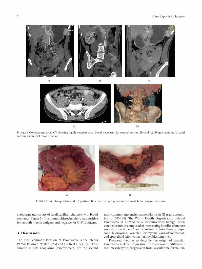
Seven days after discharge the patient was admittedagain with 6-hour severe GI bleeding with BP 70/50mmHgand Hb 4.9mg/dL when we realized that we dealt withobscure midgut GI bleeding. After stabilization by infusingIV crystalloids and colloids, 4 doses of packed RBCs, and 4doses of fresh frozen plasma the patient underwent contrast-enhanced multislice CT which revealed a highly vascularsmall bowel tumor (Figure 1). Considering high risk ofrecurrent bleeding urgent laparotomy and small bowel resec-tion were performed which proved the diagnosis of jejunalneoplasm (Figure 2). Postoperatively, oral feeds resumed onthe 2nd postoperative day, the wound stitches were takenoff on 8th postoperative day, and the patient was dischargedwith BP 120/70mmHg andHb 9.8mg/dL. At 3-month follow-up the patient was feeling well; his Hb was 15.4mg/dL.The histopathology revealed bundles of spindle cells ori-ented perpendicularly to each other with bright eosinophilic
Hindawi Publishing CorporationCase Reports in SurgeryVolume 2016, Article ID 4569781, 4 pageshttp://dx.doi.org/10.1155/2016/4569781
2 Case Reports in Surgery
(a) (b) (c)
(d) (e)
Figure 1: Contrast-enhanced CT showing highly vascular small bowel neoplasm: (a) coronal section, (b) and (c) oblique sections, (d) axialsection, and (e) 3D reconstruction.
(a) (b)
Figure 2: (a) Intraoperative and (b) postresection macroscopic appearance of small bowel angioleiomyoma.
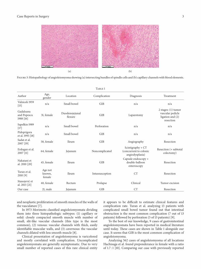
cytoplasm and variety of small capillary channels with bloodelements (Figure 3).The immunohistochemistry was positivefor smooth muscle antigens and negative for GIST antigens.
3. Discussion
The most common location of leiomyoma is the uterus(95%), followed by skin (3%) and GI tract (1.5%) [4]. Truesmooth muscle neoplasms (leiomyomata) are the second
most common mesenchymal neoplasms in GI tract account-ing for 32% [5]. The World Health Organization definedleiomyoma in 1969 to be a “circumscribed benign, oftencutaneous tumor composed of intersecting bundles ofmaturesmooth muscle cells” and classified it into three groups:solid leiomyoma, vascular leiomyoma (angioleiomyoma),and epithelioid leiomyoma (leiomyoblastoma) [6].
Proposed theories to describe the origin of vascularleiomyoma include progression from aberrant undifferenti-ated mesenchyme, progression from vascular malformation,
Case Reports in Surgery 3
200MKM
(a)
200MKM
(b)
Figure 3: Histopathology of angioleiomyoma showing (a) intersecting bundles of spindle cells and (b) capillary channels with blood elements.
Table 1
Author Age,gender Location Complication Diagnosis Treatment
Valnicek 1959[15] n/a Small bowel GIB n/a n/a
Gadaleanuand Popescu1988 [16]
31, female Duodenojejunalflexure GIB Laparotomy
2 stages: (1) tumorvascular pedicleligation and (2)
resectionSapelkin 1989[17] n/a Small bowel Perforation n/a n/a
Pidoprigoraet al. 1995 [18] n/a Small bowel GIB n/a n/a
Sadat et al.2007 [19] 58, female Ileum GIB Angiography Resection
Erdogan et al.2007 [11] 64, female Jejunum Noncomplicated
Scintigraphy + CT(concurrent to colonic
angiodysplasia)
Resection (+ subtotalcolectomy)
Nakatani etal. 2010 [20] 45, female Ileum GIB
Capsule endoscopy +double-balloonenteroscopy
Resection
Turan et al.2010 [9]
Age notknown,female
Ileum Intussusception CT Resection
Stanojevic etal. 2013 [21] 40, female Rectum Prolapse Clinical Tumor excision
Our case 21, male Jejunum GIB CT Resection
and neoplastic proliferation of smoothmuscles of the walls ofthe vasculature [7].
In 1973 Morimoto classified angioleiomyomata dividingthem into three histopathologic subtypes: (1) capillary orsolid: closely compacted smooth muscle with number ofsmall, slit-like vascular channels (this type is the mostcommon), (2) venous: vascular channels with thick, easilyidentifiable muscular walls, and (3) cavernous: the vascularchannels dilated with less smooth muscle [8].
Clinical presentation of angioleiomyoma is varicoloredand mostly correlated with complication. Uncomplicatedangioleiomyomata are generally asymptomatic. Due to verysmall number of reported cases of this rare clinical entity
it appears to be difficult to estimate clinical features andcomplication rate. Turan et al. analyzing 13 patients withcomplicated small bowel tumor found out that intestinalobstruction is the most common complication (7 out of 13patients) followed by perforation (5 of 13 patients) [9].
To the best of our knowledge, 9 cases of gastrointestinalangioleiomyomata have been reported in medical literatureuntil today. These cases are shown in Table 1 alongside ourcase. It seems that GIB is the most common complication ofangioleiomyoma.
Analyzing 562 cases of angioleiomyoma of all locationsHachisuga et al. found preponderance in female with a ratioof 1.7 : 1 [10]. Comparing our case with previously reported

4 Case Reports in Surgery
cases with known data, our case appears to be the first casewith male patient (Table 1). Moreover, our patient is theyoungest. Most of the patients are older than 40 years.
In most previously reported cases radiology emerged tobe more common. Contrast-enhanced CT appeared to beaccurate in three cases including our case. Previous studiesshowed CT scan and scintigraphy to be sensitive for smallbowel tumors [11, 12]. Takeshita et al. showed video capsuleendoscopy and double-balloon enteroscopy to be beneficialfor small bowel lesions [13]. Immunohistochemistry is crucialin the diagnosis of mesenchymal tumor and differentiation ofmalignant and suspicious high risk tumors [14].Thedefinitivetreatment of angioleiomyoma is resection.
To conclude, small bowel angioleiomyoma is rare butlife-threatening cause of midgut gastrointestinal bleeding.Contrast-enhanced tomography should be performed to apatient with obscure gastrointestinal bleeding after negativegastroscopy and colonoscopy.
Competing Interests
The authors declare that there is no conflict of interestsregarding the publication of this paper.
References
[1] K. Liu and A. J. Kaffes, “Review article: the diagnosis andinvestigation of obscure gastrointestinal bleeding,” AlimentaryPharmacology andTherapeutics, vol. 34, no. 4, pp. 416–423, 2011.
[2] A. Szold, L. B. Katz, and B. S. Lewis, “Surgical approach to occultgastrointestinal bleeding,”TheAmerican Journal of Surgery, vol.163, no. 1, pp. 90–93, 1992.
[3] B. Keum and H. J. Chun, “Capsule endoscopy and double bal-loon enteroscopy for obscure gastrointestinal bleeding: which isbetter?” Journal of Gastroenterology and Hepatology, vol. 26, no.5, pp. 794–795, 2011.
[4] M. Veeresh, M. Sudhakara, G. Girish, and C. Naik, “Leiomy-oma: a rare tumor in the head and neck and oral cavity:Report of 3 cases with review,” Journal of Oral andMaxillofacialPathology, vol. 17, no. 2, pp. 281–287, 2013.
[5] A. Agaimy and P. H. Wunsch, “True smooth muscle neo-plasms of the gastrointestinal tract: morphological spectrumand classification in a series of 85 cases from a single institute,”Langenbeck’s Archives of Surgery, vol. 392, no. 1, pp. 75–81, 2007.
[6] F. M. Enzinger, R. Lattes, and H. Torloni, Histological Typingof Soft Tissue Tumours, World Health Organization, Geneva,Switzerland, 1969.
[7] A. Alshwareb, J. Bhati, Y. Al-Nufaily, N. Alaudah, and Z.AlQudehy, “Middle ear leiomyoma presenting as granuloma-tous otitis media,” Global Journal of Otolaryngology, vol. 1, no.1, Article ID 555551, 2015.
[8] N. Morimoto, “Angiomyoma (vascular leiomyoma): a clinico-pathological study,” Medical Journal of Kagoshima University,vol. 24, pp. 663–683, 1973.
[9] M. Turan, K. Karadayi, M. Duman et al., “Small bowel tumorsin emergency surgery,” Ulus Travma Acil Cerrahi Derg, vol. 16,no. 4, pp. 327–333, 2010.
[10] T. Hachisuga, H. Hashimoto, and M. Enjoji, “Angioleiomyoma:a clinicopathologic reappraisal of 562 cases,”Cancer, vol. 54, no.1, pp. 126–130, 1984.
[11] S. Erdogan, E. Ozkara, K. Koseoglu, H. Yasa, H. Cevikel, and H.Afsin, “Incidental detection of a jejunal angioleiomyoma by Tc-99m RBC study during evaluation of gastrointestinal bleedingdue to angiodysplasia,” Turkish Journal of NuclearMedicine, vol.16, pp. 32–36, 2007.
[12] W. H. Schwesinger, K. R. Sirinek, H. V. Gaskill III, J. P. Velez,J. J. Corea, and W. E. Strodel, “Jejunoileal causes of overt gas-trointestinal bleeding: diagnosis, management and outcome,”The American Surgeon, vol. 67, no. 4, pp. 383–387, 2001.
[13] N. Takeshita, Y. Otsuka, S. Nara et al., “Utility of preoperativesmall-bowel endoscopy for hemorrhagic lesions in the smallintestine,” Surgery Today, vol. 42, no. 6, pp. 536–541, 2012.
[14] G. O. Ogun, “Mesenchymal tumours of the gastrointestinaltract: the importance and use of immunhistochemistry incharacterizing specific tumour entities,” Nigerian Journal ofMedicine, vol. 24, no. 2, pp. 150–154, 2015.
[15] V. Valnicek, “Angioleiomyoma of the small intestine as a causeof severe melena,” Rozhledy v Chirurgii, vol. 38, pp. 848–880,1959.
[16] V. Gadaleanu and V. Popescu, “Angiomyoma and vascularectasia of the small bowel as a cause of intestinal bleeding,”Pathology Research and Practice, vol. 183, no. 4, pp. 519–521,1988.
[17] O. S. Sapelkin, “Angioleiomyoma of the small intestine compli-cated by perforation,”Klinicheskaya Khirurgiya, no. 2, pp. 61–62,1989 (Russian).
[18] A. P. Pidoprigora, S. P. Chmeruk, L. I. Fedorchuk, and S. I.Shuldik, “Angioleiomyoma of the small intestine as a cause ofrecurrence of hemorrhage,” Klinichna Khirurhiia, no. 4, article48, 1995.
[19] U. Sadat, N. S. Theivacumar, J. Vat, and A. Jah, “Angioleiomy-oma of the small intestine-a rare cause of gastrointestinalbleeding,”World Journal of Surgical Oncology, vol. 5, article 129,2007.
[20] M. Nakatani, Y. Fujiwara, N. Kameda et al., “Angioleiomyomaof the small intestine detected by double-balloon enteroscopy,”Gastrointestinal Endoscopy, vol. 72, no. 1, pp. 187–188, 2010.
[21] G. Z. Stanojevic, D. S. Mihailovic, M. D. Nestorovic et al., “Caseof rectal angioleiomyoma in a female patient,”World Journal ofGastroenterology, vol. 19, no. 13, pp. 2114–2117, 2013.

Submit your manuscripts athttp://www.hindawi.com
Stem CellsInternational
Hindawi Publishing Corporationhttp://www.hindawi.com Volume 2014
Hindawi Publishing Corporationhttp://www.hindawi.com Volume 2014
MEDIATORSINFLAMMATION
of
Hindawi Publishing Corporationhttp://www.hindawi.com Volume 2014
Behavioural Neurology
EndocrinologyInternational Journal of
Hindawi Publishing Corporationhttp://www.hindawi.com Volume 2014
Hindawi Publishing Corporationhttp://www.hindawi.com Volume 2014
Disease Markers
Hindawi Publishing Corporationhttp://www.hindawi.com Volume 2014
BioMed Research International
OncologyJournal of
Hindawi Publishing Corporationhttp://www.hindawi.com Volume 2014
Hindawi Publishing Corporationhttp://www.hindawi.com Volume 2014
Oxidative Medicine and Cellular Longevity
Hindawi Publishing Corporationhttp://www.hindawi.com Volume 2014
PPAR Research
The Scientific World JournalHindawi Publishing Corporation http://www.hindawi.com Volume 2014
Immunology ResearchHindawi Publishing Corporationhttp://www.hindawi.com Volume 2014
Journal of
ObesityJournal of
Hindawi Publishing Corporationhttp://www.hindawi.com Volume 2014
Hindawi Publishing Corporationhttp://www.hindawi.com Volume 2014
Computational and Mathematical Methods in Medicine
OphthalmologyJournal of
Hindawi Publishing Corporationhttp://www.hindawi.com Volume 2014
Diabetes ResearchJournal of
Hindawi Publishing Corporationhttp://www.hindawi.com Volume 2014
Hindawi Publishing Corporationhttp://www.hindawi.com Volume 2014
Research and TreatmentAIDS
Hindawi Publishing Corporationhttp://www.hindawi.com Volume 2014
Gastroenterology Research and Practice
Hindawi Publishing Corporationhttp://www.hindawi.com Volume 2014
Parkinson’s Disease
Evidence-Based Complementary and Alternative Medicine
Volume 2014Hindawi Publishing Corporationhttp://www.hindawi.com


![Case Report Giant Verrucous Haemangioma with Linear ...angiokeratoma, angioma serpiginosum, lymphangioama and pigmented tumours.[7] Recurrent bleeding and infection along with increase](https://static.fdocuments.net/doc/165x107/5e3b758d6f248601c355512e/case-report-giant-verrucous-haemangioma-with-linear-angiokeratoma-angioma-serpiginosum.jpg)


![Untitled-1 [] · Unexplained weight loss. Recurrent fevers Recurrent Infections Excessive bleeding, bruising ort-ash. Constant lack of energy and paleness. Lump or mass anywhere in](https://static.fdocuments.net/doc/165x107/5fc7c204da0911017a0d49b9/untitled-1-unexplained-weight-loss-recurrent-fevers-recurrent-infections-excessive.jpg)