identification and characterization of midgut digestive proteases
A r t i c l e
SERINE PROTEASE FROM MIDGUTOF Bombus terrestris MALESJana Brabcova, Jirı Kindl, Irena Valterova, IvaPichova, and Marie ZarevuckaInstitute of Organic Chemistry and Biochemistry, Academy of Sciences ofthe Czech Republic, Flemingovo nam. 2, 166 10 Prague 6, Czech Republic
Jana BrabcovaInstitute of Chemical Technology Prague, Faculty of Food and BiochemicalTechnology, Technicka 5, 166 28 Prague 6, Czech Republic
Michal Jagr and Ivan MiksıkInstitute of Physiology, Academy of Sciences of the Czech Republic,Vıdenska 1083, 142 20 Prague 4, Czech Republic
A serine protease was isolated from midguts of the bumblebee male Bombusterrestris by a combination of precipitation procedures with columnchromatography. The purified enzyme exhibited two bands with molecularmasses of 25 and 26 kDa as determined by sodium dodecyl sulfatepolyacrylamide gel electrophoresis. These bands showed a proteolytic activityin zymography assay. Midgut enzymes showed optimum proteolytic activityat pH 9 and 35◦C using N-succinyl-L-alanyl-L-alanyl-L-prolyl-L-phenyl-alanine 4-nitroanilide as a substrate. The Michaelis constant (Km) andmaximum reaction rate (Vmax) were 0.55 ± 0.042 mM and 0.714 ±0.056 μmol p-nitroalanine produced min−1 mg protein−1, respectively.Inhibition was affected by trypsin inhibitor, but not byphenylmethylsulfonyl fluoride and N-tosyl-L-phenylalanine chloromethylketone, which indicated the trypsin-like but not chymotrypsin-likespecificity. The identity of the serine protease was confirmed bynanoliquid-tandem mass spectrometry. Eleven unique peptides of the B.terrestris serine protease were found. It shows high homology to a previouslyreported B. ignitus serine protease covering more than 65% of the proteinamino acid sequence. C© 2012 Wiley Periodicals, Inc.
Grant sponsor: Institute of Organic Chemistry and Biochemistry; Grant number: RVO: 61388963; Grant sponsor:The Czech Science Foundation; Grant number: 203/09/1446; Grant sponsor: Technology Agency of the CzechRepublic; Grant number: TA01020969.Correspondence to: Marie Zarevucka, IOCB AS CR, Flemingovo n. 2, 166 10 Prague 6, Czech Republic.E-mail: [email protected]
ARCHIVES OF INSECT BIOCHEMISTRY AND PHYSIOLOGY, Vol. 82, No. 3, 117–128 (2013)Published online in Wiley Online Library (wileyonlinelibrary.com).C© 2012 Wiley Periodicals, Inc. DOI: 10.1002/arch.21075

118 � Archives of Insect Biochemistry and Physiology, March 2013
Keywords: Bombus terrestris; midgut; serine protease; bumblebee
INTRODUCTION
Serine proteases play critical roles in an insect physiology. They serve as major insectgut enzymes in the digestion of dietary proteins, inactivate protein toxins, and showantimicrobial activity (Hegedus et al., 2003; Prabhakar et al., 2007; Chougule et al., 2008).Serine proteases also play critical roles in several insect biological processes such as bloodcoagulation, signal transduction, hormone activation, and development (Hedstrom, 2002;Clynen et al., 2005).
The catalytic function of serine proteases is accomplished via a catalytic triad (reactiveserine, histidine, and aspartic acid). The degree and the type of substrate specificity isdetermined by many specific chemicals within the substrate-binding cleft (Perona andCraik, 1995). Serine proteases belong to a large group of endopeptidases that require aserine residue for their catalytic activity. They are equally important for biological systems(Kini, 2005; Muller et al., 2007; Yoon et al., 2007) as well as for commercial purposes(Saeki et al., 103).
Insects produce serine proteases within gut cells and release the enzymes into thegut lumen, where they digest food proteins into short peptides and amino acids suitablefor their absorption by the midgut (Pauchet et al., 2008). As a defense mechanism,host plants produce proteinase inhibitors to suppress the activity of serine proteases(e.g., trypsin and chymotrypsin; Jongsma et al., 1995). Some polyphagous insects such asHelicoverpa zea, Agrotis ipsilon, and Spodoptera frugiperda can overcome this defense actionby expressing trypsins and chymotrypsins that are insensitive to host proteinase inhibitors(Mazumdar-Leighton and Broadway, 2001; Brioschi et al., 2007). In addition to proteindigestion, chymotrypsins may be involved in other physiological processes. For example,in Manduca sexta a chymotrypsin is involved in synthesis of chitin which is necessary forperitrophic matrix formation (Broehan et al., 2008).
The most frequently isolated protease from insects has been trypsin (Kalhok et al.,1993; Noriega et al., 1996), specifically a serine proteinase, demonstrably or presumablyof the chymotrypsin-trypsin-like family. There have also been numerous reports on insectchymotrypsins (Elpidina et al., 2005; Lopes et al., 2009).
Insect species have individual digestive serine proteases. From Locusta migratoria (Lamet al., 2000), both trypsin and chymotrypsin have been isolated. Similarly, from the honey-bee Apis mellifica, gut proteinases with trypsin-like and chymotrypsin-like specificities havebeen purified (Burgess et al., 1996). Some enzymes were present only in adult workersand drones, possibly reflecting the composition of the diets of different honeybee castes.
There is relatively little information on bumblebee digestive proteases. Here, we ad-dress this gap by characterizing the major digestive proteolytic enzyme(s) of bumblebeemale Bombus terrestris, important glasshouse pollinators. The information helps to deter-mine the possible function of serine proteases.
MATERIALS AND METHODS
Insect
The B. terrestris males (2 days old) were obtained from laboratory colonies during thewinter season 2010–2011 as described by Ptacek et al. (2000). Briefly, the midguts wereisolated immediately after collecting the males and kept at −18◦C until use.
Archives of Insect Biochemistry and Physiology
Serine Protease of Bombus terrestris � 119
Preparation of Crude Enzymes
After decapitation, midgut tissues were collected in ice-cold saline solution (0.90% NaCl,w/v). Isolated midguts were stored at 0◦C until use. The tissues were homogenized, ata ratio: 25 μl of saline solution per organ, using a Potter–Elvehjem homogenizer with aTeflon R© pestle. The homogenate was centrifuged at 20,000 × g for 20 min. The pelletwas resuspended in saline solution (25 μl per individual) and centrifuged at 20,000 × gfor 20 min. The supernatants were collected and centrifuged at 20,000 × g for 30 min.All steps were carried out at 4◦C and glycerol was added to a final concentration of 50%(w/v). The suspension was stored at –20◦C until further used.
Purification
Protease activity in each fraction was monitored by the release of p-nitroanilide fromproteinase substrate (as described below). The midgut extract was prepared from12 g crude enzyme material obtained from 500 individuals of 2-day-old B. terrestris males.Solid ammonium sulfate was added to the extract to make the concentration at 70%saturation and stirred overnight at 4◦C. Next day, the insoluble material was removedby centrifugation at 15,000 × g for 30 min. The precipitate was resuspended in 2 mlof 1 M (NH4)2SO4 in 50 mM Tris/HCl buffer (pH 7.8) and dialyzed against the samebuffer overnight using a dialysis sack. Subsequently, the dialyzed sample was applied toa Phenyl-Sepharose column (GE Healthcare Life Science, Uppsala, Sweden, formerlyAmersham Biosciences) preequilibrated with the same buffer. After washing the column,proteins were eluted by a linear gradient of 50 mM Tris/HCl buffer (pH 7.8) containing(NH4)2SO4 from 1 to 0 M at a flow rate of 1 ml/min. The active fractions were collected(5 ml) and then analyzed by sodium dodecyl sulfate polyacrylamide gel electrophoresis(SDS-PAGE) and LC-MS/MS. All purification steps were carried out at 4◦C.
Determination of Protein Concentration
Protein concentration was determined according to Bradford assay using bovine serumalbumine as a standard (Bradford, 1976).
Enzyme Activity Assay
Serine proteinase activity of the enzyme was measured by the release of p-nitroanilidinefrom the typical serine proteinase substrate N-succinyl-Ala-Ala-Pro-Phe-pNA and moni-tored by spectrophotometry at 405 nm. The assay was performed in 50 mM Tris-HClbuffer (pH 8.2) at 25◦C for 30 min in a reaction volume of 1,150 μl containing 100 mlof 0.2 mM substrate and 50 μl of the crude enzyme. One unit of protease activity (U) isdefined as the amount of enzyme releasing 1 μmol of p-nitroaniline per minute underthe given assay conditions.
Effect of Temperature and pH
The optimum temperature was determined under standard assay conditions by incubatingthe reaction mixtures at temperatures ranging from 25◦C to 65◦C. To determine thermalstability, the enzyme was preincubated for 30 min in the assay buffer at temperaturesranging from 25◦C to 65◦C and then chilled on ice for 10 min. The remaining enzyme
Archives of Insect Biochemistry and Physiology
120 � Archives of Insect Biochemistry and Physiology, March 2013
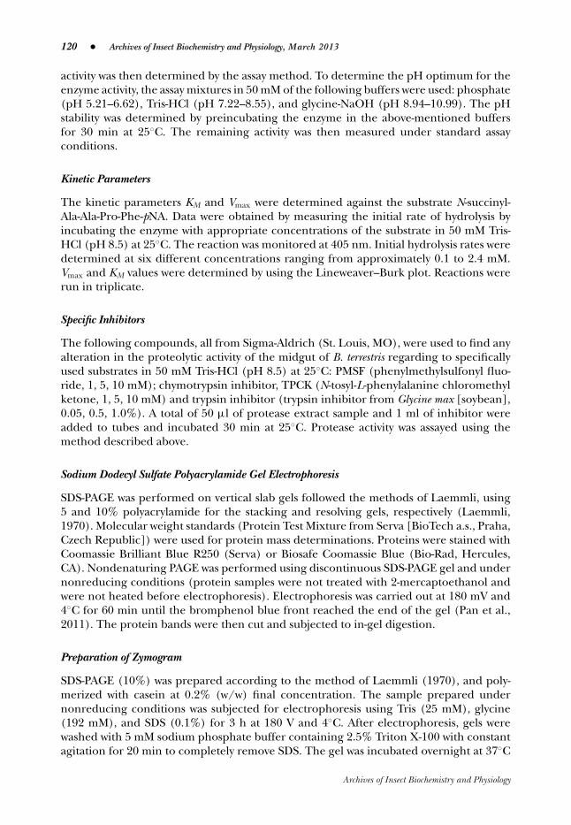
activity was then determined by the assay method. To determine the pH optimum for theenzyme activity, the assay mixtures in 50 mM of the following buffers were used: phosphate(pH 5.21–6.62), Tris-HCl (pH 7.22–8.55), and glycine-NaOH (pH 8.94–10.99). The pHstability was determined by preincubating the enzyme in the above-mentioned buffersfor 30 min at 25◦C. The remaining activity was then measured under standard assayconditions.
Kinetic Parameters
The kinetic parameters KM and Vmax were determined against the substrate N-succinyl-Ala-Ala-Pro-Phe-pNA. Data were obtained by measuring the initial rate of hydrolysis byincubating the enzyme with appropriate concentrations of the substrate in 50 mM Tris-HCl (pH 8.5) at 25◦C. The reaction was monitored at 405 nm. Initial hydrolysis rates weredetermined at six different concentrations ranging from approximately 0.1 to 2.4 mM.Vmax and KM values were determined by using the Lineweaver–Burk plot. Reactions wererun in triplicate.
Specific Inhibitors
The following compounds, all from Sigma-Aldrich (St. Louis, MO), were used to find anyalteration in the proteolytic activity of the midgut of B. terrestris regarding to specificallyused substrates in 50 mM Tris-HCl (pH 8.5) at 25◦C: PMSF (phenylmethylsulfonyl fluo-ride, 1, 5, 10 mM); chymotrypsin inhibitor, TPCK (N-tosyl-L-phenylalanine chloromethylketone, 1, 5, 10 mM) and trypsin inhibitor (trypsin inhibitor from Glycine max [soybean],0.05, 0.5, 1.0%). A total of 50 μl of protease extract sample and 1 ml of inhibitor wereadded to tubes and incubated 30 min at 25◦C. Protease activity was assayed using themethod described above.
Sodium Dodecyl Sulfate Polyacrylamide Gel Electrophoresis
SDS-PAGE was performed on vertical slab gels followed the methods of Laemmli, using5 and 10% polyacrylamide for the stacking and resolving gels, respectively (Laemmli,1970). Molecular weight standards (Protein Test Mixture from Serva [BioTech a.s., Praha,Czech Republic]) were used for protein mass determinations. Proteins were stained withCoomassie Brilliant Blue R250 (Serva) or Biosafe Coomassie Blue (Bio-Rad, Hercules,CA). Nondenaturing PAGE was performed using discontinuous SDS-PAGE gel and undernonreducing conditions (protein samples were not treated with 2-mercaptoethanol andwere not heated before electrophoresis). Electrophoresis was carried out at 180 mV and4◦C for 60 min until the bromphenol blue front reached the end of the gel (Pan et al.,2011). The protein bands were then cut and subjected to in-gel digestion.
Preparation of Zymogram
SDS-PAGE (10%) was prepared according to the method of Laemmli (1970), and poly-merized with casein at 0.2% (w/w) final concentration. The sample prepared undernonreducing conditions was subjected for electrophoresis using Tris (25 mM), glycine(192 mM), and SDS (0.1%) for 3 h at 180 V and 4◦C. After electrophoresis, gels werewashed with 5 mM sodium phosphate buffer containing 2.5% Triton X-100 with constantagitation for 20 min to completely remove SDS. The gel was incubated overnight at 37◦C
Archives of Insect Biochemistry and Physiology
Serine Protease of Bombus terrestris � 121
in Tris-HCl buffer (50 mM) pH 7.6 containing 10 mM CaCl2 and 150 mM NaCl. Gels werethen stained with Coomassie Brilliant Blue R-250 (0.1%) for 30 min and finally destainedwith 25% ethanol in 8% acetic acid and water (30:10:60, v/v) three times for 10 min toobserve the translucent activity bands.
Two-Dimensional Gel Electrophoresis (2-DE)
Lyophilized protein sample (4.65 mg) was solubilized in 130 μl of rehydration so-lution (7 M urea, 2 M thiourea, 2% 3-((3-cholamidopropyl)dimethylammonium)-1-propanesulfonate (CHAPS, w/v), 0.2% Bio-Lyte R© ampholytes (3–10 buffer, w/v), 1%dithithreitol (w/v). Proteins were then transferred to Ready Strip
TMIPG Strips (3–10 NL,
7 cm, Bio-Rad) overnight by active in-gel rehydration (50 V, 20◦C).Isoelectric focusing was carried out at 20◦C with Protean R© IEF cell system (Bio-Rad)
under mineral oil. Proteins were focused in four steps (250 V for 20 min, linear gradient;500 V for 1 h, linear gradient; 1,000 V for 1 h, linear gradient; 4,000 V for 20,000 Vh,rapid gradient). Prior to separation in the second dimension, the strips were equilibratedaccording to Gorg et al. (2004). After the equilibration step, strips were rinsed in Tris-glycine buffer (pH 8.3), transferred to a homogeneous 12.5% SDS-polyacrylamide gel(with the same composition as in the previous section), and gel strips were overlaidwith 0.5% (w/v) CertifiedTM Low Melt Agarose (Bio-Rad) in SDS-PAGE running buffercontaining trace of bromophenol blue. Finally, 5 μl of Precision Plus Protein
TMStandards
(molecular weight range 10–250 kDa, Bio-Rad) was added at the top end of the gel. Gelswere run in the Mini-Protean Tetra Cell system. The spots were then stained by CoomassieBlue, excised, and subjected to in-gel digestion. Protein bands or spots were excisedfrom the Coomassie-stained gels, and then processed as described in Shevchenko et al.(2006). Before the in-gel digestion, the gel pieces were cooled in an ice-cold bath andswollen in a 50 μl of digestion buffer (50 mM ammonium bicarbonate) containing trypsin(30 μg/ml, type IX-S, lot 51K72501, Sigma-Aldrich, St. Louis, MO) in 50 mM ammoniumbicarbonate. After 1 h of cooling at 4◦C, the gel pieces were incubated overnight at 37◦C.
The resulting tryptic peptides were extracted from the gel pieces by adding 150 μl ofextraction buffer (5% formic acid, 30% acetonitrile in water) and sonicated for 15 min.The buffer was then replaced and the gel pieces were sonicated again for additional 15min. After each extraction step, the solutions were spun and the supernatants withdrawn,pooled, and concentrated to dryness in a vacuum centrifuge. Dried extracts were storedat −80◦C before analysis.
Analysis of Tryptic Digests by LC-MS/MS
Dried protein digests were dissolved in 20 μl of 0.1% formic acid, centrifuged, and thesupernatant transferred to inserts in vials. The nano-HPLC apparatus used for proteindigest analysis was a Proxeon Easy-nLC (Proxeon, Odense, Denmark) coupled to a maXisQ-TOF (quadrupole-time of flight) mass spectrometer with ultra-high resolution (BrukerDaltonics, Bremen, Germany) by nanoelectrosprayer. The nLC-MS/MS instruments werecontrolled with the software packages HyStar 3.2 and micrOTOF-control 3.0. The datawere collected and manipulated with the software packages ProteinScape 2.0 and Data-Analysis 4.0 (Bruker Daltonics).
A total of 5 μl of the peptide mixture were injected into an NS-AC-11-C18 BiosphereC18 column (particle size: 5μm, pore size: 12 nm, length: 150 mm, inner diameter:
Archives of Insect Biochemistry and Physiology
122 � Archives of Insect Biochemistry and Physiology, March 2013
75 μm), with an NS-MP-10 Biosphere C18 precolumn (particle size: 5 μm, pore size: 12nm, length: 20 mm, inner diameter: 100 μm), both manufactured by NanoSeparations(Nieuwkoop, the Netherlands). The separation of peptides was achieved via a lineargradient between mobile phase A (water) and B (acetonitrile), both containing 0.1%(v/v) formic acid. Separation was started by running the system with 5% mobile phase B,followed by gradient elution to 30% B at 70 min. The next step was a gradient elution to50% B in 10 min, and then a gradient to 100% B in 8 min was used. Finally, the columnwas eluted with 100% B for 2 min. Equilibration before the next run was achieved bywashing the column with 5% mobile phase B for 10 min. The flow rate was 0.25 μl/min,and the column was held at ambient temperature (25◦C).
Online nanoelectrospray ionization (easy nano-ESI) was operated in positive mode.The ESI voltage was set at +4.5 kV, scan time 1.3 Hz. Operating conditions: drying gas(N2), 1 l/min; drying gas temperature, 160◦C; nebulizer pressure, 0.4 bar. Experimentswere performed by scanning from 100 to 2,200 m/z. The reference ion used (internalmass lock) was a monocharged ion of C24H19F36N3O6P3 (m/z 1,221.9906). Mass spectracorresponding to each signal from the total ion current chromatogram were averaged,enabling an accurate molecular mass determination. All LC-MS/MS analyses were donein duplicates.
Bioinformatic Analysis
Data were processed using ProteinScape software. Peptides were identified by correlatingtandem mass spectra to the SwissProt database, using the MASCOT searching engine(www.matrixscience.com). Trypsin was chosen as the enzyme parameter. One missedcleavage was allowed, and an initial peptide mass tolerance of ±10.0 ppm was used for MSand ±0.05 Da for MS/MS analysis. Cysteines were assumed to be carbamidomethylated,proline and lysine to be hydroxylated, and methionine was allowed to be oxidated. Allthese possible modifications were set to be variable. Monoisotopic peptide charge was set1+, 2+, and 3+. The Peptide Decoy option was selected during the data search processto remove false-positive results. Only significant hits with score ≥80 were accepted. In thenext stage of the database searching, amino acid sequence corresponding to the genefor B. terrestris serine protease was processed using the MASCOT searching engine. Otherparameters remained the same.
RESULTS
The serine protease was extractable from midgut homogenate in buffer. Digestive proteasefrom the midgut was purified by a multiple-step procedure (Table 1). After the final step,
Table 1. Purification of Serine Protease from Midgut of Male Bombus terrestris
Purification step Total activity (U) Protein (mg) Specific activity (U/mg) Purification factor
Homogenate 0.18 3,885.0 9.4 1Dialyzate 0.07 2,568.0 5.7 0.6(NH4)2SO4 precipitation 0.25 2,539.0 19.3 2.1Dialyzate 0.28 2,025.0 27.8 3.0Phenyl-Sepharose 0.11 26.8 787.6 83.9
Archives of Insect Biochemistry and Physiology
Serine Protease of Bombus terrestris � 123
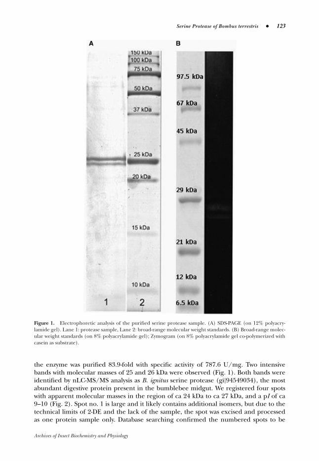
Figure 1. Electrophoretic analysis of the purified serine protease sample. (A) SDS-PAGE (on 12% polyacry-lamide gel). Lane 1: protease sample, Lane 2: broad-range molecular weight standards. (B) Broad-range molec-ular weight standards (on 8% polyacrylamide gel); Zymogram (on 8% polyacrylamide gel co-polymerized withcasein as substrate).
the enzyme was purified 83.9-fold with specific activity of 787.6 U/mg. Two intensivebands with molecular masses of 25 and 26 kDa were observed (Fig. 1). Both bands wereidentified by nLC-MS/MS analysis as B. ignitus serine protease (gi|94549034), the mostabundant digestive protein present in the bumblebee midgut. We registered four spotswith apparent molecular masses in the region of ca 24 kDa to ca 27 kDa, and a pI of ca9–10 (Fig. 2). Spot no. 1 is large and it likely contains additional isomers, but due to thetechnical limits of 2-DE and the lack of the sample, the spot was excised and processedas one protein sample only. Database searching confirmed the numbered spots to be
Archives of Insect Biochemistry and Physiology

124 � Archives of Insect Biochemistry and Physiology, March 2013
Figure 2. Two-dimensional electrophoresis of the purified serine protease sample. The numbered spots (1–4)correspond to the serine protease isoforms as identified by nLC-MS/MS analysis.
Table 2a. Parameters of the Matched Peptides
�m/z MASCOT score Calculated pIPeptide m/z meas. [ppm] z (total ion score) [ppm] for peptide Range
1 492.2738 − 1.85 2 63.3 2.54 9.76 35–422 554.5870 0.88 3 77.5 1.94 6.74 43–573 895.4732 0.78 2 97.8 1.77 6.91 58–734 773.3847 1.03 3 95.1 2.99 6.47 74–955 802.9545 0.64 2 122.3 2.42 5.84 101–1156 685.9125 1.67 2 74.6 2.39 10.00 119–1307 1,158.5333 1.43 2 116.0 1.15 5.99 131–1528 966.0167 1.46 2 123.9 1.37 6.05 157–1739 662.3224 1.02 2 84.1 2.04 8.18 181–19110 516.5931 0.73 3 89.5 3.34 8.20 218–23111 691.8514 0.39 2 102.8 2.75 5.81 232–243
identical with B. ignitus serine protease. Eleven unique peptides with MASCOT scorehigher than 60 were identified (Tables 2a, b). The peptide mass measurement accuracywas lower than ±1.5 ppm.
No specific peptides for B. terrestris serine protease were detected, because all peptidesmatched well to the sequence of B. ignitus serine protease covering more than 65% oftheir sequence (Fig. 3).
Enzyme activity increased with reaction temperature to an optimum at 35◦C thendecreased slowly at higher temperatures. Serine protease was stable up to 55◦C and lostactivity at 60◦C. The B. terrestris serine proteases displayed a broad pH range of activity(from 6 to 9). The proteases were stable in a pH range of 6–10. We studied the effectof proteinase inhibitors phenylmethanesulfonyl fluoride (PMSF), trypsin inhibitor from
Archives of Insect Biochemistry and Physiology
Serine Protease of Bombus terrestris � 125
Table 2b. Parameters of the Matched Peptides
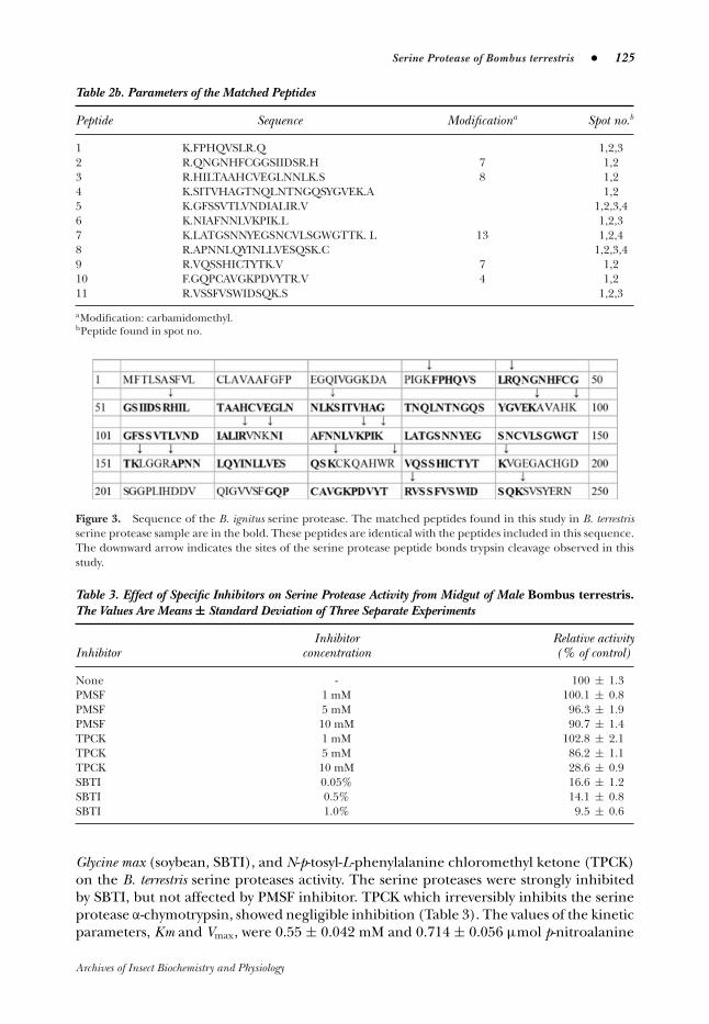
Peptide Sequence Modificationa Spot no.b
1 K.FPHQVSLR.Q 1,2,32 R.QNGNHFCGGSIIDSR.H 7 1,23 R.HILTAAHCVEGLNNLK.S 8 1,24 K.SITVHAGTNQLNTNGQSYGVEK.A 1,25 K.GFSSVTLVNDIALIR.V 1,2,3,46 K.NIAFNNLVKPIK.L 1,2,37 K.LATGSNNYEGSNCVLSGWGTTK. L 13 1,2,48 R.APNNLQYINLLVESQSK.C 1,2,3,49 R.VQSSHICTYTK.V 7 1,210 F.GQPCAVGKPDVYTR.V 4 1,211 R.VSSFVSWIDSQK.S 1,2,3
aModification: carbamidomethyl.bPeptide found in spot no.
Figure 3. Sequence of the B. ignitus serine protease. The matched peptides found in this study in B. terrestrisserine protease sample are in the bold. These peptides are identical with the peptides included in this sequence.The downward arrow indicates the sites of the serine protease peptide bonds trypsin cleavage observed in thisstudy.
Table 3. Effect of Specific Inhibitors on Serine Protease Activity from Midgut of Male Bombus terrestris.The Values Are Means ± Standard Deviation of Three Separate Experiments
Inhibitor Relative activityInhibitor concentration (% of control)
None - 100 ± 1.3PMSF 1 mM 100.1 ± 0.8PMSF 5 mM 96.3 ± 1.9PMSF 10 mM 90.7 ± 1.4TPCK 1 mM 102.8 ± 2.1TPCK 5 mM 86.2 ± 1.1TPCK 10 mM 28.6 ± 0.9SBTI 0.05% 16.6 ± 1.2SBTI 0.5% 14.1 ± 0.8SBTI 1.0% 9.5 ± 0.6
Glycine max (soybean, SBTI), and N-p-tosyl-L-phenylalanine chloromethyl ketone (TPCK)on the B. terrestris serine proteases activity. The serine proteases were strongly inhibitedby SBTI, but not affected by PMSF inhibitor. TPCK which irreversibly inhibits the serineprotease α-chymotrypsin, showed negligible inhibition (Table 3). The values of the kineticparameters, Km and Vmax, were 0.55 ± 0.042 mM and 0.714 ± 0.056 μmol p-nitroalanine
Archives of Insect Biochemistry and Physiology

126 � Archives of Insect Biochemistry and Physiology, March 2013
produced mg min−1 protein−1, respectively. Concentration–velocity curve followed theMichaelis–Menten kinetic model.
DISCUSSION
The major digestive enzymes include serine proteases such as trypsins and chymotrypsins,cysteine proteases, elastase-like enzymes, and various carboxypeptidases or aminopep-tidases (Hegedus et al., 2003; Prabhakar et al., 2007; Chougule et al., 2008). Serineproteases are typical for lepidopteran larvae (Chougule et al., 2008). The first study ofthe mechanism of enzyme secretion in Lepidoptera was done using larval Bombyx moriand indicated membrane-bound trypsin-like proteases. Some of the trypsin-like proteasesare incorporated in the peritrophic membrane (Kuriyama and Eguchi, 1985; Terra andFereira, 1994). On the contrary, we found the serine protease in midgut of 2-day-old B.terrestris males was extractable from midgut homogenate in buffer. Similar findings werereported by Choo et al. (2007), who described the cDNA cloning and characterization ofmidgut-specific serine protease of B. ignitus. The gene was specifically expressed in themidgut of B. ignitus queens, males, and workers, suggesting that the BiSP is a gut enzymeinvolved in the digestion of dietary proteins (Choo et al., 2007).
The termostability of the B. terrestris serine protease was comparable to results obtainedwith crab digestive lipase and pancreatic lipases for which the presence of the substrateincreases the thermostability of the lipolytic enzymes (Cherif et al., 2007; Verger, 1984).Insect serine protease pH optima are always alkaline (most between 8 and 9), irrespec-tive of the pH prevailing in midguts from which the enzymes were isolated (Reeck etal., 1999). Nevertheless, trypsins isolated from lepidopteran insects have higher pH op-tima, corresponding to the higher pH values found in their midguts (Sasaki and Suzuki,1982). The proteolytic enzymes of B. terrestris were fully active at pH 9 as reported forchymotrypsin-like proteases from scallop (Pecten maximus; Le Chevalier et al., 110) andyellow mealworm, Tenebrio monitor (Elpidina et al., 2005). However, trypsin from the gutfluid of B. mori (Sasaki et al., 1993) had maximum activity about pH 11.
Our results show that bumblebee serine protease is actually a mixture of severalisotypes with the same molecular mass, but different pIs. Only one isotype has a bithigher molecular mass. A couple of hypotheses may explain the presence of multiplespots identified as serine protease in the 2-DE gel (Fig. 2). The observed spots couldbe due to alternative splicing or to a combination of posttranslational modifications.Also, there may be small differences in amino acid composition between the serineproteases.
ACKNOWLEDGMENTS
We thank the research programme of the Institute of Organic Chemistry and BiochemistryRVO: 61388963 the Czech Science Foundation (grant no. 203/09/1446) and TechnologyAgency of the Czech Republic (grant no. TA01020969). The authors also wish to thankProf. Vladimır Ptacek for supplying the insects.
LITERATURE CITED
Bradford M. 1976. A rapid and sensitive method for the quantitation of microgram quantities ofprotein utilizing the principle of protein-dye binding. Anal Biochem 72:248–254.
Archives of Insect Biochemistry and Physiology
Serine Protease of Bombus terrestris � 127
Brioschi D, Nadalini LD, Bengtson MH, Sogayar MC, Moura DS, Silva-Filho MC. 2007. General upregulation of Spodoptera frugiperda trypsins and chymotrypsins allows its adaptation to soybeanproteinase inhibitor. Insect Biochem Mol Biol 37:1283–1290.
Broehan G, Kemper M, Driemeier D, Vogelpohl I, Merzendorfer H. 2008. Cloning and expressionanalysis of midgut chymotrypsin-like proteinases in the tobacco hornworm. J Insect Physiol54:1243–1252.
Burgess EPJ, Malone LA, Christeller JT. 1996. Effects of two proteinase inhibitors on the digestiveenzymes and survival of honey bees (Apis mellifera). J Insect Physiol 42:823–828.
Cherif S, Fendri A, Miled N, Trabelsi H, Mejdoub H, Gargouri Y. 2007. Crab digestive lipase actingat high temperature: purification and biochemical characterization. Biochimie 89:1012–1018.
Chougule NP, Doyle E, Fitches E, Gatehouse JA. 2008. Biochemical characterization of midgutdigestive proteases from Mamestra brassicae (cabbage moth; Lepidoptera: Noctuidae) and effectof soybean Kunitz inhibitor (SKTI) in feeding assays. J Insect Physiol 54:563–572.
Choo YM, Lee KS, Yoon HJ, Lee SB, Kim JH, Sohn HD, JIN BR. 2007. A serine protease fromthe midgut of the bumblebee, Bombus ignitus (Hymenoptera: Apidae): cDNA cloning, genestructure, expression and enzyme activity. Eur J Entomol 104:1–7.
Clynen E, Schoofs L, Salzet M. 2005. A review of the most important classes of serine proteaseinhibitors in insects and leeches. Med Chem Rev 2:197–206.
Elpidina EN, Tsybina TA, Dunaevsky YE, Belozersky MA, Zhuzhikov DP, Oppert B. 2005. Achymotrypsin-like proteinase from the midgut of Tenebrio molitor larvae. Biochimie 87:771–779.
Gorg A, Weiss W, Dunn MJ. 2004. Current two-dimensional electrophoresis technology for pro-teomics. Proteomics 12:3667–3685.
Hedstrom L. 2002. Serine protease mechanism and specificity. Chem Rev 102:4501–4523.Hegedus D, Baldwin D, O’Grady M, Braun L, Gleddie S, Sharpe A, Lydiate D, Erlandson M. 2003.
Midgut proteases from Mamestra configurata (Lepidoptera: Noctuidae) larvae: characterization,cDNA cloning, and expressed sequence tag analysis. Arch Insect Biochem Physiol 53:30–47.
Jongsma MA, Bakker PL, Peters J, Bosch D, Stiekema WJ. 1995. Adaptation of Spodoptera exigua larvaeto plant proteinase-inhibitors by induction of gut proteinase activity insensitive to inhibition.Proc Natl Acad Sci USA 92:8041–8045.
Kalhok SE, Tabak LM, Prosser DE, Brook W, Downe AE, White BN. 1993. Isolation, sequencing andcharacterization of two cDNA clones coding for trypsin-like enzymes from the midgut of Aedesaegypti. Insect Mol Biol 2:71–9.
Kini RM. 2005. Serine proteases affecting blood coagulation and fibrinolysis from snake venoms.Pathophysiol Haemo T 34:200–204.
Kuriyama K, Eguchi M. 1985. Conversion of the molecular form alkaline treatment of gut proteasefrom the silkworm Bombyx mori. Comp Biochem Physiol B 82:575–579.
Laemmli UK. 1970. Cleavage of structural proteins during the assembly of the head of bacteriophageT4. Nature 227:680–685.
Lam W, Coast GM, Rayne RC. 2000. Characterisation of multiple trypsins from the midgut of Locustamigratoria. Insect Biochem Mol Biol 30:85–94.
Le Chevalier P, Sellos D, Van Wormhoudt A. 1995. Purification and partial characterization ofchymotrypsin-like proteases from the digestive gland of the scallop Pecten maximus. CompBiochem Physiol B 110:4777–4784.
Lopes AR, Sato PM, Terra WR. 2009. Insect chymotrypsins: chloromethyl ketone inactivation andsubstrate specificity relative to possible coevolutional adaptation of insects and plants. ArchInsect Biochem Physiol 70:188–203.
Mazumdar-Leighton S, Broadway RM. 2001. Identification of six chymotrypsin cDNAs from larvalmidguts of Helicoverpa zea and Agrotis ipsilon feeding on the soybean (Kunitz) trypsin inhibitor.Insect Biochem Mol Biol 31:633–644.
Archives of Insect Biochemistry and Physiology
128 � Archives of Insect Biochemistry and Physiology, March 2013
Muller A, Voswinkel J, Gottschlich S, Csernok E. 2007. Human proteinase 3 (PR3) and its bindingmolecules: implications for inflammatory and PR3-related autoimmune responses. Ann N YAcad Sci 1109:84–92.
Noriega FG, Wang XY, Pennington JE, Barillas-Mury CV, Wells MA. 1996. Early trypsin, a female-specific midgut protease in Aedes aegypti: isolation, aminoterminal sequence determination,and cloning and sequencing of the gene. Insect Biochem Mol Biol 26:119–26.
Pan D, Hill AP, Kashou A, Wilson KA, Tan-Wilson A. 2011. Electrophoretic transfer protein zymog-raphy. Anal Biochem 411:277–283.
Pauchet Y, Muck A, Svatos A, Heckel DG, Prelss S. 2008. Mapping the larvalmidgut lumen proteomeof Helicoverpa armigera, a generalist herbivorous insect. J Proteome Res 7:1629–1639.
Perona JJ, Craik CS. 1995. Structural basis of substrate specificity in the serine proteases. ProteinSci 4:337–360.
Prabhakar S, Chen MS, Elpidina EN, Vinokurov KS, Smith CM, Marshall J, Oppert B. 2007. Sequenceanalysis and molecular characterization of larval midgut cDNA transcripts encoding peptidasesfrom the yellow mealworm, Tenebrio molitor L. Insect Mol Biol 16:455–468.
Ptacek V, Pernova E, Borovec R. 2000. The two-queen cascade method as an alternative techniquefor starting bumblebee (Bombus, Hymenoptera, Apidae) colonies in laboratory (preliminarystudy). Pcelnicze Zeszyty Naukowe 44:305–309.
Reeck G, Oppert B, Denton M, Kanost M, Baker J, Kramer K. 1999. Insect proteinases. In: Turk I,editor. Proteases: new perspectives basel. Switzerland: Birhauser Verlag. p 125–148.
Saeki K, Ozaki K, Kobayashi T, Ito S. 2007. Detergent alkaline proteases: enzymatic properties,genes, and crystal structures. J Biosci Bioeng 103:501–508.
Sasaki T, Hishida T, Ichikava K, Asari S. 1993. Amino acid sequence of alkaliphilic serine proteasefrom silkworm, Bombyx mori, larval digestive juice. FEBS Lett 320:35–37.
Sasaki T, Suzuki Y. 1982. Alkaline proteases in digestive juice of the silkworm, Bombyx mori. BiochimBiophys Acta 703:1–10.
Shevchenko A, Tomas H, Havlis J, Olsen JV, Mann M. 2006. In-gel digestion for mass spectrometriccharacterization of proteins and proteomes. Nat Protocol 1:2856–2860.
Terra WR, Ferreira C. 1994. Insect digestive enzymes: properties, compartmentalization and func-tion. Comp Biochem Physiol B 109:1–62.
Verger R. 1984. Pancreatic lipases. In: Borgstrom B, Brockman HL, editors. Lipase Amsterdam. TheNetherlands: Elsevier. p 121–150.
Yoon H, Laxmikanthan G, Lee J, Blaber SI, Rodriguez A, Kogot JM, Scarisbrick IA, Blaber M. 2007.Activation profiles and regulatory cascades of the human kallikrein-related peptidases. J BiolChem 282:31852–31864.
Archives of Insect Biochemistry and Physiology