Case Report Pancreatic Neuroendocrine Tumor in the Setting of...
5
Case Report Pancreatic Neuroendocrine Tumor in the Setting of Dorsal Agenesis of the Pancreas Samih Nassif, 1 Cecilia Ponchiardi, 2 and Teviah Sachs 3 1 Boston University School of Medicine, 72 East Concord St., Boston, MA 02118, USA 2 Department of Pathology and Laboratory Medicine, Boston University School of Medicine, 72 East Concord St., Boston, MA 02118, USA 3 Boston University School of Medicine, Moakley Building, 3rd Floor, 830 Harrison Avenue, Boston, MA 02118, USA Correspondence should be addressed to Teviah Sachs; [email protected] Received 4 July 2016; Revised 29 August 2016; Accepted 30 August 2016 Academic Editor: Haruhiko Sugimura Copyright © 2016 Samih Nassif et al. is is an open access article distributed under the Creative Commons Attribution License, which permits unrestricted use, distribution, and reproduction in any medium, provided the original work is properly cited. Dorsal agenesis of the pancreas (DAP) is an uncommon embryological abnormality where there is absence of the distal pancreas. DAP is mostly asymptomatic, but common presenting symptoms include diabetes mellitus, abdominal pain, pancreatitis, enlarged pancreatic head, and, in a few cases, polysplenia. MRCP and ERCP are the gold standard imaging techniques to demonstrate the absence of the dorsal pancreatic duct. e literature on the association of pancreatic neoplasia and DAP is limited. We present the case of a pancreatic neuroendocrine tumor in a patient with dorsal agenesis of the pancreas, with a review of the related literature. 1. Introduction Dorsal agenesis of the pancreas (DAP) is an uncommon embryological abnormality where there is absence of the distal pancreas. Here, we present the case of a 48-year-old female who was referred to our surgical oncology clinic for a pancreatic mass and was found to have concurrent DAP. We then discuss the embryology of DAP, as well as the most common clinical presentations of DAP and other established reports of DAP in the literature. 2. Case Report A 48-year-old female was referred to the surgical oncology clinic for evaluation of a pancreatic mass. is was found incidentally on workup for an endometrial stromal sarcoma, for which she had undergone a total abdominal hysterec- tomy with bilateral salpingo-oophorectomy. e patient was asymptomatic. Her past medical history was significant for uterine sarcoma and for venous thromboembolism which led to a pulmonary embolus but was otherwise unremarkable. Her physical exam was unrevealing, as was her serum labora- tory evaluation, with normoglycemia, normal hepatobiliary function, normal pancreatic enzymes, and no elevation in carbohydrate antigen 19-9, carbohydrate antigen 125, or carcinoembryonic antigen. CT and MRI imaging (Figure 1) revealed a mass at the neck of the pancreas, measuring 2.9 cm in its largest dimension, as well as the absence of the distal body and tail of the pancreas. e mass closely abutted the confluence of the portal vein and superior mesenteric vein, but there was no invasion. She underwent biopsy of this mass via endoscopic ultrasound which revealed features consistent with a well differentiated neuroendocrine tumor. e tumor was determined to be nonfunctioning given the absence of systemic symptoms and laboratory data to suggest hormone production. e patient underwent resection of this mass via spleen preserving laparoscopic approach. Intraoperative images confirmed the absence of the distal body and tail of the pancreas (Figure 2). Negative margins were achieved with this resection, and the pancreatic head and uncinate process were preserved, as were the splenic vein and artery. e pancreatic parenchyma was transected using a linear cutting stapler, with a closed staple height of 1.5 mm, and the remnant pancreatic neck was buttressed with an omental patch. A 19 Fr fluted Blake drain was placed at the resection margin at Hindawi Publishing Corporation Case Reports in Gastrointestinal Medicine Volume 2016, Article ID 3801962, 4 pages http://dx.doi.org/10.1155/2016/3801962
Transcript of Case Report Pancreatic Neuroendocrine Tumor in the Setting of...

Case ReportPancreatic Neuroendocrine Tumor in the Setting ofDorsal Agenesis of the Pancreas
Samih Nassif,1 Cecilia Ponchiardi,2 and Teviah Sachs3
1Boston University School of Medicine, 72 East Concord St., Boston, MA 02118, USA2Department of Pathology and Laboratory Medicine, Boston University School of Medicine, 72 East Concord St.,Boston, MA 02118, USA3Boston University School of Medicine, Moakley Building, 3rd Floor, 830 Harrison Avenue, Boston, MA 02118, USA
Correspondence should be addressed to Teviah Sachs; [email protected]
Received 4 July 2016; Revised 29 August 2016; Accepted 30 August 2016
Academic Editor: Haruhiko Sugimura
Copyright © 2016 Samih Nassif et al. This is an open access article distributed under the Creative Commons Attribution License,which permits unrestricted use, distribution, and reproduction in any medium, provided the original work is properly cited.
Dorsal agenesis of the pancreas (DAP) is an uncommon embryological abnormality where there is absence of the distal pancreas.DAP is mostly asymptomatic, but common presenting symptoms include diabetes mellitus, abdominal pain, pancreatitis, enlargedpancreatic head, and, in a few cases, polysplenia. MRCP and ERCP are the gold standard imaging techniques to demonstrate theabsence of the dorsal pancreatic duct. The literature on the association of pancreatic neoplasia and DAP is limited. We present thecase of a pancreatic neuroendocrine tumor in a patient with dorsal agenesis of the pancreas, with a review of the related literature.
1. Introduction
Dorsal agenesis of the pancreas (DAP) is an uncommonembryological abnormality where there is absence of thedistal pancreas. Here, we present the case of a 48-year-oldfemale who was referred to our surgical oncology clinic fora pancreatic mass and was found to have concurrent DAP.We then discuss the embryology of DAP, as well as the mostcommon clinical presentations of DAP and other establishedreports of DAP in the literature.
2. Case Report
A 48-year-old female was referred to the surgical oncologyclinic for evaluation of a pancreatic mass. This was foundincidentally on workup for an endometrial stromal sarcoma,for which she had undergone a total abdominal hysterec-tomy with bilateral salpingo-oophorectomy. The patient wasasymptomatic.
Her past medical history was significant for uterinesarcoma and for venous thromboembolism which led to apulmonary embolus but was otherwise unremarkable. Herphysical exam was unrevealing, as was her serum labora-tory evaluation, with normoglycemia, normal hepatobiliary
function, normal pancreatic enzymes, and no elevation incarbohydrate antigen 19-9, carbohydrate antigen 125, orcarcinoembryonic antigen. CT and MRI imaging (Figure 1)revealed amass at the neck of the pancreas, measuring 2.9 cmin its largest dimension, as well as the absence of the distalbody and tail of the pancreas. The mass closely abutted theconfluence of the portal vein and superior mesenteric vein,but there was no invasion. She underwent biopsy of this massvia endoscopic ultrasound which revealed features consistentwith a well differentiated neuroendocrine tumor. The tumorwas determined to be nonfunctioning given the absence ofsystemic symptoms and laboratory data to suggest hormoneproduction.
The patient underwent resection of this mass via spleenpreserving laparoscopic approach. Intraoperative imagesconfirmed the absence of the distal body and tail of thepancreas (Figure 2). Negative margins were achieved withthis resection, and the pancreatic head and uncinate processwere preserved, as were the splenic vein and artery. Thepancreatic parenchyma was transected using a linear cuttingstapler, with a closed staple height of 1.5mm, and the remnantpancreatic neck was buttressed with an omental patch. A19 Fr fluted Blake drain was placed at the resection margin at
Hindawi Publishing CorporationCase Reports in Gastrointestinal MedicineVolume 2016, Article ID 3801962, 4 pageshttp://dx.doi.org/10.1155/2016/3801962

2 Case Reports in Gastrointestinal Medicine
Distal pancreas absent
Figure 1: Axial image of an MRCP demonstrating the relevantanatomy of the tumor, vessels, and proximal pancreas, with theabsence of the dorsal pancreas (white arrow). Outlines represent thepancreatic head and neck (yellow), the pancreatic neuroendocrinetumor (red), the superior mesenteric vein (purple), and the splenicvein (blue).
Figure 2: Laparoscopic image of the retroperitoneum as seenthrough a window created in the gastrocolic omentum.The stomachis elevated (white arrow), and the pancreatic neuroendocrine tumor(red arrow) can be seen with the absence of any pancreatic tissuedistal to the tumor (black arrow).
the time of surgery. Final pathology revealed a grade 1 welldifferentiated pancreatic neuroendocrine tumor (Figure 3).Despite our intraoperative efforts to avoid it, the patient’spostoperative course was significant for a pancreatic ductleak, which was well controlled by her drain, and she wasdischarged home on POD 4. Her drain was removed on POD23. She had no evidence of diabetes or pancreatic insuffi-ciency on follow-up evaluation. Her case was discussed at ourmultidisciplinary tumor board and no further treatment forthis tumor was recommended.
3. Discussion
3.1. Embryology of the Pancreas. During the fourth week ofgestation, the dorsal (cranial) and ventral (caudal) buds ofthe pancreas develop from the endoderm at the junction ofthe foregut and the midgut. While the dorsal bud developsonly into pancreatic tissue (anterior head, body, and tail),the ventral bud also contributed to the liver, gallbladder,bile ducts, and ventral pancreas (posterior neck and head)[1]. The ventral pancreatic duct (duct of Wirsung) and thecommon bile duct thus share a common entry point to theduodenum at the major papilla. Eventually, the ventral budrotates clockwise and fuses with the dorsal bud at the seventhweek of gestation. At this time, the dorsal pancreatic duct
Figure 3:Histopathological description of the tumor. It is composedof multiple nests with hyalinized fibrovascular stroma. Tumor cellsare relatively uniform with finely granular eosinophilic cytoplasmand centrally located round to oval nucleus with “salt and pepper”chromatin pattern.There were less than 2mitoses per high-poweredfield. The tumor was chromogranin positive after immunohisto-chemical analysis (not shown).
(duct of Santorini) fuses with the ventral pancreatic ductto create the main pancreatic duct [2]. Islets of Langerhansprimarily develop in the dorsal pancreas, at week twelve ofgestation.
3.2. Dorsal Agenesis. Dorsal agenesis occurs when there isabnormal development of the dorsal pancreatic bud, but thereis intact development of the ventral pancreatic bud. Thus,there is absence of the anterior head, body, and the tail ofthe pancreas with intact formation of the liver, gallbladder,bile ducts, and posterior neck and head of the pancreas. Thedorsal pancreatic duct never forms, and pancreatic secretionscourse from the ventral pancreatic duct into the common bileduct and eventually pass through the major papilla into thesecond portion of the duodenum.
Thefirst case of dorsal agenesis of the pancreas (DAP)wasreported in 1911 as an autopsy finding, and since then therehave been relatively few reported cases in the literature [1].DAP can be complete or partial. In patients with completeDAP, the dorsal duct system and the body and the tailof the pancreas are all missing. However, in partial DAP,the accessory papilla, the terminal end of the main dorsalduct of Santorini, or the pancreatic body is present. FamilialDAP has been described in association with other congenitaldeformities, as well as alone. The molecular basis for DAP isnot well defined; however, certain homeobox genes have beenassociated with DAP in rodent models [10].
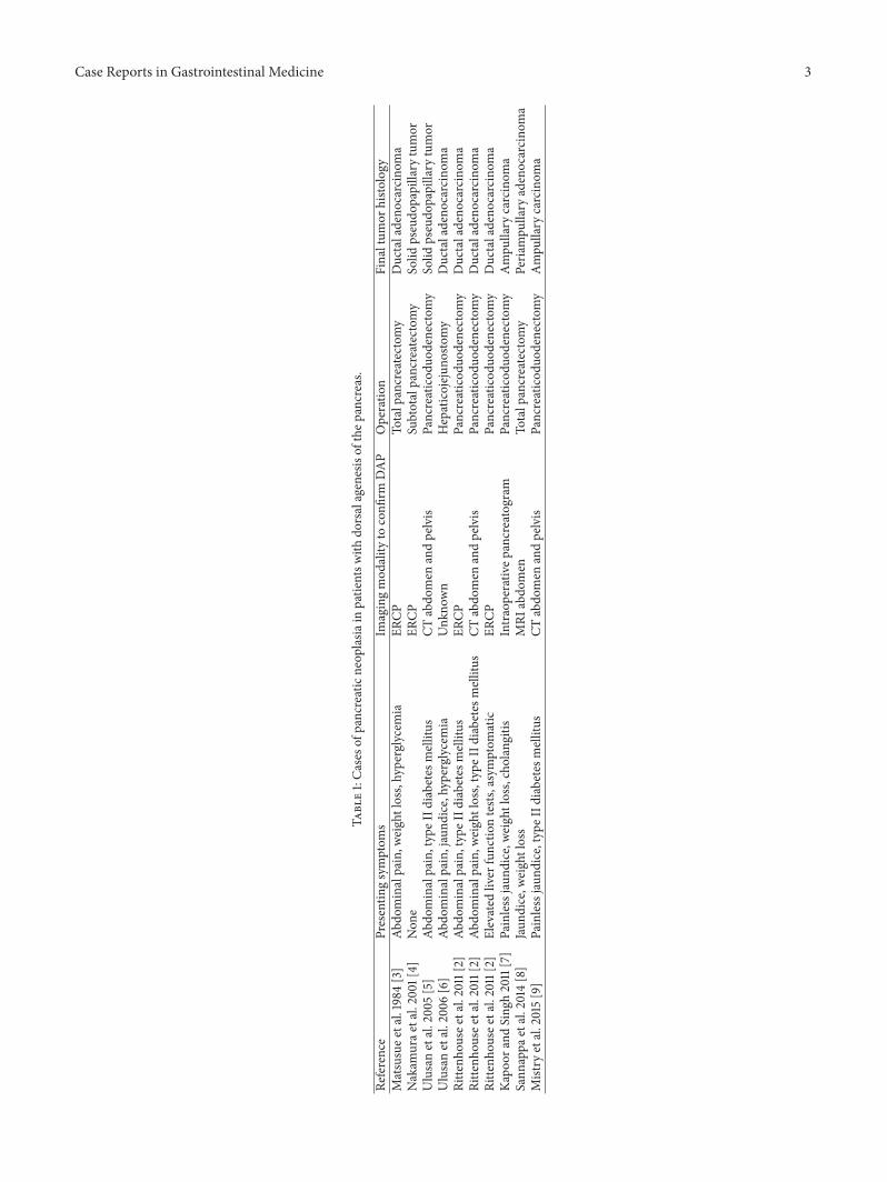
DAP can be asymptomatic due to exocrine and endocrinefunctional reserve in the remaining pancreas. However, giventhat most of the islets of Langerhans develop in the bodyand tail of the pancreas, diabetes mellitus can occur [1].Other common findings in association with DAP includeabdominal pain, pancreatitis, enlarged pancreatic head, and,in a few cases, polysplenia [3, 11]. Diagnosis of DAP requiresdemonstration of the absence of dorsal pancreatic tissue.CT can be useful as an initial study to delineate the sizeof the pancreas. MRCP and ERCP are the gold standardimaging techniques to demonstrate the absence of the dorsal
Case Reports in Gastrointestinal Medicine 3
Table1:Ca
seso
fpancreatic
neop
lasia
inpatientsw
ithdo
rsalagenesisof
thep
ancreas.
Reference
Presentin
gsymptom
sIm
agingmod
ality
toconfi
rmDAP
Operatio
nFinaltum
orhisto
logy
Matsusuee
tal.1984
[3]
Abdo
minalpain,w
eightloss,hyperglycemia
ERCP
Totalp
ancreatectom
yDuctaladeno
carcinom
aNakam
urae
tal.2001
[4]
Non
eER
CPSubtotalpancreatectomy
Solid
pseudo
papillary
tumor
Ulusanetal.2005[5]
Abdo
minalpain,typeIId
iabetesm
ellitus
CTabdo
men
andpelvis
Pancreaticod
uodenectom
ySolid
pseudo
papillary
tumor
Ulusanetal.200
6[6]
Abdo
minalpain,jaund
ice,hyperglycemia
Unk
nown
Hepaticojeju
nosto
my
Ductaladeno
carcinom
aRitte
nhou
seetal.2011[2]
Abdo
minalpain,typeIId
iabetesm
ellitus
ERCP
Pancreaticod
uodenectom
yDuctaladeno
carcinom
aRitte
nhou
seetal.2011[2]
Abdo
minalpain,w
eightloss,type
IIdiabetes
mellitus
CTabdo
men
andpelvis
Pancreaticod
uodenectom
yDuctaladeno
carcinom
aRitte
nhou
seetal.2011[2]
Elevated
liver
functio
ntests
,asymptom
atic
ERCP
Pancreaticod
uodenectom
yDuctaladeno
carcinom
aKa
poor
andSing
h2011[7]
Painlessjaun
dice,w
eightloss,cholangitis
Intraoperativ
epancreatogram
Pancreaticod
uodenectom
yAmpu
llary
carcinom
aSann
appa
etal.2014[8]
Jaun
dice,w
eightloss
MRI
abdo
men
Totalp
ancreatectom
yPeria
mpu
llary
adenocarcino
ma
Mistry
etal.2015[9]
Painlessjaun
dice,typeIId
iabetesm
ellitus
CTabdo
men
andpelvis
Pancreaticod
uodenectom
yAmpu
llary
carcinom
a

4 Case Reports in Gastrointestinal Medicine
pancreatic duct [12]. Treatment of patients with DAP isguided by the symptomatologywithwhich they presented [1].
3.3. Pancreatic Neoplasia and Dorsal Agenesis. The associa-tion of pancreatic neoplasia and DAP has not been stud-ied extensively; a PubMed search identified 10 such casespublished since 2000 (Table 1) [2–9]. The mechanism of thisassociation is uncertain however. Some theorize that DAPincreases the risk of chronic pancreatitis, which in andof itselfis a risk factor for pancreatic tumors.
Treatment of pancreatic neoplasia in the setting of DAPdoes not deviate from current management guidelines [13].Surgical resection of pancreatic tumors in patients with DAPoften requires resection of the remaining pancreatic tissue,with a high rate of insulin dependent diabetes mellitus andexocrine insufficiency. In our case, we were able to preservethe majority of the proximal pancreas, mitigating the risks ofpostoperative diabetes.
4. Conclusion
We present the case of a pancreatic neuroendocrine tumor ina patient with dorsal agenesis of the pancreas, which, to ourknowledge, has not previously been reported in the literature.The patient presented with an asymptomatic, incidentallydiscovered pancreatic mass at the neck of the pancreas thatwas resected with negative margins via spleen preserving,laparoscopic approach.This is one of the few cases of pancre-atic neoplasia identified in patients with dorsal agenesis of thepancreas (DAP), a rare developmental anomaly where thereis absence of the distal pancreas. DAP is most commonlyasymptomatic but can present with symptoms of new-onsetdiabetes mellitus, abdominal pain, or chronic pancreatitis.Because of its silent presentation, there are very few cases ofDAP reported in the literature and even fewer cases of DAPwith concurrent pancreatic neoplasia.
Competing Interests
The authors declare that there are no competing interestsregarding the publication of this paper.
References
[1] W. J. Schnedl, C. Piswanger-Soelkner, S. J. Wallner et al., “Age-nesis of the dorsal pancreas and associated diseases,” DigestiveDiseases and Sciences, vol. 54, no. 3, pp. 481–487, 2009.
[2] D. W. Rittenhouse, E. P. Kennedy, A. A. Mascaro et al., “Thenovel triad of dorsal agenesis of the pancreas with concurrentpancreatic ductal adenocarcinoma and nonalcoholic chroniccalcific pancreatitis: a case series and review of the literature,”Journal of Gastrointestinal Surgery, vol. 15, no. 9, pp. 1643–1649,2011.
[3] S. Matsusue, S. Kashihara, and S. Koizumi, “Pancreatectomy forcarcinoma of the head of the pancreas associated with multipleanomalies including the preduodenal portal vein,”The JapaneseJournal of Surgery, vol. 14, no. 5, pp. 394–398, 1984.
[4] Y. Nakamura, K. Egami, S. Maeda, M. Hosone, and M. Onda,“Solid and papillary tumor of the pancreas complicating agene-sis of the dorsal pancreas,” Journal of Hepato-Biliary-PancreaticSurgery, vol. 8, no. 5, pp. 485–489, 2001.
[5] S. Ulusan, N. Bal, O. Kizilkilic et al., “Solid-pseudopapillarytumour of the pancreas associated with dorsal agenesis,” BritishJournal of Radiology, vol. 78, no. 929, pp. 441–443, 2005.
[6] S. Ulusan, T. Yakar, Z. Koc, F. Kayaselcuk, and N. Torer, “Ade-nocarcinoma of the pancreas associated with dorsal agenesis,”Pancreas, vol. 33, no. 4, pp. 437–439, 2006.
[7] A. Kapoor and R. Singh, “Periampullary carcinoma in a patientwith agenesis of dorsal pancreas,” Journal of Surgical CaseReports, vol. 2011, no. 9, article 4, 2011.
[8] R. M. Sannappa, J. Buragohain, D. Sarma, U. K. Saikia, andB. K. Choudhury, “Agenesis of dorsal pancreas associatedwith periampullary pancreaticobiliary type adenocarcinoma,”Journal of the Pancreas, vol. 15, no. 5, pp. 489–492, 2014.
[9] J. H. Mistry, A. Yadav, and S. Nundy, “Ampullary carcinoma ina patient with agenesis of the dorsal pancreas: a case report,”Indian Journal of Surgery, vol. 77, supplement 1, pp. 32–34, 2015.
[10] M. Mart́ın, J. Gallego-Llamas, V. Ribes et al., “Dorsal pancreasagenesis in retinoic acid-deficient Raldh2 mutant mice,” Devel-opmental Biology, vol. 284, no. 2, pp. 399–411, 2005.
[11] J. Low, D. Williams, and J. R. Chaganti, “Polysplenia syndromewith agenesis of the dorsal pancreas and preduodenal portalvein presenting with obstructive jaundice—a case report andliterature review,” British Journal of Radiology, vol. 84, no. 1007,pp. e217–e220, 2011.
[12] M.Mohapatra, S. Mishra, P. C. Dalai et al., “Imaging findings inagenesis of the dorsal pancreas. Report of three cases,” Journalof the Pancreas, vol. 13, no. 1, pp. 108–114, 2012.
[13] N. Toyama, H. Kamiyama, Y. Suminaga, K. Namai, M. Ota, andF. Konishi, “Pancreas head carcinomawith total fat replacementof the dorsal exocrine pancreas,” Journal of Gastroenterology,vol. 39, no. 1, pp. 76–80, 2004.

Submit your manuscripts athttp://www.hindawi.com
Stem CellsInternational
Hindawi Publishing Corporationhttp://www.hindawi.com Volume 2014
Hindawi Publishing Corporationhttp://www.hindawi.com Volume 2014
MEDIATORSINFLAMMATION
of
Hindawi Publishing Corporationhttp://www.hindawi.com Volume 2014
Behavioural Neurology
EndocrinologyInternational Journal of
Hindawi Publishing Corporationhttp://www.hindawi.com Volume 2014
Hindawi Publishing Corporationhttp://www.hindawi.com Volume 2014
Disease Markers
Hindawi Publishing Corporationhttp://www.hindawi.com Volume 2014
BioMed Research International
OncologyJournal of
Hindawi Publishing Corporationhttp://www.hindawi.com Volume 2014
Hindawi Publishing Corporationhttp://www.hindawi.com Volume 2014
Oxidative Medicine and Cellular Longevity
Hindawi Publishing Corporationhttp://www.hindawi.com Volume 2014
PPAR Research
The Scientific World JournalHindawi Publishing Corporation http://www.hindawi.com Volume 2014
Immunology ResearchHindawi Publishing Corporationhttp://www.hindawi.com Volume 2014
Journal of
ObesityJournal of
Hindawi Publishing Corporationhttp://www.hindawi.com Volume 2014
Hindawi Publishing Corporationhttp://www.hindawi.com Volume 2014
Computational and Mathematical Methods in Medicine
OphthalmologyJournal of
Hindawi Publishing Corporationhttp://www.hindawi.com Volume 2014
Diabetes ResearchJournal of
Hindawi Publishing Corporationhttp://www.hindawi.com Volume 2014
Hindawi Publishing Corporationhttp://www.hindawi.com Volume 2014
Research and TreatmentAIDS
Hindawi Publishing Corporationhttp://www.hindawi.com Volume 2014
Gastroenterology Research and Practice
Hindawi Publishing Corporationhttp://www.hindawi.com Volume 2014
Parkinson’s Disease
Evidence-Based Complementary and Alternative Medicine
Volume 2014Hindawi Publishing Corporationhttp://www.hindawi.com