Changes neuroendocrine - BMJ
4
Thorax 1995;50:551-554 Changes in neuroendocrine elements in bronchial mucosa in chronic lung disease in adults M Pilmane, A Luts, F Sundler Abstract Background - It is not clear whether there is any association between metaplasia of the bronchial epithelium and changes in the distribution of neuroendocrine cells. This study examined, by immuno- histological techniques, the distri- bution of neuroendocrine cells and juxtamucoscal nerve fibres in bronchial biopsies showing metaplastic changes. Methods - Bronchial biopsies from 12 sub- jects with epithelial metaplasia associated with bronchiectasis and diffuse pulmonary fibrosis were examined by conventional light microscopy and immunohistological techniques for protein gene product 9*5 (PGP), chromogranin A and B (CAB), serotonin, vasoactive intestinal peptide (VIP), substance P (SP), calcitonin gene- related peptide (CGRP), calcitonin (CT), and gastrin releasing peptide (GRP). Results - Regions of non-metaplastic epi- thelium contained numerous PGP and serotonin immunoreactive cells. Sub- populations of these cells displayed CAB, CGRP, CT, and GRP immunoreactivity. Metaplastic epithelium contained only a few weakly stained PGP, serotonin, CAB, GRP, CT and CGRP immunoreactive cells in six cases. Metaplastic epithelium was characterised by a high number of CAB- containing cells in six cases and in these biopsies prominent PGP-contaiIiing nerve bundles were seen in the subepithelial layer beneath the metaplastic epithelium. Conclusions - The distribution patterns of neuroendocrine cells and neuronal ele- ments vary between areas of normal and metaplastic epithelium and within areas of metaplastic epithelium. Neuronal hyper- plasia was associated with an increase in the number of CAB-containing cells within the metaplastic epithelium. (Thorax 1995;50:551-554) Keywords: neuroendocrine cells, metaplasia, bronchial mucosa, chronic non-specific lung disease. Elements of the diffuse neuroendocrine system are well represented in the human lung, espe- cially in the large bronchi.'2 The density of neuropeptide-containing nerve fibres and the number of pulmonary neuroendocrine cells change only marginally from childhood to old age,34 but the density of cells increases sig- nificantly in chronic non-specific lung disease.5 It is not known whether there is any as- sociation between changes in the occurrence and distribution of neuroendocrine cells and the presence of metaplastic bronchial epi- thelium. Using immunocytochemical methods we have investigated biopsy samples from 12 patients with chronic non-specific lung disease associated with metaplastic epithelium of large bronchi in order to identify neuroendocrine cells and juxtamucosal nerve fibres. Antisera against the general neuroendocrine markers protein gene product 9 5 (PGP) and chromo- granin A and B (CAB), as well as the neuro- hormonal messengers vasoactive intestinal peptide (VIP), substance P (SP), calcitonin gene-related peptide (CGRP), serotonin, cal- citonin (CT), and gastrin releasing peptide (GRP) were used. Methods Mucosal biopsy specimens from large bronchi were obtained at bronchoscopy from 12 patients (aged 20-65 years) with bronchiectasis and diffuse pulmonary fibrosis. Patients with bronchiectasis had had the condition for at least 10 years, and four patients with diffuse pulmonary fibrosis for more than 11 months. The biopsy specimens were fixed in a mixture of 2% formaldehyde and 0 2% picric acid in 0 1 M phosphate buffer (pH 7 2). The tissues were then rinsed in a Tyrode buffer containing sucrose. The specimens were frozen on dry ice and sectioned in a cryostat. Sections from each bronchial biopsy specimen were routinely stained with haematoxylin and eosin for light microscopic examination of epithelial meta- plasia. For immunocytochemistry6 we used antisera against the following substances: PGP (rabbit polyclonal, working dilution 1:1600, Ultraclone, Cambridge, UK); serotonin (rabbit polyclonal, 1:1600, Inc Star, Stillwater, USA); CAB (rabbit polyclonal antiserum dem- onstrating both chromogranins,7 1:640, Milab, Malmo, Sweden); chromogranin A (mouse monoclonal, 1:320, Boehringer Mannheim, Germany); VIP (rabbit polyclonal, 1:640, Mi- lab, Malmo, Sweden), CGRP (rabbit poly- clonal, 1:1280, Milab, Malmo, Sweden); SP (rabbit polyclonal, 1:320, gift of Dr PC Emson, MRC, Cambridge, UK); calcitonin (rabbit polyclonal, 1:640, Milab, Malmo, Sweden); GRP (rabbit polyclonal 1:640, gift of Professor N Yanaihara, Shizuoka, Japan). The specimens were incubated overnight with primary anti- serum at + 4°C. After thorough rinsing in phosphate buffered saline (PBS) the sections Department of Histology and Embryology, Medical Academy of Latvia, 16 Dzirciema Street, Riga LV 1007, Latvia M Pilmane Department of Medical Cell Research, University of Lund, Biskopsgaten 5, S-223 62 Lund, Sweden A Luts F Sundler Reprint requests to: Dr M Pilmane. Received 30 November 1993 Returned to authors 16 February 1994 Revised version received 10 June 1994 Accepted for publication 31 January 1995 551 on February 22, 2022 by guest. Protected by copyright. http://thorax.bmj.com/ Thorax: first published as 10.1136/thx.50.5.551 on 1 May 1995. Downloaded from
Transcript of Changes neuroendocrine - BMJ
Thorax 1995;50:551-554
Changes in neuroendocrine elements inbronchial mucosa in chronic lung disease inadults
M Pilmane, A Luts, F Sundler
AbstractBackground - It is not clear whether thereis any association between metaplasia ofthe bronchial epithelium and changes inthe distribution of neuroendocrine cells.This study examined, by immuno-histological techniques, the distri-bution of neuroendocrine cells andjuxtamucoscal nerve fibres in bronchialbiopsies showing metaplastic changes.Methods - Bronchial biopsies from 12 sub-jects with epithelial metaplasia associatedwith bronchiectasis and diffuse pulmonaryfibrosis were examined by conventionallight microscopy and immunohistologicaltechniques for protein gene product 9*5(PGP), chromogranin A and B (CAB),serotonin, vasoactive intestinal peptide(VIP), substance P (SP), calcitonin gene-related peptide (CGRP), calcitonin (CT),and gastrin releasing peptide (GRP).Results - Regions of non-metaplastic epi-thelium contained numerous PGP andserotonin immunoreactive cells. Sub-populations of these cells displayed CAB,CGRP, CT, and GRP immunoreactivity.Metaplastic epithelium contained only afew weakly stained PGP, serotonin, CAB,GRP, CT and CGRP immunoreactive cellsin six cases. Metaplastic epithelium wascharacterised by a high number of CAB-containing cells in six cases and in thesebiopsies prominent PGP-contaiIiing nervebundles were seen in the subepitheliallayer beneath the metaplastic epithelium.Conclusions - The distribution patterns ofneuroendocrine cells and neuronal ele-ments vary between areas of normal andmetaplastic epithelium and within areas ofmetaplastic epithelium. Neuronal hyper-plasia was associated with an increasein the number of CAB-containing cellswithin the metaplastic epithelium.(Thorax 1995;50:551-554)
Keywords: neuroendocrine cells, metaplasia, bronchialmucosa, chronic non-specific lung disease.
Elements of the diffuse neuroendocrine systemare well represented in the human lung, espe-cially in the large bronchi.'2 The density ofneuropeptide-containing nerve fibres and thenumber of pulmonary neuroendocrine cellschange only marginally from childhood to oldage,34 but the density of cells increases sig-nificantly in chronic non-specific lung disease.5
It is not known whether there is any as-sociation between changes in the occurrenceand distribution of neuroendocrine cells andthe presence of metaplastic bronchial epi-thelium. Using immunocytochemical methodswe have investigated biopsy samples from 12patients with chronic non-specific lung diseaseassociated with metaplastic epithelium of largebronchi in order to identify neuroendocrinecells and juxtamucosal nerve fibres. Antiseraagainst the general neuroendocrine markersprotein gene product 9 5 (PGP) and chromo-granin A and B (CAB), as well as the neuro-hormonal messengers vasoactive intestinalpeptide (VIP), substance P (SP), calcitoningene-related peptide (CGRP), serotonin, cal-citonin (CT), and gastrin releasing peptide(GRP) were used.
MethodsMucosal biopsy specimens from large bronchiwere obtained at bronchoscopy from 12patients (aged 20-65 years) with bronchiectasisand diffuse pulmonary fibrosis. Patients withbronchiectasis had had the condition for atleast 10 years, and four patients with diffusepulmonary fibrosis for more than 11 months.The biopsy specimens were fixed in a mixtureof 2% formaldehyde and 0 2% picric acid in0 1 M phosphate buffer (pH 7 2). The tissueswere then rinsed in a Tyrode buffer containingsucrose. The specimens were frozen on dry iceand sectioned in a cryostat. Sections from eachbronchial biopsy specimen were routinelystained with haematoxylin and eosin for lightmicroscopic examination of epithelial meta-plasia. For immunocytochemistry6 we usedantisera against the following substances: PGP(rabbit polyclonal, working dilution 1:1600,Ultraclone, Cambridge, UK); serotonin (rabbitpolyclonal, 1:1600, Inc Star, Stillwater, USA);CAB (rabbit polyclonal antiserum dem-onstrating both chromogranins,7 1:640, Milab,Malmo, Sweden); chromogranin A (mousemonoclonal, 1:320, Boehringer Mannheim,Germany); VIP (rabbit polyclonal, 1:640, Mi-lab, Malmo, Sweden), CGRP (rabbit poly-clonal, 1:1280, Milab, Malmo, Sweden); SP(rabbit polyclonal, 1:320, gift ofDr PC Emson,MRC, Cambridge, UK); calcitonin (rabbitpolyclonal, 1:640, Milab, Malmo, Sweden);GRP (rabbit polyclonal 1:640, gift of ProfessorN Yanaihara, Shizuoka, Japan). The specimenswere incubated overnight with primary anti-serum at + 4°C. After thorough rinsing inphosphate buffered saline (PBS) the sections
Departmentof Histology andEmbryology, MedicalAcademy of Latvia,16 Dzirciema Street,Riga LV 1007, LatviaM Pilmane
Departmentof Medical CellResearch,Universityof Lund,Biskopsgaten 5,S-223 62 Lund,SwedenA LutsF Sundler
Reprint requests to:Dr M Pilmane.Received30 November 1993Returned to authors16 February 1994Revised version received10 June 1994Accepted for publication31 January 1995
551
on February 22, 2022 by guest. P
rotected by copyright.http://thorax.bm
j.com/
Thorax: first published as 10.1136/thx.50.5.551 on 1 M
ay 1995. Dow
nloaded from

Pilmane, Luts, Sundler
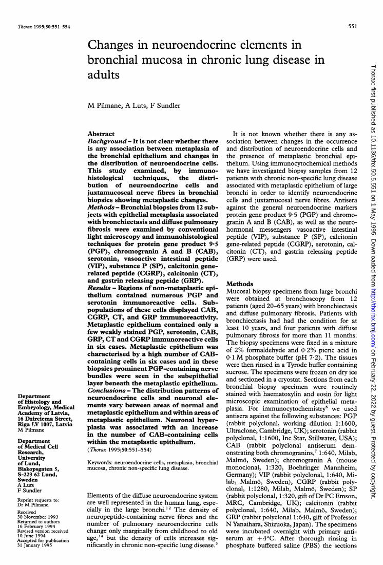
Figure 1 Immunofluorescencein patients with chronic non-spneuroendocrine cells in normalneuroendocrine cells. (b) Occaneuroendocrine ceUls (arrows).metaplastic epithelium (arrowsshowing weak CGRP immunoin the subepithelial layer (arroorigination.
epithelium showed weak PGP, VIP, CGRP,and SP immunoreactivity.
In regions with metaplastic epithelium wedistinguished different distribution patterns ofneuroendocrine cells and neuronal elements.Few, usually weakly stained, PGP, serotonin,CAB, GRP, CT and CGRP immunoreactiveneuroendocrine cells were seen in these regionsin six patients (fig lb-e). The subepithelial layerbeneath the metaplastic epithelium harbouredfine PGP, CGRP, VIP, and SP immunoreactivenerve fibres (fig le); in some cases prominentPGP-containing nerve bundles were seen (fig2a).
In areas of metaplastic epithelium in theother six patients many weakly stained PGP-containing neuroendocrine cells were seen.Most of them stored CAB (fig 2b). A sub-
-~~~~~~~~~~~~~~~~~~~~~~~~~~~~~~~~~-
micrographs of bronchial wall with metaplastic epitheliumzecific lung disease. (a) Numerous PGP immunoreactiveepithelium; the metaplastic epithelium (arrow) is devoid ofsional weakly immunostained serotonin-containing(c) Cells containing chromogranin A and B in the;). (d) GRP immunoreactive celli. (e) Neuroendocrine cellsreactivity (arrow) in the epithelium as well as nerve fibres,w). Original magnification x 250 reduced to 63% in
were incubated in fluorescein isothiocyanate(FITC)-labelled swine antirabbit (or goat anti-mouse) IgG for 45 minutes at room tem-perature. After another rinsing in PBS thesections were mounted in phosphate bufferedglycerin and examined by fluorescence micro-scopy. The specificity of the antisera usedhas been tested and the results presentedelsewhere.27
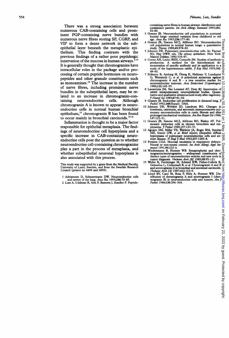
ResultsExamination of the biopsy specimens con-firmed the presence of regions of non-meta-plastic and metaplastic epithelium. In the non- Figure 2 Immunofluorescence micrographs of bronchialmetaplastic epithelium we found numerous wall with metaplastic epithelium in patients with chronicPGP immunoreactive neuroendocrine cells (fig non-specific lung disease. (a) Prominent PGP-containingla).MostofthePGPpoitiveneunerve bundles beneath the metaplastic epithelium
1la). Most of the PGP positive neuroendocrine occupying most of the subepithelial space. (b) and (c)cells were immunoreactive for serotonin; only Consecutive sections showing intensely CABoccasional serotonin positive neuroendocrine immunoreactive neuroendocrine cells (arrows) and
cells also stored CABI) CGRP CT, andGRP. serotonin immunoreactivity (c) in the same cells withincells also stored CAB, CGRP, CT, and GRP. metaplastic epithelium (arrows). Original magnificationA few nerve fibres beneath the non-metaplastic x 250 reduced to 57% in origination.
552
on February 22, 2022 by guest. P
rotected by copyright.http://thorax.bm
j.com/
Thorax: first published as 10.1136/thx.50.5.551 on 1 M
ay 1995. Dow
nloaded from

Distribution of neuroendocrine cells in bronchial mucosa
a
Figure 3 (a) Light and (b)-(d) immunofluorescence micrographs of sections from largebronchi ofpatients with chronic non-specific lung disease. (a) Characteristic bronchialmetaplastic epithelium. (b) PGP-containing neuroendocrine cells in the metaplasticepithelium (arrows) and prominent immunoreactive nerve bundles in the subepitheliallayer. (c) Numerous VIP-containing nerve fibres, some forming bundles beneath theepithelium (ep). (d) Numerous fine SP immunoreactive nerve fibres, sometimes formingnerve bundles in the subepithelium. Original magnification x 160 (a and b) and x 250(c and d) reduced to 75% in origination.
population of the CAB immunoreactive neuro-
endocrine cells also displayed immuno-reactivity for serotonin (fig 2c) in all six cases
and for chromogranin A in three cases. Gen-erally, the combined CAB antiserum detectedhigher numbers of neuroendocrine cells than
the specific chromogranin A and the serotoninantibodies together. No neuroendocrine cellscontaining CT, CGRP, or GRP were found inthe metaplastic epithelium of these patients. Inthose biopsy specimens where CAB im-munoreactive cells were numerous, PGP-con-taining nerve bundles were prominent in thesubepithelial layer beneath the metaplastic epi-thelium (fig 3a, b). Subpopulations of nervefibres within these bundles contained VIP, SP,and CGRP (fig 3c, d). Aggregates of neuro-endocrine cells (tumourlets) were not observed.We could not detect any correlation betweenthe presence and abundance ofneuroendocrinecells and neuronal elements in the bronchi andage, diagnosis, or duration of disease.
DiscussionPrevious studies have indicated a relationshipbetween neuroendocrine cells and nerve fibresin the respiratory tract.8 In the present studywe examined the presence and quantity ofneuroendocrine cells and neuropeptide-con-taining nerves in the bronchial mucosa ofpatients with chronic non-specific lung diseasedisplaying metaplastic and normal epithelium.The distribution pattern of neuroendocrinecells and neuronal elements and their markerand messenger content varied, not only be-tween areas of normal and metaplastic epi-thelium, but also within the areas ofmetaplasticepithelium. It might be that these patternschange during the process of development ofmetaplasia. This possibility is supported by thepresence of metaplastic epithelium in whichonly small numbers of neuroendocrine cellswere seen and nerve bundles were in-conspicuous in the subepithelial tissue. In othercases metaplastic epithelium possessed manyCAB-containing neuroendocrine cells as wellas prominent nerve bundles in the subepitheliallayer. There are several reports on endocrinecell proliferation in diseased lungs.910 An in-creased number of neuroendocrine cells in thelungs could be a physiological response to pul-monary injury or infection. Gosney9 has sug-gested that this response might have a tendencyto become disordered if stimuli which provokeit persist. He also described different stagesofincreases in the density ofpeptide-containingneuroendocrine cells during prolonged patho-logical processes in the lungs. The earliestdetectable change was an increase ofcalcitonin-containing neuroendocrine cells; the next stageinvolved the appearance of ACTH, VIP, SP,and growth hormone immunoreactivity. It wassuggested that proliferation of neuroendocrinecells might, if persistent, lead to the de-velopment of pulmonary tumourlets.9 Thus,there may be a relationship between the stageof tissue disorder and the number of neuro-endocrine cells and their chemical coding. Wedetected different patterns ofchanges in neuro-endocrine cells and neuronal elements whichdid not depend on age, diagnosis, or evenduration of illness. These findings suggest thatthere are other insults contributing to changesin neuropeptide-containing elements in thelungs. 10-12
553
on February 22, 2022 by guest. P
rotected by copyright.http://thorax.bm
j.com/
Thorax: first published as 10.1136/thx.50.5.551 on 1 M
ay 1995. Dow
nloaded from
Pilmane, Luts, Sundler
There was a strong association betweennumerous CAB-containing cells and prom-
inent PGP-containing nerve bundles withnumerous nerve fibres storing SP, CGRP, andVIP to form a dense network in the sub-epithelial layer beneath the metaplastic epi-thelium. This finding contrasts with theprevious findings of a rather poor peptidergicinnervation ofthe mucosa in human airways."'It is generally thought that chromogranins haveintracellular roles in the package and/or pro-
cessing of certain peptide hormones on neuro-
peptides and other granule constituents suchas monoamines.14 The increase in the numberof nerve fibres, including prominent nervebundles in the subepithelial layer, may be re-lated to an increase in chromogranin-con-taining neuroendocrine cells. Althoughchromogranin A is known to appear in neuro-endocrine cells in normal human bronchialepithelium,'5 chromogranin B has been foundto occur mainly in bronchial carcinoids.'5 6
Inflammation is thought to be a major factorresponsible for epithelial metaplasia. The find-ings of neuroendocrine cell hyperplasia and a
specific increase in CAB-containing neuro-endocrine cells pose the question as to whetherneuroendocrine cell-containing chromograninsplay a part in the process of metaplasia, andwhether subepithelial neuronal hyperplasia isalso associated with this process.
This study was supported by a grant from the Medical Faculty,University of Lund, Sweden, and from the Swedish ResearchCouncil (project no 4499 and 6859).
1 Adriaensen D, Scheuermann DW. Neuroendocrine cellsand nerves of the lung. Anat Rec 1993;236:70-85.
2 Luts A, Uddman R, Alm P, Basterra J, Sundler F. Peptide-
containing nerve fibres in human airways: distribution andcoexistence pattern. Int Arch Alley Immunol 1993;101:52-60.
3 Gosney JR. Neuroendocrine cell populations in postnatalhuman lungs: minimal variation from childhood to oldage. Anat Rec 1993;236:177-80.
4 Gosney JR, Sissons MCJ, Allibone RO. Neuroendocrinecell populations in normal human lungs: a quantitativestudy. Thorax 1988;43:878-82.
5 Johnson DE. Pulmonary neuroendocrine cells. In: FarmerSG, Hay DWP, eds. The airway epithelium. New York:Marcel Dekker, 1990:335-97.
6 Coons AH, Leduc REH, Connolly JM. Studies of antibodyproduction. A method for the histochemical de-monstration of specific antibody and its application to astudy of the hyperimmune rabbit. J Exp Med 1955;102:49-60.
7 Eriksson B, Amberg H, Oberg K, Hellman V, LundquistG, Wernstedt C, et al. A polyclonal antiserum againstchromogranin A and B - a new sensitive marker forneuroendocrine tumours. Acta Endocrinol (Copenhagen)1990;122:145-55.
8 Lauweryns JM, Van Lommel AT, Dom RJ. Innervation ofrabbit intrapulmonary neuroepithelial bodies. Quant-itative and qualitative ultrastructural study after vagotomy.J Neurol Sci 1985;67:81-92.
9 Gosney JR. Endocrine cell proliferation in diseased lung. YPathol 1992;168(Suppl): 106A.
10 Johnson DE, Wobken JD, Iandrum BG. Changes inbombesin, calcitonin, and serotonin immunoreactive pul-monary neuroendocrine cells in cystic fibrosis and afterprolonged mechanical ventilation.Am Rev RespirDis 1988;137:123-31.
11 Gosney JR, Sissons MCJ, Allibone RO, Blakey AF. Pul-monary endocrine cells in chronic bronchitis and em-physema. Y Pathol 1989;157:123-33.
12 Aguayo SM, Miller YE, Waldron JA, Bogin RM, SundayME, Staton GW, et al. Brief report; idiopathic diffusehyperplasia of pulmonary neuroendocrine cells and air-ways disease. N Engi Y Med 1992;327:1285-8.
13 Persson CGA. Mucosal exudation in respiratory defence.Neural or non-neural control. Int Arch Alergy Appl Im-munol 1991;94:222-6.
14 Wiedenmann B, Hutmer WB. Synaptophysin and chro-mogranins/secretogranins - widespread constituents ofdistinct types of neuroendocrine vesicles and new tools intumor diagnosis. Virchows Arch [B] 1989;58:95-121.
15 Weiler R, Feichtinger M, Schmid KW, Fisher-Colbrie R,Grimelius L, Cedermark B, et al. Chromogranin A and Band secretogranin II in bronchial and intestinal carcinoids.Virchows Arch [A] 1987;412:103-9.
16 Lloyd RV, Cano M, Rosa P, Hille A, Huttner WB. Dis-tribution of chromogranin A and secretogranin I (chro-mogranin B) in neuroendocrine cells and tumors. Am JPathol 1988;130:296-304.
554
on February 22, 2022 by guest. P
rotected by copyright.http://thorax.bm
j.com/
Thorax: first published as 10.1136/thx.50.5.551 on 1 M
ay 1995. Dow
nloaded from