Case Report Overlap of Acute Cholecystitis with Gallstones...
5
Case Report Overlap of Acute Cholecystitis with Gallstones and Squamous Cell Carcinoma of the Gallbladder in an Elderly Patient Ehsan YJldJz, Yavuz Savas Koca, and Ebrahim Barut Department of Genaral Surgery, School of Medicine, Suleyman Demirel University, Isparta, Turkey Correspondence should be addressed to ˙ Ihsan Yıldız; [email protected] Received 30 April 2015; Revised 29 July 2015; Accepted 10 August 2015 Academic Editor: Alexander R. Novotny Copyright © 2015 ˙ Ihsan Yıldız et al. is is an open access article distributed under the Creative Commons Attribution License, which permits unrestricted use, distribution, and reproduction in any medium, provided the original work is properly cited. Introduction. e incidence of gallbladder cancer presenting with acute cholecystitis is 2.3%, squamous cell carcinoma is rarely seen, and overlap of acute cholecystitis and squamous cell carcinoma is a very rare condition in the literature. Presentation of Case.A 75-year-old woman was admitted to emergency service with a pain in the right upper quadrant, nausea, and vomiting. e patient was considered as having acute cholecystitis. During the exploration because Hartman’s pouch was not dissected, it was adhered to the cystic duct and had fibrotic adhesion. It could not be understood whether this adhesion was a tumor or a fibrotic tumor and thus we performed cholecystectomy with a 1cm resection of the choledochus. Pathological examination revealed the presence of (R0), T1 N0 M0 squamous cell carcinoma with clean resection borders and there was no in tumor five dissected lymph nodes. e patient has been followed up for about two years and no clinical problem has been observed throughout the follow-up. Discussion. Acute cholecystitis with gallstones may overlap with gallbladder cancer and generally presents nonspecific symptoms. No additional imaging techniques were performed since no clinical sign except for the wall thickening was detected and no suspected malignancy such as mass was detected on USG. Squamous cell carcinoma of the gallbladder shows poor diagnosis, but since its overlap with cholecystitis presents early symptoms and thus leads to early diagnosis and effective treatment, the localization of the carcinoma is of prime importance. Conclusion. Gallbladder cancer should be kept in mind in elderly patients with acute cholecystitis. 1. Introduction Acute cholecystitis with gallstones is the most common sur- gical problem of the gallbladder. e incidence of gallbladder cancer presenting with acute cholecystitis is 2.3% [1, 2]. Among the gallbladder cancers, squamous cell carcinoma is rarely seen [2]. e overlap of acute cholecystitis and squamous cell carcinoma is a very rare condition in the literature [3–6]. Gallbladder cancers, comprising 90% adenocarcinoma and 0.5–3% squamous cell carcinoma (SCC), account for 3-4% of all malignant tumors. SCC is more common at advanced ages and the female to male ratio is 1/2 [7–9]. SCC presents no specific symptom except for the clinical presenta- tion of acute cholecystitis. Nevertheless, there are a number of studies reporting the presentation of a palpable mass in the right upper quadrant at advanced ages [8, 9]. Early diagnosis of SCC accounts for 10–50% of the cases. Ultrasonography (USG) is the method of choice in the diagnosis but advanced techniques such as tomography are also used, particularly for the suspected cases. In the diagnosis of gallbladder cancer, the sensitivity of USG is 34%, whereas it is ∼40 for tomography [8]. Surgical treatment of SCC is followed by radiotherapy and chemotherapy. Early stages of SCC are treated by cholecystec- tomy and the late stages are treated by extensive hepatectomy including resection of segment 4 or 5 [8, 9]. Survival rate following surgery is 20% in the early-stage patients and 10% in the late-stage patients [9]. In this study, we present a 75-year-old female patient who presented to our emergency service with a pain in the right upper quadrant and underwent cholecystectomy due to acute cholecystitis and then presented with squamous cell carcinoma of the gallbladder in the pathological examination. Hindawi Publishing Corporation Case Reports in Surgery Volume 2015, Article ID 767196, 4 pages http://dx.doi.org/10.1155/2015/767196
Transcript of Case Report Overlap of Acute Cholecystitis with Gallstones...

Case ReportOverlap of Acute Cholecystitis withGallstones and Squamous Cell Carcinoma ofthe Gallbladder in an Elderly Patient
Ehsan YJldJz, Yavuz Savas Koca, and Ebrahim Barut
Department of Genaral Surgery, School of Medicine, Suleyman Demirel University, Isparta, Turkey
Correspondence should be addressed to Ihsan Yıldız; [email protected]
Received 30 April 2015; Revised 29 July 2015; Accepted 10 August 2015
Academic Editor: Alexander R. Novotny
Copyright © 2015 Ihsan Yıldız et al. This is an open access article distributed under the Creative Commons Attribution License,which permits unrestricted use, distribution, and reproduction in any medium, provided the original work is properly cited.
Introduction.The incidence of gallbladder cancer presenting with acute cholecystitis is 2.3%, squamous cell carcinoma is rarely seen,and overlap of acute cholecystitis and squamous cell carcinoma is a very rare condition in the literature. Presentation of Case. A75-year-old woman was admitted to emergency service with a pain in the right upper quadrant, nausea, and vomiting. The patientwas considered as having acute cholecystitis. During the exploration because Hartman’s pouch was not dissected, it was adheredto the cystic duct and had fibrotic adhesion. It could not be understood whether this adhesion was a tumor or a fibrotic tumor andthus we performed cholecystectomy with a 1 cm resection of the choledochus. Pathological examination revealed the presence of(R0), T1 N0 M0 squamous cell carcinoma with clean resection borders and there was no in tumor five dissected lymph nodes. Thepatient has been followed up for about two years and no clinical problem has been observed throughout the follow-up. Discussion.Acute cholecystitis with gallstonesmay overlapwith gallbladder cancer and generally presents nonspecific symptoms.No additionalimaging techniques were performed since no clinical sign except for the wall thickening was detected and no suspectedmalignancysuch as mass was detected on USG. Squamous cell carcinoma of the gallbladder shows poor diagnosis, but since its overlap withcholecystitis presents early symptoms and thus leads to early diagnosis and effective treatment, the localization of the carcinoma isof prime importance. Conclusion. Gallbladder cancer should be kept in mind in elderly patients with acute cholecystitis.
1. Introduction
Acute cholecystitis with gallstones is the most common sur-gical problem of the gallbladder.The incidence of gallbladdercancer presenting with acute cholecystitis is 2.3% [1, 2].Among the gallbladder cancers, squamous cell carcinomais rarely seen [2]. The overlap of acute cholecystitis andsquamous cell carcinoma is a very rare condition in theliterature [3–6].
Gallbladder cancers, comprising 90% adenocarcinomaand 0.5–3% squamous cell carcinoma (SCC), account for3-4% of all malignant tumors. SCC is more common atadvanced ages and the female to male ratio is 1/2 [7–9]. SCCpresents no specific symptom except for the clinical presenta-tion of acute cholecystitis. Nevertheless, there are a numberof studies reporting the presentation of a palpable mass in theright upper quadrant at advanced ages [8, 9].
Early diagnosis of SCC accounts for 10–50% of thecases. Ultrasonography (USG) is the method of choice inthe diagnosis but advanced techniques such as tomographyare also used, particularly for the suspected cases. In thediagnosis of gallbladder cancer, the sensitivity of USG is 34%,whereas it is ∼40 for tomography [8].
Surgical treatment of SCC is followed by radiotherapy andchemotherapy. Early stages of SCC are treated by cholecystec-tomy and the late stages are treated by extensive hepatectomyincluding resection of segment 4 or 5 [8, 9].
Survival rate following surgery is 20% in the early-stagepatients and 10% in the late-stage patients [9].
In this study, we present a 75-year-old female patientwho presented to our emergency service with a pain in theright upper quadrant and underwent cholecystectomy dueto acute cholecystitis and then presented with squamous cellcarcinoma of the gallbladder in the pathological examination.
Hindawi Publishing CorporationCase Reports in SurgeryVolume 2015, Article ID 767196, 4 pageshttp://dx.doi.org/10.1155/2015/767196
2 Case Reports in Surgery
2. Case
A 75-year-old woman presented to our emergency servicewith a pain in the right upper quadrant, nausea, and vomiting.The patient had no clinical features and had no specific signor symptom except for a temperature of 37.5∘C and a positiveMurphy’s sign in the right upper quadrant.
Biochemical parameters were as follows: BK: 12,000(4,000–8,000), AST: 45 (0–35U/L), ALT: 56 (0–35U/L), andAMLZ: 120 (24–151U/L).The other parameters were normal.
Abdominal USG revealed swollen gallbladder with a wallthickness of 2 cm and echogenicity suggesting a 3 cm stone inHartman’s pouch. Depending on these signs, the patient wasconsidered as having acute cholecystitis.
3. Intraoperative Findings
Thesurgery startedwith laparoscopy. During the exploration,the gallbladder was swollen, the wall was thickened andinflamed, pericholecysticminimal fluid was detected, and theother intra-abdominal organs were normal. The gallbladderwas suspended from the fundus. The Calot’s triangle wasdissected but it was difficult to dissect the Hartman’s pouchsince it was adhered to the cystic duct and to the 1 cm segmentof the choledochus.We had no available conditions for frozenbiopsy during the surgery.
Afterwards, the procedurewas converted to open surgery,and cholecystectomy was performed by the resection of1 cm segment of the choledochus (because Hartman’s pouchwas not dissected since it was adhered to the cystic ductand had fibrotic adhesion). The choledochus was primarilyanastomosed over the T-tube. The patient was uneventfullydischarged on postoperative day 6. On postoperative day 14, aT-tube cholangiography was performed and no clinical prob-lem was observed.The T-tube was removed on postoperativeday 21.
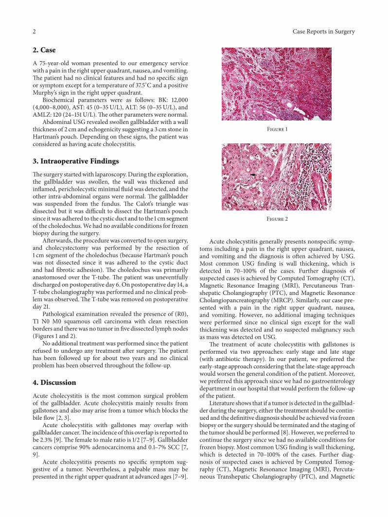
Pathological examination revealed the presence of (R0),T1 N0 M0 squamous cell carcinoma with clean resectionborders and therewas no tumor in five dissected lymphnodes(Figures 1 and 2).
No additional treatment was performed since the patientrefused to undergo any treatment after surgery. The patienthas been followed up for about two years and no clinicalproblem has been observed throughout the follow-up.
4. Discussion
Acute cholecystitis is the most common surgical problemof the gallbladder. Acute cholecystitis mainly results fromgallstones and also may arise from a tumor which blocks thebile flow [2, 3].
Acute cholecystitis with gallstones may overlap withgallbladder cancer.The incidence of this overlap is reported tobe 2.3% [9].The female to male ratio is 1/2 [7–9]. Gallbladdercancers comprise 90% adenocarcinoma and 0.1–7% SCC [7,9].
Acute cholecystitis presents no specific symptom sug-gestive of a tumor. Nevertheless, a palpable mass may bepresented in the right upper quadrant at advanced ages [7–9].
Figure 1
Figure 2
Acute cholecystitis generally presents nonspecific symp-toms including a pain in the right upper quadrant, nausea,and vomiting and the diagnosis is often achieved by USG.Most common USG finding is wall thickening, which isdetected in 70–100% of the cases. Further diagnosis ofsuspected cases is achieved by Computed Tomography (CT),Magnetic Resonance Imaging (MRI), Percutaneous Tran-shepatic Cholangiography (PTC), and Magnetic ResonanceCholangiopancreatography (MRCP). Similarly, our case pre-sented with a pain in the right upper quadrant, nausea,and vomiting. However, no additional imaging techniqueswere performed since no clinical sign except for the wallthickening was detected and no suspected malignancy suchas mass was detected on USG.
The treatment of acute cholecystitis with gallstones isperformed via two approaches: early stage and late stage(with antibiotic therapy). In our patient, we preferred theearly-stage approach considering that the late-stage approachwould worsen the general condition of the patient. Moreover,we preferred this approach since we had no gastroenterologydepartment in our hospital that would perform the follow-upof the patient.
Literature shows that if a tumor is detected in the gallblad-der during the surgery, either the treatment should be contin-ued and the definitive diagnosis should be achieved via frozenbiopsy or the surgery should be terminated and the staging ofthe tumor should be performed [8]. However, we preferred tocontinue the surgery since we had no available conditions forfrozen biopsy. Most commonUSG finding is wall thickening,which is detected in 70–100% of the cases. Further diag-nosis of suspected cases is achieved by Computed Tomog-raphy (CT), Magnetic Resonance Imaging (MRI), Percuta-neous Transhepatic Cholangiography (PTC), and Magnetic

Case Reports in Surgery 3
Resonance Cholangiopancreatography (MRCP). Neverthe-less, no surgical deficit occurred in our patient since nodifferent surgical procedure is suggested when a tumor isdetected on frozen biopsy. Surgery is the method of choicein the treatment. The first and second stages of SCC aretreated by cholecystectomy, whereas the late stages are treatedby extensive cholecystectomy with wedge resection andhepatectomy including resection of segment 4 or 5 [8].
In our patient, the Hartman’s pouch was not dissectedsince it was adhered to the cystic duct and to the 1 cm segmentof the choledochus. Nevertheless we could not understandwhether this adhesion was a tumor or a fibrotic tumor andthus we performed cholecystectomy with a 1 cm resection ofthe choledochus and choledochocholedochostomy over theT-tube.
Squamous cell carcinoma of the gallbladder has a poorprognosis since it is a rare condition and leads to delayeddiagnosis [2, 3, 6, 8]. The involvement of serosal and lym-phatic glands is a major factor for the poor prognosis. SCC ismostly diagnosed in the wall (25%), through the involvementof the lymphatic gland (35%), and through distant metastasis(40%) [8, 9]. In the cases with lymphatic gland involvement,the prognosis remains poor despite extensive surgery [9].The5-year survival rate is 20% after surgery, whereas it is 0% inthe nonoperated patients and 10% in the late-stage patientsundergoing surgery. In stages 2 and 3, the 5-year survivalrate is 0% after cholecystectomy and 29% after extensivecholecystectomy [3, 9].
Radiotherapy and chemotherapy are suggested after thesurgical treatment of SCC. However, our patient refused toundergo any of these treatments. Nevertheless, the patient hasbeen followed up for about two years and no clinical problemhas been observed throughout the follow-up.
Gallbladder cancer, as in most cancers, is more commonin advanced ages. Gallbladder cancer is more frequent inwomen than in men and it generally presents nonspecificsymptoms such as acute cholecystitis [1–3, 7, 8]. Similarly, ourpatient was 75 years old and she presented no clinical signsuggestive of gallbladder cancer. In the patients of gallbladdercancer, the detection of a palpable mass in the right upperquadrant is not unique to cancer but it may be a significantsign since it may suggest the presence of a malignity [4, 8].However, our patient had no palpable mass in the right upperquadrant.
The laboratory findings of the patients with no suspicionof cancer present no findings other than the findings of acutecholecystitis [7–9]. Similarly, our patient had no clinical orlaboratory finding suggestive of cancer.
A number of studies have shown that terminating thesurgical procedure may be beneficial in the patients whoare preoperatively suspected with gallbladder cancer andundergo biopsy [6]. However, we preferred to continue thesurgery sincewe hadno available conditions for frozen biopsyduring the surgery and the procedure was completed with nocomplication.
The staging of gallbladder cancer is performed accordingto the Japanese Biliary Surgical Society system [8]. Ourpatient presented with (R0) T1 N0M0 squamous cell carci-noma with clean resection borders.
Previous studies have shown that gallbladder cancershould be suspected in the advanced-age patients admittedto hospital with the symptoms of cholecystitis. However, thelocalization and diameter of the tumor are a key factor for theprognosis of the cancer [2, 8]. The patients presenting withgallstones in the Hartman’s pouch and proximal to the cysticduct present symptoms at early stages and thus enable earlydiagnosis and effective treatment [7, 8].
Squamous cell carcinoma of the gallbladder shows poordiagnosis, but since its overlap with cholecystitis presentsearly symptoms and thus leads to early diagnosis and effectivetreatment, the localization of the carcinoma is of primeimportance [2, 8].
5. Conclusion
Gallbladder cancer should be kept in mind in the advanced-age patients presenting with the symptoms of cholecystitis.
Disclosure
Thesource of studywas SuleymanDemirel University, SchoolofMedicine, Department of Genaral Surgery, Isparta, Turkey.
Conflict of Interests
The authors declare that there is no conflict of interestsregarding the publication of this paper.
Authors’ Contribution
Ihsan Yıldız contributed approximately 60%, Yavuz SavasKoca contributed approximately 30%, and Ibrahim Barutcontributed approximately 10% to this study.
References
[1] K. L. Huguet, C. B. Hughes, andW. R. Hewitt, “Gallbladder car-cinosarcoma: a case report and literature review,” Journal ofGastrointestinal Surgery, vol. 9, no. 6, pp. 818–821, 2005.
[2] C. M. Lam, A. W. Yuen, R. M. Leung et al., “Gallbladder cancerpresenting with acute cholecystitis: a population-based study,”Surgical Endoscopy and Other Interventional Techniques, vol. 19,no. 5, pp. 697–701, 2005.
[3] U. Damian, B. Rumstadt, andD. Schilling, “Acute squamous cellcarcinoma of the gallbladder: a rare cause of acute cholecystitis,”Deutsche Medizinische Wochenschrift, vol. 136, no. 27, pp. 1422–1425, 2011.
[4] N. Gomez, I. Urrea, and R. Astudillio, “Primary epidermoidcarcinoma of the gallbladder,” Acta Gastroenterologica Lati-noamericana, vol. 20, no. 3, pp. 169–173, 1990.
[5] M. Bouassida, B.Mroua, A. Douggaz,M.M.Mighri, H. Touinsi,and S. Sassi, “Primary pure squamous cell carcinoma of thegallbladder revealed by an acute angiocholitis,” Presse Medicale,vol. 42, no. 1, pp. 110–113, 2013.
[6] P. E. Lada, B. Taborda, M. Sanchez et al., “Adenosquamous andsquamous carcinoma of the gallbladder,” Cirugia Espanola, vol.81, no. 4, pp. 202–206, 2007.
[7] S. Gupta, S. K. Gupta, and N. C. Aryya, “Primary squamouscell carcinoma of gallbladder presenting as acute cholecystitis,”
4 Case Reports in Surgery
Indian Journal of Pathology and Microbiology, vol. 47, no. 2, pp.231–233, 2004.
[8] M. Yıldırım, N. Erkan, and K. Kayapınar, “Dıagnosıs and treat-ment ın gallbladder cancer: worst prognosıs,” Ege Klinikleri TıpDergisi, no. 2, pp. 107–110, 2005.
[9] M. Hosseinzadeh, M. Shokripur, and H. Salahi, “Primary puresquamous cell carcinoma of gallbladder presenting as acutecholecystitis,” Iranian Journal of Medical Sciences, vol. 37, no. 4,pp. 271–273, 2012.

Submit your manuscripts athttp://www.hindawi.com
Stem CellsInternational
Hindawi Publishing Corporationhttp://www.hindawi.com Volume 2014
Hindawi Publishing Corporationhttp://www.hindawi.com Volume 2014
MEDIATORSINFLAMMATION
of
Hindawi Publishing Corporationhttp://www.hindawi.com Volume 2014
Behavioural Neurology
EndocrinologyInternational Journal of
Hindawi Publishing Corporationhttp://www.hindawi.com Volume 2014
Hindawi Publishing Corporationhttp://www.hindawi.com Volume 2014
Disease Markers
Hindawi Publishing Corporationhttp://www.hindawi.com Volume 2014
BioMed Research International
OncologyJournal of
Hindawi Publishing Corporationhttp://www.hindawi.com Volume 2014
Hindawi Publishing Corporationhttp://www.hindawi.com Volume 2014
Oxidative Medicine and Cellular Longevity
Hindawi Publishing Corporationhttp://www.hindawi.com Volume 2014
PPAR Research
The Scientific World JournalHindawi Publishing Corporation http://www.hindawi.com Volume 2014
Immunology ResearchHindawi Publishing Corporationhttp://www.hindawi.com Volume 2014
Journal of
ObesityJournal of
Hindawi Publishing Corporationhttp://www.hindawi.com Volume 2014
Hindawi Publishing Corporationhttp://www.hindawi.com Volume 2014
Computational and Mathematical Methods in Medicine
OphthalmologyJournal of
Hindawi Publishing Corporationhttp://www.hindawi.com Volume 2014
Diabetes ResearchJournal of
Hindawi Publishing Corporationhttp://www.hindawi.com Volume 2014
Hindawi Publishing Corporationhttp://www.hindawi.com Volume 2014
Research and TreatmentAIDS
Hindawi Publishing Corporationhttp://www.hindawi.com Volume 2014
Gastroenterology Research and Practice
Hindawi Publishing Corporationhttp://www.hindawi.com Volume 2014
Parkinson’s Disease
Evidence-Based Complementary and Alternative Medicine
Volume 2014Hindawi Publishing Corporationhttp://www.hindawi.com