Case Report Oncocytic lipoadenoma of the parotid gland: a report of
J Toxicol Pathol 2015; 28: 229–232
Case Report
Lipomatosis of the canine parotid gland: case report with a literature review
Hiroo Madarame1*, Ryuuji Harada1, Shinpei Kawarai1, Haruo Takeda2, and Takuo Shida3
1 Laboratory of Small Animal Clinics, Veterinary Teaching Hospital, Azabu University, 1-17-71 Fuchinobe, Chuo, Sagamihara, Kanagawa 252-5201, Japan
2 Veterinary Teaching Hospital, Azabu University, 1-17-71 Fuchinobe, Chuo, Sagamihara, Kanagawa 252-5201, Japan3 Laboratory of Radiology, School of Veterinary Medicine, Azabu University, 1-17-71 Fuchinobe, Chuo, Sagamihara, Kanagawa 252-5201, Japan
Abstract: In this report, we describe a case of lipomatosis in the left parotid gland of an eight-year-old female Shetland sheepdog and review the relevant literature. Preoperative diagnosis of lipomatosis with Tru-Cut biopsy presented difficulties in this case. The post-operative diagnosis was based on the gross appearance of a non-infiltrative, circumscribed swollen mass in the parotid gland and the histological appearance of normal adipocytes that infiltrated into the parotid gland without involving surrounding structures. Addition-ally, flotation of the whole parotid gland in formalin solution together with well-maintained residual lobulation and a well-maintained configuration on the cut surface were identified as subsidiary characteristics of lipomatosis of the parotid gland. Lipomatosis of the parotid gland has been rarely reported in dogs and is a poorly understood condition. (DOI: 10.1293/tox.2015-0027; J Toxicol Pathol 2015; 28: 229–232)
Key words: dog, lipomatosis, parotid gland
Fat-containing tumors and tumor-like lesions of sali-vary glands are rare in domestic animals1–3, with the overall incidence of salivary tumors in dogs and cats reported to be 0.17%1. Referring to the WHO International Histological Classification of Tumors of Domestic Animals, no benign or malignant lipomatous tumors have been reported in ani-mals4, and it is probable that cases reported as lipoma were examples of uncommon tumor-like lesions, that is, lipo-matosis (syn. fatty infiltration, lipomatous infiltration)5. A comparative review of salivary gland conditions in humans indicates that fat-containing tumors and tumor-like lesions of these glands are uncommon and that lipomatosis (syn. in-terstitial lipomatosis, diffuse lipomatosis, fatty infiltration, fatty replacement) leading to clinical swelling of a gland or even necessitating surgery is also rare6.
A unilateral mass (9.0×7.0×1.5 cm) found in the left pa-rotid gland of an eight-year-old female Shetland sheepdog was excised surgically at the Veterinary Teaching Hospital, Azabu University, Japan. A preoperative duration of more than 20 months had passed since the owner’s first observa-
tion of a subcutaneous mass with a maximum dimension of less than 1 cm in the left parotid region (Fig. 1). The mass grew slowly prior to excision, and the patient was adminis-tered a nonsteroidal anti-inflammatory drug (piroxicam) as the only treatment, with follow-up before surgery. The dog was not obese (BW 12.9 kg), and lipomas at other sites were not reported. On the first presentation at Azabu University, the dog exhibited no clinical symptoms. Clinicopathologi-cally, ultrasonography showed that the mass had an appar-ent circumscribed capsule that radiography indicated did not infiltrate into the bone. Tru-Cut biopsy was performed a week before surgery. Preoperative diagnosis with Tru-Cut biopsy was difficult and the histology is not confirmative for this condition. The histopathological findings were “scat-tered serous lobules (probably of salivary gland origin) be-tween mature lipid cells and hemorrhagic foci.”
The surgically excised swollen left parotid gland had a flat and smooth surface without distinct fibrous encapsula-tion. The cut surface of the parotid gland was swollen, al-though its residual lobulation and configuration were well maintained (Fig. 2). When the entire parotid gland was fixed in 10% neutral-buffered formalin, the parotid gland floated in the fixative solution, probably because of a high fat con-tent.
Paraffin-embedded tissue samples were processed rou-tinely for histopathological examination. Three- to four-mi-crometer-thick sections were stained with hematoxylin and eosin (H. E.), and selected sections were stained with azan stain and Watanabe’s reticulin impregnation. Additional
Received: 18 May 2015, Accepted: 17 June 2015Published online in J-STAGE: 17 July 2015*Corresponding author: H Madarame (e-mail: [email protected])©2015 The Japanese Society of Toxicologic PathologyThis is an open-access article distributed under the terms of the Cre-ative Commons Attribution Non-Commercial No Derivatives (by-nc-nd) License <http://creativecommons.org/licenses/by-nc-nd/3.0/>.

Canine Parotid Lipomatosis230
sections were also subjected to immunohistochemistry in order to analyze the proliferative activity, using primary antibodies against the proliferating cell nuclear antigen (PCNA) (prediluted; Nichirei Biosciences, Tokyo, Japan) and Ki-67 (prediluted; Dako Japan, Kyoto, Japan). Labeled antigens were detected by using a Histofine Simple Stain MAX PO (MULTI) kit (Nichirei Biosciences). Each anti-body was visualized using 3-3′-diaminobenzidine (DAB, Nichirei Biosciences), and slides were counterstained with Mayer’s hematoxylin.
On microscopic examination, individual or various-ly-sized sheets of mature adipocytes, with abundant clear cytoplasm and eccentrically located blunt nuclei, diffusely infiltrated both inter- and intralobularly. Adipose tissue oc-
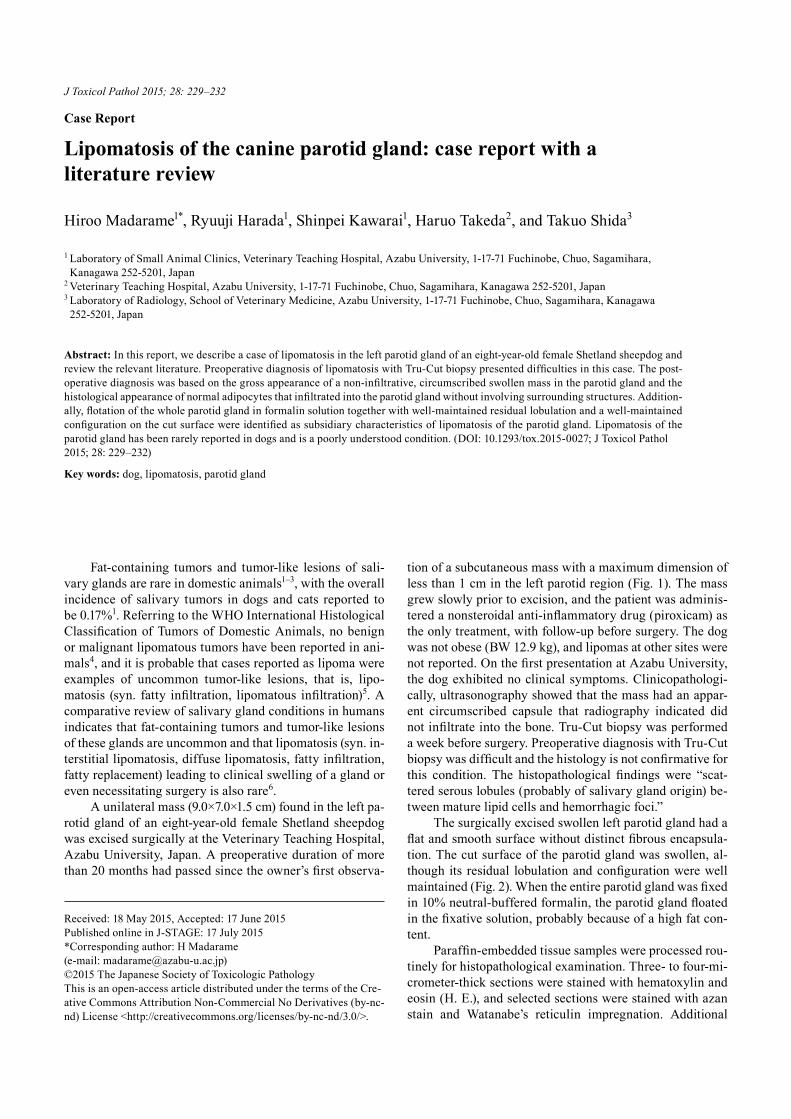
cupied the greater proportion of the parotid gland (Fig. 3), but little morphological change was observed in the remain-ing serous acini and ducts (Fig. 4). The periphery of the pa-rotid gland was smooth and covered with a thin collagen fiber capsule at intervals (Fig. 5). There was no infiltration of adipocytes to adjacent tissue. The swollen parotid gland was diagnosed as a lipomatosis.
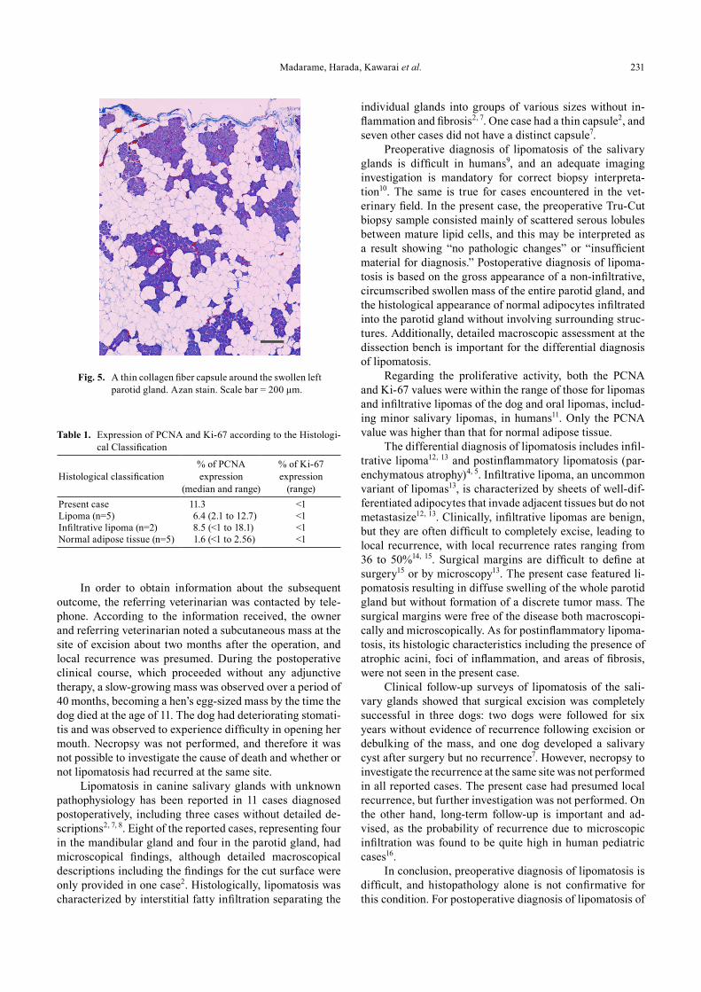
The immunohistochemically stained PCNA and Ki-67 slides were analyzed by calculating the average percent-age of positive cells in thirty randomly selected high-power fields for each marker for the present case, lipomas (n= 5), infiltrative lipomas (n=2), and normal adipose tissue (n=5). The mean percentage of immunohistochemical PCNA ex-pression was 11.3%, and that for Ki-67 was less than 1%. The data are summarized in Table 1.
Fig. 1. Dog with parotid infiltrative lipoma. The image was taken at first presentation at Azabu University. The dog was presented for surgery of a unilateral subcutaneous mass located near the base of the left ear.
Fig. 2. A surgically excised swollen left parotid gland. The surface is flat and smooth and without distinct encapsulation. The lobulation and configuration on the cut surface are well main-tained. Scale bar = 2 cm.
Fig. 3. Adipose tissue occupied the greater proportion of the parotid gland. H. E. Scale bar = 500 μm.
Fig. 4. Well-differentiated adipocytes diffusely infiltrated into and between serous lobules. There are few morphological changes in the remaining serous acini and ducts. H. E. Scale bar = 50 μm.
Madarame, Harada, Kawarai et al. 231
In order to obtain information about the subsequent outcome, the referring veterinarian was contacted by tele-phone. According to the information received, the owner and referring veterinarian noted a subcutaneous mass at the site of excision about two months after the operation, and local recurrence was presumed. During the postoperative clinical course, which proceeded without any adjunctive therapy, a slow-growing mass was observed over a period of 40 months, becoming a hen’s egg-sized mass by the time the dog died at the age of 11. The dog had deteriorating stomati-tis and was observed to experience difficulty in opening her mouth. Necropsy was not performed, and therefore it was not possible to investigate the cause of death and whether or not lipomatosis had recurred at the same site.
Lipomatosis in canine salivary glands with unknown pathophysiology has been reported in 11 cases diagnosed postoperatively, including three cases without detailed de-scriptions2, 7, 8. Eight of the reported cases, representing four in the mandibular gland and four in the parotid gland, had microscopical findings, although detailed macroscopical descriptions including the findings for the cut surface were only provided in one case2. Histologically, lipomatosis was characterized by interstitial fatty infiltration separating the
individual glands into groups of various sizes without in-flammation and fibrosis2, 7. One case had a thin capsule2, and seven other cases did not have a distinct capsule7.
Preoperative diagnosis of lipomatosis of the salivary glands is difficult in humans9, and an adequate imaging investigation is mandatory for correct biopsy interpreta-tion10. The same is true for cases encountered in the vet-erinary field. In the present case, the preoperative Tru-Cut biopsy sample consisted mainly of scattered serous lobules between mature lipid cells, and this may be interpreted as a result showing “no pathologic changes” or “insufficient material for diagnosis.” Postoperative diagnosis of lipoma-tosis is based on the gross appearance of a non-infiltrative, circumscribed swollen mass of the entire parotid gland, and the histological appearance of normal adipocytes infiltrated into the parotid gland without involving surrounding struc-tures. Additionally, detailed macroscopic assessment at the dissection bench is important for the differential diagnosis of lipomatosis.
Regarding the proliferative activity, both the PCNA and Ki-67 values were within the range of those for lipomas and infiltrative lipomas of the dog and oral lipomas, includ-ing minor salivary lipomas, in humans11. Only the PCNA value was higher than that for normal adipose tissue.
The differential diagnosis of lipomatosis includes infil-trative lipoma12, 13 and postinflammatory lipomatosis (par-enchymatous atrophy)4, 5. Infiltrative lipoma, an uncommon variant of lipomas13, is characterized by sheets of well-dif-ferentiated adipocytes that invade adjacent tissues but do not metastasize12, 13. Clinically, infiltrative lipomas are benign, but they are often difficult to completely excise, leading to local recurrence, with local recurrence rates ranging from 36 to 50%14, 15. Surgical margins are difficult to define at surgery15 or by microscopy13. The present case featured li-pomatosis resulting in diffuse swelling of the whole parotid gland but without formation of a discrete tumor mass. The surgical margins were free of the disease both macroscopi-cally and microscopically. As for postinflammatory lipoma-tosis, its histologic characteristics including the presence of atrophic acini, foci of inflammation, and areas of fibrosis, were not seen in the present case.
Clinical follow-up surveys of lipomatosis of the sali-vary glands showed that surgical excision was completely successful in three dogs: two dogs were followed for six years without evidence of recurrence following excision or debulking of the mass, and one dog developed a salivary cyst after surgery but no recurrence7. However, necropsy to investigate the recurrence at the same site was not performed in all reported cases. The present case had presumed local recurrence, but further investigation was not performed. On the other hand, long-term follow-up is important and ad-vised, as the probability of recurrence due to microscopic infiltration was found to be quite high in human pediatric cases16.
In conclusion, preoperative diagnosis of lipomatosis is difficult, and histopathology alone is not confirmative for this condition. For postoperative diagnosis of lipomatosis of
Fig. 5. A thin collagen fiber capsule around the swollen left parotid gland. Azan stain. Scale bar = 200 μm.
Table 1. Expression of PCNA and Ki-67 according to the Histologi-cal Classification
Histological classification% of PCNA expression
(median and range)
% of Ki-67 expression
(range)Present case 11.3 <1Lipoma (n=5) 6.4 (2.1 to 12.7) <1Infiltrative lipoma (n=2) 8.5 (<1 to 18.1) <1Normal adipose tissue (n=5) 1.6 (<1 to 2.56) <1
Canine Parotid Lipomatosis232
the salivary glands, detailed gross findings are important along with routine histopathology. As lipomatosis of the sal-ivary glands is uncommon, all reports of lipomatosis of the salivary glands in dogs are individual case reports or small case series, including this case report. Data from a larger number of cases with long-term follow-up are necessary to provide the basis for a critical understanding of the etiology and biological behaviors of lipomatosis in salivary glands. At present, lipomatosis is merely of clinical significance, but it should be kept in mind as an additional differential diagnosis in cases showing swelling of the salivary glands.
Acknowledgments: The authors thank Ms. Kanako Satoh and Ms. Megumi Mori for their expert technical assistance. We also thank Mr. Jonathan Lynch for proofreading the original manuscript. This research work was partially sup-ported by a Grant-in-Aid for Matching Fund Subsidy for Private Universities from the Promotion and Mutual Aid Corporation for Private Schools of Japan.
Disclosure of potential conflicts of interest: The authors declare that they have no competing interests.
References
1. Carberry CA, Flanders JA, Harvey HJ, and Ryan AM. Sali-vary gland tumors in dogs and cats: a literature and case review. J Am Anim Hosp Assoc. 24: 561–567. 1988.
2. Bindseil E, and Madsen JS. Lipomatosis causing tumour-like swelling of a mandibular salivary gland in a dog. Vet Rec. 140: 583–584. 1997. [Medline] [CrossRef]
3. Hammer A, Getzy D, Ogilvie G, Upton M, Klausner J, and Kisseberth WC. Salivary gland neoplasia in the dog and cat: survival times and prognostic factors. J Am Anim Hosp As-soc. 37: 478–482. 2001. [Medline] [CrossRef]
4. Head KW, Cullen JM, Dubielzig RR, Else RW, Misdorp W, Patnaik AK, Tateyama S, and van der Gaag I. Histological Classification of Salivary Gland Tumors of Domestic Ani-mals. In: World Health Organization International Histo-logical Classification of Tumors of Domestic Animals, Sec-ond Series, Volume X, Armed Forces Institute of Pathology, American Registry of Pathology, Washington, DC. 58–72.
2003. 5. Head KW, and Else RW. Tumors of the Salivary Glands. In:
Tumors in Domestic Animals, 4th ed. DJ Meuten (ed). Iowa State Press, Ames. 410–420. 2002.
6. Agaimy A. Fat-containing salivary gland tumors: a review. Head Neck Pathol. 7(Suppl 1): S90–S96. 2013. [Medline] [CrossRef]
7. Brown PJ, Lucke VM, Sozmen M, Whitbread TJ, and Wyatt JM. Lipomatous infiltration of the canine salivary gland. J Small Anim Pract. 38: 234–236. 1997. [Medline] [Cross-Ref]
8. Spangler WL, and Culbertson MR. Salivary gland disease in dogs and cats: 245 cases (1985-1988). J Am Vet Med As-soc. 198: 465–469. 1991. [Medline]
9. Mittari E, Aletra CH, Charalabopoulos K, and Batistatou A. Lipomatosis of the parotid gland in a 9-year-old male. Int J Clin Pract. 63: 335–336. 2009. [Medline] [CrossRef]
10. Ethunandan M, Vura G, Umar T, Anand R, Pratt CA, Macpherson DW, and Wilson AW. Lipomatous lesions of the parotid gland. J Oral Maxillofac Surg. 64: 1583–1586. 2006. [Medline] [CrossRef]
11. Fregnani ER, Pires FR, Falzoni R, Lopes MA, and Vargas PA. Lipomas of the oral cavity: clinical findings, histologi-cal classification and proliferative activity of 46 cases. Int J Oral Maxillofac Surg. 32: 49–53. 2003. [Medline] [Cross-Ref]
12. Hendrick MJ, Mahaffey EA, Moore FM, Vos JH, and Walder EJ. Tumors of Adipose Tissue. In: World Health Organization International Histological Classification of Tumors of Domestic Animals, Second Series, Volume II, Armed Forces Institute of Pathology, American Registry of Pathology, Washington, DC. 19–20. 1998.
13. Gross TL, Ihrke PJ, Walder EJ, and Affolter VK. Lipocytic tumors. In: Skin diseases of the dog and cat, 2nd ed. Black-well Publishing Company, Oxford. 766–777. 2005.
14. McChesney AE, Stephens LC, Lebel J, Snyder S, and Fer-guson HR. Infiltrative lipoma in dogs. Vet Pathol. 17: 316–322. 1980. [Medline]
15. Bergman PJ, Withrow SJ, Straw RC, and Powers BE. Infil-trative lipoma in dogs: 16 cases (1981-1992). J Am Vet Med Assoc. 205: 322–324. 1994. [Medline]
16. Sinha DD, Joshi M, Sharma C, and Chaturvedi V. Infantile congenital parotid lipomatosis: a rare case report. J Pediatr Surg. 40: e15–e16. 2005. [Medline] [CrossRef]