A CASE OF PRIMARY HODGKIN’S LYMPHOMA OF THE PAROTID GLAND
7
A CASE OF PRIMARY HODGKIN’S LYMPHOMA OF THE PAROTID GLAND . CASE REPORT AND DIFFERENTIAN DIAGNOSIS FROM KUTTNER’S SYNDROM Izzo P.*, Izzo S.*, Gabriele R.*, Di Cello P.*, Millarelli F.*, Pugliese F.*, Izzo L.* *Department of Surgery “P. Valdoni” Policlinico Umberto I, University of Rome “Sapienza” Corresponding authors Luciano Izzo Dipartimento di Chirurgia “P. Valdoni” Università degli Studi di Roma “La Sapienza” [email protected] Via G.Tomasi di Lampedusa 9,00144, Rome, Italy Abstract We report a rare case of primary Hodgkin’s lymphoma (HL) of the submandibular gland, with initially diagnosis of Kuttner’s Syndrom. A 48-years-old man was referred to our hospital foe evaluation of a submandibular mass. Although the initial Fine Needle Aspiration and subsequent cytology was highly suggestive for a cronic sialadenitis with lymphoid cells. After surgical gland removal we obtained a definitive diagnosis of Hodgkin’s lymphoma in submandibular gland a seat where the most common lymphoma tipe is B-cell. Key words: Submandibular gland, Hodgkin lymphoma, Kuttner syndrome.
Transcript of A CASE OF PRIMARY HODGKIN’S LYMPHOMA OF THE PAROTID GLAND
A CASE OF PRIMARY HODGKIN’S LYMPHOMA OF THE PAROTID GLAND . CASE REPORT AND DIFFERENTIAN DIAGNOSIS FROM KUTTNER’S SYNDROM
Izzo P.*, Izzo S.*, Gabriele R.*, Di Cello P.*, Millarelli F.*, Pugliese F.*, Izzo L.* *Department of Surgery “P. Valdoni” Policlinico Umberto I, University of Rome “Sapienza” Corresponding authors Luciano Izzo Dipartimento di Chirurgia “P. Valdoni” Università degli Studi di Roma “La Sapienza” [email protected] Via G.Tomasi di Lampedusa 9,00144, Rome, Italy
Abstract
We report a rare case of primary Hodgkin’s lymphoma (HL) of the submandibular
gland, with initially diagnosis of Kuttner’s Syndrom. A 48-years-old man was referred to our
hospital foe evaluation of a submandibular mass. Although the initial Fine Needle Aspiration
and subsequent cytology was highly suggestive for a cronic sialadenitis with lymphoid cells.
After surgical gland removal we obtained a definitive diagnosis of Hodgkin’s lymphoma in
submandibular gland a seat where the most common lymphoma tipe is B-cell.
Key words: Submandibular gland, Hodgkin lymphoma, Kuttner syndrome.

INTRODUCTION The submandibolar triangle is delimited among the anterior and posterior side of the digastric muscle and the inferior edge of the body of the mandibolar bone. To its inside there are the submandibolar gland, the ipoglossal nerve, lingual and marginal branch of the facial over that a varying number from 3 to 6 lymph nodes adjacent the gland. The greatest part of the primary malignant tumors that interest the submandibolar triangle, are lymphomas of the variety not-Hodgkin type B cells (38%) followed by the adenoideo-cystic (15,5%) carcinoma, mucoepidermoyd carcinoma (15,5%), Hodgkin lymphomas (7%), other varieties (24%)1. Clinically they go to differential diagnosis with other benign formations that interest the great salivary glands and with the chronic flogosis. The diagnostic certainty is given by a careful histological examination post-surgery that allows to make differential diagnosis in comparison to the simple FNAB that has relative importance in the definitive diagnosis not allowing clear hystological differentiation and underestimating cancer cellular presence. CASE REPORT A 48 year old man came to our observation with the presence of swollen right submandibular mass painless and not tender for about four months, with onset of the skin overlying the region free from alterations. The patient did not present at the time of clinical observation fever, weight loss, sweats or pruritus sine materia. The patient underwent an MRI scan with DP T2, T1, T2 SPIR and neck with and without contrast. The images showed the presence of a large mass in the right submandibular region. The tissue was meeting in inhomogeneous enhancement after intravenous infusion of paramagnetic contrast agent.
Fig. 1. X-ray image (MRI) of Kuttner tumor of the submaxillary right gland. Needle aspiration was performed by FNAB (fine needle aspiration) of the submandibular gland in question. The cytologic report described the presence of red blood cells and lymphocytes of varying size compatible with a chronic inflammation of the submandibular gland. We proceeded to surgical removal of the gland and two growths were observed with dimensions of about 3x6 cm and 4x2 cm adhering to the submandibular gland. The surgical specimen was thus sent to the pathologist for histological examination. The gland is presented with dimensions of 4 x 2.1 x 2 cm, with a nodular formation with smooth whitish in color, compact and elastic consistency of the cutting diameter of 3.6

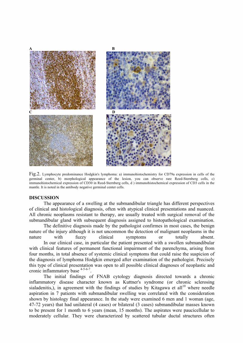
cm, the second formation appeared smooth gray color with a firm texture and with a diameter of 4 cm. Microscopically the 2 nodular growths showed, after immunohistochemical investigations, impaired neoplastic Hodgkin's lymphoma variant of "lymphocyte-dominated", but the remaining parenchyma was free of cancer and chronic inflammatory phenomena. The immunohistochemical investigations were performed on tissue sections of thickness between 3 and 5 mm using slides treated with polylysine, fixed in buffered formalin solution at 10% and then included in paraffin. The slides thus prepared were left for 24-48 hours in an oven thermostat at 37 ° C and endogenous peroxidase activity to prevent the sections were treated for 15-20 minutes with a 3% solution of hydrogen peroxide and washing in TBS buffer. The immunostains were subjected to a series of incubations with antibodies and margining. The slides thus treated were placed in a humid chamber and covered with sections with anti-CD30 (a marker of Reed-Steenberg plurinucleate cells), anti-CD3 (T lymphocyte marker), anti-CD79a (marker associated with B) and finally, anti-CD15 (marker of lymphoblastic cells may be an expression of non-Hodgkin's lymphoma with high-grade malignancy). After conducting various washes in distilled water and running water, microscopic histological examination both of the two neoplasms showed a picture of the reaction with complete immunoassay, which showed positivity for CD-30, CD79a and CD-3 and the presence Reed-Sternberg cells with accompanying T and B lymphocytes, while the negativity of the CD-15 has allowed us to exclude the non-Hodgkin's lymphoma with high-grade malignancy as well as the negative Cytokeratin ruled out the possibility that you could speak about metastatic localization (Fig. 2).
The diagnosis of lymphoma, suggested to perform a total body CT scan with contrast, which revealed no additional sites affected by cancer. The patient was sent to the Institute of Hematology, where he performed additional diagnostic tests that confirmed, given the unique location gland, the diagnosis of Hodgkin's disease stage 1, according to the Ann Arbor classification. Based on the staging, the patient was treated with a regimen of four cycles of chemotherapy performed with Adriamycin, Bleomycin, Vinblastine and Dacarbazine on a monthly basis, followed by radiation therapy (30 Gy) for four weeks of consolidation on the cervical lymph nodes and supraclavicular that led to complete remission of the disease the patient. Multidisciplinary follow-up was performed at 6, 12, 18 and 24-month with radiological controls, otorhinolaryngologist and hematology visit, that until today have shown no recurrence of disease.
A B
C D
Fig.2. Lymphocyte predominance Hodgkin's lymphoma: a) immunohistochemistry for CD79a expression in cells of the germinal center, b) morphological appearance of the lesion, you can observe rare Reed-Sternberg cells, c) immunohistochemical expression of CD30 in Reed-Sternberg cells, d ) immunohistochemical expression of CD3 cells in the mantle. It is noted in the antibody negative germinal center cells. DISCUSSION The appearance of a swelling at the submandibular triangle has different perspectives of clinical and histological diagnosis, often with atypical clinical presentations and nuanced. All chronic neoplasms resistant to therapy, are usually treated with surgical removal of the submandibular gland with subsequent diagnosis assigned to histopathological examination. The definitive diagnosis made by the pathologist confirmes in most cases, the benign nature of the injury although it is not uncommon the detection of malignant neoplasms in the nature with fuzzy clinical symptoms or totally absent. In our clinical case, in particular the patient presented with a swollen submandibular with clinical features of permanent functional impairment of the parenchyma, arising from four months, in total absence of systemic clinical symptoms that could raise the suspicion of the diagnosis of lymphoma Hodgkin emerged after examination of the pathologist. Precisely this type of clinical presentation was open to all possible clinical diagnoses of neoplastic and cronic inflammatory base 4-5-6-7. The initial findings of FNAB cytology diagnosis directed towards a chronic inflammatory disease character known as Kuttner's syndrome (or chronic sclerosing sialadenitis,), in agreement with the findings of studies by Kitagawa et all10 where needle aspiration in 7 patients with submandibular swelling was correlated with the consideration shown by histology final appearance. In the study were examined 6 men and 1 woman (age, 47-72 years) that had unilateral (4 cases) or bilateral (3 cases) submandibular masses known to be present for 1 month to 6 years (mean, 15 months). The aspirates were paucicellular to moderately cellular. They were characterized by scattered tubular ductal structures often

enveloped by collagen bundles or lymphoplasmacytic infiltrate, isolated fragments of fibrous stroma, a background rich in lymphoid cells, and paucity or absence of acini. Histologic examination of the excised submandibular glands revealed preserved lobular architecture, thickening of interlobular septa by sclerotic tissue, dense lymphoplasmacytic infiltrate, preservation of ducts with periductal fibrosis, and variable loss of acini. In combination with the clinical findings, the fine-needle aspiration cytologic findings can strongly suggest the diagnosis of Kuttner tumor and may obviate the need of surgical intervention.
Despite the report of our cytological finding leaded to a benign lesion, histology showed a parenchyma-free histological with features of a chronic inflammatory process, with a partial impairment of the gland, affected by intense lymphocytic infiltrate that, properly immunohystochimically colored, highlighted the presence of a type lymphoma Hodgkin's lymphocytic predominance in a place where the most frequently occurring type B lymphoma, non Hodgkin's. Even more, the variants of Hodgkin's lymphoma that expressed in our clinical case was predominantly lymphocytic and has a frequency of 23.8%. This variant is characterized by an abundant infiltration of T lymphocytes to medium-small sized CD 3 +, moreover with reactive nature, and only the presence of some tumor cells with the immunophenotype CD30 + and CD15-cells with Reed Steinberg. In addition the degree of impairment was located as not to allow the determination of the origin of the tumor as a primary concern of this or secondary involvement of the parenchyma by a neoplasm arising in a lymph node intraparenchymal. Only the completion of light microscopy with immunohistochemistry, has allowed the identification of a systemic neoplastic disease, still at a localized stage establishing an appropriate treatment that allowed the current patient survival free of disease. CONCLUSIONS
In our case report the patient had a swelling of the submandibular gland, which gave the initial cytologic of inflammation which imposed differential diagnosis with Kuttner's syndrome (a common disease of the submandibular gland), with the histiocytic tumor, sclerosing lymphoma, sarcoidosis and from other chronic inflammation of the salivary glands1-2-3-4. The diagnosis obtained by histology of surgical specimen was by surprise a lynfatic neoplastic disease, in particular a Hodgkin's lymphoma of lymphocyte predominance. Hodgkin's disease is an infrequent disease (7%)1 in the salivary glands, where it is most frequent a localization of non-Hodgkin's lymphoma, especially in B-cell variant. In our case, the preoperative use of the method of Aspirative Citology with FN was not diagnostic, pointing out only the reactive lymphocytic amount directing to an inflammatory disease, which in the case of locating the submandibular gland directed towards to Kuttner syndrome rather than to Hodgkin's lymphoma, a potentially systemic disease, with complete absence of a set of symptoms characteristic of the disease. Even the histological findings on surgical piece, despite having shown the presence of a frank malignancy, did not allow to determine the true origin of the lesion if starting from parenchyma or lymph nodes with secondary involvement of the gland intraglandular. Even more characteristic is the histological type of Hodgkin's type lymphoproliferative malignancy in a place where Literature reports more frequently the onset of Non-Hodgkin's lymphoma type. The element that allowed in our clinical case to rule out this syndrome was the presence of a lymphocytic predominance Hodgkin's lymphoma type (5-6%) instead of a non-

Hodgkin's lymphoma, but also the absence of a real impairment of the remaining sclerotic and inflammatory parenchyma despite the initial success of cytology, confirming the diagnostic limitations of FNAB for these locations, as cited in accord with Literature1.
BIBLIOGRAPHY
1. Izzo L, Casullo A, Caputo M, Costi U, Guerrisi A, Stasolla A, Basso L, Marini M, De Toma G.Space occupying lesions of parotid gland. Comparative diagnostic imaging and pathological analysis of echo color/power Doppler and of magnetic resonance imaging.Acta Otorhinolaryngol Ital. 2006 Jun;26(3):147-53.
2. Bialek EJ, Osmolski A, Karpinska G, et all. Us-appearance of a Kuttner tumour resembling a malignant lesion: US-histopathologic correlation. European Journal of Ultrasound 2001: 14: 167-80.
3. Ochoa ER, Harris NL, Pilch BZ. Marginal zone B-cell lynphoma of the salivary gland arising in chronic sclerosing sialadenitis (Kuttner tumor). Am J Surg Pathol;2001: 12: 1546-50.
4. Huang C, Damrose E, Bhuta S, Abemayor E. Kuttner tumor (Chronic Sclerosin Sialadenitis). Am J of Otolaryng, 2002: 23 (6): 394-397.
5. Lima I; Suellen A; Carneiro B et all. Hodgkin lymphoma as complication of primari Sjogren’s Sindrome. Mod Rheumathol. 2008: 18; 200-202.
6. Muneyuki M, Yuichi S, Andrei KJ et all. A case of primary Hodgkin’s lymphoma of the parotid gland. Auris Nasus Larynx; 2008 : 35: 440-442.
7. Satoshi K, Yoh Z, Kenichi H et all. Abundant IgG4-Positive plasma cell infiltration characterizes chronic sclerosing sialadenitis (Kuttner’s Tumor). Am J Surg. Pathol, 2005; 29: 783-791.
8. Wah C, Hunter KL, Yuen M. Lymphadenopathy of IgG4-related Sclerosing Disease. Am J Surg Pathol. 2008; 32 (5); 671-681.
9. Cheuk W, John KC. Kuttner tumor of the submandibular gland. Fine-Needle aspiration cytologic findings of seven cases. Am J Clin Pathol, 2002: 117: 103-108.
10. Kitagawa S; Zen Y; Harada K et all. Abundant IgG4-Positive Plasma Cell Infiltration Characterizes Chronic Sclerosing Sialadenitis (Kuttner's Tumor).The American Journal of Surgical Pathology: June 2005; 29 (6); 783-791