Cardioplegia at subnormothermia facilitates rapid ...
11
Cardioplegia at subnormothermia facilitates rapid functional resuscitation of hearts preserved in SOMAH for transplants Citation Lowalekar, Samar K, Patrick R Treanor, and Hemant S Thatte. 2014. “Cardioplegia at subnormothermia facilitates rapid functional resuscitation of hearts preserved in SOMAH for transplants.” Journal of Cardiothoracic Surgery 9 (1): 155. doi:10.1186/s13019-014-0155-z. http://dx.doi.org/10.1186/s13019-014-0155-z. Published Version doi:10.1186/s13019-014-0155-z Permanent link http://nrs.harvard.edu/urn-3:HUL.InstRepos:13347506 Terms of Use This article was downloaded from Harvard University’s DASH repository, and is made available under the terms and conditions applicable to Other Posted Material, as set forth at http:// nrs.harvard.edu/urn-3:HUL.InstRepos:dash.current.terms-of-use#LAA Share Your Story The Harvard community has made this article openly available. Please share how this access benefits you. Submit a story . Accessibility
Transcript of Cardioplegia at subnormothermia facilitates rapid ...

Cardioplegia at subnormothermia facilitates rapid functional resuscitation of hearts preserved in SOMAH for transplants
CitationLowalekar, Samar K, Patrick R Treanor, and Hemant S Thatte. 2014. “Cardioplegia at subnormothermia facilitates rapid functional resuscitation of hearts preserved in SOMAH for transplants.” Journal of Cardiothoracic Surgery 9 (1): 155. doi:10.1186/s13019-014-0155-z. http://dx.doi.org/10.1186/s13019-014-0155-z.
Published Versiondoi:10.1186/s13019-014-0155-z
Permanent linkhttp://nrs.harvard.edu/urn-3:HUL.InstRepos:13347506
Terms of UseThis article was downloaded from Harvard University’s DASH repository, and is made available under the terms and conditions applicable to Other Posted Material, as set forth at http://nrs.harvard.edu/urn-3:HUL.InstRepos:dash.current.terms-of-use#LAA
Share Your StoryThe Harvard community has made this article openly available.Please share how this access benefits you. Submit a story .
Accessibility

Lowalekar et al. Journal of Cardiothoracic Surgery 2014, 9:155http://www.cardiothoracicsurgery.org/content/9/1/155
RESEARCH ARTICLE Open Access
Cardioplegia at subnormothermia facilitates rapidfunctional resuscitation of hearts preserved inSOMAH for transplantsSamar K Lowalekar1,2,3, Patrick R Treanor3 and Hemant S Thatte1,2,3*
Abstract
Objectives: Hearts preserved ex vivo at 4°C undergo time-dependent irreversible injury due to extremehypothermia. Studies using novel organ preservative solution SOMAH, suggest that hearts are optimally‘preserved’ at subnormothermic temperature of 21°C. Present study evaluates relative efficacy of SOMAH‘cardioplegia’ at 4 and 21°C in preservation of optimum heart function after in vitro storage at subnormothermia.
Methods: Porcine hearts arrested with SOMAH cardioplegia at 4 or 21°C were stored in SOMAH for 5-hour at 21°C(n = 5). At the end of storage, the weight of hearts was recorded and biopsies taken for cardiac tissue high energyphosphate level measurements. The hearts were then attached to a reperfusion apparatus and biochemicalparameters including cardiac enzyme release and myocardial oxygen consumption and lactate production weredetermined in perfusate samples at regular intervals during ex vivo perfusion experiment. Functional evaluationof the hearts intraoperatively and ex vivo was performed by 2D echocardiography using trans-esophagealechocardiography probe.
Results: Post-storage heart weights were unaltered in both groups, while available high-energy phosphates (HEP)were greater in the 21°C group. Upon ex vivo reperfusion, coronary flow was significantly greater (p < 0.05) in 21°Cgroup. 2D echo revealed a greater cardiac output, fractional area change and ejection fraction in 21°C group thatwas not significantly different than the 4°C group. However, unlike 4°C hearts, 21°C hearts did not require inotropicintervention. Upon reperfusion, rate of cardiac enzyme release temporally resolved in 21°C group, but not in the4°C group. 21°C working hearts maintained their energy state during the experimental duration but not the 4°Cgroup; albeit, both groups demonstrated robust metabolism and function during this period.
Conclusions: Rapid metabolic switch, increased synthesis of HEP, decreased injury and optimal function providesevidence that hearts arrested at 21°C remain viably and functionally superior to those arrested at 4°C when storedin SOMAH at ambient temperature pre-transplant.
Ultramini-abstract: Cardioplegic arrest and preservation of hearts in SOMAH at ambient temperature efficientlyconserves metabolism and function in in vitro porcine model of heart transplant.
Keywords: Cardioplegia, Transplantation, Reperfusion injury, High energy phosphates
* Correspondence: [email protected] Surgery Division, Brigham and Women’s Hospital, VA BostonHealthcare System, 1400 V. F. W. Parkway, West Roxbury, Massachusetts02132 Boston, USA2Department of Surgery, Brigham and Women’s Hospital, VA BostonHealthcare System, Boston, Massachusetts, USAFull list of author information is available at the end of the article
© 2014 Lowalekar et al.; licensee BioMed Central Ltd. This is an Open Access article distributed under the terms of the CreativeCommons Attribution License (http://creativecommons.org/licenses/by/4.0), which permits unrestricted use, distribution, andreproduction in any medium, provided the original work is properly credited. The Creative Commons Public DomainDedication waiver (http://creativecommons.org/publicdomain/zero/1.0/) applies to the data made available in this article,unless otherwise stated.

Lowalekar et al. Journal of Cardiothoracic Surgery 2014, 9:155 Page 2 of 10http://www.cardiothoracicsurgery.org/content/9/1/155
BackgroundCardioplegia, a technique to stop the heart intraopera-tively, is an integral part of open-heart surgeries and isalso a fundamental requisite in the field of heart trans-plant. During surgery it is important for cardioplegiasolution to not only arrest the heart adequately but alsoprevent damage to cardiac tissue during and/or after infu-sion. Several strategies to attain this ideal situation havebeen attempted by altering cardioplegia temperature, orusing blood, crystalloid or blood-crystalloid mixture thathave produced equivocal results in their ability to arrestthe heart [1-6]. However, the meta-analysis of these stud-ies does not provide much evidence in favor of one or theother [7,8]. Nevertheless, of all these strategies by far, theuse of high K+ cardioplegia at extreme hypothermia(4°C) has become most widely accepted, especially inthe procurement of donor hearts for transplant [9].This is despite the growing awareness of the damagingeffects of extreme hypothermia on solid organs [10,11]and failure of presently used modalities in extracorporealpreservation of hearts to extend the ex vivo storage timebeyond 4–6 hours.Recent results from our laboratory using animal models
of heart transplant have demonstrated that hearts pre-served in SOMAH solution of our design, demonstrategreater accumulation of high energy phosphates (HEP) incomparison to the comparator solutions [12-16]. Further-more, hearts preserved in SOMAH for 5–24 hours andnot exposed to extreme hypothermia (4°C) demonstrate arobust maintenance of organ viability and function, andminimal injury upon reperfusion and reanimation [12-14].Additionally, these studies also demonstrate that heartsstored in SOMAH at 21°C outperform those preserved at4 and 13°C upon reanimation. However, in these investiga-tions SOMAH cardioplegia was delivered at 4°C for arrestprior to excision of donor hearts. Since preservation ofhearts in SOMAH at 21°C was superior than that at 4°C,the present study was undertaken to assess whether deliv-ery of high K+ SOMAH cardioplegia at 21°C compared tothat at 4°C would further augment and improve viabilityand energy metabolism, and also prevent injury in heartsstored ex vivo for 5 hours at 21°C, that were then reper-fused and reanimated for evaluation of cardiac function.
MethodsAnimal protocolHeart procurement surgery and cardioplegiaTen female Yorkshire swine (45–54 Kg) were used inthis comparative study. Hearts were divided into twogroups of either 4°C (n = 5) or 21°C cardioplegia (n = 5),as per the protocol approved by our Animal StudiesCommittee (Institutional Animal Care and Use Commit-tee). Hearts were extracted using mediastinal approach asdescribed [12]. The animals were bled from femoral vessels
to collect blood for ex vivo experiments, and aorta wasclamped when systolic pressure fell below 40 mmHg.1000 ml of SOMAH cardioplegia (SOMAH [12] modifiedby addition of 20 mM K+, final concentration), at 4 or 21°Cwas infused into the aortic root at a pressure of 75–100 mmHg at a flow rate of 300–400 ml/minute usingroller pump and pressure transducer (Myotherm Cardio-plegia System, Medtronics, Minneapolis, MN, USA) andthe data was recorded using iWorks system (Dover, NH,USA). After cardioplegic arrest, heart was dissected fromall attachments and rinsed with normal saline beforestoring in SOMAH for 5-hours at 21°C. Hearts were trans-ported to the lab within 15 minutes of excision.
Extracorporeal storage of heartHearts were placed in sterile zip-lock bags containing 2 Lof SOMAH in water-jacketed water bath at 21 ± 2°C. Thetemperature of preservation solution was checked regu-larly during the entire storage period. Hearts were main-tained in a non-contractile state by increasing SOMAH’splegia potential by supplementing the solution with20 mM K+ complemented by 37 mM Mg2+ [17,18], duringstorage. Hearts in each group were weighed prior to andafter 5-hour storage upon carefully emptying the heartchambers. Tissue punch biopsies (2X4 mm) were taken inthe lab from the posterior wall of LV, 15 minutes into(0 hour; control) and after 5 hour storage for HEP assays.
ATP and creatine phosphate assayATP and creatine phosphate (CP) were measured in tis-sue extracts as described [12,19]. In brief, tissue biopsieswere flash frozen and stored at −80°C; 20 mg of tissuewas suspended in 400 μl of 0.4 M ice-cold perchloricacid and homogenized twice for 30 seconds. Homogen-ate was centrifuged at 1970 g for 10 minutes at 0°C. Analiquot of supernatant was neutralized with equal volumeof ice-cold 0.4 M KHCO3 and centrifuged as above. Thesupernatant was stored at −80°C for ATP and CP mea-surements. The pellet was dissolved in equal volume of0.1 M NaOH, centrifuged and used for protein assay.ATP and CP were measured using a bioluminescentassay kit (Sigma-Aldrich and GloMax-Multi + DetectionSystem, Promega), according to the protocol provided bythe manufacturer.
Preparation of heart for ex vivo resuscitation andfunctional studiesAorta and pulmonary artery (PA) were separated. Aortawas cannulated (1/2-3/8 inch tubing connector) and cor-onaries were gently flushed with 100 ml of SOMAH inboth 4°C and 21°C cardioplegia groups at 40–50 mmHgpressure, carefully avoiding entry of air into the aorta.Pulmonary veins (PV) were separated and cannulatedwith 1/2-1/4 inch tubing connector. PA was cannulated

Lowalekar et al. Journal of Cardiothoracic Surgery 2014, 9:155 Page 3 of 10http://www.cardiothoracicsurgery.org/content/9/1/155
for sample collection while superior and inferior venacavas were ligated.
Preparation of blood for ex vivo studiesSystemically heparinized blood was collected intraopera-tively, leukodepleted (Pall Leukoguard filter) and storedat 4°C. Prior to experiments, perfusate was prepared byadjusting the hematocrit of blood to 20% using SOMAHsolution (1:1 ratio to reduce viscous strain on heart) andwarmed to 21°C. The perfusate, pH, glucose, K+, Ca2+
and HCO3− were adjusted for swine blood levels (7.5;100 mg/dl; 3.7, 1.38, and 32 mmol/l respectively), using10% dextrose, KCl, CaCl2 and NaHCO3, respectively, asrequired.
The SOMAH deviceA custom-built apparatus was used for extra-corporealreanimation of hearts (Figure 1, Additional file 1: Video).CDI monitor (Clinical Documentation Improvementmonitoring system 500, Terumo cardiovascular systemscorporation, Ann Arbor, MI), was used for real-timemonitoring of perfusate pH, temperature, pO2, pCO2, K
+
and HCO3−. These parameters were also analyzed in in-
flow/outflow samples using i-STAT analyzer (Abaxis Ltd,Union city, CA). Pressures and flows were recorded atvarious points in the circuits (Figure 2). A trans-esophageal
Figure 1 Circuit diagram of the SOMAH Device. A custom-built apparatu(green) visualizes antegrade coronary perfusion through aorta during initialthrough PV in the working state. In circuit 1, the perfusate is pumped frominto aorta for coronary perfusion. The return of perfusate to heart chamberheart to oxygenator-heat-exchanger system is collected in pre-load bag froadjusted by altering the height of pre-load bag. This circuit is diverted intocoronaries and returns to heart chamber through PA. The second componentafter-load chamber, from where the perfusate returns to heart chamber by grasystem for real-time monitoring of perfusate pH, temperature, pO2, pCO2, K
+ ancircuits and monitored using HMI software specifically written for SOMAH Devi
echocardiography (TEE) probe was used to assess thecontractile function of heart using the 2D-Echo sys-tem. 3-lead ECG was recorded during 2D Echo usingbrass crocodile leads immersed in the perfusate surround-ing the heart. Pressures and flow data was acquired andmonitored in real time using HMI software, specificallywritten for SOMAH Device (Comdel Inc, Wahpeton, ND).
Ex vivo functional studiesHearts were attached to SOMAH Device and perfusedthrough the aorta at 40–60 mmHg for 5 minutes, with1–1.5 liters of SOMAH at 21°C (pH 7.5), and then withperfusate, till pH, blood gases and electrolyte equilibriumwas established. The perfusate pH, glucose, K+, Ca2+,HCO3− were adjusted for swine blood levels as mentionedabove. Strong cardiac contractions were noted in bothgroups as the system temperature was gradually raised to37°C over a 30 minute period. Hemodynamic steady state(with respect to pH, blood gases, and electrolytes) wasachieved within 40 minutes. Total duration of the experi-mental perfusion was approximately 180 minutes. Heartswere perfused through aortic root (no workload) until thesystem temperature reached 37°C, after which PV perfu-sion (full workload) proceeded until end of experiment.Coronary blood flow was determined during the initialantegrade perfusion by the amount of perfusate flowing to
s was designed for extra-corporeal reanimation of hearts. The circuit 1reperfusion of hearts and circuit 2 (red), for perfusion of heartsheart chamber to oxygenator-heat-exchanger system and eventuallythrough PA completes the circuit. In circuit 2, blood pumped fromm where it drained into the PV by gravity. The pressures/flows aretwo components. First component is the part of perfusate that entersis formed by the perfusate that continues through aorta into thevity. A CDI monitor was incorporated beyond oxygenator-heat-exchangerd HCO3−. Pressures and flows were recorded at various points in the twoce.

Figure 2 Flow diagram of experimental design. Illustration shows the general experimental design of this study, starting from intraoperativecardioplegia for cardiac arrest to the end of ex vivo heart reperfusion experiment.
Lowalekar et al. Journal of Cardiothoracic Surgery 2014, 9:155 Page 4 of 10http://www.cardiothoracicsurgery.org/content/9/1/155
the heart through aorta per minute, and in the workingheart by the amount of perfusate collected from pulmon-ary artery (both cavas ligated) per minute. Electrocon-version (40-50 J) and/or epinephrine (1:50,000-1:100,000)were used if required [14-16]. Epicardial 2D Echo wasperformed using TEE probe for functional assessment at60 minutes (baseline) and at peak performance, approxi-mately 90 minutes after initiation of perfusate perfusion inthe two groups with hearts under full workload; and every30 minutes thereafter. Peak cardiac performance wasdefined by the maximum contractile activity observed by2D Echo. The data at peak performance was used forcomparisons between the two groups.
Enzyme assays and blood chemistryQuantitative levels of cardiac creatine kinase (CK), aspar-tate aminotransferase (AST), troponin-I (cTnI), lactateand gases (pO2/pCO2) were measured intra-operativelyand in SOMAH samples taken at 10 minute, 2-hour and
at end of 5-hour heart storage using Vetscan VS2 or iStat(Abaxis Ltd, Union City, CA). Inflow (aortic) and outflow(PA) samples were collected for enzyme assays andpost perfusion assessment of myocardial O2 consump-tion (MVO2) and lactate levels using Vetscan VS2 ori-Stat System, at 5 and 90 minutes for enzyme assays,and at 60 minutes (baseline) and 90 minutes (peak per-formance) for MVO2 and lactate, after start of perfusateperfusion with Vetscan or iStat. MVO2 was calculated asdescribed [20].
Epicardial echocardiographyA trans-esophageal (TEE) probe was used for 2D echoevaluation of cardiac function intra-operatively and ex vivousing the Acuson Cypress system (Acuson, MountainView, CA) and images were analyzed using Cypress viewersoftware provided with the system. Hearts were connectedto SOMAH Device and suspended in a chamber contain-ing 2 L of perfusate that covered 2/3rd surface of the heart.

Lowalekar et al. Journal of Cardiothoracic Surgery 2014, 9:155 Page 5 of 10http://www.cardiothoracicsurgery.org/content/9/1/155
ECG was recorded during entire course of the experimentand 2D Echo acquisition was begun approximately 45–60minute after perfusion, when good cardiac contractionswere observed, and repeated at 30-minute intervals. Probewas placed in direct contact with heart and angle of probeand direction of pulse were adjusted as to obtain short-axisand long-axis views for calculations of cardiac functionalparameters, and ventricular wall and septal thickness.
Statistical analysesEqual number of animals (n = 5), were assigned to 4°Cand 21°C cardioplegia groups for comparative analysis forbiochemical, hemodynamic and functional measurementsfrom each group. Statistical comparison for significantdifferences between the two groups was performed usingSigmaPlot software. Paired t-test was used for all com-parisons. P-value of <0.05 was considered significant. Allvalues are expressed as mean ± SEM. Flow diagram of theexperimental design is shown in Figure 2.The authors had full access to the data and take full
responsibility for its integrity. All authors have read andagreed to the manuscript as written. The funding agenciesdid not play any role in influencing data collection, extrac-tion and interpretation.
ResultsIntraoperative cardioplegiaCardiac arrest was dependent on the temperature ofcardioplegia and occurred within 10–15 seconds in 4°Cgroup and 20–25 seconds in the 21°C group, likely be-cause the hypothermic (4°C) component of cardiac arrestwas deliberately eliminated in the latter.
Gross morphology, heart weights and release of enzymesduring storageIrrespective of temperature of cardioplegia, all heartspresented normal gross morphology without any discol-oration. Hearts were pliable with no signs of hardeningor stiffness. Weights of the hearts during 5-hour storagein the two groups were not altered between pre and poststorage, demonstrating lack of storage-induced gross edema(not shown). A time dependent release of cardiac enzymeswas minimally apparent in both groups that were notsignificantly different (not shown).
Cardiac tissue HEP levels after arrest and during storageAs shown in Figure 3, concentration of ATP, CP andtotal high-energy phosphates (HEP), within 15 minutes(control) of cardiac arrest were significantly greater in 4°Cas compared to 21°C cardioplegia hearts (P < 0.001) pos-sibly due to the greater consumption of energy in 21°Cgroup hearts as they required approximately 10 secondslonger for total arrest. Both groups actively synthesizedCP and ATP during storage, Figure 3B. While the total
concentrations of HEP at the end of storage were signifi-cantly greater in 4°C hearts (P < 0.01), normalization ofvalues at 5 hour with respect to those at 0 hour dem-onstrated greater availability of HEP in 21°C hearts thanin 4°C hearts at the end of storage, Figure 3C.
Ex vivo cardiac functional studiesCoronary flow upon reperfusionCoronary flow through the aorta, upon initial antegradeperfusion at similar perfusion pressures, was signifi-cantly greater in 21°C hearts (P < 0.05) than the 4°C group(Table 1). The hearts in both groups demonstrated slowfour chamber contractions immediately upon initiation ofperfusion. Coronary flow decreased initially when systemtemperature was raised to 30°C, as the hearts started con-tracting vigorously. Both pressure and flow increased assystem temperature was raised to 37°C. Coronary flowwas highest in both groups at 37°C (Table 1).
Release of enzymes upon reperfusionRate of CK, AST and cTnI release increased in 4°Chearts during the perfusion period (Figure 4). Both CKand cTnI release increased significantly with the time ofperfusion, but the AST did not. In contrast, there was atemporal decrease in release of these three enzymes inthe 21°C hearts during the same period (Figure 4). How-ever, release of CK and cTnI, but not the AST, was signifi-cantly greater by the 21°C hearts upon initiation ofperfusion than by the 4°C hearts.
Metabolism in reperfused heartsConsistent with our previous observations [14-16], therewas a rapid switch in metabolism from anaerobic toaerobic upon reperfusion in both 4°C and 21°C cardio-plegia groups, demonstrated by an increase in oxygenconsumption and reversal of lactate ratios at peak perform-ance, 90 minutes into reperfusion (Figures 5A and B). Oxy-gen extraction, lactate production and utilization, reacheda steady state in both groups at peak performance thatwere not significantly different. However, upon reperfusion,21°C group hearts demonstrated robust synthesis of HEPin the working hearts. In contrast, production was attenu-ated in 4°C hearts and the HEP continued to decline dur-ing course of the experiment. Ratios (post perfusion/preperfusion) of ATP, CP and total HEP were 1.10, 1.97 and1.17, respectively in 21°C hearts which were significantlygreater (p < 0.01) than ratios of 0.47, 0.32 and 0.38 obser-ved in the 4°C hearts at end of experiment, when postperfusion biopsies were taken for HEP assays.
Functional revival upon reperfusionImmediate spontaneous activity of both the atria andventricles was apparent upon commencement of reper-fusion in both groups. With increased temperature, force
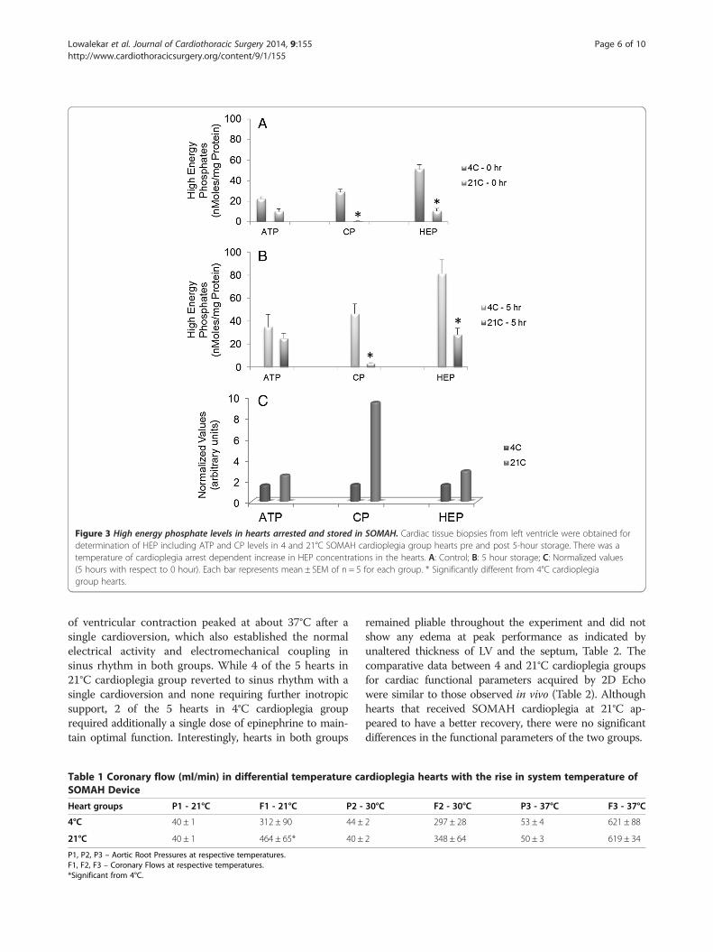
Figure 3 High energy phosphate levels in hearts arrested and stored in SOMAH. Cardiac tissue biopsies from left ventricle were obtained fordetermination of HEP including ATP and CP levels in 4 and 21°C SOMAH cardioplegia group hearts pre and post 5-hour storage. There was atemperature of cardioplegia arrest dependent increase in HEP concentrations in the hearts. A: Control; B: 5 hour storage; C: Normalized values(5 hours with respect to 0 hour). Each bar represents mean ± SEM of n = 5 for each group. * Significantly different from 4°C cardioplegiagroup hearts.
Lowalekar et al. Journal of Cardiothoracic Surgery 2014, 9:155 Page 6 of 10http://www.cardiothoracicsurgery.org/content/9/1/155
of ventricular contraction peaked at about 37°C after asingle cardioversion, which also established the normalelectrical activity and electromechanical coupling insinus rhythm in both groups. While 4 of the 5 hearts in21°C cardioplegia group reverted to sinus rhythm with asingle cardioversion and none requiring further inotropicsupport, 2 of the 5 hearts in 4°C cardioplegia grouprequired additionally a single dose of epinephrine to main-tain optimal function. Interestingly, hearts in both groups
Table 1 Coronary flow (ml/min) in differential temperature caSOMAH Device
Heart groups P1 - 21°C F1 - 21°C P2 -
4°C 40 ± 1 312 ± 90 44 ±
21°C 40 ± 1 464 ± 65* 40 ±
P1, P2, P3 – Aortic Root Pressures at respective temperatures.F1, F2, F3 – Coronary Flows at respective temperatures.*Significant from 4°C.
remained pliable throughout the experiment and did notshow any edema at peak performance as indicated byunaltered thickness of LV and the septum, Table 2. Thecomparative data between 4 and 21°C cardioplegia groupsfor cardiac functional parameters acquired by 2D Echowere similar to those observed in vivo (Table 2). Althoughhearts that received SOMAH cardioplegia at 21°C ap-peared to have a better recovery, there were no significantdifferences in the functional parameters of the two groups.
rdioplegia hearts with the rise in system temperature of
30°C F2 - 30°C P3 - 37°C F3 - 37°C
2 297 ± 28 53 ± 4 621 ± 88
2 348 ± 64 50 ± 3 619 ± 34

Figure 4 Release of creatine kinase and cardiac troponin-I upon reperfusion. CK (A), AST (B) and cTnI (C) levels were determined in perfusate,5 min and 90 min (peak performance) after start of reperfusion of 4 and 21°C cardioplegia hearts; n = 5 for each SOMAH group. *Significant changefrom 5 minutes (p <0.05); † Significantly different from 4°C cardioplegia group hearts at similar time point.
Lowalekar et al. Journal of Cardiothoracic Surgery 2014, 9:155 Page 7 of 10http://www.cardiothoracicsurgery.org/content/9/1/155
DiscussionWe have designed and evaluated SOMAH, a synergisticextracellular organ storage solution that includes variousenergy substrates, metabolic modulators, free radicalscavengers and anti-oxidants, ammonia chelators andnitric oxide synthase substrates, intra and extracellular
Figure 5 Cardiac metabolism in working hearts. Myocardial O2 Consumphearts. MVO2 and Lactate ratios were determined from the differences in theBaseline = 60 min after reperfusion, at hemodynamic steady state; 90 mineach group.
H+ chelators and physiological concentration of calcium,components that help prevent edema, ionic imbalanceand energy depletion [12]. SOMAH provides a favorableenvironment and cellular support during ex-vivo storageof hearts, lungs and abdominal organs, and helps attenuatereperfusion injury upon reanimation [13-16,21]. Promising
tion (A) and Lactate Ratio (B) upon perfusion of 4 and 21°C cardioplegiarespective parameters in the outflow and inflow perfusate samples.= at peak performance. Each bar represents mean ± SEM of n = 5 for

Table 2 Cardiac functional parameters during surgery and at peak performance upon extracorporeal reperfusion in 4°Cand 21°C SOMAH cardioplegia group hearts
Parameters In vivo 4°C hearts 21°C hearts
LV ant wall thickness (cm) 1.48 ± 0.07 1.55 ± 0.06 1.51 ± 0.04
Septal thickness (cm) 1.43 ± 0.12 1.61 ± 0.04 1.49 ± 0.07
Heart rate 98 ± 12 100 ± 10 110 ± 10
LV systolic pressure (mm Hg) 116 ± 8 100 ± 20 110 ± 10
LV diastolic pressure (mm Hg) 68 ± 7 40 ± 10 55 ± 10
Left atrial pressure (mm Hg) <10 0-5 0-5
Cardiac output 3300 ± 400 2500 ± 300 2640 ± 250
Fractional area change (%) >40 42 ± 13 51 ± 4
Ejection fraction (%) 65 ± 5 58 ± 4 66 ± 5
Stroke volume (ml) 29 ± 4 25 ± 2 24 ± 3
Lowalekar et al. Journal of Cardiothoracic Surgery 2014, 9:155 Page 8 of 10http://www.cardiothoracicsurgery.org/content/9/1/155
results with organ preservation encouraged us to un-dertake this study to evaluate SOMAH as a cardioplegiasolution.Recent results from our laboratory show that extra-
corporeal preservation of hearts for transplant at 21°C(ambient temperatures) in SOMAH is ideal for the high-est promotion of synthesis and accumulation of HEP inheart tissue and preservation of ideal function [13-16].Based on these results and on the reported advantagesof crystalloid solution [4,22] and tepid temperatures forcardioplegia [15], the present study was undertaken to eva-luate whether using crystalloid SOMAH at 21°C insteadof standard temperature of 4°C for cardioplegia couldfurther improve the quality of hearts preserved at ambienttemperature in SOMAH, and their eventual reanimationin vitro into optimal function.Myocardial edema has been reported in hearts arrested
at perfusion pressures of cardioplegia as low as 50 mmHg[23]. However, in present and past studies we haveconsistently used a crystalloid SOMAH cardioplegiainfusion pressure of 100 mmHg irrespective of cardio-plegia temperature, but edema was not seen in any ofthe hearts [14-16]. SOMAH cardioplegia provides allthe advantages of blood cardioplegia, in terms of protec-tion from cardiac edema and provision of substrates forenergy metabolism, and also provides clear surgical field.Additionally, provision of physiological concentrationsof calcium in SOMAH solution, also prevents the likeli-hood of myocardial damage due to ‘calcium paradox’[6,24]. Furthermore, the disadvantages of blood such asthe presence of leukocytes and platelets, culpable in reper-fusion injury [25], are also averted. In contrast, the othercrystalloid solutions (Celsior and UWS), when used forcardioplegia in our recent studies did not prevent lossof high-energy phosphates in the stored hearts, edemaupon reperfusion and potential high K+ mediated calciumoverload and stiffness that resulted in non-functioninghearts [14-16].
In this study, HEP were conserved in 4°C heartsbecause of rapid arrest, as a result total concentration ofHEP was also greater in these hearts at the end of stor-age. In contrast, despite K+ concentration (20 mM) beingequal in the two groups, the 21°C hearts took longerfor total arrest because of absence of the hypothermiccomponent, resulting in depletion of HEP. Both groupssynthesized HEP during storage in SOMAH, however, thefunctional availability of HEP was greater in 21°C heartsthan the 4°C hearts at the end of 5 hour (Figure 3). Simi-larly, upon reperfusion, 21°C hearts continued to synthesizeHEP to meet the demands of the working heart, unlike the4°C hearts. At peak performance the available HEP in 21°Chearts was significantly greater than 4°C hearts, and contin-ued to be so during course of the experiments. In contrast,4°C hearts were unable to synthesize HEP to keep withthe energy demands, thus HEP continued to decrease inthe working hearts. These results are in agreement withour previous observations that unlike in hearts preservedin SOMAH at ambient temperatures, exposure to severehypothermia leads to attenuation of HEP synthesis uponreperfusion in these hearts [13].Antegrade perfusion was significantly lower in the 4°C
heart than in 21°C hearts, and remained diminished,even at higher perfusion pressures until the system tem-perature stabilized at 37°C (Table 1). It is plausible thatsudden shock of encountering 4°C cardioplegia by thenormothermic beating heart leads to profound vasocon-striction that does not resolve during storage and onlydoes so upon initiation of reperfusion and raising oftemperature to 37°C, and potentially because of activerelease of vasodilators nitric oxide and prostacyclins [12].Increased vasodilation, greater coronary vascular patencyand a favorable metabolic status provides for rapid nour-ishment and H+ washout, resulting in robust synthesis ofHEP and swift recovery of function in the 21°C hearts.These hearts reverted to sinus rhythm with a single car-dioversion and rapidly attained cardiac and hemodynamic

Lowalekar et al. Journal of Cardiothoracic Surgery 2014, 9:155 Page 9 of 10http://www.cardiothoracicsurgery.org/content/9/1/155
parameters approaching in vivo range (Table 2), not requir-ing any inotropic support. On the other hand, 4°C heartsdemonstrated strong contraction only when warmed to37°C, some of the hearts requiring additional electrover-sion and/or inotropic intervention, albeit ten times lessthan that reported in human hearts in vitro [26] to main-tain cardiac output.Release of cardiac enzymes was observed in both the
groups upon reperfusion. An important mechanism ofrelease of enzymes from the cardiomyocyte is by cytosolleakage during intracellular vesicular trafficking and in-corporation (such as vesicles harboring glucose transpor-ters; GLUT) into cell membrane, by a HEP dependentprocess, in response to external stimuli like insulin and in-creased metabolic demands in working hearts [27]. There-fore, release of cellular enzymes can occur even in absenceof actual damage to the cardiomyocytes (enzyme paradox).Initial burst of enzyme release in 21°C cardioplegia groupupon initiation of reperfusion likely resulted from greateravailability of HEP at the end of storage for vesiculartransport as the metabolic demands were ramped up withincrease in system temperature and cardiac contractility(Figure 3). However, upon reaching metabolic steady stateany further release of enzyme was temporally attenuated.In contrast, in the 4°C hearts, the rate of release of en-zymes increased with time as more HEP became availablefor these functions. Even though the present data does notdifferentiate between progressions of enzyme release andneeds further investigation, the fact, that the cardiacfunctional parameters in both groups were similar and ap-proach the physiological values observed in vivo (Table 2)indicate that the releases of enzymes by the SOMAHhearts is a marker of metabolism rather than tissueinjury and hence these hearts would perform well upontransplant.Certain limitations were inherent to this study. While
the present findings indicate a certain advantage of usingSOMAH at 21°C for clinical cardioplegia either intraoper-atively or during heart procurement for transplantation, inthe present preliminary study we have performed only amodest number of experiments in each group and used2D echocardiography for the functional evaluation ofreperfused hearts. While this provided convincing evi-dence in favor of 21°C cardioplegia, further studies usingPressure-Volume loops acquisition and analysis for precisedetermination and interpretation of the significant bio-physiological effects of SOMAH on extracorporeal cardiacreperfusion under varying conditions, are required.
ConclusionEfficient HEP metabolism, decreased rate of enzymerelease, and diminished requirement for stimulatory inter-ventions suggest that use of SOMAH for cardioplegia andstorage at 21°C optimally protects the heart function and
thus may positively influence post operative outcomesupon transplant. Extrapolating ex vivo 5 hour preservationas analogous to cross clamp time, use of SOMAH cardio-plegia at ambient temperatures during open heart surgery,where the cross clamp times generally do not exceed3 hours (average 60–90 minutes); where the hearts areperiodically flushed with 500 ml of fresh cardioplegiaevery 20 to 30 minutes during cross clamp, may signifi-cantly improve post surgical outcomes. However, furtherstudies are needed to translate this inferential hypothesisinto a clinical reality.
Additional file
Additional file 1: Somah Device - Representative of a Working Heart.
Abbreviations2D Echo: 2 Dimensional echocardiography; AST: Aspartate aminotransferase;ATP: Adenosine tri-phosphate; CK: Creatine kinase; CP: Creatine phosphate;cTnI: Troponin-I; ECG: Electrocardiograph; HEP: High energy phosphate;MVO2: Myocardial O2 consumption; PA: Pulmonary artery; PV: Pulmonaryvein; TEE: Trans-esophageal echocardiography; UWS: University of WisconsinSolution.
Competing interestsThe authors declare that they have no competing interests.
Authors’ contributionsSKL and HST designed the experiments and analyzed the data. SKL acquiredthe 2D Echo data intraoperatively and ex vivo, conducted the experimentsand wrote the manuscript. HST performed the heart procurement surgeries,helped in overall conduct of experiments and preparation of the manuscript.PRT assisted in infusion of cardioplegia, set up of ex vivo perfusion apparatusand in conduct of experiments. All authors read and approved the finalmanuscript.
AcknowledgementsWe thank Peter Hirsch, MS, for his assistance during pre and post surgery inthe OR. We gratefully acknowledge the assistance of Diane Ghera, BS, andher staff at the VABHS animal care facility. We thank Aditi Thatte for herencouragement and support. We greatly appreciate Susan Greely, LouiseConnelly, Michael Charpak and Dr. Frances Achee for their continuingadministrative support.
SupportThis work was supported by VA Merit Review grant (Dr. Thatte), Departmentof Veterans Affairs, Office of Research and Development, Washington DC,and an unrestricted gift from Somahlution (Jupiter, Fl) to Boston VA ResearchInstitute for research (Dr. Thatte).
Author details1Cardiothoracic Surgery Division, Brigham and Women’s Hospital, VA BostonHealthcare System, 1400 V. F. W. Parkway, West Roxbury, Massachusetts02132 Boston, USA. 2Department of Surgery, Brigham and Women’s Hospital,VA Boston Healthcare System, Boston, Massachusetts, USA. 3Harvard MedicalSchool, Brigham and Women’s Hospital, VA Boston Healthcare System,Boston, Massachusetts, USA.
Received: 12 May 2014 Accepted: 25 August 2014
References1. Hosenpud JD, Bennett LE, Keck BM, Fiol B, Boucek MM, Novick RJ: The
registry of the international society for heart and lung transplantation:fifteenth official report – 1998. J Heart Lung Transplant 1998, 17:656–668.

Lowalekar et al. Journal of Cardiothoracic Surgery 2014, 9:155 Page 10 of 10http://www.cardiothoracicsurgery.org/content/9/1/155
2. Cohen G, Borger MA, Weisel RD, Rao V: Intraoperative myocardialprotection: current trends and future prospects. Ann Thorac Surg 1999,68:1995–2001.
3. Fazel S, Yau TM: Myocardial protection in reoperative coronary arterybypass grafting: toward decreasing morbidity and mortality. J Card Surg2004, 19:291–295.
4. Ovrum E, Tangen G, Tollofsrud S, Oystese R, Ringdal ML, Istad R: Cold bloodversus cold crystalloid cardioplegia: a prospective randomized study of345 aortic valve patients. Eu A Cardio-thorac Surg 2010, 38:745–749.
5. Luciani GB, Forni A, Rigatelli G, Chiominto B, Cardaioli P, Mazzucco A,Faggian G: Myocardial protection in heart transplantation using bloodcardioplegia: 12-year outcome of a prospective randomized trial.J Heart Lung Transplant 2011, 30:29–36.
6. Yamamoto H, Yamamoto F: Myocardial protection in cardiac surgery: ahistorical review from the beginning to the current topics. Gen ThoracCardiovasc Surg 2013, 61:485–496.
7. Fan Y, Zhang AM, Xiao YB, Weng YG, Hetzer R: Warm versus coldcardioplegia for heart surgery: a meta analysis. Eur J Cardio-thorac Surg2010, 37:912–919.
8. Sa MP, Rueda FG, Ferraz PE, Chalegre ST, Vasconcelos FP, Lima RC: Is thereany difference between blood and crystalloid cardioplegia formyocardial protection during cardiac surgery? A meta-analysis of 5576patients from 36 randomized trials. Eu A Cardio-thorac Surg 2012,27(6):535–546.
9. White CW, Ali A, Hasanally D, Xiang B, Li Y, Mundt P, Lytwyn M, Colah S,Klein J, Ravandi A, Arora RC, Lee TW, Hryshko L, Large S, Tian G, Freed DH:A cardioprotective preservation strategy employing ex vivo heartperfusion facilitates successful transplant of donor hearts aftercardiocirculatory death. J Heart Lung Transplant 2013, 32:734–743.
10. Belzer FO, Southard JH: Principles of solid-organ preservation by coldstorage. Transplantation 1988, 45(4):673–676.
11. Stefanovich P, Ezzel RM, Sheehan SJ, Tompkins RG, Yarmush ML, Toner M:Effects of hypothermia on the function, membrane integrity, andcytoskeletal structure of hepatocytes. Cryobiology 1995, 32:389–403.
12. Thatte HS, Rousou L, Hussaini BE, Lu XG, Treanor PR, Khuri SF: Developmentand evaluation of a novel solution, Somah, for the procurement andpreservation of beating and non-beating donor hearts for transplantation.Circulation 2009, 120:1704–1713.
13. Lowalekar SK, Lu XG, Thatte HS: Further evaluation of somah: long-termpreservation, temperature effect and prevention of ischemia-reperfusioninjury in rat hearts harvested after cardiocirculatory death. Transplant Proc2013, 45(9):3192–3197.
14. Lowalekar SK, Cao H, Lu XG, Treanor R, Thatte HS: Subnormothermicpreservation in SOMAH: a novel approach for enhanced functionalresuscitation of donor hearts for transplant. Am J Transplant 2014.doi:10.1111/ajt.12846.
15. Lowalekar SK, Cao H, Lu XG, Treanor PR, Thatte HS: Hearts preserved inSomah at sub-normothermia demonstrate rapid functional restorationand are less likely to develop heart failure upon transplantation.Abstract. J Heart Lung Transplant 2014, 33(4):S176.
16. Lowalekar SK, Cao H, Lu XG, Treanor PR, Thatte HS: Sub-normothermicpreservation of donor hearts for transplantation using a novel solution,SOMAH: a comparative pre-clinical study. J Heart Lung Transplant 2014,33(9):963–970.
17. Fukuhiro Y, Wowk M, Ou R, Rosenfeldt F, Pepe S: Cardioplegic strategiesfor calcium control: low Ca2+, high Mg2+, citrate, or Na+/H + exchangeinhibitor HOE-642. Circulation 2000, 102(19–3):III319–III325.
18. Osaki S, Ishino K, Kotani Y, Honjo O, Suezawa T, Kanki K, Sano S:Resuscitation of non-beating donor hearts using continuous myocardialperfusion: the importance of controlled initial reperfusion. Ann Thorac Surg2006, 81:2167–2171.
19. Bessho M, Ohsuzu F, Yanagida S, Sakata N, Aosaki N, Tajima T, Nakamura H:Differential extractability of creatine phosphate and ATP from cardiacmuscle with ethanol and perchloric acid solution. Anal Biochem 1991,192:117–124.
20. Klabunde R: Cardiac function. In Cardiovascular Physiology Concepts.Baltimore, MD USA: Lippincott Williams & Wilkins; 2011:84–88.
21. Louis AV, Hemphill C, Schipper D, Qu N, Stavoe K, Penick K, Thatte HS,Khalpey Z: Extended preservation of lungs at subnormothermia with anovel organ storage solution “SOMAH”: salvage, reconditioning andfunctional evaluation. Abstract. J Heart Lung Transplant 2014, 33(4):S71.
22. Charniot JC, Bonnefont-Rousselot D, Albertini JP, Dever S, Vignat N, Nataf P,Pavie A, Monsuea JJ, Delattre J, Artigou JY: Oxidative stress implicationafter prolonged storage donor heart with blood versus crystalloidcardioplegia and reperfusion versus static storage. J Sur Res 2009,160:308–314.
23. Mehlhorn U, Geissler HJ, Laine GA, Allen SJ: Myocardial fluid balance.Eur J Cardiothorac Surg 2001, 20:1220–1230.
24. Piper HM: The calcium paradox revisited: an artifact of great heuristicvalue. Cardiovasc Res 2000, 45:123–127.
25. Han S, Huang W, Liu Y, Pan S, Feng Z, Li S: Does leukocyte-depleted bloodcardioplegia reduce myocardial reperfusion injury in cardiac surgery?A systematic review and meta-analysis. Perfusion 2013, 28(6):474–483.
26. Hill AJ, Laske TG, Coles JA, Sigg DC, Skadsberg ND, Vincent SA, Soule CL,Gallagher BA, Iaizzo PA: In vitro studies in human hearts. Am Thorac Surg2005, 79:168–177.
27. Ferrera R, Benhabbouche S, Bopassa JC, Li B: One hour reperfusion isenough to assess function and infarct size with TTC staining inLangendorff rat model. Cardiovasc Drugs Ther 2009, 23:327–331.
doi:10.1186/s13019-014-0155-zCite this article as: Lowalekar et al.: Cardioplegia at subnormothermiafacilitates rapid functional resuscitation of hearts preserved in SOMAHfor transplants. Journal of Cardiothoracic Surgery 2014 9:155.
Submit your next manuscript to BioMed Centraland take full advantage of:
• Convenient online submission
• Thorough peer review
• No space constraints or color figure charges
• Immediate publication on acceptance
• Inclusion in PubMed, CAS, Scopus and Google Scholar
• Research which is freely available for redistribution
Submit your manuscript at www.biomedcentral.com/submit