CARDIAC LYMPH AND LYMPHATICS - PubMed Central Canada
17
CARDIAC LYMPH AND LYMPHATICS EXPERIMENTAL OBSERVATIONS AND CLINICAL SIGNIFICANCE S. R. ULLAL M.S. F.R.C.S. Associate Professor of Surgery, Kasturba Medical College, and Cardiothoracic Surgeon, Government Wenlock Hospital, Mangalore, India. SCIENTIFIC EXPERIMENTATION WAS John Hunter's way of life, and no- where is its value better illustrated than in the field of cardiac surgery, which has seen phenomenal advances thanks to experimental work in the laboratory. In an oft-quoted letter to his good friend Edward Jenner in 1775 John Hunter wrote these famous words-'Why think? Why not try the experiment?' It is therefore appropriate that this lecture should deal with experimental work on a subject that was of great interest to both John and William Hunter-namely, the lymphatics, or 'absorbents', to use the Hunterian term. Since lymph originates from the intercellular fluid, it is a step closer to the cells than blood. Cardiac lymph may therefore be expected to reflect closely the changes occurring in heart muscle in health and in disease. Unfortunately, this subject has been a long-neglected one, and it is only in recent years that there has been an appreciation of the pos- sible significance of the cardiac lymph and lymphatics in heart disease. The cardiac lymphatics have been shown to be affected in rheumatic heart disease'. Recent experimental work has suggested that lymphatic obstruction may produce functional as well as pathological changes in the heart, some not unlike those seen in rheumatic and other affections of the heart2' 3. The story of the discovery of the cardiac lymphatics is practically as old as that of the discovery of the lymphatic system itself. On 30th October 1651 Olaus Rudbeck, of Sweden, observed for the first time lymphatic 'glands' receiving afferent vessels from the heart in a dog. In 1692 Nuck used mercurial injections to demonstrate the lymphatics in the dead heart-a method which fell into disuse until nearly a century later, when William Hunter and his assistant Cruikshank in England, and Mascagni in Italy, employed this technique to give the first detailed account of the superficial cardiac lymphatics, with relevant illustrations. The method of injecting coloured substances into the myocardium of the living heart was introduced by Albrecht in 1887. Using this technique Hunterian Lecture delivered on 6th July 1972 (Ann. Roy. Coll. Surg. Engl. 1972, vol. 51) 282
Transcript of CARDIAC LYMPH AND LYMPHATICS - PubMed Central Canada
CARDIAC LYMPH AND LYMPHATICS
EXPERIMENTAL OBSERVATIONS AND CLINICAL SIGNIFICANCE
S. R. ULLAL M.S. F.R.C.S.
Associate Professor of Surgery, Kasturba Medical College, and Cardiothoracic Surgeon,Government Wenlock Hospital, Mangalore, India.
SCIENTIFIC EXPERIMENTATION WAS John Hunter's way of life, and no-where is its value better illustrated than in the field of cardiac surgery,which has seen phenomenal advances thanks to experimental work inthe laboratory. In an oft-quoted letter to his good friend Edward Jennerin 1775 John Hunter wrote these famous words-'Why think? Why nottry the experiment?' It is therefore appropriate that this lecture shoulddeal with experimental work on a subject that was of great interest toboth John and William Hunter-namely, the lymphatics, or 'absorbents',to use the Hunterian term.
Since lymph originates from the intercellular fluid, it is a step closerto the cells than blood. Cardiac lymph may therefore be expected toreflect closely the changes occurring in heart muscle in health and indisease. Unfortunately, this subject has been a long-neglected one, andit is only in recent years that there has been an appreciation of the pos-sible significance of the cardiac lymph and lymphatics in heart disease.The cardiac lymphatics have been shown to be affected in rheumaticheart disease'. Recent experimental work has suggested that lymphaticobstruction may produce functional as well as pathological changes inthe heart, some not unlike those seen in rheumatic and other affectionsof the heart2' 3.The story of the discovery of the cardiac lymphatics is practically as
old as that of the discovery of the lymphatic system itself. On 30thOctober 1651 Olaus Rudbeck, of Sweden, observed for the first timelymphatic 'glands' receiving afferent vessels from the heart in a dog. In1692 Nuck used mercurial injections to demonstrate the lymphatics inthe dead heart-a method which fell into disuse until nearly a centurylater, when William Hunter and his assistant Cruikshank in England,and Mascagni in Italy, employed this technique to give the first detailedaccount of the superficial cardiac lymphatics, with relevant illustrations.The method of injecting coloured substances into the myocardium of
the living heart was introduced by Albrecht in 1887. Using this techniqueHunterian Lecture delivered on 6th July 1972
(Ann. Roy. Coll. Surg. Engl. 1972, vol. 51)
282

CARDIAC LYMPH AND LYMPHATICS
Aagaard, in 1924, was able to demonstrate the subepicardial, myocardial,and subendocardial lymphatic plexuses. The most exhaustive study of themorphology of the cardiac lymphatics was not made until 1939, whenPatek4 established beyond doubt the presence of intercommunicatinglymph capillary networks in the subendocardial, myocardial, and sub-epicardial layers of the ventricles. He was unable, however, todemonstrate lymphatics in the atrial muscle or atrioventricular valves. Ayear later Drinker and his colleagues5 succeeded in cannulating a lymph-atic vessel leaving the heart in a dog and studied the flow rate andpartial composition of cardiac lymph. Nothing more was attempteduntil the late fifties, when Miller and his colleagues2' 3 in Chicago pro-vided the stimulus for further work on this subject. They producedchronic lymphatic obstruction of the dog heart and reported markedpathological changes in the myocardium and endocardium in con-sequence. Their suggestion that lymphatic obstruction may be ofsignificance in the aetiology of certain cardiac disorders focused theattention of clinicians and research workers on the subject of cardiaclymph and lymphatics. Since then anatomical studies of the cardiaclymphatics have been carried out with hydrogen peroxide' 7 and dyeinjection techniques8' 9 10 on the beating dog heart.The present study was undertaken at the Institute of Medical Sciences,
Pacific Medical Center, San Francisco. Its main purpose was to evaluatethe use of cardiac lymph as a parameter of myocardial behaviour andto assess the functional and pathological changes in the heart followinglymphatic obstruction. To achieve this a preliminary study of the an-atomy of the cardiac lymphatic drainage into the mediastinum, as wellas a baseline study of the flow characteristics and composition of normalcardiac lymph, was essential. Thus the project was divided into fivesections: (A) anatomical studies of the lymph drainage of the heart;(B) the flow and composition of cardiac lymph; (C) the effect of anoxiaon cardiac lymph; (D) cardiac lymph during extracorporeal circulation;and (E) functional and pathological changes in the heart followingchronic cardiac lymphatic obstruction.
Lymph drainage of the heartMethods. Anatomical studies were conducted on 35 anaesthetized adult mon-
grel dogs. The heart was exposed through a left anterolateral thoracotomy in thefourth intercostal space. The pericardium was opened and small amounts of Evansblue (T 1824) were injected at multiple sites into the myocardium of all the fourchambers through a 27-gauge needle. Care was taken to avoid contamination ofthe pericardial cavity by the dye. Ten dogs were kililed and a detailed and meti-culous mediastinal dissection performed to outline the extracardiac course ofthe lymphatics. In some cases attempts were made to demonstrate lymphaticsin the mitral and tricuspid valve leaflets in the beating heart, employing thetechnique described by Miller et al.3 Tissue specimens from the myocardiumand valves were processed for microscopical examination.
283

S. R. ULLAL
Results. A dense network of subepicardial lymphatics was demon-strated immediately after dye injection. These converged on to largervessels, supplied with valves, which coursed alongside the coronaryvessels in the atrioventricular sulcus and terminated in the left tracheo-bronchial node. The efferents from this node, usually two or three inniumber, ran diagonally upwards across the trachea and behind theinnominate artery to enter the 'cardiac node' situated between thesuperior vena cava and the innominate artery (Fig. 1). Efferents fromthis lymph node ran upwards towards the neck to join the right lymph-atic duct.Though this was the pattern in the majority of the dogs, anatomical
2 Fig. 1. Schematic diagram of1< ^ lymphatic drainage pathways in
the dog heart. (1) Tracheobron-chial lymph node. (2) Cardiacnode. (3) Right lymphatic duct.(4) Thoracic duct. (5) Posterior
mediastinal node.
variations were found in nearly a third of the animals. Quite frequentlya couple of efferent vessels from the tracheobronchial node enteredsmall paratracheal nodes at the apex of the chest, sometimes completelybypassing the cardiac node. In a few cases several small vessels wereseen communicating directly with the thoracic duct. The frequentpresence of such anatomical variations in lymph drainage makes itessential, in all experiments involving total cardiac lymph obstruc-tion or collection of all the cardiac lymph, for the lymphatic pathwaysto be studied first in each animal. Otherwise some of the cardiac lymphmay escape analysis or lymph originating elsewhere may be included.
In 4 dogs it was possible to demonstrate the presence of fine lymph-atic vessels in the wall of the atria, the main difficulty, as with the valves,
284
CARDIAC LYMPH AND LYMPHATICS
being the injection of dye into these thin structures in a beating heart.Very delicate lymphatic vessels were also demonstrated on the atrialsurfaces of the mitral and tricuspid leaflets.
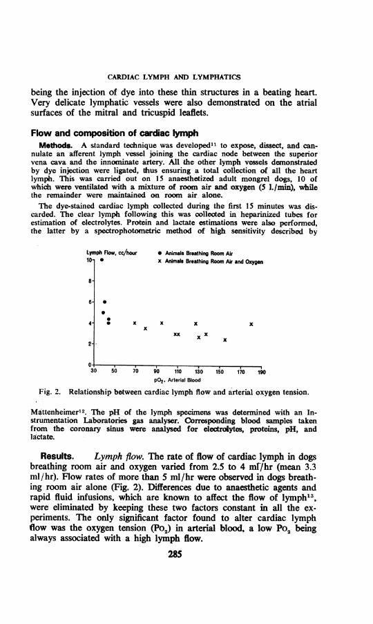
Flow and composition of cardiac lymphMethods. A standard technique was developed"' to expose, dissect, and can-
nulate an afferent lymph vessel joining the cardiac node between the superiorvena cava and the innominate artery. All the other lymph vessels demonstratedby dye injection were ligated, thus ensuring a total collection of all the heartlymph. This was carried out on 15 anaesthetized adult mongrel dogs, 10 ofwhich were ventilated with a mixture of room air and oxygen (5 l./min), whilethe remainder were maintained on roon air alone.The dye-stained cardiac lymph collected during the first 15 minutes was dis-
carded. The clear lymph following this was oollected in heparinized tubes forestimation of electrolytes. Protein and lactate estimations were also perfonned,the latter by a spectrophotometric method of high sensitivity described by
Lymph Flow, cc/hour * Animals Breathing Room Air10- * x Animals Breathing Room Air and Oxygn
8-
6- -
4- * x x x xx
xx x xx2-
030 50 70 90 110 130 150 170 190
P02, Arterial Blood
Fig. 2. Relationship between cardiac lymph flow and arterial oxygen tension.
MOttenheimer 2. The pH of the lymph specimens was determined with an In-strumentation Laboratories gas analyser. Corresponding blood samples takenfrom the coronary sinus were analysed for electrolytes, proteins, pH, andlactate.
Results. Lymph flow. The rate of flow of cardiac lymph in dogsbreathing room air and oxygen varied from 2.5 to 4 ml/hr (mean 3.3ml/hr). Flow rates of more than 5 ml/hr were observed in dogs breath-ing room air alone (Fig. 2). Differences due to anaesthetic agents andrapid fluid infusions, which are known to affect the flow of lymph'3,were eliminated by keeping these two factors constant in all the ex-periments. The only significant factor found to alter cardiac lymphflow was the oxygen tension (Po2) in arterial blood, a low Po2 beingalways associated with a high lymph flow.
285

S. RF. ULLAL
Electrolytes and proteins. The average chloride and sodium valuestended to be higher and the potassium values lower in cardiac lymphthan in coronary sinus blood. All the protein fractions were present incardiac lymph but in different proportions from those in plasma. Thealbumin:globulin ratio was consistently higher in cardiac lymph thanin plasma.pH. In all well-oxygenated dogs the pH of cardiac lymph was con-
sistently on the alkaline side (8.0 or higher).Lactate. The mean lactate concentrations in cardiac lymph were
significantly higher than in coronary sinus blood (Fig. 3). Pyruvateestimations were not carried out owing to the technical difficulty ofcollecting the quantities of lymph needed for such tests. Besides, recent
Lactates, mg %60 * Lymph
* Blood
50
40-
30-
20-
10-
Room Air Only Room Air and Oxygen Room Ar Only Room Air and OxygenCoronary Sinus Blood - Systemic Venous Blood
Fig 3. Lactate concentrations in cardiac lymph and blood.
work14 has shown that the lactate level alone serves as a good indica-tor of oxygen deficit and that neither the pyruvate level nor the lactate:pyruvate ratio is more reliable.
Effect of anoxia on cardiac lymphBlood is one step away from the tissue cells, with the interstitial
fluid and lymph intervening. For this reason it is believed that thevalidity of blood lactate and pyruvate levels as true indicators of theintracellular state of oxygenation is open to doubt15. The same argumenthas been advanced against relying on the electrolyte levels in theblood as reflecting the electrolyte state in the myocardiuml6. The con-sistently higher level of lactate found in cardiac lymph than in coronarysinus blood suggests that lymph may perhaps provide a more sensitive
286

CARDIAC LYMPH AND LYMPHATICS
indicator of changes in myocardial oxygenation. To test this hypothesisa set of experiments were devised to study the changes in cardiac lymphresulting from oxygen lack.
Methods. Cardiac lymphatic cannulation was carried out on 15 anaesthetizeddogs and the first hour's collection of lymph was used to determine baseline flowrates and lactate concentrations. Generalized anoxia was then produced in 10 ofthe dogs by stopping the respirator and clamping the endotracheal tube. As soonas cardiac arrest occurred the dog was resuscitated by oxygenation, cardiacmassage, and correction of acid-base problems. The cardiac lymph was collectedfor another 2-3 hours and analysed for lactate. Corresponding samples of bloodfrom the coronary sinus were also analysed. The arterial blood gases weremonitored throughout the experiments.
In 2 dogs generalized hypoxia was induced by ventilation with a mixture of10% oxygen and 90% nitrogen. Cardiac arrest did not occur in either.In 3 others the left anterior descending coronary artery was occluded for an
hour, producing localized myocardial anoxia. The occlusion was released after thisperiod and both lymph and coronary sinus blood analysed for lactate duringthe next 2-3 hours.
Results. The most noticeable change was in the flow rate and colourof the cardiac lymph. Both generalized hypoxia and anoxia produceda sharp rise in the lymph flow rate. This was noticeable within 5 minutesof the anoxic episode and continued for 1 hours, after which itstarted to decline slowly. Even 3 hours after the anoxic episode thelymph flow rate was still higher than the pre-anoxia level. The cardiaclymph also became grossly blood-stained in the dogs subjected to anoxia,and the blood-staining persisted during the next 3 hours right up to theend of the experiments. The blood-staining was either absent or veryfaint in the dogs subjected to generalized hypoxia.
In the dogs with occlusion of the anterior descending coronary arterythe lymph remained clear and the flow rate practically stable duringthe period of occlusion. Immediately after restoration of coronary per-fusion the lymph became grossly blood-stained and the flow rate rosesteeply, both the changes persisting during the rest of the experiment.The lactate concentration increased sharply in both cardiac lymph
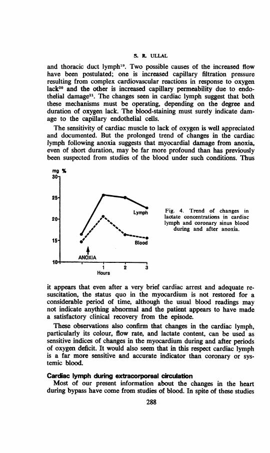
and coronary sinus blood during the first hour after anoxia, the lymphshowing the higher readings. The blood value thereafter decreased,reaching baseline level within 1-2 hours. The pattern of change incardiac lymph was markedly different; the lymph lactate level remainedhigh for 2 hours or more before starting to decline slowly, and re-mained considerably above the baseline level in all the dogs at the endof the experiments (Fig. 4).
Discussion. Hypoxia and anoxia have been reported to produceincreased flow and protein concentrations in cervical17, pulmonary'8,
287
S. L ULLAL
and thoracic duct lymphl9. Two possible causes of the increased flowhave been postulated; one is increased capillary filtration pressureresulting from complex cardiovascular reactions in response to oxygenlack20 and the other is increased capillary permeability due to endo-thelial damage21. The changes seen in cardiac lymph suggest that boththese mechanisms must be operating, depending on the degree andduration of oxygen lack. The blood-staining must surely indicate dam-age to the capillary endothelial cells.The sensitivity of cardiac muscle to lack of oxygen is well appreciated
and documented. But the prolonged trend of changes in the cardiaclymph following anoxia suggests that myocardial damage from anoxia,even of short duration, may be far more profound than has previouslybeen suspected from studies of the blood under such conditions. Thus
mg %30
25
Lymph Fig. 4. Trend of changes in20 / , * lactate concentrations in cardiac
/, t, lymph and coronary sinus blood/w' ', during and after anoxia.
15 Blood
ANOXIA10 I
1 2 3Hours
it appears that even after a very brief cardiac arrest and adequate re-suscitation, the status quo in the myocardium is not restored for aconsiderable period of time, although the usual blood readings maynot indicate anything abnormal and the patient appears to have madea satisfactory clinical recovery from the episode.These observations also confirm that changes in the cardiac lymph,
particularly its colour, flow rate, and lactate content, can be used assensitive indices of changes in the myocardium during and after periodsof oxygen deficit. It would also seem that in this respect cardiac lymphis a far more sensitive and accurate indicator than coronary or sys-temic blood.
Cardiac lymph during extracorporeal circulationMost of our present information about the changes in the heart
during bypass have come from studies of blood. In spite of these studies
288

CARDIAC LYMPH AND LYMPHATICS
there are still several practical problems which remain unsolved or aboutwhich there is no unanimity of opinion. Thus anoxic cardiac arrestis considered by many to be detrimental to myocardial function22,while others are impressed with the apparent clinical safety of this pro-cedure23. Differences of opinion exist about the use of coronary per-fusion during aortic occlusion, on the role of hypothermia duringextracorporial circulation, and on whether the heart should be fibril-lated or left beating during bypass and surgery. As cardiac lymph hadproved to be a very sensitive indicator of myocardial damage due toanoxia, it was logical to use it to study the changes in the myocardiumduring bypass and the above-mentioned procedures.
Methods. Adult mongrel dogs weighing 21-28 kg were anaesthetized anda stable flow of cardiac lymph was established. The external jugular vein andthe external iliac vessels were cannulated for bypass (Fig. 5). This reducedmanipulations in the vicinity of the heart to a bare minimum, preventing dis-
'4 Venous Infusion & Sampling
Fig. 5. Schematic diagram showing the experimental set-up for cardio-pulmonary bypass and lymph cannulation.
lodgement of the lymph cannula. The superior and inferior venae cavae wereencircled with tapes and the azygos vein ligated before lymph cannulation, sothat total bypass could be achieved whenever indicated. A cannula insertedthrough the left atrial appendage served as a left heart vent. To avoid disturbingthe lymph cannula the coronary perfusion cannula was introduced into the supra-valvular portion of the ascending aorta through an opening made in the leftsubclavian artery in the chest. The aortic lumen could be occluded around this
289

S. R. ULLAL
cannula by means of a snare. Catheters were inserted into the femoral veinand artery for infusions, blood gas sampling, and continuous monitoring ofblood pressure.A medium-sized Temptrol disposable bubble oxygenator equipped with De-
Bakey roller pumps was used for cardiopulmonary bypass. The circuit wasprimed with 1,500 ml of fresh acid-citrate-dextrose dog blood and 1,000 ml of5% dextrose in 0.2% normal saline. An average flow rate of 2 1./mi at an aver-age pressure of 120 mm Hg was maintained. The coronary perfusion line wasdriven by a separate pump with a calculated flow of 250 ml/min at an averagepressure of 200 mm Hg.
Results. Group 1 (8 dogs). These were subjected to partial andtotal bypass at normal temperature for -1 hour, with the heart beating.The flow rate of cardiac lymph tended to decrease slightly during totalbypass, but otherwise remained stable. The lymph remained clear dur-ing and after bypass. There was a slow but steady rise in lymph lactateconcentration during bypass, the maximum values being seen im-mediately after the end of perfusion, following which there was a slowdecline back to baseline values. Moderate respiratory alkalosis waspresent in all the dogs during bypass.Group 2 (5 dogs). After initiation of bypass the heart was fibrillated
electrically for 30 minutes, with decompression of the left heart by aleft atrial vent. The heart was then defibrillated with AC shock andbypass continued for another 30 minutes. The cardiac lymph flow de-creased by more than 50% during fibrillation. It remained clear forthe first 10-15 minutes, after which it became faintly blood-stained andremained so to the end of the experiment. After defibrillation and re-storation of the heart beat the lymph flow increased sharply, exceedingthe baseline flow rate, and remained so even after bypass was discon-tinued. The steep and persistent rise in the lactate concentrations ofcardiac lymph during and after fibrillation is shown in Figure 6.Group 3 (4 dogs). After initiation of total bypass at normal tem-
perature and left atrial venting the ascending aorta was clamped for15 minutes. After the release of the clamp and restoration of heartaction bypass was continued for another 30 minutes. Aortic occlusioncoincided with a sharp decrease in cardiac lymph flow. Release of theocclusion was immediately followed by a copious flow of lymph whichwas franily blood-stained, both these changes persisting till afterbypass and the end of the experiment. The highest and sharpest rise oflactate concentrations in lymph in the whole series was recorded in thesedogs (Fig. 7).Group 4 (4 dogs). The same experiment was performed as in Group
3 but with the blood cooled to 30°C during the period of aortic oc-clusion. Though the lymph flow changes were essentially similar tothose seen in Group 3, the blood-staining was very faint. The lactate
290

CARDIAC LYMPH AND LYMPHATICS
concentrations rose slowly and steadily during bypass but remainedat significantly lower levels than in Group 3 (Fig. 7).
50-
40 , Lymph
CD 30 _ '
, 20-
101_
I I A A I ABypass Fib. Defib. Off 1st Hr. 2nd Hr.
Bypass
Fig. 6. Lactate concentrations in cardiac lymph and blood during bypassand fibrillation (Fib.).
80
70
60
501Fig. 7. Changes in lac-tate content of cardiaclymph and blood duringand after anoxic cardiac ar-rest at normothermia and
moderate hypothermia.
I IBypass Aorta Aorta Off 1st Hr.
Clamped Open Bypass2nd Hr.
Group 5 (4 dogs). The aorta was occluded for 15 minutes at normaltemperature with simultaneous coronary perfusion. No significantchanges were noticed in the flow rate of cardiac lymph, which re-
291
E, 40
-j
30 _
20 F
101-
0--
nl1

S. R. ULLAL
mained perfectly clear throughout the experiment. The lactateconcentrations in lymph were the lowest recorded in the series (Fig. 8).
Discussion. The cardiac lymph changes during anoxic cardiac ar-rest clearly point to severe damage to the capillary endothelium in themyocardium, as well as to pronounced anaerobic metabolism. The pro-gression and persistence of these changes must be regarded as indicatinglong-lasting and perhaps permanent damage to the myocardium, eventhough the anoxic arrest period was only 15 minutes, which is reportedto be within the 'safety range' for dogs23. The so-called safety of a sur-gical procedure is often assessed from the ability of the heart to take overafter bypass or the immediate survival rate. This is evidently an un-satisfactory method of assessment, as such 'safe' procedures may wellproduce long-lasting changes in the myocardium which cannot be
50
40
30 LymphE
_ Blood
820 -
10
O A A A A A ABypass Cor. Perfusion Off 1st Hr. 2nd Hr.
On Off BypassFig. 8. Influence of coronary perfusion during bypass on lactate content of
cardiac lymph and blood.
appreciated by the usual battery of tests, most of them conducted onblood. Ultrastructural studies24 have demonstrated cellular damage inthe myocardium after 20 minutes of anoxic arrest during clinical open-heart surgery. The apparent clinical safety of anoxic arrest, as reportedby some, is not supported by our findings in cardiac lymph. The studyalso indicates that a beating heart is preferable to fibrillation duringbypass surgery and that moderate hypothermia does confer a significant,though not complete, degree of protection against anoxic damage tothe heart muscle.
Changes in the heart following chronic cardiac lymphatic obstructionMethods. In 22 adult dogs the cardiac lymph drainage was studied and all
the lymph vessels leaving the heart were divided between ligatures. The cardiac
292

CARDIAC LYMPH AND LYMPHATICS
node was dissected out and excised after ligation and division of all the ves-sels entering and leaving it. The dogs were killed at intervals of 1-36 weeks andhlistopathological studies of cardiac tissue carried out. In 14 of these dogs haemo-dynamic data were collected before lymphatic obstruction and again at thesecond operation. Six dogs were subjected to 'sham operations'-thoracotomyand injection, dissection, and identification of the cardiac lymphatics, whichwere left intact. The haemodynamic data included heart rate, arterial bloodpressure, left atrial pressure, left ventricular systolic and diastolic pressures,and the maximum rate of rise of intraventricular pressure (dp/dt). Cardiac out-put was measured by the Cardiogreen dilution technique at three different levelsof left atrial pressure. The stroke volume and the left ventricular work (g/m)were calculated and ventricular function curves constructed.
60
40
20
0.
BBEEcm
-j
80
60-
40-
20
0
IxIx
lo
.
0-10 WEEKS
x'x
10-20 WEEKS
/
.20-30 WEEKS
x = Before*=After
Control
x
/
Control
x/
ControlI
0 10 20 10 20 10 20L.A. Pressure (cm H20)
Fig. 9. Changes in ventricular function curves following cardiac lymphaticobstruction.
Haemodynamic changes. The ventricular function curves were de-pressed in all dogs after cardiac lymphatic obstruction, as well as inthose controls which were evaluated more than 10 weeks after the in-itial operation. The control dogs evaluated within the first 10 weeksshowed no significant changes in their ventricular function curves(Fig. 9).
293
X.,
X
X

S. R. ULLAL
A significant reduction in maximum dp/dt was also observed in themajority of animals with cardiac lymphatic obstruction (Fig. 10) andwas definitely more marked than in the controls.No significant changes in the electrocardiogram or serum enzyme
levels were noticed, as reported by Foldi and Braun25.Discussion. The reduction in maximum dp/dt must be assessed with
caution in view of the various factors which are known to affect suchreadings2" and which could not be controlled during these experiments.Functional abnormalities have been observed in the postoperative periodafter heart transplantation, and interruption of lymph pathways has
x= Controls *=With Lymph Obstruction60
0-o
R 40 -
.c2 Fig. 10. Changes in maximumg 30 - dp/dt in dogs with cardiac
* lymphatic obstruction.
c"- 20-0)
10 -xx ex
o 10 20 30 40Weeks
been one of the reasons suggested27. The changes in ventricular functioncurves in the first 10 weeks after lymphatic obstruction would appearto support this theory.
Pathological changes. No evidence of cardiac enlargement, peri-cardial effusion, or gross cardiac failure was found in any of the dogs.Surprisingly, no collateral or aberrant lymph pathways could bedemonstrated. The presence of lymphovenous communications was sus-pected, but could not be confirmed. In the majority of dogs withlong-standing cardiac lymphatic obstruction the atrioventricular valveleaflets were found to be thickened and irregular, with a waxy appear-ance. In vivo injection of dye into these leaflets demonstrated a farmore extensive network of lymph capillaries than is found normally
294

CARDIAC LYMPH AND LYMPHATICS
(Fig. 11). However, gross endocardial thickening and subendocardialhaemorrhage, as reported by Miller et al.2, were not observed in thisseries.
Microscopically, significant changes appeared to be confined to theatrioventricular valves and, to a slight extent, the endocardium. Theseconsisted of dilated interstitial spaces and lymph capillaries during thefirst 3 weeks. In more chronic lymph obstruction, thickening of thevalve leaflets became apparent and was quite marked after 12 weeks.The thickening was caused by loose mesenchymal connective tissue and
..w.~~~~~~~~~~~~~~~~e..... 0........
~~~~~~~~~~~~~~~~~~~~~~~~~~~~~~~ .. ...
'MMAN.~~~~~~~~~~~~~~~~~~~~~~~~~~~~~~~~~~~~~~~~~~~~~~~~~~~~~~~~~~~~~~~~~~~~~~
Fig. 11. Preparation showing the extensive network of lymph vessels on theatrial surface of the mitral valve in a dog with lymphatic obstruction of
the heart.
also by accumulation of an amorphous 'myxoid-like' substance whichwas faintly basophilic on staining with haematoxylin and eosin andstained positively with Alcian blue (Fig. 12). The same changes, alongwith some degree of fibrosis, were sometimes found in the subendocardiallayer, though these were patchy in distribution and far less pronouncedthan in the valve leaflets.
Discussion. Occasional deposits of myxoid are known to occur inold dogs, but never to the extent observed in this series. Moreover,these dogs were young and no such lesions were present in the controls.
295

S. R. ULLAL
These findings may have some clinical significance. Myxomatous de-generation of the mitral valve has been recognized as a cause of isolatedacquired mitral regurgitation28. Similar changes have been shown tocause mitral insufficiency and cardiac failure in dogs, with increasedsusceptibility to endocarditis and to rupture of the chordae tendineae29.Electron microscopical studies have demonstrated that rheumatic heartlesions are associated with deposits of acid mucopolysaccharides, similarto the 'myxoid' seen in these experiments, in the endocardium.
?.P~ ~ ~
~~~~~~~~. ........
Fig. 12. Microscopic appearance of a section through the mitral valveleaflet, showing deposition of 'myxoid' and connective tissue following
lymphatic obstruction.
The absence of collateral lymph pathways was puzzling. Lymphaticregeneration occurs after transection or ligation of lymph vessels30.After resection of a length of lymphatic, however, lymph flow can beestablished only through the opening up of preexisting collateral chan-nels31. It is possible that in the heart a substantial amount of lymphis drained through lymphovenous communications, which are known tostart functioning under the stress of increased lymph pressure orvolume32. This may also explain the absence of gross and progressivechanges in the myocardium and endocardium even after long-standing
296

CARDIAC LYMPH AND LYMPHATICS
lymphatic obstruction. A thorough investigation of lymphovenous com-munications in the heart is long overdue.
ConclusionsCardiac lymph appears to provide a very sensitive indicator of anoxic
changes in the myocardium. Such changes can be appreciated in cardiaclymph even when the usual blood studies present an apparently satis-factory picture. The use of blood to assess the metabolic state of themyocardium has several drawbacks. The blood stream is a step awayfrom the heart cells. Any arterial or venous blood sample used foranalysis during bypass will necessarily reflect the state of whole bodyperfusion rather than local changes in the myocardium alone. Evensamplings of coronary venous return are unsatisfactory, as they mayreflect regional differences in the heart muscle, depending on the siteof sampling. Cardiac lymph is virtually free of these defects.
It also appears that lymphatic obstruction impairs myocardial ac-tivity by producing changes in structure and function. It is tempting tosurmise that some form of lymphatic dysfunction may be of significancein rheumatic and other forms of heart disease. Unfortunately, ob-servations on laboratory animals need not always be applicable to man,and we have yet to study the normal lymphatic drainage of the heartof man in detail, for want of a satisfactory method of demonstratingthese lymphatics post mortem. Now that the attention of research work-ers has been drawn to this field, no doubt satisfactory techniques willbe developed and the results may provide new insight into the workingof the heart and the diseases affecting this most fascinating muscle inthe human body.
ACKNOWLEDGEMENTSI acknowledge with gratitude the constant encouragement and guidance of Dr.
Frank Gerbode throughout this research project. I am indebted to my colleagueand friend, Dr. Trond Kluge, of Oslo, who actively participated in the wholeproject and whose vast research experience in the field of lymph was invaluable inthis study. My grateful thanks are also due to Mr. George Pyfrom for histechnical assistance during the experiments, particularly those involving cardio-pulmonary bypass, and to Mr. W. R. Hoehne for his very accurate and pains-taking estimations of lactate concentration in -all the lymph samples.
REFERENCES1. EFIMTSEVA, A. F. (1964) Arkh. Pat. 26, 30.2. MILLER, A. J., PIcK, R., and KATZ, L. N. (1960) Circulat. Res., 8, 291.3. MILLER, A. J., PICK, R., and KATZ, L. N. (1961) Circulat. Res., 9, 1005.4. PATEK, P. R. (1939) Amer. J. Anat., 64, 203.5. DRINKER, C. K., WARREN, M. F., MAURER, F. W., and MCCARRELL, J. D. (1940) Amer. J. Physiol.,
130, 43.6. PARKE, W. P., and MICHELS, N. A. (1963) Anat. Rec., 146, 163.7. JOHNSON, R. A., and BLAKE, T. M. (1966) Circulation, 33, 137.8. MILLER, A. J. (1963) Arch. intern. Med., 112, 501.
297

S. R. ULLAL
9. SYMBAS, P. N... COOPER, T., GANTNER, G. A., JR., and WILLMAN, V. L. (1963) Surg.,Forum, 14, 254.10. BRADHAM, R. R., PARKER, E. F., BARRINGTON, B. A., JR., WEBB, C. M., and STALLWORTH, J. M.
(1970) Ann. Surg., 171, 899.11. KLUGE, T., and ULLAL, S. R. (1971) Acta physiol. scand., 83, 433.12. MATTENHEIMER, H. (1970) Micromethods for the Clinical and Biochemical Laboratory, p. 143. Ann
Arbor, Mich., Ann Arbor Science Publishers.13. WASSERMANN, K., and MAYERSON, H. S. (1952) Amer. J. Physiol., 170, 1.14. WEIL, M. H., and ABDELMONEN, A. A. (1970) Circulation, 41, 989.15. ALPERT, N. R. (1965) Ann. N.Y. Acad. Sci., 119, 995.16. ARESKOG, N. H., ARTURSON, G., and GRoTTE, G. (1965) Biochem. Pharmacol., 14, 783.17. MAURER, F. W. (1941) Amer. J. Physiol., 131, 331.18. WARREN, M. F., and DRINKER, C. K. (1942) Amer. J. Physiol., 136, 207.19. BEZNAK, A. B. L., and LILuEsTRAND, G. (1949) Acta physiol. scand., 19, 170.20. ADAMSON, T. M., BOYD, R. H. D., HILL, J. R., NORMAND, I. C. S., REYNOLDS, E. 0. R., and STANG,
L. B. (1970) J. Physiol., 207, 493.21. HENDLEY, E. D., and SCHILLER, A. A. (1954) Amer. J. Physiol., 179, 216.22. SARIN, C. L., HALL, R. W., and Ross, D. N. (1968) J. thorac. cardiovasc. Surg., 56, 395.23. GOLDMAN, B. S., TRIMBLE, A. S., SHEVERINI, M. A., TEASDALE, S. J., SILVER, M. D., and ELLIOTr, R. N.
(1971) Ann. thorac. Surg., 11, 123.24. VITALI, M. L., ANVERSA, P., MORGUTrI, L., and Toso, A. (1969) J. cardiovasc. Surg. (Torino), 10, 212.25. FOLDI, M., BRAUN, P., PAPP, M., and HORVATH, I. (1959) Nature (Lond.), 183, 1333.26. WALLACE, A. G., SKINNER, N. S., JR., and MITCHELL, J. H. (1963) Amer. J. Physiol., 205, 30.27. WELLMAN, V. L., COOPER, T., CIAN, L. G., and HANLON, C. R. (1962) Surg. Forum, 13, 93.28. SCHUSTER, B., DAVIS, R. H., KOHLER, J. H., and KNOEBEL, S. B. (1970) Amer. J. Cardiol., 25, 127.29. POMERANCE, A., and WHrFNEY, J. C. (1970) Cardiovasc. Res., 4, 61.30. DANESE, C. A., HOWARD, J. M., and BOwER, R. (1962) Ann. Surg., 156, 61.31. DANESE, C. A. (1968) Lymph and the Lymphatic System, p. 53. Springfield, Ill., Thomas.32. THREEFOOT. S. A., and KOSSOVER, M. F. (1966) Arch. intern. Med., 117, 213.
PRIMARY F.R.C.S. COURSE IN BIRMINGHAMA COURSE FOR candidates taking the Primary F.R.C.S. Examination will be heldat the Birmingham Medical School on each Wednesday from 2.30 to 7.00 p.m.,from 10th January to 6th June 1973.The syllabus will include lectures, tutorials, and demonstrations in anatomy,
physiology, general pathology. bacteriology, immunology, and pharmacology.The course will not replace systematic reading in these subjects.
All those attending the course will meet at the Medical School at 2.00 p.m.on Wednesday 10th January for a preliminary discussion of arrangements beforethe first lecture.
Refreshments during the 4.30-5.00 pm. break can be taken in the canteenin the New Block of the Medical School.A trial examination consisting of multiple choice and written essay questions
will be held on the last afternoon, 6th June, to be taken by all those who haveattended the course.The course fee is £21 and the number of places available is limited to 20.
Applications should be made to Professor A. G. W. Whitfield, Director, Boardof Graduate Studies, The Medical School, Birmingham B15 2TJ, and should beaccompanied by the names of two referees.
FINAL F.R.C.S. COURSE IN BIRMINGHAMTHIs COURSE IS run every Thursday from 2.30 until 7.03 p.m. and is held inturn at the various Birmingham hospitals. The next term will be from 1IthJanuary to 26th April 1973. There will be a four-day intensive course from 30thApril to 3rd May 1973 designed for those candidates who are taking the EnglishF.R.C.S. at this time.A fee of £15 will be charged for each term and an additional fee of £10
for the four intensive days in May.Applications should be made to Professor A. G. W. Whitfield, Board of
Graduate Studies, The Medical School, Birmingham B1 5 2TJ, enclosing thecourse fee. It is hoped that those signing on for this course will attend regularly.
298


![Veins and Lymphatics - Tagungsmanagement · Veins and Lymphatics 2013; volume 2:e1 [Veins and Lymphatics 2013; 2:e1] [page 1] Stiffness of compression devices Giovanni Mosti Angiology](https://static.fdocuments.net/doc/165x107/5f0ee5c27e708231d44179f9/veins-and-lymphatics-veins-and-lymphatics-2013-volume-2e1-veins-and-lymphatics.jpg)


