Lymphatic System and Immunity. Lymphatics outline Pathways Tissue Fluids and Lymph Lymph Movement...
84
Lymphatic System and Immunity
-
Upload
allyson-barber -
Category
Documents
-
view
227 -
download
0
Transcript of Lymphatic System and Immunity. Lymphatics outline Pathways Tissue Fluids and Lymph Lymph Movement...

Lymphatic System
and Immunity
Lymphatics outline
Pathways Tissue Fluids and Lymph Lymph Movement Thymus and Spleen Body Defenses Against
Infection
Nonspecific Defenses Specific Defenses
Antigens Lymphocyte origins Lymphocyte Function T-Cell B-Cell Classification of Immunity Allergic Reactions Transplantation and Tissue
Rejection

Introduction
A. The lymphatic system is comprised of a network of vessels that transport body
fluids, the cells and chemicals in those vessels and the organs and glands that produce them.
CopyrightThe McGraw-Hill Companies, Inc. Permission required for reproduction or display.


B. Lymphatic vessels collect and carry away excess fluid from interstitial spaces & special vessels called lacteals
transport fats to the circulatory system.
C. The organs of the lymphatic system help defend against disease.
CopyrightThe McGraw-Hill Companies, Inc. Permission required for reproduction or display.

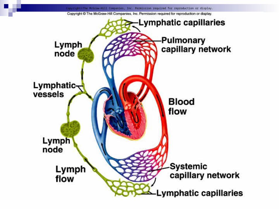
Lymphatic Pathways
A. Lymphatic pathways start as lymphatic capillaries that merge to form larger vessels that empty into the
circulatory system.
CopyrightThe McGraw-Hill Companies, Inc. Permission required for reproduction or display.

B. Lymphatic Capillaries
1. Lymphatic capillaries are tiny, closed-ended tubes that extend into interstitial spaces.
2. They receive tissue fluid through their thin walls; once inside, tissue fluid is
called lymph.
CopyrightThe McGraw-Hill Companies, Inc. Permission required for reproduction or display.

CopyrightThe McGraw-Hill Companies, Inc. Permission required for reproduction or display.
C. Lymphatic Vessels1. The walls of lymphatic vessels are
thinner than those of veins but are constructed with the same
three layers with semilunar valves on the inside.
2. Larger lymphatic vessels pass through lymph nodes and
merge to form lymphatic trunks.
CopyrightThe McGraw-Hill Companies, Inc. Permission required for reproduction or display.
CopyrightThe McGraw-Hill Companies, Inc. Permission required for reproduction or display.

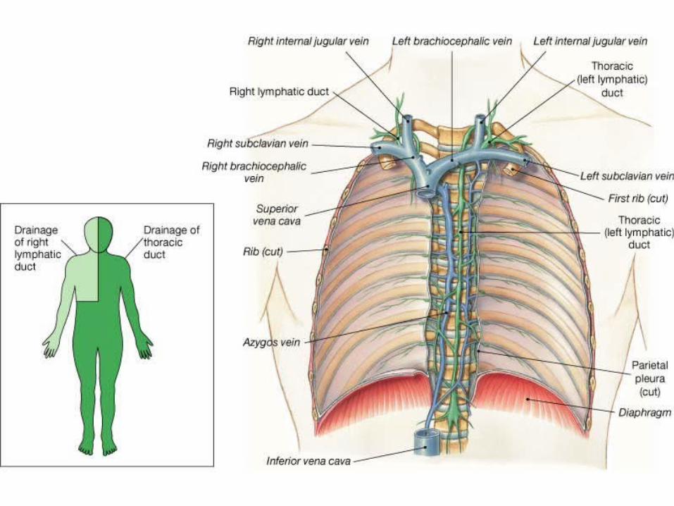
D. Lymphatic Trunks and Collecting Ducts 1. The lymphatic trunks drain
lymph from the body and are named for the regions they drain.
2. These trunks join one of two collecting ducts---either the
thoracic duct or right lymphatic duct.
CopyrightThe McGraw-Hill Companies, Inc. Permission required for reproduction or display.

3.The thoracic duct drains into the left subclavian vein, while the right lymphatic duct drains into the right subclavian vein.
CopyrightThe McGraw-Hill Companies, Inc. Permission required for reproduction or display.

Tissue Fluid and LymphA. Tissue fluid becomes lymph once it has
entered a lymphatic capillary; lymph formation depends on tissue fluid
formation.
CopyrightThe McGraw-Hill Companies, Inc. Permission required for reproduction or display.

B. Tissue Fluid Formation 1. Tissue fluid is made up of water and
dissolved substances that leave blood capillaries by filtration and diffusion.
2. During filtration, some smaller proteins leak from capillaries into the tissues and are not returned to the bloodstream, thus increasing osmotic pressure within the tissues.
CopyrightThe McGraw-Hill Companies, Inc. Permission required for reproduction or display.
C. Lymph Formation and Function
1. Rising osmotic pressure in tissues interferes with the return of
fluids to the bloodstream.
2. Increasing interstitial pressure forces some of the fluid into
lymphatic capillaries.
CopyrightThe McGraw-Hill Companies, Inc. Permission required for reproduction or display.
Lymph MovementA. The hydrostatic pressure of tissue fluid drives the entry of lymph into lymphatic capillaries.
B. Forces that move blood in veins (skeletal muscle contraction, breathing movements,
and contraction of smooth muscle in the walls of lymphatic trunks) are the forces that propel
lymph through lymphatic vessels.
CopyrightThe McGraw-Hill Companies, Inc. Permission required for reproduction or display.
C. A condition that interferes with the flow in lymph will result in edema.
D. During surgery, lymphatic vessels or tissues may be removed or disturbed, resulting in edema.
CopyrightThe McGraw-Hill Companies, Inc. Permission required for reproduction or display.
Lymphoid Nodules
Lymphoid Nodules are masses of lymphoid tissue that are not surrounded by a fibrous capsule.
Found beneath the epithelia lining of various organs of respiratory, digestive and urinary systems
Tonsils consist of pharyngeal or adenoids, pair of palatine, and a pair of lingual tonsils
Payer’s patch within the intestines Also within the appendix
Lymph Nodes A. Lymph nodes, which contain lymphocytes and macrophages, are located along lymphatic pathways.
CopyrightThe McGraw-Hill Companies, Inc. Permission required for reproduction or display.

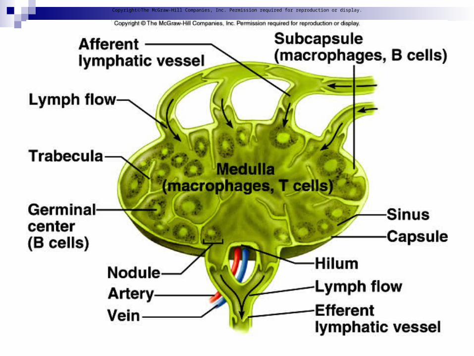
B. Structure of a Lymph Node
1. Lymph nodes are bean-shaped, with blood vessels, nerves,
and efferent lymphatic vessels attached to the indented hilum, and with afferent lymphatic vessels entering on the convex surface.
CopyrightThe McGraw-Hill Companies, Inc. Permission required for reproduction or display.
2. Lymph nodes are covered with connective tissue that extends
inside the node and divides it into nodules and spaces called sinuses.
3. These contain both lymphocytes and macrophages which clean the
lymph as it flows through the node.
CopyrightThe McGraw-Hill Companies, Inc. Permission required for reproduction or display.
CopyrightThe McGraw-Hill Companies, Inc. Permission required for reproduction or display.

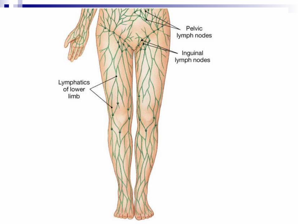
C. Locations of Lymph Nodes
1. The lymph nodes generally occur in chains along the parts of the larger lymphatic vessels.
CopyrightThe McGraw-Hill Companies, Inc. Permission required for reproduction or display.

D. Functions of Lymph Nodes
1. The macrophages and lymphocytes within lymph nodes filter lymph and remove bacteria and cellular debris before lymph is returned to the blood.
2. Lymph nodes are also centers of lymphocyte production; these cells function in immune surveillance.
CopyrightThe McGraw-Hill Companies, Inc. Permission required for reproduction or display.

CopyrightThe McGraw-Hill Companies, Inc. Permission required for reproduction or display.

3. Lobules contain lymphocytes, some of which mature into T lymphocytes
(T cells) that leave the thymus to provide immunity.4. The thymus secretes the hormone
thymosin, which influences the maturation of T lymphocytes
once they leave the thymus.
CopyrightThe McGraw-Hill Companies, Inc. Permission required for reproduction or display.
CopyrightThe McGraw-Hill Companies, Inc. Permission required for reproduction or display.
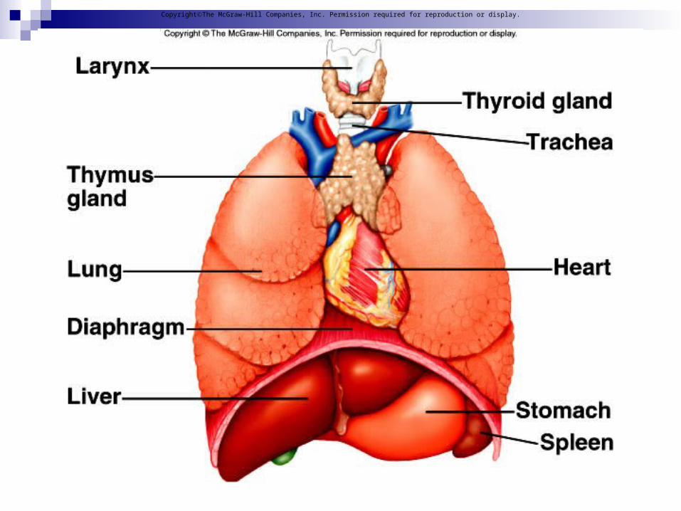
C. Spleen1. The spleen lies in the upper left
abdominal cavity and is the body’s largest lymphatic organ.
2. The spleen resembles a large lymph node except that it contains blood instead of lymph.
CopyrightThe McGraw-Hill Companies, Inc. Permission required for reproduction or display.

3. Inside the spleen lies white pulp (containing many lymphocytes)
and red pulp (containing red blood cells, macrophages, and lymphocytes).
4. The spleen filters the blood and removes damaged blood cells
and bacteria.
CopyrightThe McGraw-Hill Companies, Inc. Permission required for reproduction or display.

CopyrightThe McGraw-Hill Companies, Inc. Permission required for reproduction or display.


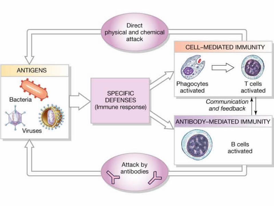
Body Defenses Against InfectionA. Diseases-causing agents, also called
pathogens, can produce infections within the body.
CopyrightThe McGraw-Hill Companies, Inc. Permission required for reproduction or display.

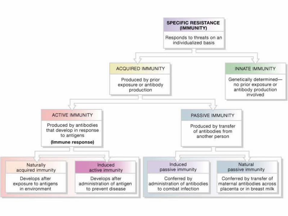
B. The body has two lines of defense against pathogens: nonspecific defenses that guard against any pathogen, and specific defenses (immunity) that mount a response against a
very specific target.
CopyrightThe McGraw-Hill Companies, Inc. Permission required for reproduction or display.
1. Specific defenses are carried out by lymphocytes that recognize a
specific invader.
2. Nonspecific and specific defenses work together to protect the body
against infection.
CopyrightThe McGraw-Hill Companies, Inc. Permission required for reproduction or display.
Nonspecific DefensesA. Species Resistance1. A species is resistant to diseases that
affect other species because it has a unique chemical environment or temperature that fails to provide the conditions required by the pathogens of another species.
CopyrightThe McGraw-Hill Companies, Inc. Permission required for reproduction or display.

B. Mechanical Barriers
1. The unbroken skin and mucous membranes of the body create
mechanical barriers that prevent the entry of certain pathogens.
2. Mechanical barriers represent the body’s first line of defense.
CopyrightThe McGraw-Hill Companies, Inc. Permission required for reproduction or display.

C. Chemical Barriers1. Chemical barriers, such as the highly
acidic and caustic environment provided by gastric juice, or lyzozyme in tears, kill many pathogens.
2. Interferons, hormone-like peptides that serve as antiviral substances, are produced by cells when they are infected with viruses and induce nearby cells to produce antiviral enzymes that protect them from infection.
CopyrightThe McGraw-Hill Companies, Inc. Permission required for reproduction or display.
D. Fever1. Fever offers powerful protection against infection by interfering with the proper conditions that promote bacterial growth.
a. During fever, the amount of iron in the blood is reduced, and thus
fewer nutrients are available to support the growth of pathogens.
b. Phagocytic cells attack with greater vigor when the temperature rises.
CopyrightThe McGraw-Hill Companies, Inc. Permission required for reproduction or display.
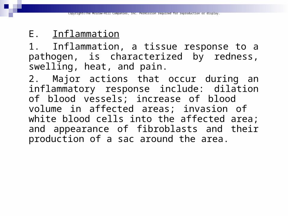
E. Inflammation1. Inflammation, a tissue response to a pathogen, is characterized by redness, swelling, heat, and pain.2. Major actions that occur during an inflammatory response include: dilation of blood vessels; increase of blood volume in affected areas; invasion of white blood cells into the affected area; and appearance of fibroblasts and their production of a sac around the area.
CopyrightThe McGraw-Hill Companies, Inc. Permission required for reproduction or display.

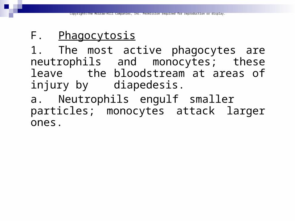
F. Phagocytosis1. The most active phagocytes are
neutrophils and monocytes; these leave the bloodstream at areas of injury by diapedesis.
a. Neutrophils engulf smaller particles; monocytes attack
larger ones.
CopyrightThe McGraw-Hill Companies, Inc. Permission required for reproduction or display.

2. Monocytes give rise to macrophages, which become fixed in various
tissues.
3. Monocytes, macrophages, and neutrophils constitute the
mononuclear phagocytic system.
4. Phagocytosis also removes foreign particles from the lymph.
CopyrightThe McGraw-Hill Companies, Inc. Permission required for reproduction or display.


Specific Defenses (Immunity) A. The body’s third line of defense, immunity refers to the response mounted by the body against specific, recognized foreign molecules.
CopyrightThe McGraw-Hill Companies, Inc. Permission required for reproduction or display.
B. Antigens1. Before birth, the body makes an
inventory of "self" proteins and other large molecules.
2. Antigens are generally larger molecules that elicit an immune response.a. Sometimes small molecules
called haptens combine with larger molecules and become antigenic.
CopyrightThe McGraw-Hill Companies, Inc. Permission required for reproduction or display.
C. Lymphocyte Origins
1. During fetal development, red bone marrow releases lymphocytes into
circulation, 70-80% of which become T lymphocytes (T cells) and the remainder of which become B lymphocytes (B cells).
CopyrightThe McGraw-Hill Companies, Inc. Permission required for reproduction or display.

2. Undifferentiated lymphocytes that reach the thymus become T cells; B
cells are thought to mature in the bone marrow.
3. Both B and T cells reside in lymphatic organs.
CopyrightThe McGraw-Hill Companies, Inc. Permission required for reproduction or display.

CopyrightThe McGraw-Hill Companies, Inc. Permission required for reproduction or display.

D. Lymphocyte Functions 1. T cells attack foreign, antigen-bearing
cells, such as bacteria, by direct cell-to- cell contact, providing cell-mediated immunity.2. T cells also secrete cytokines
(lymphokines) that enhance cellular response to antigens.
CopyrightThe McGraw-Hill Companies, Inc. Permission required for reproduction or display.
3. T cells may also secrete toxins that kill target cells, or produce growth-inhibiting factors or interferon to interfere with viruses and tumor cells.
4. B cells attack pathogens by differentiating into plasma cells that secrete antibodies (immunoglobulins).
CopyrightThe McGraw-Hill Companies, Inc. Permission required for reproduction or display.

5. Body fluids attack and destroy specific antigens or antigen-bearing
particles through antibody-mediated immunity also called humoral immune response.
CopyrightThe McGraw-Hill Companies, Inc. Permission required for reproduction or display.

E. T Cells and the Cellular Immune Response
1. T cell activation requires the presence of an antigen-presenting cell,
such as a B cell or macrophage, that has already encountered the antigen.
CopyrightThe McGraw-Hill Companies, Inc. Permission required for reproduction or display.

2. In order for a helper T cell to become activated, it must first encounter a macrophage displaying the antigen on its major histocompatibility complex (MHC) proteins; if the antigen fits the helper T cell's antigen receptor, it becomes activated and stimulates B cells to produce antibodies.
CopyrightThe McGraw-Hill Companies, Inc. Permission required for reproduction or display.

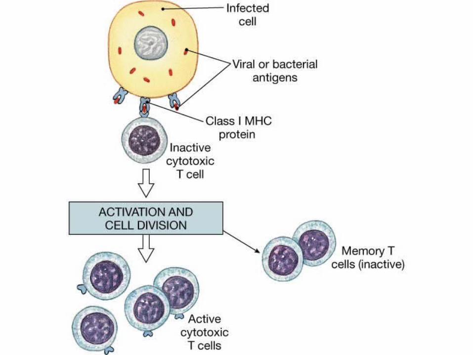
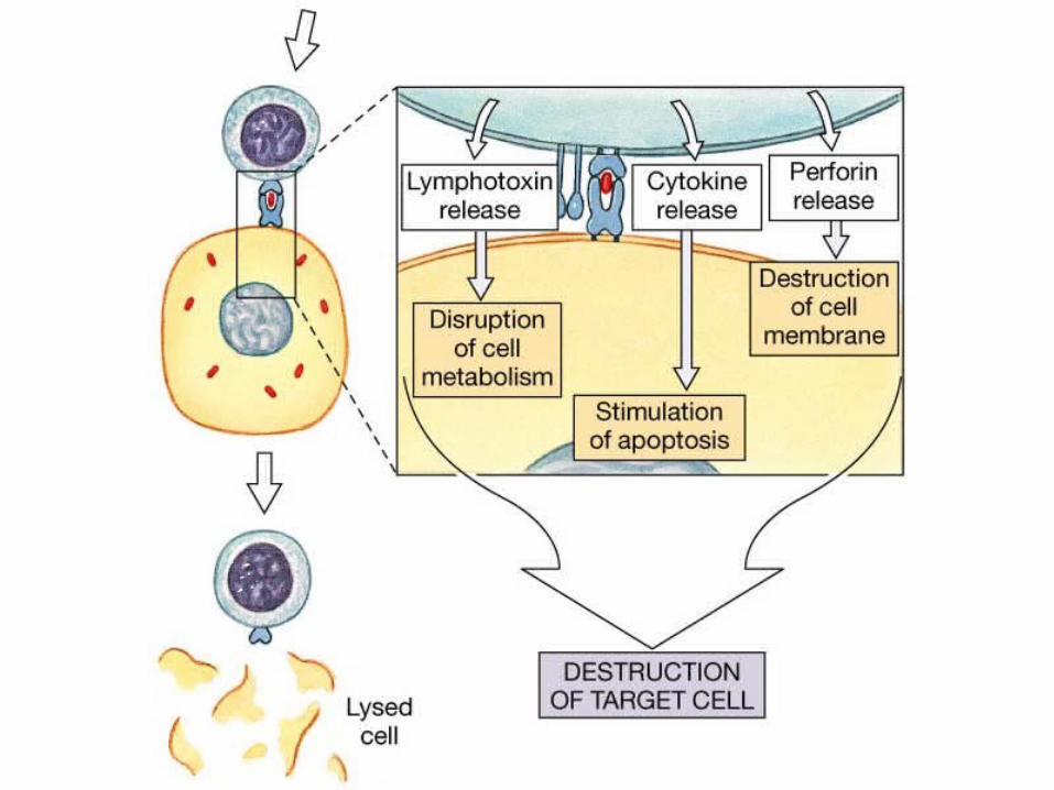
3. Cytotoxic T cells continually monitor the body's cells, recognizing and eliminating tumor cells and virus-infected cells by release of proteins, cutting holes and by other means.a. Cytotoxic T cells become
activated when a antigen binds to its receptors.
4. Memory T cells provide a no-delay response to any future exposure to the same antigen.
CopyrightThe McGraw-Hill Companies, Inc. Permission required for reproduction or display.

CopyrightThe McGraw-Hill Companies, Inc. Permission required for reproduction or display.
F. B Cells and the Humoral Immune Response
1. A B cell may become activated and produce a clone of cells when its
antigen receptor encounters its matching antigen, but most B cells need helper T cells for activation.
CopyrightThe McGraw-Hill Companies, Inc. Permission required for reproduction or display.

2. When a helper T cell encounters a B cell that has itself encountered an
antigen, the helper T cell releases cytokines that activate the B cell so that it can divide and form a clone.
CopyrightThe McGraw-Hill Companies, Inc. Permission required for reproduction or display.

3. Some of the B cells become plasma cells, producing and secreting antibodies.
4. Like T cells, some of the B cells become memory cells to
respond to future encounters with the antigen.
CopyrightThe McGraw-Hill Companies, Inc. Permission required for reproduction or display.

CopyrightThe McGraw-Hill Companies, Inc. Permission required for reproduction or display.


G. Types of Antibodies 1. There are five major types of antibodies
(immunoglobulins) that constitute the gamma globulin fraction of the
plasma.a. IgG is in tissue fluid and plasma
and defends against bacterial cells, viruses, and toxins and activates complement.
CopyrightThe McGraw-Hill Companies, Inc. Permission required for reproduction or display.
b. IgA is in exocrine gland secretions (breast milk,
saliva, tears) and defends against bacteria and viruses.
c. IgM is found in plasma and activates complement and
reacts with blood cells during transfusions.
CopyrightThe McGraw-Hill Companies, Inc. Permission required for reproduction or display.
d. IgD is found on the surface of most B lymphocytes and
functions in B cell activation.
e. IgE is found in exocrine gland secretions and promotes
allergic reactions
CopyrightThe McGraw-Hill Companies, Inc. Permission required for reproduction or display.

H. Antibody Actions1. Antibodies can react to antigens in three ways: direct attack, activation of
complement, or stimulation of changes in areas that help prevent the spread of the pathogens.2. Direct attack methods include
agglutination, precipitation, and neutralization of antigens.
CopyrightThe McGraw-Hill Companies, Inc. Permission required for reproduction or display.

3. The activation of complement can produce opsonization,
chemotaxis, inflammation, or lysis in target cells or antigens.
CopyrightThe McGraw-Hill Companies, Inc. Permission required for reproduction or display.

I. Immune Responses
1. When B or T cells become activated the first time, their actions constitute a
primary immune response, after which some cells remain as memory cells.
CopyrightThe McGraw-Hill Companies, Inc. Permission required for reproduction or display.
2. If the same antigen is encountered again, more numerous memory
cells can mount a more rapid response, known as the secondary immune response.
a. The ability to produce a secondary immune
response may be long-lasting.
CopyrightThe McGraw-Hill Companies, Inc. Permission required for reproduction or display.

J. Practical Classification of Immunity 1. Naturally acquired active immunity
occurs after exposure to the antigen itself.2. Artificially acquired active immunity
occurs through the use of vaccines, without the person becoming ill from the disease.
CopyrightThe McGraw-Hill Companies, Inc. Permission required for reproduction or display.

3. Artificially acquired passive immunity involves the injection of gamma
globulin containing antibodies and isshort-lived.
4. Naturally acquired passive immunity occurs as antibodies are passed from mother to fetus and is short-lived.
CopyrightThe McGraw-Hill Companies, Inc. Permission required for reproduction or display.

K.Allergic Reactions1. Allergic reactions to allergens are excessive immune responses that may lead to tissue damage.2. Delayed-reaction allergy results from
repeated exposure to substances that cause inflammatory reactions in the skin.
CopyrightThe McGraw-Hill Companies, Inc. Permission required for reproduction or display.

3. Immediate-reaction allergy is an inherited ability to overproduce IgE.4. During allergic reactions, mast cells release histamine and leukotrienes, producing a variety of effects.5. Allergy mediators sometimes flood the body, resulting in anaphylactic shock, a severe form of immediate-reaction allergy.
CopyrightThe McGraw-Hill Companies, Inc. Permission required for reproduction or display.

L.Transplantation and Tissue Rejection1. A transplant recipient's immune system may react with foreign antigens on the surface of the transplanted tissue, causing a tissue rejection reaction.2. Close matching of donor and recipient tissues can reduce the chances of tissue rejection, and use of immunosuppressive drugs may reduce rejection, although the individual may be more susceptible to infection.
CopyrightThe McGraw-Hill Companies, Inc. Permission required for reproduction or display.

M. Autoimmunity1. In autoimmune disorders, the immune
system manufactures antibodies against some of its own antigens.2. Autoimmune disorders may result from
viral infection, faulty T cell development, or reaction to
a nonself antigen that bears close resemblance to a self antigen.
CopyrightThe McGraw-Hill Companies, Inc. Permission required for reproduction or display.

![Veins and Lymphatics - Tagungsmanagement · Veins and Lymphatics 2013; volume 2:e1 [Veins and Lymphatics 2013; 2:e1] [page 1] Stiffness of compression devices Giovanni Mosti Angiology](https://static.fdocuments.net/doc/165x107/5f0ee5c27e708231d44179f9/veins-and-lymphatics-veins-and-lymphatics-2013-volume-2e1-veins-and-lymphatics.jpg)