CARDIAC ARREST AND VENTRICULAR - Thoraxthorax.bmj.com/content/thoraxjnl/7/3/205.full.pdfoperations...
8
Thorax (1952), 7, 205. CARDIAC ARREST AND VENTRICULAR FIBRILLATION * A METHOD OF TREATMENT BY ELECTRICAL SHOCK BY I. K. R. MCMILLANt F. B. COCKETT, AND P. STYLES Froml the Departnment of Cardiology, tile Professorial Surgical Unit, and tile Departalzent of Phlysical Medicine, St. Thomlas's Hospital, London (RECEIVED FOR PUBLICATION APRIL 30, 1952) Cardiac arrest during operation is a subject of importance to all surgeons. During intra-thoracic operations cardiac arrest and ventricular fibrilla- tion can be differentiated by direct observation of the heart. Ventricular fibrillation is an incoordinated type of contraction which produces no useful beats. The typical rippling movements of the ventricular muscle are unmistakable when seen or felt with the hand directly on the heart. The treatment of a heart that has stopped is cardiac massage and adequate oxygenation. The treatment of ventri- cular fibrillation which we recommend is, first, massage to maintain the oxygen supply to the heart through the coronary arteries, followed rapidly by electrical shocking to restore normal rhythm In abdominal operations, however, or during operations elsewhere in the body, once the pulse becomes undetectable most surgeons waste no time in performing a quick midline upper abdominal incision and massaging the heart through the dia- phragm. At the same time adrenaline is given either intravenously, intramuscularly, or more usually directly into the ventricle. The latter is a most dangerous procedure, as it is now known that one of the effects of adrenaline on a heart which is already anoxic may be the production of ventricular fibrillation. Until recent years ventri- cular fibrillation was almost always fatal. The purpose of this paper is twofold: (1) to emphasize once again the dangerous effects of adrenaline ; (2) to describe the experiments lead- ing up to the development of a satisfactory elec- trical instrument for defibrillating the heart. * This work was assisted by a grant from the Endowment Fund of St. Thomas's Hospital. t MacKenzie MacKinnon Research Fellow of the Royal College of Physicians and the Royal College of Surgeons. 0 VENTRICULAR FIBRILLATION The treatment of ventricular fibrillation bv electrical shock therapy is not new and was first tried experimentally in 1899 (Prevost and Battelli). In recent years this method has been extensively investigated in the United States and France (Kouwenhoven and Kay, 1951; Johnson and Kirby, 1951 ; Santy and Marion, 1950). The treatment of ventricular fibrillation by zlectrical shock gives encouraging results in the experimental animal, and may be a useful adjunct in the operating theatre during cardiac and general surgery. Under these conditions shock therapy can be rapidly applied. Provided that adequate oxygenation is maintained and direct cardiac mas- sage promptly initiated and maintained, shock therapy can be applied without undue haste. METHOD The principle of the method is to apply a powerful shock across the ventricles and produce complete arrest of the fibrillating heart. Once asystole occurs a regular ventricular beat may start spontaneously or cardiac massage may be needed to initiate it. The apparatus comprises a double-wound trans- former having a tapped secondary capable of supply- ing I ampere from any tapping. The primary circuit is supplied from 50 c/s mains via a foot-operated switch and a Londex motor-driven interrupter whose contacts close every 1.5 seconds for 0.5 second. The output voltage tappings are selected by means of a rotary switch on the front panel. Available voltages are 85, 125, 185, 250. 270, and 300. A buzzer operated from an auxiliary winding on the transformer gives an audible warning that the electrodes are alive. Closure of the mains switch merely sets in motion the interrupter. In order to administer a shock the foot switch must be depressed. The electrodes consist of two large curved plates which are shaped to fit the curve of the ventricles. on 21 June 2018 by guest. Protected by copyright. http://thorax.bmj.com/ Thorax: first published as 10.1136/thx.7.3.205 on 1 September 1952. Downloaded from
Transcript of CARDIAC ARREST AND VENTRICULAR - Thoraxthorax.bmj.com/content/thoraxjnl/7/3/205.full.pdfoperations...

Thorax (1952), 7, 205.
CARDIAC ARREST AND VENTRICULAR FIBRILLATION *A METHOD OF TREATMENT BY ELECTRICAL SHOCK
BY
I. K. R. MCMILLANt F. B. COCKETT, AND P. STYLESFroml the Departnment of Cardiology, tile Professorial Surgical Unit, and tile Departalzent of Phlysical
Medicine, St. Thomlas's Hospital, London
(RECEIVED FOR PUBLICATION APRIL 30, 1952)
Cardiac arrest during operation is a subject ofimportance to all surgeons. During intra-thoracicoperations cardiac arrest and ventricular fibrilla-tion can be differentiated by direct observation ofthe heart.
Ventricular fibrillation is an incoordinated typeof contraction which produces no useful beats.The typical rippling movements of the ventricularmuscle are unmistakable when seen or felt withthe hand directly on the heart. The treatment ofa heart that has stopped is cardiac massage andadequate oxygenation. The treatment of ventri-cular fibrillation which we recommend is, first,massage to maintain the oxygen supply to theheart through the coronary arteries, followedrapidly by electrical shocking to restore normalrhythm
In abdominal operations, however, or duringoperations elsewhere in the body, once the pulsebecomes undetectable most surgeons waste no timein performing a quick midline upper abdominalincision and massaging the heart through the dia-phragm. At the same time adrenaline is giveneither intravenously, intramuscularly, or moreusually directly into the ventricle. The latter isa most dangerous procedure, as it is now knownthat one of the effects of adrenaline on a heartwhich is already anoxic may be the production ofventricular fibrillation. Until recent years ventri-cular fibrillation was almost always fatal.The purpose of this paper is twofold: (1) to
emphasize once again the dangerous effects ofadrenaline ; (2) to describe the experiments lead-ing up to the development of a satisfactory elec-trical instrument for defibrillating the heart.
* This work was assisted by a grant from the Endowment Fund ofSt. Thomas's Hospital.
t MacKenzie MacKinnon Research Fellow of the Royal Collegeof Physicians and the Royal College of Surgeons.
0
VENTRICULAR FIBRILLATIONThe treatment of ventricular fibrillation bv
electrical shock therapy is not new and was firsttried experimentally in 1899 (Prevost and Battelli).In recent years this method has been extensivelyinvestigated in the United States and France(Kouwenhoven and Kay, 1951; Johnson andKirby, 1951 ; Santy and Marion, 1950).The treatment of ventricular fibrillation by
zlectrical shock gives encouraging results in theexperimental animal, and may be a useful adjunctin the operating theatre during cardiac and generalsurgery. Under these conditions shock therapycan be rapidly applied. Provided that adequateoxygenation is maintained and direct cardiac mas-sage promptly initiated and maintained, shocktherapy can be applied without undue haste.
METHODThe principle of the method is to apply a powerful
shock across the ventricles and produce completearrest of the fibrillating heart. Once asystole occursa regular ventricular beat may start spontaneously orcardiac massage may be needed to initiate it.The apparatus comprises a double-wound trans-
former having a tapped secondary capable of supply-ing I ampere from any tapping. The primary circuitis supplied from 50 c/s mains via a foot-operatedswitch and a Londex motor-driven interrupter whosecontacts close every 1.5 seconds for 0.5 second.The output voltage tappings are selected by means
of a rotary switch on the front panel. Availablevoltages are 85, 125, 185, 250. 270, and 300. A buzzeroperated from an auxiliary winding on the transformergives an audible warning that the electrodes are alive.Closure of the mains switch merely sets in motion theinterrupter. In order to administer a shock the footswitch must be depressed.The electrodes consist of two large curved plates
which are shaped to fit the curve of the ventricles.
on 21 June 2018 by guest. Protected by copyright.
http://thorax.bmj.com
/T
horax: first published as 10.1136/thx.7.3.205 on 1 Septem
ber 1952. Dow
nloaded from

1. K. R. McMILLAN, F. B. COCKETT, and P. STYLES
FOOT SWITCH
MAINS
BUZERINDACTTOINICATOICATO
FIG. I .-Circuit diagram.
The voltage required varies with the size of the heartand its electrical resistance, and this latter factor isalso governed by the efficiency of the contact betweenelectrodes and heart surface. Single shocks may besufficient to stop ventricular fibrillation or, if they areinsufficient, the voltage may be increased and furthersingle shocks applied. If these are still inadequatea series of about six shocks at 1.5-second intervalsmay be given (Wiggers, 1940).
RESULTS IN DoGsAll dogs were under nembutal anaesthesia.Seven dogs were given ventricular fibrillation elec-
trically by means of an induction coil. Three dogshave been given ventricular fibrillation by the appli-cation of aconitine (one drop 0.2% solution inbenzene) to the auricle, which causes auricularfibrillation and later ventricular fibrillation. Todefibrillate the heart of a 20-kg. dog 185 volts wassufficient when given in a series of five shocks.Nine dogs have been successfully defibrillated at
periods of up to five minutes after the onset ofventricular fibrillation, but in all cases cardiac mas-sage has been continued through the interveningperiod. Of the three dogs chemically stimulated, twowere defibrillated with a voltage of 300. The third,due to other procedures on the heart, was in verypoor condition and inadequately oxygenated.Ventricular fibrillation was not arrested.
_ _ ~~~~*. 11P~~~~~~~~~~~~~~~~~~~~~~~~~~~~~~~~~~~~~~~~~~~~~~~~~~~~~. ......... ..............
FIG. 2a.-The defibrillator.
Another dog in which ven-tricular fibrillation occurredduring a thoracic sympath-ectomy failed to revert tonormal, probably because ofinadequate oxygenation owing
-0 SELECTOR to collapse of both lungs.0 E.C.G.s taken after defibrilla-° tion in one dog showed a regu-_ELECTRODES lar rhythm (Fig. 3). After
electrical shocking sections ofventricular muscle show some
E______ superficial coagulation but noburning. Ventricular fibrilla-tion is a terminal event, andany procedure with a chance
of success is worth attempting in spite of possibleminor damage. Later experiments with large elec-trodes showed no coagulation or burning.
RESULTS IN MANCase 1.-Mrs. T., aged 65 years, who had been
attending as an out-patient, had gross left ventricularenlargement, anginal attacks, and multiple ventricularextrasystoles. During a visit to the hospital shecollapsed while climbing a flight of stairs. She wasfound to be pulseless and respiration had almoststopped. She was put on a couch, and within threeminutes the upper abdomen was opened and sub-diaphragmatic cardiac massage started. The heartthen started to beat again, but weakly, and soonfaded away. She was intubated and oxygen givenby means of controlled respiration from an anaes-thetic apparatus. Next the diaphragm was incised,the pericardium was entered, and direct cardiacmassage started. A few minutes later, as the strengthof the -beat was poor, adrenaline (1 fml. of 1/1,000)was given intracardially. This produced a strong beattemporarily, but was rapidly superseded by ventricu-lar fibrillation. Procaine amide hydrochloride (" pro-nestyl ") was injected intravenously without effect.Cardiac massage was continued, and 10 minutes laterthe defibrillator was used. The electrodes were
FIG. 2b.--The electrodes.
206
on 21 June 2018 by guest. Protected by copyright.
http://thorax.bmj.com
/T
horax: first published as 10.1136/thx.7.3.205 on 1 Septem
ber 1952. Dow
nloaded from

CARDIAC ARREST AND VENTRICULAR FIBRILLATION
.f4
; .- }; t tz -- .*- X -- e 0 - 8 <-;4'*Li .+ . _, . i. s .; * - s - - *,, . * * e + +-^' t-' >I ' , . . o .-< . f _v=+>S_.e t t t V j + -1 i t i tM 1*-J-.--.o ; +. 1 . , _. _ \ X = > W 9 9* A s v > \¢ S vl . t w z .- . F a z t- .--te t..4t/ ' * *t . . . * . 4 * t * . ._ . tt B
;7'j:1 t ~- II ~~~~~~~~~~~~~~~~~~~~~~~~~~~~~~~~~~. .|......................?.......
._: t .......
:^ 0 I r,--,FIG. 3.-Electrocardiograms taken from Dog 3 to show effects of
defibrillation: (A) before shock normal; (B) ventricular fibrilla-tion; (C) I mn. after shock; (D) 10 mi. after shock; (E)15 mmi. after shock; (F) I hour after shock.
inserted through the abdomen and up through the
diaphragm and one placed on each side of the
ventricle. The heart was given four shocks at 1.5-
second intervals, each shock lasting 0.5 second, of
300 volts (this voltage was based on the voltagerequired in large dogs). This abolished fibrillation,
and on massage a regular ventricular beat started.
The heart beat regularly for the next hour when aided
by massage and showed no sign of returning tofibrillation. The strength of the beat, however, waspoor and the heart would only beat spontaneouslyfor about two minutes following massage.
After an hour the strength of the beat was gettingprogressively weaker and asystole followed the cessa-tion of massage. Further measures were considereduseless. She had not had a spontaneous respirationfor half an hour and the heart was no longer fillingfollowing inspiration.The shock produced only slight localized muscle
contraction outside the heart.Necropsy confirmed the above findings, and section
of the ventricles showed no evidence of damage dueto electrical shocks or burns.Case 2.-A man of 62, suffering from polycythaemia
vera, was taken to the theatre for the evacuation ofa haematoma following a rib biopsy. After 60 mg.of pethidine he was given gas, oxygen, and a littlecyclopropane. During this minor procedure respira-tion ceased, and at about the same time the pulsecould not be felt.At once the patient was turned on to his back, a
midline upper abdominal incision was made, and thecessation of the heart beat confirmed. A transverseslit in the diaphragm opposite the pericardial attach-ment was then made, four fingers thrust through this.and direct cardiac massage started. A regular butweak beat was started without difficulty. In orderto strengthen this 25 at 1/1,000 adrenaline was givenintracardially. Almost at once the rhythm changedfrom normal to ventricular fibrillation. Intracardiac& procaine " was then given without effect. Massagewas continued and the defibrillator was sent for. Thisarrived, from another hospital, and was set up readyfor use within 45 minutes of the start of cardiacmassage. During the whole of this period massagewas maintained and oxygen was given. The patientmade spontaneous respiratory efforts and a cornealreflex was present for the whole of this time.The electrodes of the defibrillator were introduced
through the wound under the diaphragm and peri-cardium and applied to the ventricles.Two shocks of 270 volts were given with only
momentary cessation of fibrillation. Accordinglyfour shocks of 300 volts at 1.5-second intervals last-ing about 0.5 second each were given and againfibrillation was only momentarily stopped. Fourmore shocks were then given and this producedasystole which lasted about three minutes. Duringthis time massage was continued, and after threeminutes the heart started spontaneously and beatfairly strongly. Massage was continued, and afterabout half an hour a steady beat persisted at a rateof about 50 per minute. At this stage many beatswere coupled and appeared to be ventricular inorigin. Blood pressure by a cuff was unrecordable,the corneal reflex was present, the pupils were fixed,central, but not dilated, spontaneous respirations werepresent, and the colour was less cyanosed thanpreviously.
To-.ri 1-*--: i:i '--T'J---~ti;+1 ! ; qz;!1r
;i ri-. i__-ia -4 i - i;
"Z.07
on 21 June 2018 by guest. Protected by copyright.
http://thorax.bmj.com
/T
horax: first published as 10.1136/thx.7.3.205 on 1 Septem
ber 1952. Dow
nloaded from

I. K. R. McMILLAN, F. B. COCKETT, and P. STYLESr * * ;_ ..
sL- ffi
_._jlFb- j.l-lllW_t 1-1.'t;.+1'>_^ * . - ^ . ew - -; * + t - ' - ' f - ' '; - ' - -. ' ' r. . . . t s . . : . . . . . . : w..
t * ...... + _ f *'
_fE_t Tr ''-'T' ''-'' - ' e ' ' ' - fi -' '''---'t _f =.. ..... ., ....... .. 1, . .. _ . . . . _ . _ .................... . t '1. ~~~ Aup- W4mm
:t j:* s ; -. .. . . . . . . . . . . . . . . . .. . . . .......... 1.^
,mm.
_~~~~~ c-
FiG. 4.-Electrocardiographs taken from Case 3, 10 days after defibrillation.
-.-I 1 III," IN
208
I
I
on 21 June 2018 by guest. Protected by copyright.
http://thorax.bmj.com
/T
horax: first published as 10.1136/thx.7.3.205 on 1 Septem
ber 1952. Dow
nloaded from

CARDIAC ARREST AND VENTRICULAR FIBRILLATION
f iftttj4:49~,11t4114 ..,4 LtI¼'II I th ±-4 'th
AVR
]i II01. 4.1.1 . i! :.it -4 i. ..i - +. I1 .. . 4+i .
I- i L ...!Hi kffiflfIfiffiffifflfil".fI Li fiill t. ;1._.i
AVL
{t- t- rt 4221 4T 4Ti T I
K,SvA IL LULit--; i.
AVF
itMk1HiHt::I IH-4
V3
V5
*XiT.tifgi: 1IJ*: i~ I. Ta IiI i
FIG. 5.-Electrocardiographs taken from Case 3, 15 days after defibrillation.
_Ht+ + .. t.__s__4t._. pa._.HFj;f_
~_s _ __ + _ .. _.__.. _ _- _ _._..+__i.v. .
__ _...*.. = _ _ ,, _.$,,, ___
47_____0 r_~_~
..I.- i 4.1 ti :1s. II . I . i 4 4 - i t1'. I
-,1 1 ': iI -. I I -,- - - -r I I - 1 .I
,,.I
I---
t I 1'
20-9
.-r T -T
!+it4It ' ' +
Tw,4 ti .,Ii o
on 21 June 2018 by guest. Protected by copyright.
http://thorax.bmj.com
/T
horax: first published as 10.1136/thx.7.3.205 on 1 Septem
ber 1952. Dow
nloaded from

1. K. R. McMILLAN, F. B. COCKETT, and P. STYLES
As the heart seemed to be well started it was decidedto close the wounds in the diaphragm and abdomen.During the sewing-up procaine amide hydrochloride(" pronestyl ") 500 mg. was given intravenously inorder to reduce the number of ectopic beats ifpossible. After the incision had been closed theoriginal biopsy wound was closed. At this stage thecarotid pulse rate was 44 per minute. It was decidedto leave the patient on the table and continue givingoxygen.
During the next half-hour the heart rate slowly fellto about 30 per minute, and at the end of this periodcardiac arrest occurred again. Cardiac massage wastried once more, and apart from a few beats pro-duced no effect, but at no stage after defibrillationwas ventricular fibrillation observed.Post-mortem section of the heart showed no evidence
of damage, such as superficial coagulation, which couldbe associated with the application of electrodes.
Case 3v--Mr. W., aged 32; on June 23, 1952, wasundergoing a left lower lobectomy for bronchiectasisunder a general anaesthetic.
Dissection of the left lower lobe was difficult owingto adhesions, and there had been free oozing but nogross haemorrhage. His general condition was satis-factory until the hilum of the lower lobe wasapproached and one of the lower lobe arteries hadbeen ligated. At this point the anaesthetist reportedthat the pulse was impalpable.The heart was examined and found to have stopped,
but was not fibrillating. The pericardium was opened.Cardiac massage was started and the patient respiredwith 100% oxygen. Noradrenaline, 40 y, was givenintravenously. Although as a result of cardiacmassage no pulse could be felt, there was goodcapillary filling after pressure on the forehead.
Cardiac massage was continued for eight or nineminutes without effect and then ventricular fibrillationstarted.The defibrillator was applied and three shocks of
250 volts given. This produced asystole again andmassage had no effect. Then -1 ml. of 1/1,000adrenaline was given into the left ventricle, and twominutes later (11 minutes from the start) ventricularfibrillation appeared again. No blood pressure orperipheral pulse was recordable.Pulmonary ventilation had been carried out all this
time.After 12+ minutes, as the heart was still fibrillating,
two further shocks of 250 volts were given. Thisproduced complete asystole once more and cardiacmassage was continued. After 15 minutes, however,the heart started with a regular rhythm. A bloodpressure of 80/60 immediately became recordable andbleeding started from two vessels in the hilum.The general circulation was very rapidly re-estab-
lished, and within half a minute hilar' blood wasbright red.The pulse and strength of the heart beat improved
steadily until the end of the operation.After this the lobectomy was completed and the
wound closed with temporary drainage.
The patient was kept in the operating theatre forthe next five hours. During this time his blood pres-sure fluctuated considerably and could not be main-tained without frequent doses of noradrenaline andmethedrine until about nine hours after defibrillation,when it remained steady at about 90/60. At that timean electrocardiogram showed sinus rhythm withnumerous extrasystoles. The T waves were upright.He regained consciousness two hours after defibrilla-tion, but was found to have a complete right hemi-paresis, including aphasia. At this time he respondedto simple commands but was confused.
After five hours the right leg began to twitch andhe was a little less confused.The next morning his general condition was good,
with no evidence of cardiac failure. He was movingthe right leg a little, the right facial palsy hadimproved, but speech was still very slurred. Theright arm was spastic and immovable. His under-standing was better, and he could talk a little. Hispulse was regular and blood pressure steady at 90/60.
After 48 hours his condition had further improved.He moved the right leg and face well. The right armwas still immovable.He made very steady progress after this, and by
the tenth day was up and walking slowly. His speechwas much better but still slurred, and he appeared tofollow a simple conversation but had echolalia. Theright arm showed no improvement, and his bloodpressure was still 90/60.An E.C.G. 14 days after operation showed evidence
of anterior myocardial infarction. Movement in theright arm started to return about the fourteenth dayand improved rapidly, but at this time there waspartial motor aphasia and echolalia.An electroencephalogram was within normal limits.Twenty-two days after operation screening showed
no evidence of aneurysm of the ventricles and move-ment of the ventricular borders was normal.
DISCUSSION
From the experience gained with these threecases in which the defibrillator was used, certainconclusions and suggestions concerning the sur-gical treatment of acute cardiac arrest can be putforward.
The Action of Adrenaline.-In all three casesdescribed ventricular fibrillation started soon afterthe intracardiac injection of adrenaline, given topotentiate the weak, irregular beat of the failingheart. This dangerous action of adrenaline, parti-cularly when the heart muscle is anoxic, or in thepresence of chloroform or cyclopropane, is beingincreasingly recognized by surgeons and anaes-thetists. This action is not prevented or reversedby procaine.The important thing, once a regular beat has
been established after cardiac arrest, is to ensurethat the coronary blood flow is maintained in
210
on 21 June 2018 by guest. Protected by copyright.
http://thorax.bmj.com
/T
horax: first published as 10.1136/thx.7.3.205 on 1 Septem
ber 1952. Dow
nloaded from

CARDIAC ARREST AND VENTRICULAR FIBRILLATION
order that the heart muscle may pay off its oxygendebt accumulated during the period of arrest.Coronary flow depends mainly on the level of thediastolic blood pressure. Although cardiac mas-sage may keep up an efficient peripheral circula-tion, the blood pressure may be unrecordableduring the procedure. We have been unable toraise the systolic blood pressure of dogs above40 mm. of mercury by cardiac massage alone.For the purpose of raising the blood pressurenoradrenaline is probably the drug of choice. Bar-croft and Konzett (1949) and Swan (1949) haverecently described its properties, and Churchill-Davidson (1951) has reported on its clinical pos-
sibilities. In brief, it has a powerful peripheralvasoconstrictor action, no action at all on theheart muscle, and, if anything, a vasodilator actionon the coronary arteries. Thus it raises the bloodpressure as effectively as adrenaline but does nothave the dangerous cardiac side-effects. Fromexperience with these three cases we feel that theadministration of intracardiac adrenaline in cases
of acute cardiac arrest is very likely to precipitateventricular fibrillation. In this hospital ampoulescontaining 2 ml. of noradrenaline (strength 20 ,Ag.per ml.) are being issued to all theatres for trial inanaesthetic emergencies instead of adrenaline.
In Case 3 noradrenaline was given into a peri-pheral vein in a subject with a very poor peri-pheral circulation. In these circumstances it isnot likely to be very effective. It should be givendirectly into a vena cava near the heart if feasible;it may also be given into a pulmonary vein or an
auricle. It is possible that ventricular fibrillationis more likely to follow intraventricular injection.A dose of up to 5 ml. (2{ ampoules, or 100 y) can
be given by this route in an emergency.A further point arises from Case 3. Autoregula-
tion of the blood pressure did not return untilabout six hours after cardiac arrest. During thistime the blood pressure had to be maintainedby the continuous intravenous infusion of nor-
adrenaline and methedrine. This emphasizes theimportance of the after-care of these cases. Theblood pressure fell away to 30 mm. Hg systolic ifthese drugs were discontinued during this period.
This patient was the only one who had a normalheart pre-operatively.The treatment of ventricular fibrillation, once
established and recognized, is a practical possibilitywith the defibrillator. Whether it occurs de novo
or as a sequel to cardiac arrest, this method isreadily applicable.The approach through the upper abdomen and
then straight into the pericardium by incising the
diaphragm enables the operator to appreciate with-out doubt the onset or presence of ventricularfibrillation. It is not always possible to be surewhether the ventricles are fibrillating if they areonly felt through the diaphragm. The diaphrag-matic incision can be easily enlarged to insert theelectrodes of the defibrillator.
In such cases a direct transthoracic approachhas been recommended for the exposure of theheart, but we have found the abdominal exposuredescribed quite adequate, and possibly easier as anemergency measure in general. Where the chestis already open, however, as in Case 3, theapproach is no problem.Most of the apparatus for defibrillation
described has been designed to work from UnitedStates mains of 110 volts 60 cycles A.C. Thismachine is designed to work from English mainsof 220 volts 50 cycles A.C. It appears from ourresults that higher voltages are required than thosereported by United States workers.
In the operating theatre, where, if the machineis at hand, a delay of no more than a minute isnecessary, we feel that it has a definite place,together with cardiac massage, in treating thishitherto irreversible condition. These results arenecessarily incomplete, but in view of the workalready done by other workers we feel that know-ledge of the method and the relative simplicityof the machine should encourage others to try it.
It should be emphasized that in the first two casesthere was a long and unavoidable delay beforedefibrillation could be initiated which undoubtedlyreduced the chance of permanent recovery. Ifthe interval is short, complete recovery is possible,as shown by American workers. Case 3, wherethe time interval was very short but the total timeof arrest was about 15 minutes, led to recoverywith cerebral damage; but the period of arrestwas longer than is usually considered compatiblewith survival.
SUMMARY
A machine for giving electric shocks sufficientto defibrillate hearts is described
Results on experimental animals and -threehuman cases are discussed.The danger of giving large doses of intracardiac
adrenaline to cases of acute cardiac arrest ispointed out, and the alternative use of nor-adrenaline is suggested.The importance of maintaining the blood pres-
sure post-operatively, if necessary with drugs, isemphasized.
211
on 21 June 2018 by guest. Protected by copyright.
http://thorax.bmj.com
/T
horax: first published as 10.1136/thx.7.3.205 on 1 Septem
ber 1952. Dow
nloaded from
1. K. R. McMILLAN, F. B. COCKETT, and P. STYLES
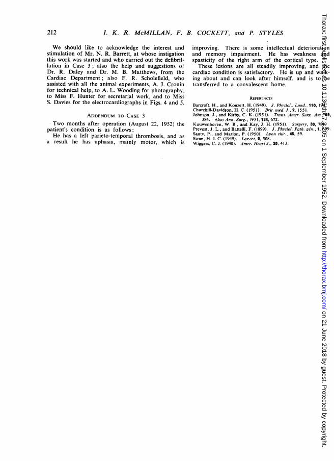
We should like to acknowledge the interest andstimulation of Mr. N. R. Barrett, at whose instigationthis work was started and who carried out the defibril-lation in Case 3; also the help and suggestions ofDr. R. Daley and Dr. M. B. Matthews, from theCardiac Department; also F. R. Scholefield, whoassisted with all the animal experiments, A. J. Croninfor technical help, to A. L. Wooding for photography,to Miss F. Hunter for secretarial work, and to MissS. Davies for the electrocardiographs in Figs. 4 and 5.
ADDENDUM TO CASE 3Two months after operation (August 22, 1952) the
patient's condition is as follows:He has a left parieto-temporal thrombosis, and as
a result he has aphasia, mainly motor, which is
improving. There is some intellectual deteriorationand memory impairment. He has weakness andspasticity of the right arm of the cortical type.These lesions are all steadily improving, and the
cardiac condition is satisfactory. He is up and walk-ing about and can look after himself, and is to betransferred to a convalescent home.
REFERENCES
Barcroft, H., and Konzett, H. (1949). J. Phvsiol., Lond., 110, 194.Churchill-Davidson, H. C. (1951). Brit. med. J., 2, 1551.Johnson, J., and Kirby, C. K. (1951). Trans. Amer. Surg. Ass., 69,
384. Also Ann. Surg., 1951,134, 672.Kouwenhoven, W. B., and Kay, J. H. (1951). Surgery, 30, 781.Prevost, J. L., and Battelli, F. (1899). J. Physiol. Path. gen., 1, 399.Santv, P., and Marion, P. (1950). Lyon chir., 45, 59.Swan, H. J. C. (1949). Larcet, 2, 508.Wiggers, C. J. (1940). Amer. Heart J., 20, 413.
212
on 21 June 2018 by guest. Protected by copyright.
http://thorax.bmj.com
/T
horax: first published as 10.1136/thx.7.3.205 on 1 Septem
ber 1952. Dow
nloaded from