Carbonyl groups anchoring for the water dispersibility of magnetite nanoparticles
9
ORIGINAL CONTRIBUTION Carbonyl groups anchoring for the water dispersibility of magnetite nanoparticles Ke Tao & Sheng Song & Jing Ding & Hongjing Dou & Kang Sun Received: 4 November 2010 / Revised: 7 January 2011 /Accepted: 13 January 2011 /Published online: 2 February 2011 # Springer-Verlag 2011 Abstract Magnetite nanoparticles, which were synthesized by using diethylene glycol (DEG) as both solvent and reductant, displayed excellent hydrophilicity and water dispersibility. The influences of the amount of carboxy- methyl-dextran sodium salt and NaOH on nanoparticles dispersibility were evaluated in terms of zeta potentials, concentration variation under centrifugal field, Fourier transform infrared, and X-ray photoelectron spectra, re- spectively. The results showed that the water dispersibility is not related to the amount of carboxymethyl-dextran, but depends on that of NaOH. Further evaluation revealed that DEG was partially oxidized with the assistance of NaOH, and thanks to the resultant carbonyl groups, the flexible oxidized DEG chains were anchored on the nanoparticles surface, which provides sufficient protection and high water-dispersibility of the as-prepared magnetite nano- particles. Furthermore, we suggest that the binding between carbonyl groups and nanoparticles is related to the electron-withdrawing effect. The current study may provide more selectiveness of modifying molecules for magnetite nanoparticles. Keywords Diethylene glycol . Carboxymethyl-dextran . Carbonyl group . Magnetite nanoparticle . Water dispersibility Introduction Due to the high biocompatibility and superparamagnetic behavior, magnetite nanoparticles (MNPs) have attracted intensive attentions not only for fundamental scientific interest but also for their potential applications especially, but not exclusively, in biomedical fields, such as magnetic resonance imaging (MRI), hyperthermia, and biomedical separation [1–5]. For most of these applications, e.g., cellular or even molecular resolution in MRI diagnosis [6], and the tracking of cancer cell migration [7], the quality and performance of MNPs not only relate to the magnetic properties, size, and size distribution but also crucially depend on their dispersibility, stabilizing agents, and control over their surface chemistry [8, 9]. Unfortunately, pristine nanoparticles would spontaneously aggregate and deposit from solvents due to its large surface energy, which results in the change of physiochemical properties and the risk of blood vessel blocking [10, 11]. Aiming at biomedical applications, the surface of MNPs is generally modified to provide nanoparticles the capability of stably dispersing in water or other biological solvents [12]. Besides, the modifying molecules should possess good biocompatibility and the capability of binding to other biomolecules [13]. Therefore, choosing the modifying molecules and elucidating their modification mechanism are of great importance for the subsequent biomedical applications. A series of molecules have been chosen to cap on the surface of magnetite nanoparticles to provide water dispersibility. Two strategies were generally used: syn- thesizing magnetite nanoparticles with the existence of water-soluble molecules [14–16] and modifying the surface of pre-synthesized oil-soluble MNPs by the ligand-exchange reaction [17] or the second layer coating K. Tao (*) : S. Song : J. Ding : H. Dou : K. Sun (*) State Key Lab of Metal Matrix Composites, Shanghai Jiao Tong University, Shanghai 200240, People’ s Republic of China e-mail: [email protected] K. Sun e-mail: [email protected] Colloid Polym Sci (2011) 289:361–369 DOI 10.1007/s00396-011-2380-5
Transcript of Carbonyl groups anchoring for the water dispersibility of magnetite nanoparticles

ORIGINAL CONTRIBUTION
Carbonyl groups anchoring for the water dispersibilityof magnetite nanoparticles
Ke Tao & Sheng Song & Jing Ding & Hongjing Dou &
Kang Sun
Received: 4 November 2010 /Revised: 7 January 2011 /Accepted: 13 January 2011 /Published online: 2 February 2011# Springer-Verlag 2011
Abstract Magnetite nanoparticles, which were synthesizedby using diethylene glycol (DEG) as both solvent andreductant, displayed excellent hydrophilicity and waterdispersibility. The influences of the amount of carboxy-methyl-dextran sodium salt and NaOH on nanoparticlesdispersibility were evaluated in terms of zeta potentials,concentration variation under centrifugal field, Fouriertransform infrared, and X-ray photoelectron spectra, re-spectively. The results showed that the water dispersibilityis not related to the amount of carboxymethyl-dextran, butdepends on that of NaOH. Further evaluation revealed thatDEG was partially oxidized with the assistance of NaOH,and thanks to the resultant carbonyl groups, the flexibleoxidized DEG chains were anchored on the nanoparticlessurface, which provides sufficient protection and highwater-dispersibility of the as-prepared magnetite nano-particles. Furthermore, we suggest that the bindingbetween carbonyl groups and nanoparticles is related tothe electron-withdrawing effect. The current study mayprovide more selectiveness of modifying molecules formagnetite nanoparticles.
Keywords Diethylene glycol . Carboxymethyl-dextran .
Carbonyl group .Magnetite nanoparticle .
Water dispersibility
Introduction
Due to the high biocompatibility and superparamagneticbehavior, magnetite nanoparticles (MNPs) have attractedintensive attentions not only for fundamental scientificinterest but also for their potential applications especially,but not exclusively, in biomedical fields, such as magneticresonance imaging (MRI), hyperthermia, and biomedicalseparation [1–5]. For most of these applications, e.g.,cellular or even molecular resolution in MRI diagnosis[6], and the tracking of cancer cell migration [7], the qualityand performance of MNPs not only relate to the magneticproperties, size, and size distribution but also cruciallydepend on their dispersibility, stabilizing agents, andcontrol over their surface chemistry [8, 9]. Unfortunately,pristine nanoparticles would spontaneously aggregate anddeposit from solvents due to its large surface energy, whichresults in the change of physiochemical properties and therisk of blood vessel blocking [10, 11]. Aiming atbiomedical applications, the surface of MNPs is generallymodified to provide nanoparticles the capability of stablydispersing in water or other biological solvents [12].Besides, the modifying molecules should possess goodbiocompatibility and the capability of binding to otherbiomolecules [13]. Therefore, choosing the modifyingmolecules and elucidating their modification mechanismare of great importance for the subsequent biomedicalapplications.
A series of molecules have been chosen to cap on thesurface of magnetite nanoparticles to provide waterdispersibility. Two strategies were generally used: syn-thesizing magnetite nanoparticles with the existence ofwater-soluble molecules [14–16] and modifying thesurface of pre-synthesized oil-soluble MNPs by theligand-exchange reaction [17] or the second layer coating
K. Tao (*) : S. Song : J. Ding :H. Dou :K. Sun (*)State Key Lab of Metal Matrix Composites,Shanghai Jiao Tong University,Shanghai 200240, People’s Republic of Chinae-mail: [email protected]
K. Sune-mail: [email protected]
Colloid Polym Sci (2011) 289:361–369DOI 10.1007/s00396-011-2380-5

[18, 19]. In both of these two strategies, the functionalgroups that possess binding affinity toward MNPs shouldbe contained to ensure the anchoring of modifyingmolecules or several coating layers, and the other end ofmodifying molecules extending outside should be hydro-philic. In recent research, carboxyl groups being containedin or being modified on a variety of molecules haveconfirmed their affinity toward MNPs surface [20].Therefore, hydrophilic molecules containing carboxylgroups can be selected as the stabilizer of MNPs, such aspoly(acrylic acid) (PAA) [21], citric acid [22], humic acid[23], dimercaptosuccinic acid [17, 24], etc. Carboxylgroups were also modified in some molecules to providethe binding capability to MNPs. For an instance,monocarboxyl-terminated poly(ethylene glycol) (MPEG)was coated on the MNPs after being oxidized to MPEG–COOH at first [25]. Besides, catechol derivatives, such asdopamine [26, 27], and some kinds of silanes [28, 29] canreplace oleic acid on MNPs by a ligand-exchange reaction,demonstrating the binding capability of some certain kindsof hydroxyl and silane groups. Although some otherbiocompatible molecules containing hydroxyl or other groupshave been used in the synthesis of water-soluble magnetitenanoparticles, to the best of our knowledge, there is noevidence to prove that they have strong affinity towardMNPs.The limitation in the binding functional groups stronglylimited the choice of surface modifying molecules. Moreover,the reason of variation in binding capability of differentmolecules is not clear yet, which has been a challenge for theMNPs subsequent applications.
Recently, a novel diethylene glycol (DEG)-mediatedstrategy was developed, and water-soluble magnetite nano-particles with relatively narrow size distribution can beobtained at elevated temperature. For example, Caruntu[30] reported that the coprecipitation of Fe2+ and Fe3+ couldhappen in DEG at about 210∼220 °C, Ge et. al. [31] usedPAA as the capping agent and Fe3+ as the only ironprecursor to synthesize magnetite nanoparticles in DEG,and Fe(acac)3 (acac, acetylcetonate) was decomposed inethylene glycol, diethylene glycol, and triethylene glycol,respectively [32]. Despite the successful synthesis ofMNPs, the mechanism of nanoparticles dispersing in waterhas not been addressed. In this paper, we used carbox-ymethyl (CM)-dextran in the synthesis owing to itsbiocompatibility. Unexpectedly, it was found that althoughthe carboxymethyl-dextran to some extent hybridizes withthe nanoparticles, the key of water dispersibility is thecapping of carbonyl groups of oxidized DEG. The resultslead to a proposal that besides the functional groupsmentioned above, some kinds of C=O bonds also possessthe binding capability to MNPs surface, and probablyprovide more selectiveness of modifying molecules forMNPs.
Materials and methods
Materials
FeCl3·6H2O (>99%), NaOH (>96%), dextran (Mw=20,000),polyethylene glycol (PEG, Mw=20,000), and acetone(>99.5%) were supplied by Sinopharm Chem Reagent Co.Carboxymethyl-dextran sodium salt (Mw=20,000) and dieth-ylene glycol (Reagent Plus, >99%) were purchased fromSigma-Aldrich Co. Polyvinyl pyrrolidone (PVP, K15, Mμ=10,000) was purchased from Fluka Co. All these reagentswere used without further purification.
The synthesis
CM-dextran-hybridized magnetite nanoparticles were syn-thesized via a hydrolysis-reducing process of FeCl3 byusing DEG as the solvent. Typically, A NaOH/DEG stocksolution was prepared by dissolving NaOH (50 mmol) inDEG (20 ml). This solution was heated at 120 °C for 1 hunder nitrogen, cooled down, and kept at 70 °C. Then, amixture of CM-dextran (0.15 g), FeCl3 (2 mmol), and DEG(15 ml) were heated to 220 °C for 30 min under vigorousstirring, forming a transparent light-yellow solution. ANaOH/DEG stock solution (2 ml) was injected rapidly intothe above hot mixture, and the reaction solution turnedblack immediately. The resulting mixture was furtherheated at 220 °C for 1 h to yield water-soluble magnetitenanocrystals. After that, the hot solution was carefullydropped into 50 ml acetone, and the MNPs can be easilyharvested by a magnet. The nanoparticles were rinsed byacetone for five times and dispersed in Milli-Q water.Different amount of CM-dextran and NaOH/DEG solutionwas used, as denoted in the text.
Characterization
Transmission electron microscopy (TEM) samples wereprepared by putting a drop of the as-prepared suspension ona carbon-coated copper grid and then dried in a desiccator.TEM observation was performed at 200 kV on a JEOLJEM-2100F field-emission transmission electron micro-scope. The crystallite phase of the as-prepared nanoparticleswas identified by recording X-ray diffraction pattern (XRD)on a D8 Advance diffractometer (Bruker, Germany)equipped with a CuKα radiation source. Fourier transforminfrared (FTIR) spectra were taken on a Bruker EQUINOX55 spectrometer. X-ray photoelectron spectra (XPS)were recorded by using a Perkin-Elmer PHI5000c spec-trometer with an AlKα monochromatic X-ray source.Thermogravimetric analysis (TGA) measurements wereperformed at a ramp rate of 10 °C/min under nitrogenatmosphere using TGA 2050 (TA Instruments), respectively.
362 Colloid Polym Sci (2011) 289:361–369

Zeta potentials were measured on a Malvern ZetaSizer NanoZ90 by using HCl and NaOH solution to adjust the pH value.The dispersibility of nanoparticles in water was determined bythe ultraviolet-visible (UV-vis) measurement: MNPs dis-persed in water were centrifuged at different rotational speedfor certain time, and the supernatant was extracted to recordtheir UV-vis spectra on a UV-2550 Shimadzu spectropho-tometer. The absorption at 650-nm wavelength was recordedand normalized by that of the rational MNPs solution.Magnetization measurements of nanoparticles were per-formed on a vibrating sample magnetometer (VSM) at roomtemperature.
Results and discussion
The high-temperature hydrolysis of Fe3+ upon addition ofNaOH in the presence of CM-dextran yields uniformmagnetite nanocrystals, as shown in the TEM images inFig. 1. The as-prepared nanoparticles showed the averagesize of 4.8 nm with a narrow size distribution. Clear latticefringe in high-resolution transmission electron microscopy(HRTEM) image (Fig. 1b) demonstrates the well singlecrystalline nature of resultant nanoparticles, and interplanardistance about 0.251 nm is consistent with the (311) plainof Fe3O4. Under the reductive atmosphere provided byDEG at high temperature [33, 34], Fe(OH)3 partiallytransforms to Fe(OH)2, then resulting in the formation ofFe3O4 nanocrystals through dehydration. XRD patterns(Fig. 2) confirm the phase of the Fe3O4 because the positionand relative intensity of the main peaks match well to thosefrom the JCPDS card (19-0629) for Fe3O4. With theincrease of CM-dextran amount, the intensity of XRDpeaks would be decreased because of the increase ofnoncrystallite content. The magnetic hysteresis loops wererecorded by a VSM at room temperature. Figure 3 showsthe magnetization of Fe3O4 nanocrystals prepared with
different dose of CM-dextran and NaOH. As expected, thenanocrystals are superparamagnetic at room temperature,showing no remanence or coercivity. The saturated massmagnetization of the MNPs prepared with 0.15, 0.30, and0.45 g CM-dextran are about 22.5, 20.3, and 14.8 emu/g,respectively. These values are comparable to those ofMNPs prepared by similar approach [31]. Furthermore, ifless NaOH (1 ml NaOH/DEG stocking solution) was used,the saturated mass magnetization decreased to about16.3 emu/g, indicating that the degree of crystallization isdependant on the amount of NaOH.
In our experiments, we found that the magnetite nano-crystals formed in 1 min after the injection and furtherreaction up to 8 h did not significantly enlarge the size ofnanocrystals. This result indicates that a burst of nucleationoccurs shortly after the injection of NaOH probably owingto the high reaction temperature, and the growth of particlein the current system is relatively slow. Therefore, theformation of MNPs should follow the classic LaMer model,in which the burst nucleation and slow growth is necessaryfor the preparation of monodispersed nanoparticles. Fur-thermore, we found that not only the reaction time but alsothe amounts of CM-dextran sodium salt and NaOH havealmost no influence on the MNPs size, indicating that thereaction mechanism of this synthetic process should followCaruntu's report [30], in which iron/DEG chelated com-plexes would form as the intermediate in the DEG-mediated reaction, and the size of nanoparticles is notresponsive to the reaction conditions except the chelatingsolvent.
Comparing with reported approaches, the DEG-mediatedmethods combine the synthesis and surface modification ofMNPs into one facile step, in which DEG acts as both thereaction media and reductant. Furthermore, the relativelyhigh reaction temperature allows sufficient rearrangementof atoms within a growing nanocrystal over the course ofthe synthesis, yielding highly crystalline products [35]. This
Fig. 1 TEM (a) and HRTEM(b) images of MNPs sampleprepared with 0.54 g FeCl3,0.15 g CM-dextran, and 2 mlNaOH/DEG
Colloid Polym Sci (2011) 289:361–369 363
method is also applicable for using other neutral, negative,or slightly positive macromolecules as stabilizer, such asdextran, PEG, and PVP. Especially, the resultant nano-particles can be easily redispersed in water by slightshaking after they were dried, which is convenient for theirstorage and transportation.
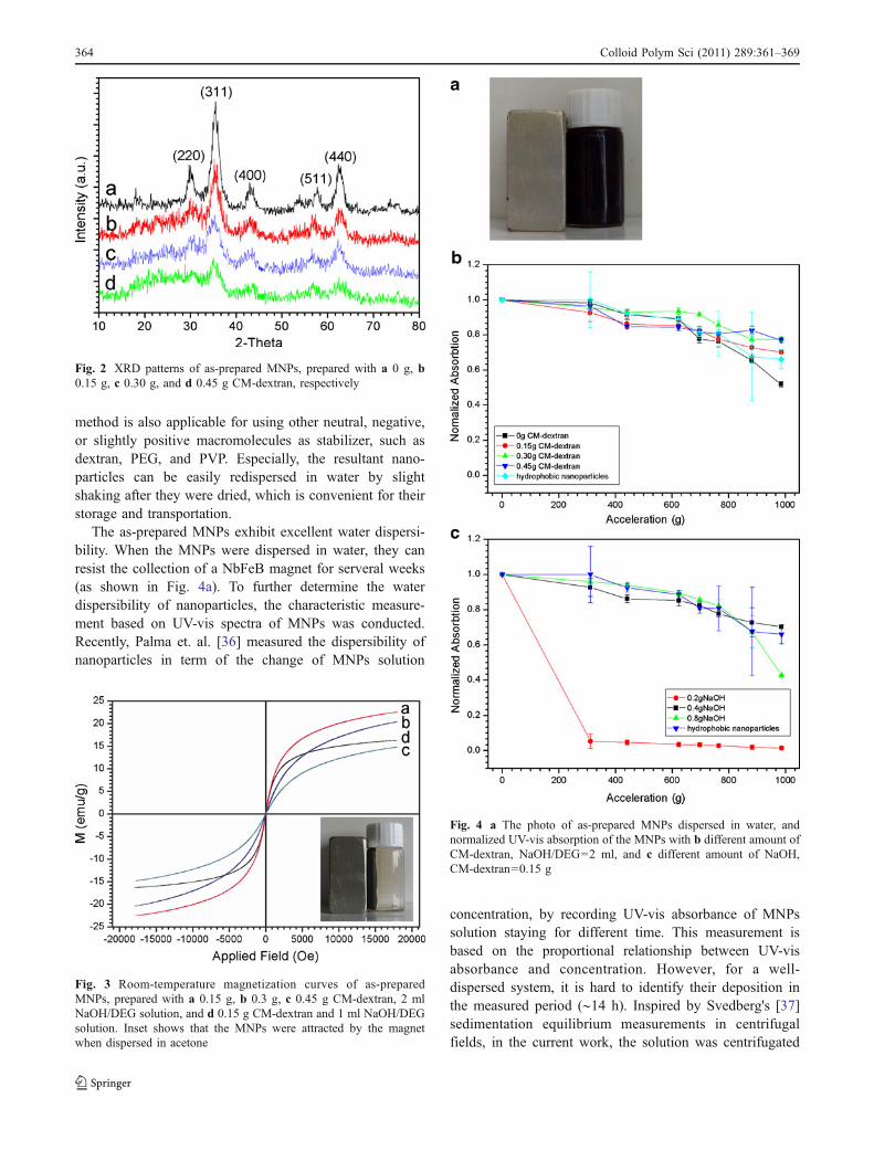
The as-prepared MNPs exhibit excellent water dispersi-bility. When the MNPs were dispersed in water, they canresist the collection of a NbFeB magnet for serveral weeks(as shown in Fig. 4a). To further determine the waterdispersibility of nanoparticles, the characteristic measure-ment based on UV-vis spectra of MNPs was conducted.Recently, Palma et. al. [36] measured the dispersibility ofnanoparticles in term of the change of MNPs solution
concentration, by recording UV-vis absorbance of MNPssolution staying for different time. This measurement isbased on the proportional relationship between UV-visabsorbance and concentration. However, for a well-dispersed system, it is hard to identify their deposition inthe measured period (∼14 h). Inspired by Svedberg's [37]sedimentation equilibrium measurements in centrifugalfields, in the current work, the solution was centrifugated
Fig. 3 Room-temperature magnetization curves of as-preparedMNPs, prepared with a 0.15 g, b 0.3 g, c 0.45 g CM-dextran, 2 mlNaOH/DEG solution, and d 0.15 g CM-dextran and 1 ml NaOH/DEGsolution. Inset shows that the MNPs were attracted by the magnetwhen dispersed in acetone
Fig. 2 XRD patterns of as-prepared MNPs, prepared with a 0 g, b0.15 g, c 0.30 g, and d 0.45 g CM-dextran, respectively
Fig. 4 a The photo of as-prepared MNPs dispersed in water, andnormalized UV-vis absorption of the MNPs with b different amount ofCM-dextran, NaOH/DEG=2 ml, and c different amount of NaOH,CM-dextran=0.15 g
364 Colloid Polym Sci (2011) 289:361–369

at first and the UV-vis absorbance of supernatant nomalizedby that of the rational solution was recorded. As areference, MNPs were also synthesized by Hyeon's classi-cal method [38] and dispersed in cyclohexane, which canbe stably dispersed for at least 1 year. The result (Fig. 4)shows that except the nanoparticles synthesized with 1 mlNaOH/DEG stock solution (containing 0.2 g NaOH), thedispersibility of MNPs in water is comparable to, if notbetter than, the reference solution. Moreover, it is foundthat the dispersibility of MNPs is not dependant on theamount of CM-dextran. Even when no macromolecule wasused in the synthesis, MNPs can still be stably dispersed.This result is not consistent with the common considerationthat macromolecule acted as the origination of waterdispersibility. However, when 1 ml NaOH/DEG stocksolution was injected, the resultant nanoparticles wouldalmost deposit even under low centrifugation speed, whichindicates that other factors rather than the macromoleculesrelated to the MNPs dispersibility.
To further confirm this, zeta potentials of nanoparticlessurface were characterized as a function of pH value. Asshown in Fig. 5a, when different amount of CM-dextran wasused in the synthesis, zeta-potential curves are almost thesame in their shape. Although little difference in acidicenvironment owing to various amount of –COOH groupscan be observed, the amount of macromolecules almost hasno influences on zeta potential in neutral–basic range. Thisphenomena also exsisted in the control experiment withoutCM-dextran or with dextran (Fig. 5b), indicating that theamount or the kind of macromolecules is not the key factorin the nanoparticles dispersibility. However, when lessNaOH (1 ml NaOH/DEG solution) was introduced, asFig. 5c shows, the zeta potential is about 5 mV in a neutralenvironment. The small potential value leads to the quickdeposition of MNPs from water, which is consistent with theUV-vis measurement. It was suggested that if the nano-particles were stabilized by steric interactions, the MNPssolution would remain stable even around its isoelectic point[39], which indicates that MNPs in our case are stabilized byelectrostatic repulsion rather than steric stabilization ofpolymeric coating layer. Meanwhile, the absolute value ofzeta potential of nanopaticles would increase with theincrease of NaOH/DEG solution, indicating that the highwater-dispersibility of resultant nanoparticles originated fromNaOH rather than macromolecules.
The influence of macromolecules and NaOH on thedispersibility of MNPs can be further evidenced by theTGA, as shown in Fig. 6. The weight loss in the range fromca. 250 to 400 °C should be ascribed to the macro-molecules, as identified by the curve of pure CM-dextran(Fig. 7 inset). When the NaOH dose is constant, the weightloss at this stage is related to the amount of macro-molecules. By comparing with weight loss curves of pure
Fig. 5 Zeta potentials of MNPs prepared with a different amount ofCM-dextran, b different kind of macromolecules, and c different dozeof NaOH/DEG solution
Colloid Polym Sci (2011) 289:361–369 365

CM-dextran and MNPs prepared without macromolecules,it can be calculated that the weight percentages of CM-dextran are about 8%, 16%, and 24% for MNPs samplesprepared with 0.15, 0.3, and 0.45 g macromolecules,respectively. This result shows that only a small part ofCM-dextran (about 8 wt.% of the added amount) washybridized with MNPs. Meanwhile, when various amountof NaOH was used, the weight loss ascribing to CM-dextran almost kept the same (about 7% weight lossbetween 250 and 400 °C). As discussed above, it can beconcluded that the high water-dispersibility is not caused byCM-dextran. Meanwhile, the weight loss between 100 and200 °C would increase with the increase of NaOH (3%,6%, and 19% weight loss for 1, 2, and 4 ml NaOH/DEG
stock solution, respectively), which makes it being consid-ered as the decomposition or evaporation of real stabilizerrelated to the high water-dispersibility of nanoparticles.
The supposition that macromolecules are not thestabilizer of nanoparticles, can explain the feasibility ofusing PAA, polyvinyl acetate, or PEG. However, whenchitosan was chosen as the macromolecule in the reaction,the sedimentation of resultant nanoparticles would bevisually observed within 1 h. Then, how does NaOH affectthe dispersibility? To elucidate this, control experiment wasperformed with the NaOH/DEG stocking solution preparedunder nitrogen protection. It is surprising that sedimentationof resultant nanoparticles can be observed within severalhours instead of a well-dispersed solution. As has beenknown, hydroxyl groups in DEG are easy to be oxidized,especially with the help of NaOH. Therefore, it is possiblethat the oxidized DEG played an important role in thestabilization of nanoparticles. Accordingly, the weight lossat the stage before 200 °C, which is related to the amount ofNaOH, should be attributed to the oxidized DEG. Thishypothesis can also be used to explain the inappropriatenessof chitosan as macromolecule. Because of the negativecharge of oxidized DEG, positive charged chitosan wouldconnect nanoparticles together by the electrostatic attrac-tion, leading to their aggregation.
To confirm the oxidization of DEG and its effect,FTIR spectra were recorded for NaOH/DEG stocksolution prepared with and without nitrogen protection,as Fig. 7 shows. The intensity of the peak at about3,357 cm−1 corresponding to –OH groups was found toweaken when the solution was prepared without nitrogenprotection, indicating the decrease of –OH groups.Importantly, the enhanced peak at about 2,697 cm−1 and
Fig. 6 a–d Weight loss curves of MNPs prepared with 2 ml NaOH/DEG solution and different amount of CM-dextran (0, 0.15, 0.30, and0.45 g for a, b, c, and d, respectively). e, f Weight loss curves of
MNPs prepared with 0.15 g CM-dextran and different dose of NaOH/DEG solution (1 and 4 ml for e and f, respectively)
Fig. 7 FTIR spectra of NaOH/DEG stock solution prepared a withoutand b with nitrogen protection
366 Colloid Polym Sci (2011) 289:361–369

shoulder peak at about 2,800 cm−1 can be observed withthe oxidation of DEG, which should be ascribed to theFermi resonance of aldehyde groups [40]. Meanwhile,peak at 1,666 cm−1, which is consistent with stretchingvibration of C=O, is also intensified. These two peaksproved that the main product of DEG oxidization containsaldehyde groups rather than carboxyl groups. Based onour results, it can be concluded that the resultant nano-particles are coated by oxidized DEG, which mainlyconsist of aldehyde groups. We further recorded thehigh-resolution XPS spectrum of C 1s of MNPs preparedwithout macromolecules. As shown in Fig. 8, a CHx peakat 284.4 eV could be observed, assigned to the alkane ofthe coating molecules. The presence of the aldehydeendgroups is confirmed by the other two peaks, whichlocated at about 285.6 and 288.4 eV, respectively. FTIRspectra of resultant nanoparticles prepared with or withoutmacromolecules were also measured to further confirm theexistence of aldehyde groups on MNPs surface. However,probably because the amount of absorption is below thedetective sensitivity of FTIR, we could not find peakscontributed from aldehyde or carboxyl groups. Our resultsalso indicate that the CM-dextran cannot efficientlystabilize the MNPs alone, probably because of theflocculation of nanoparticles being connected by theCM-dextran sodium salt [41]: Comparing with PAA, thesteric hindrance of saccharide rings and rigid chains makeCM-dextran molecules easy to bind different MNPstogether, rather than coat on one MNP, which thereforeleads to the flocculation. However, when the DEG waspartially oxidized, although some CM-dextran moleculesstill hybrid with MNPs, the soft oxidized DEG moleculesabsorbed on the surface of MNPs would prevent theaggregation of the MNPs. Therefore the water dispersi-
bility of nanoparticles improved, and it can also explainthe different stabilizing mechanism between CM-dextranand PAA as macromolecules.
For the confirmed capping agents of MNPs, the groupsanchoring on nanoparticles surface possess strong electron-withdrawing effect, or connect with electron-donatinggroups. For example, in the case of oleic acid or otherlong-chain fatty acids coating on MNPs, their electron-withdrawing carboxyl groups are directly connected withnanoparticles surface and electron-donating alkyl groupsextend outside. Also, the binding affinities of catecholderivatives toward inorganic oxides have been shown toscale with their pKa and Bronstead acidity [42, 43], whichmight be caused by the electro-withdrawing and donatingeffect. Their result also confirm this: only when electro-withdrawing substituents (nitro groups or carboxyl groups)were coupled on or near the catechol ring, the moleculescan strongly bind to the MNPs surface. In our case,carbonyl groups also possess electron-withdrawing effectand connect to the electron-donating alkyl groups (andprobably unoxidized –OH), which leads to the bindingaffinity toward Fe atoms on MNPs surface and providesthem high water-dispersibility. To further illustrate this, wesynthesized pristine MNPs dispersing in water bycoprecipitation method and mixed the solution with differentkinds of esters. It was found that the MNPs can be extractedby long-chain ester such as isopropyl myristate with slightlyshaking the mixture, but still stay in water when they mixedwith short chain esters. This result offers an evidence for therelationship between electron-withdrawing/donating effectand binding affinity toward MNPs nanoparticles, and con-firms that carbonyl groups in some cases can also be cappedon the MNPs surface. However, although elctron-withdrawing mainly results in the negative electric charge,not all molecules with negative charge can be anchored on theMNPs surface.
Conclusion
Water-soluble Fe3O4 nanoparticles were prepared by a DEG-mediated method. By varing the amount of CM-dextran andNaOH, it was found that the high water-dispersibility is notrelated to the amount of CM-dextran macromolecule, butdepends on that of NaOH. Based on the results of surfacecharacterization, it was found that the CM-dextran canhybridize with the nanoparticles to provide them a hydro-philic surface. However, the high water-dispersibility ofMNPs should be mainly attributed to the oxidized DEGanchoring on nanoparticles surface through carbonyl groups.Our results suggest that the carbonyl group is possible tobind to the MNPs surface, which probably provide moreselectiveness of modifying molecules for MNPs.
Fig. 8 Expanded XPS spectra of C 1s of the MNPs prepared withoutCM-dextran
Colloid Polym Sci (2011) 289:361–369 367

Acknowledgments This work was financially supported by NSFC(No. 30872630), Science and Technology Committee of Shanghai(No. 08ZR1415700). We thank the Instrumental Analysis Center ofSJTU for the assistance on measurements. We also thank ShanghaiSunny New Technology Development Co., Ltd. for their support.
References
1. Pankhurst QA, Thanh NKT, Jones SK, Dobson J (2009) Progressin applications of magnetic nanoparticles in biomedicine. J PhysD Appl Phys 42:224001
2. Lu AH, Salabas EL, Schüth F (2007) Magnetic nanoparticles:synthesis, protection, functionalization, and application. AngewChem Int Ed 46:1222–1244
3. Jeong U, Teng X,WangY, Yang H, Xia Y (2007) Superparamagneticcolloids: controlled synthesis and niche applications. Adv Mater19:33–60
4. Gu H, Xu K, Xu C, Xu B (2006) Biofunctional magneticnanoparticles for protein separation and pathogen detection. ChemCommun 9:941–949
5. Laurent S, Forge D, Port M, Roch A, Robic C, Elst LV, MullerRN (2008) Magnetic iron oxide nanoparticles: synthesis, stabili-zation, vectorization, physicochemical characterizations, andbiological applications. Chem Rev 108:2064–2110
6. Bulte JWM, Kraitchman DL (2004) Iron oxide MR contrastagents for molecular and cellular imaging. NMR Biomed 17:484–499
7. Shapiro EM, Skrtic S, Sharer K, Hill JM, Dunbar CE, KoretskyAP (2004) MRI detection of single particles for cellular imaging.Proc Natl Acad Sci USA 101:10901–10906
8. Gupta AK, Gupta G (2005) Synthesis and surface engineering ofiron oxide nanoparticles for biomedical applications. Biomaterials26:3995–4021
9. Kim DK, Mikhaylova M, Zhang Y, Muhammed M (2003)Protective coating of superparamagnetic iron oxide nanoparticles.Chem Mater 15:1617–1627
10. Caruso F (2001) Nanoengineering of particle surfaces. Adv Mater13:11–22
11. Häfeli U, Schütt W, Teller J, Zborowski M (1997) Scientific andclinical application of magnetic carriers. Plenum, New York
12. Bourlinos AB, Bakandritsos A, Georgakilas V, Petridis D (2002)Surface modification of ultrafine magnetic iron oxide particles.Chem Mater 14:3226–3228
13. Mornet S, Vasseur S, Grasset F, Duguet E (2004) Magneticnanoparticles design for medical diagnosis and therapy. J MaterChem 14:2161–2175
14. Pardoe H, Chua-anusorn W, St. Pierre TG, Dobson J (2001)Structural and magnetic properties of nanoscale iron oxideparticles synthesized in the presence of dextran and polyvinylalcohol. J Magn Magn Mater 225:41–46
15. Ditsch A, Laibinis PE, Wang DIC, Hatton TA (2005) Controlledclustering and enhanced stability of polymer-coated magneticnanoparticles. Langmuir 21:6006–6018
16. Si S, Kotal A, Mandal TK, Giri S, Nakamura H, Kohara T (2004)Size-controlled synthesis of magnetite nanoparticles in thepresence of polyelectrolytes. Chem Mater 16:3489–3496
17. Song HT, Choi J, Huh YM, Kim S, Jun Y, Suh JS, Cheon J (2005)Surface modulation of magnetic nanocrystals in the developmentof highly efficient magnetic resonance probes for intracellularlabeling. J Am Chem Soc 127:9992–9993
18. Fan H, Leve EW, Scullin C, Gabaldon J, Tallant D, Bunge S,Boyle T, Wilson MC, Brinker CJ (2005) Surfactant-assistedsynthesis of water-soluble and biocompatible semiconductorquantum dot micelles. Nano Lett 5:645–648
19. Prakash A, Zhu H, Jones CJ, Benoit DN, Ellsworth AZ, BryantEL, Colvin VL (2009) Bilayers as phase transfer agents fornanocrystals prepared in nonpolar solvents. ACS Nano 3:2139–2146
20. Yin Y, Alivisatos AP (2005) Colloidal nanocrystal synthesis andthe organic-inorganic interface. Nature 437:664–670
21. Lin CL, Lee CF, ChiuWY (2005) Preparation and properties of poly(acrylic acid) oligomer stabilized superparamagnetic ferrofluid. JColloid Interface Sci 291:411–420
22. Lattuada M, Hatton TA (2007) Functionalization of monodispersemagnetic nanoparticles. Langmuir 23:2158–2168
23. Illés E, Tombácz E (2006) The effect of humic acid adsorption onpH-dependent surface charging and aggregation of magnetitenanoparticles. J Colloid Interface Sci 295:115–123
24. Fauconnier N, Pons JN, Roger J, Bee A (1997) Thiolation ofmaghemite nanoparticles by dimercaptosuccinic acid. J ColloidInterface Sci 194:427–433
25. Li Z, Wei L, Gao M, Lei H (2005) One-pot reaction tosynthesize biocompatible magnetite nanoparticles. Adv Mater17:1001–1005
26. Xu C, Xu K, Gu H, Zheng R, Liu H, Zhang X, Guo Z, Xu B(2004) Dopamine as a robust anchor to immobilize functionalmolecules on the iron oxide shell of magnetic nanoparticles. J AmChem Soc 126:9938–9939
27. Xie J, Xu C, Xu Z, Hou Y, Young KL, Wang SX, Pourmand N,Sun S (2006) Linking hydrophilic macromolecules to monodis-perse magnetite (Fe3O4) nanoparticles via trichloro-s-triazine.Chem Mater 18:5401–5403
28. Kohler N, Fryxell GE, Zhang M (2004) A bifunctional poly(ethylene glycol) silane immobilized on metallic oxide-basednanoparticles for conjugation with cell targeting agents. J AmChem Soc 126:7206–7211
29. Koh I, Wang X, Varughese B, Isaacs L, Ehrman SH, English DS(2006) Preparation and characterization of antibody labeledmagnetic iron oxide nanoparticles for bioseparations. J PhysChem B 110:1553–1558
30. Caruntu D, Caruntu G, Chen Y, O'Connor CJ, Goloverda G,Kolesnichenko VL (2004) Synthesis of variable-sized nano-crystals of Fe3O4 with high surface reactivity. Chem Mater16:5527–5534
31. Ge J, Hu Y, Biasini M, Dong C, Guo J, Beyermann WP, Yin Y(2007) One-step synthesis of highly water-soluble magnetitecolloidal nanocrystals. Chem Euro J 13:7153–7161
32. Cai W, Wan J (2007) Facile synthesis of superparamagneticmagnetite nanoparticles in liquid polyols. J Colloid Interface Sci305:366–370
33. Sun Y, Xia Y (2002) Shape-controlled synthesis of gold and silvernanoparticles. Science 298:2176–2179
34. Deng H, Li XL, Peng Q, Wang X, Chen JP, Li YD (2005)Monodisperse magnetic single-crystal ferrite microspheres.Angew Chem Int Ed 44:2782–2785
35. Murray CB, Kagan CR, Bawendi MG (2000) Synthesis andcharacterization of monodisperse nanocrystals and close-packednanocrystal assemblies. Annu Rev Mater Sci 30:545–610
36. Palma RD, Peeters S, Van Bael MJ, Van den Rul H, Bonroy K,Laureyn W, Mullens J, Borghs G, Maes G (2007) Silane ligandexchange to make hydrophobic superparamagnetic nanoparticleswater-dispersible. Chem Mater 19:1821–1831
37. Svedberg T (1937) Protein molecules. Chem Rev 20:81–9838. Park J, An K, Hwang Y, Park JG, Noh HJ, Kim JY, Park JH,
Hwang NM, Hyeon T (2004) Ultra-large-scale syntheses ofmonodisperse nanocrystals. Nat Mater 3:891–895
39. Cunningham D, Littleford RE, Smith WE, Lundahl PJ, KhanI, McComb DW, Graham D, Laforest N (2006) Practicalcontrol of SERRS enhancement. Faraday Disscuss 132:135–145
368 Colloid Polym Sci (2011) 289:361–369

40. Sadtler Research Laboratories (1985) Sadtler standard spectra:standard infrared grating spectra. Sadtler Research Laboratories,Philadelphia
41. LaMer VK, Healy TW (1963) Adsorption-flocculation reactions ofmacromolecules at the solid-liquid interface. Rev Pure Appl Chem13:112–133
42. Amstad E, Gillich T, Bilecka I, Textor M, Reimhult E (2009) Ultrastableiron oxide nanoparticle colloidal suspensions using dispersants withcatechol-derived anchor groups. Nano Lett 9:4042–4048
43. Araujo PZ, Morando PJ, Blesa MA (2005) Interaction of catecholand gallic acid with titanium dioxide in aqueous suspensions:equilibrium studies. Langmuir 21:3470–3474
Colloid Polym Sci (2011) 289:361–369 369