Carbohydrates are ubiquitous modifications of eukaryotic cell surface proteins and lipids.
28
Carbohydrates are ubiquitous modifications of eukaryotic cell surface proteins and lipids. Glyc Glyc Ol
-
Upload
jazmine-pope -
Category
Documents
-
view
221 -
download
3
Transcript of Carbohydrates are ubiquitous modifications of eukaryotic cell surface proteins and lipids.
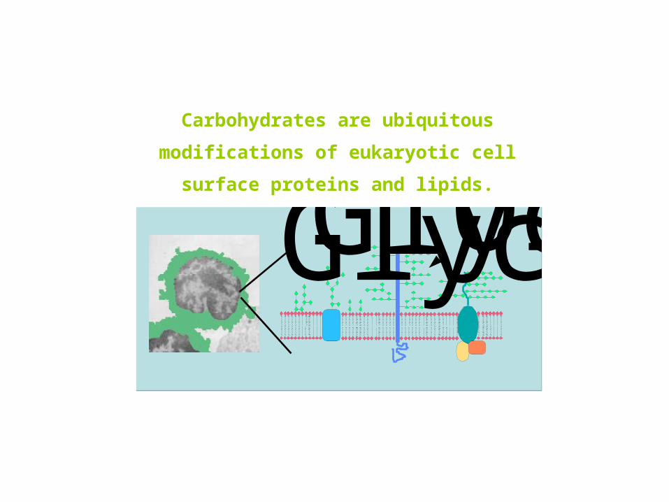
Carbohydrates are ubiquitous modifications of
eukaryotic cell surface proteins and lipids.
GlycoproteinGlycolipidOligosaccharides

Glycans
Localization of glycoconjugates in intracellular and extracellular compartments

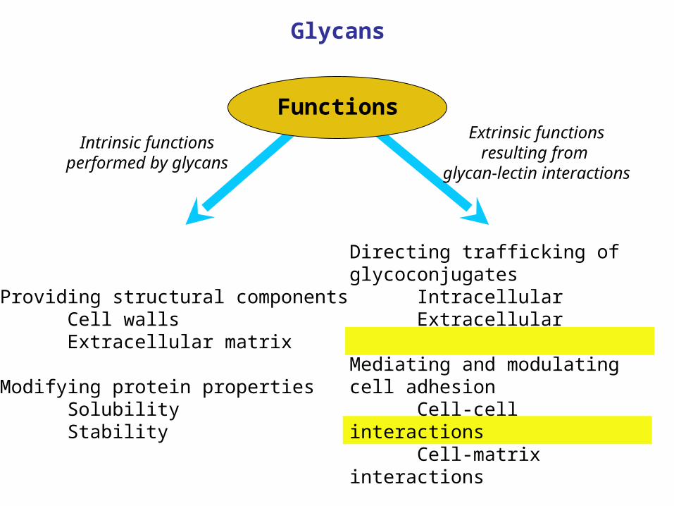
Providing structural componentsCell wallsExtracellular matrix
Modifying protein propertiesSolubilityStability
Directing trafficking of glycoconjugatesIntracellularExtracellular
Mediating and modulating cell adhesionCell-cell interactionsCell-matrix interactions
Mediating and modulating signalingIntracellularExtracellular
Functions
Extrinsic functionsresulting from
glycan-lectin interactions
Intrinsic functionsperformed by glycans
Glycans

Three major classes of glycoconjugates
Attached to proteins * through a nitrogen atom of asparagine
(N-linked)
** through an oxygen atom of serine or threonine(O-linked)
*** Attached to lipids
Glycans: Structures

Glycans are composed of monosaccharides with related chemical structures
1
2
3
4
5
6
HC=O
HCOH
HCOH
HOCH
CH2OH
HOCH
1
2
3
4
5
6
HC=O
HCOH
HCOH
HCOH
HOCH
CH2OH
Epimerization at C4
Glucose Galactose
Hexoses are the most common constituents of glycans
Glycans: Structures

Two ways to form a pyranose (6-member) ring from glucose
Glycans: Structures

Substitution of the 2-hydroxyl group of glucose or galactose with acetamido group yields N-acetylhexosamines
Glycans: Structures
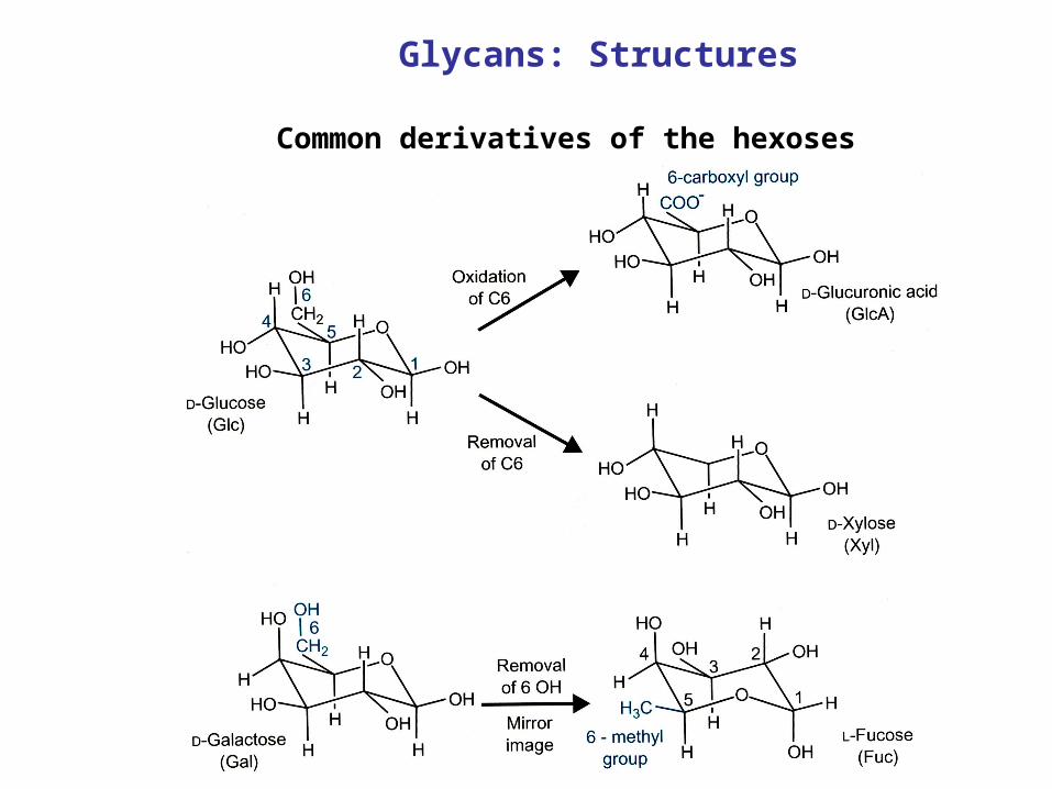
Common derivatives of the hexoses
Glycans: Structures

Structure of N-acetylneuraminic acid, the most common form of sialic acid
Glycans: Structures

Glycosidic linkages between monosaccharides exist in multiple configurations
Glycans: Structures

A typical N-linked glycan
Chemical structure
Word structure
Symbol structure
Glycans: Structures

Formation of glycosidic linkages require energy and catalyzed by specific enzymes, glycosyltransferases
Glycosyltransferase reaction
UDP-galactose, an example of
a nucleotide sugar donor
Uridine diphosphate (UDP)
Glycans: Structures

Understanding structure-function relationship for glycans can be more difficult than for other classes of biopolymers
* The functions of polypeptide and glycan portions of glycoproteins are potentially independent
Glycans: Structures
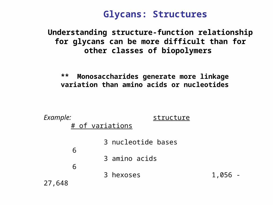
Understanding structure-function relationship for glycans can be more difficult than for other classes of biopolymers
** Monosaccharides generate more linkage variation than amino acids or nucleotides
Example: structure # of variations
3 nucleotide bases 6 3 amino acids 6 3 hexoses 1,056 - 27,648
Glycans: Structures

Glycans: Structures
Glycan structures are encoded indirectly in the genome

Providing structural componentsCell wallsExtracellular matrix
Modifying protein propertiesSolubilityStability
Directing trafficking of glycoconjugatesIntracellularExtracellular
Mediating and modulating cell adhesionCell-cell interactionsCell-matrix interactions
Mediating and modulating signalingIntracellularExtracellular
FunctionsExtrinsic functions
resulting from glycan-lectin interactions
Intrinsic functionsperformed by glycans
Glycans

I. Heparan sulfate proteoglycans
Xylose
Galactose
GlcNAc
GlcA
Disaccharide unit
Core protein
Linkage region
Sulfate
IdoA
Large O-linked chains of as many as a hundred residues

Abnormalities in heparan sulfate biosynthesis is implicated in the human multiple exostoses syndrome

Drosophilagene
Function Vertebratehomologue
Associated phenotype (mammals)
dally Cell surface proteoglycan,effects on Wg and Dppsignaling
Glypicans Overgrowth, skeletal abnormalities,tumor-susceptibility syndrome,Wilm’s tumor of the kidney
sugarless Glycosaminoglycanbiosynthesis,effects on Wg, FGFR, andDpp signaling
UDP-glucosedehydrogenase
?
sulfateless Heparan sulfatemodification,effects on Wg, Hh, andEGFR signaling
N-deacetylaseN-sulfotransferase
Mast cells: loss of heparin anddefects in secretory granules
tout-velu Heparan sulfateco–polymerase, effects onHh signaling
EXT1 & EXT2 Multiple exostoses tumors
pipe Related toglycosaminoglycan2–O–sulfotransferases,effects on ventralizingsignal in embryogenesis
Heparan sulfate2–O–sulfotransferase
Renal agenesis,eye and skeletal abnormalities

II. Notch signaling
Notch proteins mediate a wide variety of cell fate decisions during development:
in Drosophila: - Organogenesis and pattern formation - Neurogenesis - Myogenesis - Oogenesis - etc.
Malfunctioning of Notch signaling causes:
- Human T-cell lymphoblastic leukaemia (TAN-1/Notch1)- Human stroke and dementia (CADASIL, Notch3)- Spondylocostal dysostosis (Dll3) - Alagille syndrome (Jagged1)- Murine breast cancer (int-3/Notch4)
in Vertebrates (4 Notch genes, 5 ligands): - Somitogenesis - Neurogenesis - T-cell development - etc.

NotchSerrateDelta
Notch
Serrate
Delta
Fringe
O-fucose
GlcNAc
+
Providing structural componentsCell wallsExtracellular matrix
Modifying protein propertiesSolubilityStability
Directing trafficking of glycoconjugatesIntracellularExtracellular
Mediating and modulating cell adhesionCell-cell interactionsCell-matrix interactions
Mediating and modulating signalingIntracellularExtracellular
FunctionsExtrinsic functions
resulting from glycan-lectin interactions
Intrinsic functionsperformed by glycans
Glycans

III. O-mannosylation in muscular dystrophies

Normal muscle Muscular dystrophy
Extracellular
Intracellular Actin-filament
Dystrophin
Dystroglycan
Neurexin
Agrin
Laminin
Membrane
Basal Lamina
Actin-filament
Dystrophin
Dystroglycan
Neurexin
Agrin
Laminin
O-mannosyl glycans

Functions: III. O-mannosylation in muscular dystrophies
Compromised O-mannosylation of dystroglycan causes severe muscle and brain abnormalities
Example: Muscle-eye-brain disease:congenital muscular dystrophy,severe congenital myopia, hydrocephalus,
mental retardation
Muscle biopsy: Dystrophic changes
Cranial MRI: Cerebellar hypoplasiaNeuronal migration disorder
Yoshida et al., Dev. Cell 2001
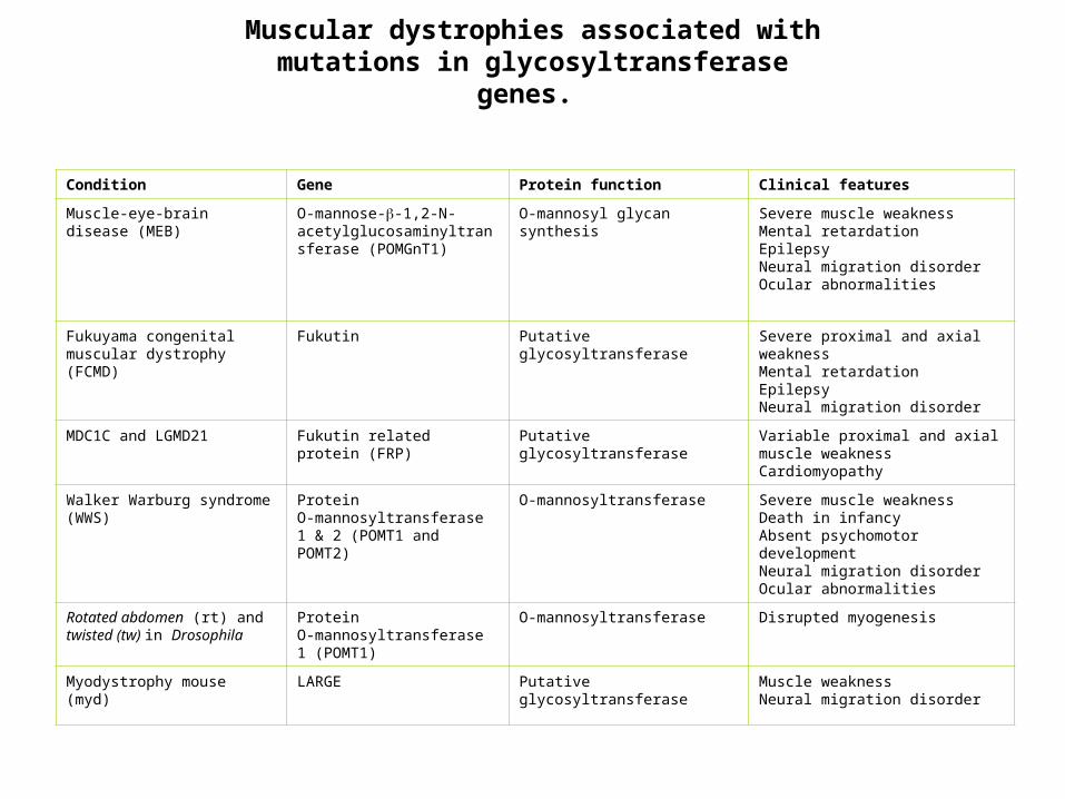
Muscular dystrophies associated with mutations in glycosyltransferase genes.
Condition Gene Protein function Clinical features
Muscle-eye-brain disease (MEB)
O-mannose--1,2-N-acetylglucosaminyltransferase (POMGnT1)
O-mannosyl glycan synthesis Severe muscle weaknessMental retardationEpilepsyNeural migration disorderOcular abnormalities
Fukuyama congenital muscular dystrophy (FCMD)
Fukutin Putative glycosyltransferase Severe proximal and axial weaknessMental retardationEpilepsyNeural migration disorder
MDC1C and LGMD21 Fukutin related protein (FRP) Putative glycosyltransferase Variable proximal and axial muscle weaknessCardiomyopathy
Walker Warburg syndrome (WWS)
Protein O-mannosyltransferase 1 & 2 (POMT1 and POMT2)
O-mannosyltransferase Severe muscle weaknessDeath in infancyAbsent psychomotor developmentNeural migration disorderOcular abnormalities
Rotated abdomen (rt) and twisted (tw) in Drosophila
Protein O-mannosyltransferase 1 (POMT1)
O-mannosyltransferase Disrupted myogenesis
Myodystrophy mouse (myd) LARGE Putative glycosyltransferase Muscle weaknessNeural migration disorder
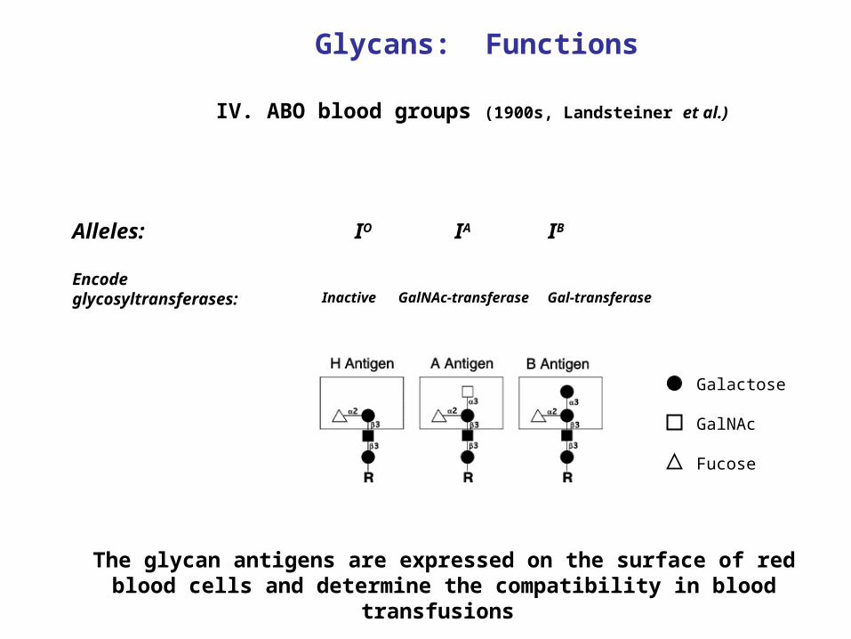
IV. ABO blood groups (1900s, Landsteiner et al.)
IO IA IBAlleles:
Encode glycosyltransferases: Inactive GalNAc-transferase Gal-transferase
Galactose
GalNAc
Fucose
Glycans: Functions
The glycan antigens are expressed on the surface of red blood cells and determine the compatibility in blood transfusions

Providing structural componentsCell wallsExtracellular matrix
Modifying protein propertiesSolubilityStability
Directing trafficking of glycoconjugatesIntracellularExtracellular
Mediating and modulating cell adhesionCell-cell interactionsCell-matrix interactions
Mediating and modulating signalingIntracellularExtracellular
StructureFunctions
Glycans