CAP Cancer Protocol Intrahepatic Bile Ducts · Web viewFor accreditation purposes, only the...
16
Protocol for the Examination of Specimens From Patients With Carcinoma of the Intrahepatic Bile Ducts Version: IntrahepaticBileDuct 4.0.0.0 Protocol Posting Date: June 2017 Includes pTNM requirements from the 8 th Edition, AJCC Staging Manual For accreditation purposes, this protocol should be used for the following procedures AND tumor types: Procedure Description Resection Includes specimens designated hepatic resection, partial or total Tumor Type Description Carcinoma Invasive carcinomas including combined hepatocellular-cholangiocarcinoma, small cell and large cell (poorly differentiated) neuroendocrine carcinoma This protocol is NOT required for accreditation purposes for the following: Procedure Biopsy Primary resection specimen with no residual cancer (eg, following neoadjuvant therapy) Cytologic specimens The following tumor types should NOT be reported using this protocol: Tumor Type Intraductal papillary neoplasm without associated invasive carcinoma Mucinous cystic neoplasm without associated invasive carcinoma Well-differentiated neuroendocrine tumors of liver Hepatocellular carcinoma and fibrolamellar carcinoma (consider the Hepatocellular Carcinoma protocol) Lymphoma (consider the Hodgkin or non-Hodgkin Lymphoma protocols) Sarcoma (consider the Soft Tissue protocol) Authors Sanjay Kakar, MD*; Chanjuan Shi, MD, PhD*; Oyedele A. Adeyi, MBBS; Wendy L. Frankel, MD; Dhanpat Jain, MD; Alyssa M. Krasinskas, MD; Mari Mino-Kenudson, MD; Timothy Pawlik, MD, MPH, PhD; Vaibhav Sahai, MD; Michael S Torbenson, MD; Jean-Nicolas Vauthey, MD; Mary K. Washington, MD, PhD © 2017 College of American Pathologists (CAP). All rights reserved. For Terms of Use please visit www.cap.org/cancerprotocols
Transcript of CAP Cancer Protocol Intrahepatic Bile Ducts · Web viewFor accreditation purposes, only the...

Protocol for the Examination of Specimens From Patients With Carcinoma of the Intrahepatic Bile DuctsVersion: IntrahepaticBileDuct 4.0.0.0 Protocol Posting Date: June 2017Includes pTNM requirements from the 8th Edition, AJCC Staging Manual
For accreditation purposes, this protocol should be used for the following procedures AND tumor types:Procedure DescriptionResection Includes specimens designated hepatic resection, partial or totalTumor Type DescriptionCarcinoma Invasive carcinomas including combined hepatocellular-
cholangiocarcinoma, small cell and large cell (poorly differentiated) neuroendocrine carcinoma
This protocol is NOT required for accreditation purposes for the following:ProcedureBiopsyPrimary resection specimen with no residual cancer (eg, following neoadjuvant therapy)Cytologic specimens
The following tumor types should NOT be reported using this protocol:Tumor TypeIntraductal papillary neoplasm without associated invasive carcinomaMucinous cystic neoplasm without associated invasive carcinomaWell-differentiated neuroendocrine tumors of liverHepatocellular carcinoma and fibrolamellar carcinoma (consider the Hepatocellular Carcinoma protocol)Lymphoma (consider the Hodgkin or non-Hodgkin Lymphoma protocols)Sarcoma (consider the Soft Tissue protocol)
AuthorsSanjay Kakar, MD*; Chanjuan Shi, MD, PhD*; Oyedele A. Adeyi, MBBS; Wendy L. Frankel, MD; Dhanpat Jain, MD; Alyssa M. Krasinskas, MD; Mari Mino-Kenudson, MD; Timothy Pawlik, MD, MPH, PhD; Vaibhav Sahai, MD; Michael S Torbenson, MD; Jean-Nicolas Vauthey, MD; Mary K. Washington, MD, PhD
With guidance from the CAP Cancer and CAP Pathology Electronic Reporting Committees.* Denotes primary author. All other contributing authors are listed alphabetically.
© 2017 College of American Pathologists (CAP). All rights reserved. For Terms of Use please visit www.cap.org/cancerprotocols

Gastrointestinal • Intrahepatic Bile DuctsIntrahepaticBileDuct 4.0.0
Accreditation RequirementsThis protocol can be utilized for a variety of procedures and tumor types for clinical care purposes. For accreditation purposes, only the definitive primary cancer resection specimen is required to have the core and conditional data elements reported in a synoptic format.
Core data elements are required in reports to adequately describe appropriate malignancies. For accreditation purposes, essential data elements must be reported in all instances, even if the response is “not applicable” or “cannot be determined.”
Conditional data elements are only required to be reported if applicable as delineated in the protocol. For instance, the total number of lymph nodes examined must be reported, but only if nodes are present in the specimen.
Optional data elements are identified with “+” and although not required for CAP accreditation purposes, may be considered for reporting as determined by local practice standards.
The use of this protocol is not required for recurrent tumors or for metastatic tumors that are resected at a different time than the primary tumor. Use of this protocol is also not required for pathology reviews performed at a second institution (ie, secondary consultation, second opinion, or review of outside case at second institution).
Synoptic ReportingAll core and conditionally required data elements outlined on the surgical case summary from this cancer protocol must be displayed in synoptic report format. Synoptic format is defined as:
Data element: followed by its answer (response), outline format without the paired "Data element: Response" format is NOT considered synoptic.
The data element must be represented in the report as it is listed in the case summary. The response for any data element may be modified from those listed in the case summary, including “Cannot be determined” if appropriate.
Each diagnostic parameter pair (Data element: Response) is listed on a separate line or in a tabular format to achieve visual separation. The following exceptions are allowed to be listed on one line:
o Anatomic site or specimen, laterality, and procedureo Pathologic Stage Classification (pTNM) elementso Negative margins, as long as all negative margins are specifically enumerated where applicable
The synoptic portion of the report can appear in the diagnosis section of the pathology report, at the end of the report or in a separate section, but all Data element: Responses must be listed together in one location
Organizations and pathologists may choose to list the required elements in any order, use additional methods in order to enhance or achieve visual separation, or add optional items within the synoptic report. The report may have required elements in a summary format elsewhere in the report IN ADDITION TO but not as replacement for the synoptic report i.e. all required elements must be in the synoptic portion of the report in the format defined above.
CAP Laboratory Accreditation Program Protocol Required Use Date: March 2018** Beginning January 1, 2018, the 8th edition AJCC Staging Manual should be used for reporting pTNM.
CAP Intrahepatic Bile Duct Protocol Summary of Changes
The following data elements were modified:Pathologic Stage Classification (pTNM, AJCC 8th Edition)Histologic TypeMarginsAdditional Pathological Findings
The following data element was deleted:Clinical History
2

CAP Approved Gastrointestinal • Intrahepatic Bile DuctsIntrahepaticBileDuct 4.0.0.0
Surgical Pathology Cancer Case Summary
Protocol posting date: June 2017
INTRAHEPATIC BILE DUCTS:
Select a single response unless otherwise indicated.
Procedure (Note A)___ Wedge resection___ Partial hepatectomy ___ Total hepatectomy___ Other (specify): _______________________________ Not specified
Tumor SizeGreatest dimension (centimeters): ___ cm+ Additional dimensions (centimeters): ___ x ___ cm___ Cannot be determined (see “Comment”)
Tumor Focality (Note B)___ Solitary (specify location): _______________________________ Multiple (specify locations): ____________________________
Histologic Type (Note C)___ Intrahepatic cholangiocarcinoma___ Combined hepatocellular-cholangiocarcinoma___ Intraductal papillary neoplasm with an associated invasive carcinoma___ Mucinous cystic neoplasm with an associated invasive carcinoma___ Large cell neuroendocrine carcinoma___ Small cell neuroendocrine carcinoma___ Neuroendocrine carcinoma (poorly differentiated)#
___ Other histologic type not listed (specify): ___________________________# Note: Select this option only if large cell or small cell cannot be determined.
Histologic Grade (Note D)___ G1: Well-differentiated___ G2: Moderately differentiated___ G3: Poorly differentiated___ Other (specify): _______________________________ GX: Cannot be assessed___ Not applicable
+ Tumor Growth Pattern (Note E)+ ___ Mass-forming+ ___ Periductal infiltrating+ ___ Mixed mass-forming and periductal infiltrating+ ___ Cannot be determined
Tumor Extension (select all that apply)___ No evidence of primary tumor___ Tumor confined to the intrahepatic bile ducts (carcinoma in situ/high-grade dysplasia)___ Tumor confined to hepatic parenchyma___ Tumor involves visceral peritoneal surface___ Tumor directly invades gallbladder
+ Data elements preceded by this symbol are not required for accreditation purposes. These optional elements may be clinically important but are not yet validated or regularly used in patient management.
3

CAP Approved Gastrointestinal • Intrahepatic Bile DuctsIntrahepaticBileDuct 4.0.0.0
___ Tumor directly invades adjacent organs other than the gallbladder (specify): ____________________________ Cannot be assessed
Margins (Note F)
Hepatic Parenchymal Margin___ Cannot be assessed___ Uninvolved by invasive carcinoma
+ Distance of invasive carcinoma from margin (millimeters or centimeters): ___ mm or ___ cm___ Involved by invasive carcinoma
Bile Duct Margin (required only if applicable)___ Not applicable___ Cannot be assessed___ Uninvolved by invasive carcinoma and high-grade intraepithelial neoplasia
+ Distance of invasive carcinoma from margin (millimeters or centimeters): ___ mm or ___cm___ Uninvolved by invasive carcinoma
+ Distance of invasive carcinoma from margin (millimeters or centimeters): ___ mm or ___cm___ Involved by invasive carcinoma___ Involved by high-grade intraepithelial neoplasia
Other Margin (required only if applicable)Specify margin: _______________________________ Cannot be assessed___ Uninvolved by invasive carcinoma___ Involved by invasive carcinoma
Lymphovascular Invasion ___ Not identified___ Present___ Cannot be determined
+ Perineural Invasion+ ___ Not identified+ ___ Present+ ___ Cannot be determined
Regional Lymph Nodes
___ No lymph nodes submitted or found
Lymph Node Examination (required only if lymph nodes are present in the specimen)
Number of Lymph Nodes Involved: _______ Number cannot be determined (explain): ______________________
Number of Lymph Nodes Examined: _______ Number cannot be determined (explain): ______________________
Pathologic Stage Classification (pTNM, AJCC 8th Edition) (Note G)Note: Reporting of pT, pN, and (when applicable) pM categories is based on information available to the pathologist at the time the report is issued. Only the applicable T, N, or M category is required for reporting; their definitions need not be included in the report. The categories (with modifiers when applicable) can be listed on 1 line or more than 1 line.
TNM Descriptors (required only if applicable) (select all that apply)___ m (multiple primary tumors)
+ Data elements preceded by this symbol are not required for accreditation purposes. These optional elements may be clinically important but are not yet validated or regularly used in patient management.
4

CAP Approved Gastrointestinal • Intrahepatic Bile DuctsIntrahepaticBileDuct 4.0.0.0
___ r (recurrent)___ y (posttreatment)
Primary Tumor (pT)___ pTX: Primary tumor cannot be assessed___ pT0: No evidence of primary tumor___ pTis: Carcinoma in situ (intraductal tumor)___ pT1: Solitary tumor without vascular invasion, ≤5 cm or >5 cm___ pT1a: Solitary tumor ≤5 cm without vascular invasion ___ pT1b: Solitary tumor >5 cm without vascular invasion___ pT2: Solitary tumor with intrahepatic vascular invasion, or multiple tumors, with or without vascular
invasion___ pT3: Tumor perforating the visceral peritoneum ___ pT4: Tumor involving local extrahepatic structures by direct invasion
Regional Lymph Nodes (pN) (Note H)___ pNX: Regional lymph nodes cannot be assessed___ pN0: No regional lymph node metastasis___ pN1: Regional lymph node metastasis present
Distant Metastasis (pM) (required only if confirmed pathologically in this case) ___ pM1: Distant metastasis
Specify site(s), if known: ____________________________
+ Additional Pathologic Findings (select all that apply) (Note I)+ Fibrosis:
+___ Cirrhosis+___ Extent of fibrosis (specify name of the scheme and scale used for assessing stage of fibrosis):
_________________________+ ___ Primary sclerosing cholangitis+ ___ Biliary stones+ ___ Chronic hepatitis (specify type): ____________________________+ ___ Other (specify): ____________________________+ ___ None identified
+ Ancillary Studies + Specify: ___________________________________
+ Comment(s)
+ Data elements preceded by this symbol are not required for accreditation purposes. These optional elements may be clinically important but are not yet validated or regularly used in patient management.
5

Background Documentation Gastrointestinal • Intrahepatic Bile DuctsIntrahepaticBileDuct 4.0.0.0
Explanatory Notes
A. ApplicationThis protocol applies only to hepatic resection specimens containing intrahepatic cholangiocarcinoma, combined hepatocellular-cholangiocarcinoma and primary high grade neuroendocrine carcinomas. Hepatocellular carcinomas and carcinomas arising in the perihilar bile ducts are staged using separate TNM systems.1
Anatomically, the intrahepatic bile ducts extend from the periphery of the liver to the second-order bile ducts (Figure 1). The perihilar bile ducts extend from the hepatic duct bifurcation to include the extrahepatic biliary tree proximal to the origin of the cystic duct. The distal extrahepatic bile duct extends from the junction of the cystic duct-common hepatic duct to the ampulla of Vater.1
Figure 1. Anatomy of the intrahepatic and extrahepatic biliary system.
B. Tumor FocalitySections should be prepared from each major tumor nodule, with representative sampling of smaller nodules. For purposes of staging, satellite nodules, multifocal primary cholangiocarcinomas, and intrahepatic metastases are considered to be multiple tumors.1 In intrahepatic cholangiocarcinoma, multiple tumor deposits have been associated with poorer survival.2,3
C. Histologic Type The protocol recommends the following modified classification of the World Health Organization (WHO).4 In the United States, approximately 30% of the primary malignant tumors of the liver are biliary carcinomas.4
WHO Classification of Carcinomas of the Intrahepatic Bile Ducts (Modified)Intrahepatic cholangiocarcinomaCombined hepatocellular-cholangiocarcinomaIntraductal papillary neoplasm with an associated invasive carcinomaMucinous cystic neoplasm with an associated invasive carcinomaPoorly differentiated neuroendocrine carcinomaLarge cell neuroendocrine carcinomaSmall cell neuroendocrine carcinoma
Combined or mixed hepatocellular-cholangiocarcinoma should show histologic evidence of both hepatocellular and biliary differentiation by morphology, and supported by immunohistochemistry.4 Hepatocellular markers with high sensitivity and specificity such as arginase-1 should be included in the panel (in addition to markers like Hep Par 1),5 and a cholangiocarcinoma component should not be diagnosed based solely on immunoreactivity with
6

Background Documentation Gastrointestinal • Intrahepatic Bile DuctsIntrahepaticBileDuct 4.0.0.0
markers like CK7, CK19, and/or MOC31, which can be positive in a subset of HCC, especially in variants like scirrhous HCC.6 Discrete gland formation with or without mucin, positive staining of these areas with CK7, CK19, and/or MOC31, and negative results in these areas with hepatocellular markers is the most reliable evidence of a cholangiocarcinoma component. The proportion of each component can be provided. The size of the entire tumor is used for staging. The demographics and clinical features of combined HCC-cholangiocarcinoma such as age, sex, viral hepatitis status, and cirrhosis tend to resemble that of HCC,7,8 while some studies have reported molecular changes similar to cholangiocarcinoma.9 Many studies show that combined HCC-cholangiocarcinoma is more aggressive compared to classical HCC and has a higher recurrence rate after liver transplantation.10,11 Carcinosarcoma is mentioned as a histologic type in the AJCC 8th edition.
D. Histologic GradeFor cholangiocarcinomas, definitive criteria for histologic grading have not been established; however, the following quantitative grading system based on the proportion of gland formation within the tumor is suggested:
Grade X Grade cannot be assessedGrade 1 Well differentiated (more than 95% of tumor composed of glands)Grade 2 Moderately differentiated (50% to 95% of tumor composed of glands)Grade 3 Poorly differentiated (less than 49% of tumor composed of glands)
Undifferentiated category is rarely used and is reserved for tumors that do not show obvious glandular, squamous, or neuroendocrine differentiation on morphology and/or immunohistochemistry. It is more appropriate to categorize these as undifferentiated carcinomas rather than cholangiocarcinoma. This category is not included in the AJCC scheme. There is no separate grading scheme for combined hepatocellular-cholangiocarcinoma; both the components can be separately graded. This grading system is not applicable to poorly differentiated neuroendocrine carcinoma.
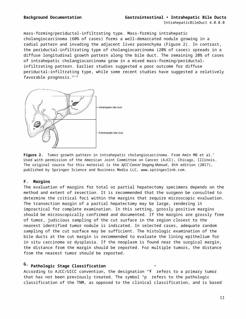
E. Tumor Growth Pattern Three tumor growth patterns of intrahepatic cholangiocarcinoma are described: the mass-forming type, the periductal infiltrating type, and mixed mass-forming/periductal-infiltrating type. Mass-forming intrahepatic cholangiocarcinoma (60% of cases) forms a well-demarcated nodule growing in a radial pattern and invading the adjacent liver parenchyma (Figure 2). In contrast, the periductal-infiltrating type of cholangiocarcinoma (20% of cases) spreads in a diffuse longitudinal growth pattern along the bile duct. The remaining 20% of cases of intrahepatic cholangiocarcinoma grow in a mixed mass-forming/periductal-infiltrating pattern. Earlier studies suggested a poor outcome for diffuse periductal-infiltrating type, while some recent studies have suggested a relatively favorable prognosis.12-15
Figure 2. Tumor growth pattern in intrahepatic cholangiocarcinoma. From Amin MB et al.1 Used with permission of the American Joint Committee on Cancer (AJCC), Chicago, Illinois. The original source for this material is the AJCC Cancer Staging Manual, 8th edition (2017), published by Springer Science and Business Media LLC, www.springerlink.com.
7

Background Documentation Gastrointestinal • Intrahepatic Bile DuctsIntrahepaticBileDuct 4.0.0.0
F. MarginsThe evaluation of margins for total or partial hepatectomy specimens depends on the method and extent of resection. It is recommended that the surgeon be consulted to determine the critical foci within the margins that require microscopic evaluation. The transection margin of a partial hepatectomy may be large, rendering it impractical for complete examination. In this setting, grossly positive margins should be microscopically confirmed and documented. If the margins are grossly free of tumor, judicious sampling of the cut surface in the region closest to the nearest identified tumor nodule is indicated. In selected cases, adequate random sampling of the cut surface may be sufficient. The histologic examination of the bile ducts at the cut margin is recommended to evaluate the lining epithelium for in situ carcinoma or dysplasia. If the neoplasm is found near the surgical margin, the distance from the margin should be reported. For multiple tumors, the distance from the nearest tumor should be reported.
G. Pathologic Stage ClassificationAccording to AJCC/UICC convention, the designation “T” refers to a primary tumor that has not been previously treated. The symbol “p” refers to the pathologic classification of the TNM, as opposed to the clinical classification, and is based on gross and microscopic examination. pT entails a resection of the primary tumor or biopsy adequate to evaluate the highest pT category, pN entails removal of nodes adequate to validate lymph node metastasis, and pM implies microscopic examination of distant lesions. Clinical classification (cTNM) is usually carried out by the referring physician before treatment during initial evaluation of the patient or when pathologic classification is not possible.
Pathologic staging is usually performed after surgical resection of the primary tumor. Pathologic staging depends on pathologic documentation of the anatomic extent of disease, whether or not the primary tumor has been completely removed. If a biopsied tumor is not resected for any reason (eg, when technically infeasible) and if the highest T and N categories or the M1 category of the tumor can be confirmed microscopically, the criteria for pathologic classification and staging have been satisfied without total removal of the primary cancer.
TNM DescriptorsFor identification of special cases of TNM or pTNM classifications, the “m” suffix and “y,” “r,” and “a” prefixes are used. Although they do not affect the stage grouping, they indicate cases needing separate analysis.
The “m” suffix indicates the presence of multiple primary tumors in a single site and is recorded in parentheses: pT(m)NM.
The “y” prefix indicates those cases in which classification is performed during or after initial multimodality therapy (ie, neoadjuvant chemotherapy, radiation therapy, or both chemotherapy and radiation therapy). The cTNM or pTNM category is identified by a “y” prefix. The ycTNM or ypTNM categorizes the extent of tumor actually present at the time of that examination. The “y” categorization is not an estimate of tumor before multimodality therapy (ie, before initiation of neoadjuvant therapy).
The “r” prefix indicates a recurrent tumor when staged after a documented disease-free interval and is identified by the “r” prefix: rTNM.
The “a” prefix designates the stage determined at autopsy: aTNM.
T Category ConsiderationsTis includes high-grade biliary intraepithelial neoplasia (BilIn-3), intraductal papillary neoplasm with high-grade dysplasia, and mucinous cystic neoplasm with high-grade dysplasia. For intraepithelial lesions, a 3-tier biliary intraepithelial neoplasia classification has been proposed.16
The T categories are based on size, vascular invasion and extrahepatic spread. For invasive carcinoma associated with intraductal papillary neoplasms and mucinous cystic neoplasms, only the size of the invasive component should be used to determine the T category. The synoptic report is not required for intraductal papillary neoplasms and mucinous cystic neoplasms in the absence of an invasive component. For invasive carcinoma associated with intraductal papillary neoplasms and mucinous cystic neoplasms, only the size of the invasive component should be used to determine the T category. The invasive portion in these cases can be
8

Background Documentation Gastrointestinal • Intrahepatic Bile DuctsIntrahepaticBileDuct 4.0.0.0
multifocal. The size of the largest focus as well as cumulative size of all invasive carcinoma foci should be included in the report. Till further data becomes available, the T category should be determined based on size of the largest invasive focus. Vascular invasion includes either gross or microscopic involvement of vessels. Major vascular invasion is defined as invasion of the branches of the main portal vein or hepatic artery (first and second order branches) or as invasion of 1 or more of the 3 hepatic veins (right, middle or left).
Direct invasion of visceral peritoneum is considered as T3, while adjacent organs, including colon, duodenum, stomach, common bile duct, portal lymph nodes, abdominal wall, and diaphragm, is considered as T4 disease. Due to inconsistent criteria for defining tumors with periductal growth pattern and its unclear association with outcome, this growth pattern is no longer a part of the T classification.
Stage GroupingsStage IA T1a N0 M0Stage IB T1b N0 M0Stage II T2 N0 M0Stage IIIA T3 N0 M0Stage IIIB T4 N0 M0
Any T N1 M0Stage IV Any T Any N M1
Additional Descriptors
Lymphovascular InvasionLymphovascular invasion (LVI) indicates whether microscopic lymphovascular invasion is identified in the pathology report. LVI includes lymphatic invasion, vascular invasion, or lymphovascular invasion.
H. Lymph NodesLymph node metastases have consistently been identified as an important predictor of outcome for intrahepatic cholangiocarcinoma.1,2,17
The lymph node involvement pattern for intrahepatic cholangiocarcinomas varies with location in the liver (Figure 3). For carcinomas arising in the right lobe of the liver (segments 5-8), the regional lymph nodes include the hilar (common bile duct, hepatic artery, portal vein, and cystic duct), periduodenal, and peripancreatic lymph nodes. For tumors arising in the left lobe, the regional lymph nodes are the hilar, inferior phrenic and gastrohepatic lymph nodes. Nodal involvement of the celiac, periaortic, or pericaval lymph nodes is considered to be distant metastasis (pM1).1
9

Background Documentation Gastrointestinal • Intrahepatic Bile DuctsIntrahepaticBileDuct 4.0.0.0
Figure 3. Segmental anatomy of the liver. From Greene et al.22 Used with permission of the American Joint Committee on Cancer (AJCC), Chicago, Illinois. The original source for this material is the AJCC Cancer Staging Atlas (2006) published by Springer Science and Business Media LLC, www.springerlink.com.
I. Additional Pathologic FindingsThe extent of fibrosis should be reported as cirrhosis or advanced fibrosis have an adverse effect on outcome. The scoring system described by Ishak18 is recommended by the AJCC Cancer Staging Manual, 8th edition,1 but other commonly used schemes (Batts-Ludwig, Metavir) can be used. The name of the staging scheme and its scale should be included.
The presence of underlying disease, such as primary sclerosing cholangitis,19 should be included in the pathology report. Biliary parasites and recurrent pyogenic cholangitismay be present along with cholangiocarcinoma in Asian countries. Hepatitis C infection, nonalcoholic fatty liver disease, obesity, and smoking are also risk factors for cholangiocarcinoma.20,21
References1. Amin MB, Edge SB, Greene FL, et al, eds. AJCC Cancer Staging Manual. 8th ed. New York, NY: Springer;
2017. 2. Ohtsuka M, Ito H, Kimura F, et al. Results of surgical treatment for intrahepatic cholangiocarcinoma and
clinicopathological factors influencing survival. Br J Surg. 2002;89(12):1525-1531.3. Sano T, Shimada K, Sakamoto Y, Ojima H, Esaki M, Kosuge T. Prognosis of perihilar carcinoma: hilar bile
duct cancer versus intrahepatic cholangiocarcinoma involving the hepatic hilus. Ann Surg Oncol. 2008;15(2):590-599.
4. Bosman FT, Carneiro F, Hruban RH, Theise ND, eds. WHO Classification of Tumours of the Digestive System. Geneva, Switzerland: WHO Press; 2010.
5. Nguyen T, Phillips D, Jain D, et al. Comparison of 5 Immunohistochemical Markers of Hepatocellular Differentiation for the Diagnosis of Hepatocellular Carcinoma. Arch Pathol Lab Med. 2015;139(8):1028-1034.
6. Krings G, Ramachandran R, Jain D, et al. Immunohistochemical pitfalls and the importance of glypican 3 and arginase in the diagnosis of scirrhous hepatocellular carcinoma. Mod Pathol. 2013;26(6):782-791.
7. Yano Y, Yamamoto J, Kosuge T, et al. Combined hepatocellular and cholangiocarcinoma: a clinicopathologic study of 26 resected cases. Jpn J Clin Oncol. 2003;33:283-287.
8. Tang D, Nagano H, Nakamura M, et al. Clinical and pathological features of Allen's type C classification of resected combined hepatocellular and cholangiocarcinoma: a comparative study with hepatocellular carcinoma and cholangiocellular carcinoma. J Gastrointest Surg. 2006;10:987-998.
10
Background Documentation Gastrointestinal • Intrahepatic Bile DuctsIntrahepaticBileDuct 4.0.0.0
9. Cazals-Hatem D, Rebouissou S, Bioulac-Sage P, et al. Clinical and molecular analysis of combined hepatocellular-cholangiocarcinomas. J Hepatol. 2004;41(2):292-298.
10. Wu CH, Yong CC, Liew EH, et al. Combined hepatocellular carcinoma and cholangiocarcinoma: diagnosis and prognosis after resection or transplantation. Transplant Proc. 2016;48(4):1100-1104.
11. Sapisochin G, Fidelman N, Roberts JP, Yao FY. Mixed hepatocellular cholangiocarcinoma and intrahepatic cholangiocarcinoma in patients undergoing transplantation for hepatocellular carcinoma. Liver Transpl. 2011;17(8):934-942.
12. Hirohashi K, Uenishi T, Kubo S, et al. Macroscopic types of intrahepatic cholangiocarcinoma: clinicopathologic features and surgical outcomes. Hepatogastroenterology. 2002;49(44):326-329.
13. Shimada K, Sano T, Sakamoto Y, Esaki M, Kosuge T, Ojima H. Surgical outcomes of the mass-forming plus periductal infiltrating types of intrahepatic cholangiocarcinoma: a comparative study with the typical mass-forming type of intrahepatic cholangiocarcinoma. World J Surg. 2007;31(10):2016-2022.
14. Imai K, Yamamoto M, Ariizumi S. Surgery for periductal infiltrating type intrahepatic cholangiocarcinoma without hilar invasion provides a better outcome than for mass-forming type intrahepatic cholangiocarcinoma without hilar invasion. Hepatogastroenterology. 2010;57(104):1333-1336.
15. Uno M, Shimada K, Yamamoto Y, et al. Periductal infiltrating type of intrahepatic cholangiocarcinoma: a rare macroscopic type without any apparent mass. Surg Today. 2012;42(12):1189-1194.
16. Zen Y, Adsay NV, Bardadin K, et al. Biliary intraepithelial neoplasia: an international interobserver agreement study and proposal for diagnostic criteria. Mod Pathol. 2007;20(6):701-709.
17. Mavros MN, Economopoulos KP, Alexiou VG and Pawlik TM (2014). Treatment and prognosis for patients with intrahepatic cholangiocarcinoma: systematic review and meta-analysis. JAMA Surg. 149(6):565-574.
18. Ishak K, Baptista A, Bianchi L, et al. Histologic grading and staging of chronic hepatitis. J Hepatol. 1995;22:696-699.
19. Shaib Y, El-Serag HB. The epidemiology of cholangiocarcinoma. Semin Liver Dis. 2004;24(2):115-125.20. Welzel TM, Graubard BI, El-Serag HB, et al. Risk factors for intrahepatic and extrahepatic
cholangiocarcinoma in the United States: a population-based case-control study. Clin Gastroenterol Hepatol. 2007;5(10):1221-1228.
21. Ben-Menachem T. Risk factors for cholangiocarcinoma [comment]. Eur J Gastroenterol Hepatol. 2007;19(8):615-617.
22. Greene FL, Compton, CC, Fritz AG, et al, eds. AJCC Cancer Staging Atlas. New York, NY: Springer; 2006.
11