CAP Breast Invasive Resection Cancer Protocol · Web viewThis protocol can be utilized for a...
49
Protocol for the Examination of Resection Specimens From Patients With Invasive Carcinoma of the Breast Version: Breast Invasive Resection 4.3.0.1 Protocol Posting Date: September 2019 CAP Laboratory Accreditation Program Protocol Required Use Date: May 2020 Includes pTNM requirements from the 8 th Edition, AJCC Staging Manual For accreditation purposes, this protocol should be used for the following procedures AND tumor types: Procedure Description Excision less than total mastectomy Includes specimens designated excision, segmental resection, lumpectomy, quadrantectomy, and segmental or partial mastectomy, with or without axillary contents Total Mastectomy Includes skin-sparing and nipple–sparing mastectomy, with or without axillary contents Tumor Type Description Invasive breast carcinoma of any type, with or without ductal carcinoma in situ (DCIS) Includes microinvasive carcinoma and carcinoma with neuroendocrine features This protocol is NOT required for accreditation purposes for the following: Procedure Needle or skin biopsies Primary resection specimen with no residual cancer (eg, following neoadjuvant therapy) Additional excision performed after the definitive resection (eg, re- excision of surgical margins) Cytologic specimens The following tumor types should NOT be reported using this protocol: Tumor Type Ductal carcinoma in situ without invasive carcinoma (consider the Breast DCIS Resection protocol) Paget disease of the nipple without invasive carcinoma (consider the Breast DCIS Resection protocol) © 2019 College of American Pathologists (CAP). All rights reserved. For Terms of Use please visit www.cap.org/cancerprotocols .
Transcript of CAP Breast Invasive Resection Cancer Protocol · Web viewThis protocol can be utilized for a...

Protocol for the Examination of Resection Specimens From Patients With Invasive Carcinoma of the BreastVersion: Breast Invasive Resection 4.3.0.1 Protocol Posting Date: September 2019
CAP Laboratory Accreditation Program Protocol Required Use Date: May 2020
Includes pTNM requirements from the 8th Edition, AJCC Staging Manual
For accreditation purposes, this protocol should be used for the following procedures AND tumor types: Procedure DescriptionExcision less than total mastectomy
Includes specimens designated excision, segmental resection, lumpectomy, quadrantectomy, and segmental or partial mastectomy, with or without axillary contents
Total Mastectomy Includes skin-sparing and nipple–sparing mastectomy, with or without axillary contents
Tumor Type DescriptionInvasive breast carcinoma of any type, with or without ductal carcinoma in situ (DCIS)
Includes microinvasive carcinoma and carcinoma with neuroendocrine features
This protocol is NOT required for accreditation purposes for the following:ProcedureNeedle or skin biopsies Primary resection specimen with no residual cancer (eg, following neoadjuvant therapy)Additional excision performed after the definitive resection (eg, re-excision of surgical margins)Cytologic specimens
The following tumor types should NOT be reported using this protocol:Tumor TypeDuctal carcinoma in situ without invasive carcinoma (consider the Breast DCIS Resection protocol) Paget disease of the nipple without invasive carcinoma (consider the Breast DCIS Resection protocol)Encapsulated or solid papillary carcinoma without invasion (consider the Breast DCIS Resection protocol)Phyllodes tumor Lymphoma (consider the Hodgkin or non-Hodgkin Lymphoma protocols)Sarcoma (consider the Soft Tissue protocol)
AuthorsPatrick L. Fitzgibbons, MD*; James L. Connolly, MD*; Shikha Bose, MD; Yunn-Yi Chen, MD, PhD; Monica E. de Baca, MD; Mary Edgerton, MD, PhD; Daniel F. Hayes, MD; Kalisha A. Hill, MD; Susan C. Lester, MD, PhD; Jean F. Simpson, MD; Ross Simpson, MD; Barbara L. Smith, MD, PhD; Lee K. Tan, MD; Donald L. Weaver, MD.With guidance from the CAP Cancer and CAP Pathology Electronic Reporting Committees.* Denotes primary author. All other contributing authors are listed alphabetically.
© 2019 College of American Pathologists (CAP). All rights reserved. For Terms of Use please visit www.cap.org/cancerprotocols.

Breast • Invasive Carcinoma 4.3.0.1Resection
Accreditation RequirementsThis protocol can be utilized for a variety of procedures and tumor types for clinical care purposes. For accreditation purposes, only the definitive primary cancer resection specimen is required to have the core and conditional data elements reported in a synoptic format.
Core data elements are required in reports to adequately describe appropriate malignancies. For accreditation purposes, essential data elements must be reported in all instances, even if the response is “not applicable” or “cannot be determined”.
Conditional data elements are only required to be reported if applicable as delineated in the protocol. For instance, the total number of lymph nodes examined must be reported, but only if nodes are present in the specimen.
Optional data elements are identified with “+” and although not required for CAP accreditation purposes, may be considered for reporting as determined by local practice standards.
The use of this protocol is not required for recurrent tumors or for metastatic tumors that are resected at a different time than the primary tumor. Use of this protocol is also not required for pathology reviews performed at a second institution (ie, secondary consultation, second opinion, or review of outside case at second institution).
Synoptic ReportingAll core and conditionally required data elements outlined on the surgical case summary from this cancer protocol must be displayed in synoptic report format. Synoptic format is defined as:
Data element: followed by its answer (response), outline format without the paired "Data element: Response" format is NOT considered synoptic.
The data element should be represented in the report as it is listed in the case summary. The response for any data element may be modified from those listed in the case summary, including “Cannot be determined” if appropriate.
Each diagnostic parameter pair (Data element: Response) is listed on a separate line or in a tabular format to achieve visual separation. The following exceptions are allowed to be listed on one line:
o Anatomic site or specimen, laterality, and procedureo Pathologic Stage Classification (pTNM) elementso Negative margins, as long as all negative margins are specifically enumerated where applicable
The synoptic portion of the report can appear in the diagnosis section of the pathology report, at the end of the report or in a separate section, but all Data element: Responses must be listed together in one location
Organizations and pathologists may choose to list the required elements in any order, use additional methods in order to enhance or achieve visual separation, or add optional items within the synoptic report. The report may have required elements in a summary format elsewhere in the report IN ADDITION TO but not as replacement for the synoptic report ie, all required elements must be in the synoptic portion of the report in the format defined above.
Summary of Changesv4.3.0.1Updated the Background Documentation (Notes)
v4.3.0.0The following data elements were modified:
Specify closest uninvolved margin – is now a non-core (optional) element Specifying the closest uninvolved DCIS margin(s) – is now conditionally required if <2mm Size of Largest Metastatic Deposit – is now a core (required) element
The following data elements were added: Distance to other margins If margins are involved Distance to other margins If margins are involved by DCIS Added Ki-67 to Ancillary Studies
2

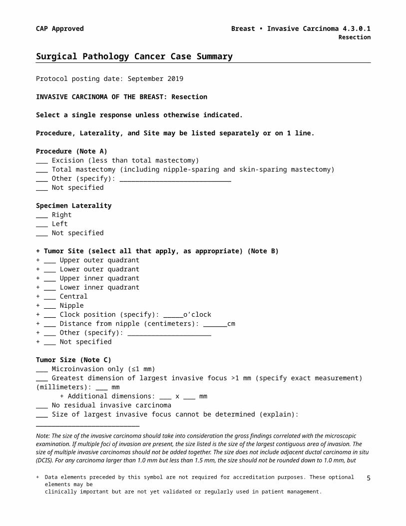
CAP Approved Breast • Invasive Carcinoma 4.3.0.1Resection
Surgical Pathology Cancer Case Summary
Protocol posting date: September 2019
INVASIVE CARCINOMA OF THE BREAST: Resection
Select a single response unless otherwise indicated.
Procedure, Laterality, and Site may be listed separately or on 1 line.
Procedure (Note A)___ Excision (less than total mastectomy)___ Total mastectomy (including nipple-sparing and skin-sparing mastectomy)___ Other (specify): _______________________________ Not specified
Specimen Laterality___ Right___ Left___ Not specified
+ Tumor Site (select all that apply, as appropriate) (Note B)+ ___ Upper outer quadrant+ ___ Lower outer quadrant+ ___ Upper inner quadrant+ ___ Lower inner quadrant+ ___ Central+ ___ Nipple+ ___ Clock position (specify): _____o’clock+ ___ Distance from nipple (centimeters): ______cm+ ___ Other (specify): _____________________+ ___ Not specified
Tumor Size (Note C) ___ Microinvasion only (≤1 mm)___ Greatest dimension of largest invasive focus >1 mm (specify exact measurement) (millimeters): ___ mm
+ Additional dimensions: ___ x ___ mm___ No residual invasive carcinoma ___ Size of largest invasive focus cannot be determined (explain): __________________________
Note: The size of the invasive carcinoma should take into consideration the gross findings correlated with the microscopic examination. If multiple foci of invasion are present, the size listed is the size of the largest contiguous area of invasion. The size of multiple invasive carcinomas should not be added together. The size does not include adjacent ductal carcinoma in situ (DCIS). For any carcinoma larger than 1.0 mm but less than 1.5 mm, the size should not be rounded down to 1.0 mm, but rather rounded up to 2.0 mm, to ensure that the tumor is not miscategorized as pT1mi. Exception to the size rule – if two histologically similar carcinomas are within 5.0 mm of each other, measure from outer edges of the two.
If there has been a prior core needle biopsy or incisional biopsy showing a larger area of invasion than in the excisional specimen, the largest dimension of the invasive carcinoma in the prior specimen should be used for T classification, if known. This also applies if the entire tumor has been removed by prior biopsy. The size of the largest foci in the two specimens should not be added together.
If there has been prior neoadjuvant treatment and no invasive carcinoma is present, the cancer is classified as ypTis if there is residual DCIS and ypT0 if there is no remaining carcinoma. A protocol is not required if no cancer is present in the specimen.
+ Data elements preceded by this symbol are not required for accreditation purposes. These optional elements may be clinically important but are not yet validated or regularly used in patient management.
3

CAP Approved Breast • Invasive Carcinoma 4.3.0.1Resection
Histologic Type (Note D)___ No residual invasive carcinoma ___ Invasive carcinoma of no special type (invasive ductal carcinoma, not otherwise specified)___ Micro-invasive carcinoma___ Invasive lobular carcinoma___ Invasive carcinoma with lobular features___ Invasive carcinoma with ductal and lobular features (“mixed type carcinoma”)___ Mucinous carcinoma___ Tubular carcinoma___ Invasive carcinoma, tubulo-lobular variant___ Invasive cribriform carcinoma___ Invasive micropapillary carcinoma___ Invasive papillary carcinoma___ Invasive carcinoma with medullary features___ Metaplastic carcinoma ___ Low-grade adenosquamous carcinoma___ Fibromatosis-like metaplastic carcinoma ___ Metaplastic carcinoma, spindle cell type___ Metaplastic carcinoma, mixed epithelial and mesenchymal type___ Invasive carcinoma with metaplastic features___ Squamous cell carcinoma___ Adenoid cystic carcinoma___ Invasive carcinoma with apocrine features___ Invasive carcinoma with clear cell (glycogen rich) features___ Invasive carcinoma with neuroendocrine features___ Invasive carcinoma, with signet-ring cell features___ Secretory carcinoma ___ Invasive carcinoma, type cannot be determined___ Other histologic type not listed (specify): ____________________________
Note: Determination of histologic type is based on routine histologic examination; special stains such as e-cadherin are not required for determining histologic type. The histologic type corresponds to the largest carcinoma. If there are smaller carcinomas of a different type, this information should be included under “Additional Pathologic Findings.”
Note: Special type carcinomas should consist of at least 90% pure pattern.
Histologic Grade (Nottingham Histologic Score) (Note E)___ No residual invasive carcinoma
Glandular (Acinar)/Tubular Differentiation___ Score 1 (>75% of tumor area forming glandular/tubular structures)___ Score 2 (10% to 75% of tumor area forming glandular/tubular structures)___ Score 3 (<10% of tumor area forming glandular/tubular structures)___ Only microinvasion present (not graded)___ No residual invasive carcinoma___ Score cannot be determined
+ Data elements preceded by this symbol are not required for accreditation purposes. These optional elements may be clinically important but are not yet validated or regularly used in patient management.
4
CAP Approved Breast • Invasive Carcinoma 4.3.0.1Resection
Nuclear Pleomorphism___ Score 1 (nuclei small with little increase in size in comparison with normal breast epithelial cells, regular
outlines, uniform nuclear chromatin, little variation in size)___ Score 2 (cells larger than normal with open vesicular nuclei, visible nucleoli, and moderate variability in both
size and shape)___ Score 3 (vesicular nuclei, often with prominent nucleoli, exhibiting marked variation in size and shape,
occasionally with very large and bizarre forms)___ Only microinvasion present (not graded)___ No residual invasive carcinoma ___ Score cannot be determined
Mitotic Rate (see Table 1)___ Score 1 ___ Score 2 ___ Score 3 ___ Only microinvasion present (not graded)___ No residual invasive carcinoma ___ Score cannot be determined
Overall Grade___ Grade 1 (scores of 3, 4, or 5) ___ Grade 2 (scores of 6 or 7) ___ Grade 3 (scores of 8 or 9) ___ Only microinvasion present (not graded)___ No residual invasive carcinoma ___ Score cannot be determined (explain: ________________)
Note: The grade corresponds to the largest area of invasion. If there are smaller foci of invasion of a different grade, this information should be included under “Additional Pathologic Findings.”
+ Tumor Focality (Note F) + ___ Single focus of invasive carcinoma+ ___ Multiple foci of invasive carcinoma
+ ___ Number of foci: ___+ ___ Number of foci is at least: ____+ ___ Number of foci cannot be determined+ Sizes of individual foci (millimeters): ____mm
+ ___ Cannot be determined
Note: If there are multiple invasive carcinomas, size, grade, histologic type, and the results of studies for estrogen receptor (ER), progesterone receptor (PgR), and HER2 should pertain to the largest invasive carcinoma. If smaller invasive carcinomas differ in any of these features, this information may be included in the “Comments” section.
Note: Sizes of individual foci may be repeated as needed or recorded on a single line.
Ductal Carcinoma In Situ (DCIS) (Note G)___ Not identified___ Present
+ ___ Negative for extensive intraductal component (EIC) + ___ Positive for extensive intraductal component (EIC) + ___ Only DCIS is present after presurgical (neoadjuvant) therapy
Note: If there has been prior neoadjuvant treatment and only residual DCIS, the cancer is classified as ypTis.___ Cannot be excluded
+ Size (Extent) of DCIS + Estimated size (extent) of DCIS is at least (millimeters) ___ mm
+ Additional dimensions (millimeters): ___ x ___ mm+ Number of blocks with DCIS: ___
+ Data elements preceded by this symbol are not required for accreditation purposes. These optional elements may be clinically important but are not yet validated or regularly used in patient management.
5

CAP Approved Breast • Invasive Carcinoma 4.3.0.1Resection
+ Number of blocks examined: ___
Note: The size (extent) of DCIS (greatest dimension using gross and microscopic evaluation) is an estimation of the volume of breast tissue occupied by DCIS. This information may be helpful for cases with a predominant component of DCIS (eg, DCIS with microinvasion) but may not be necessary for cases of EIC-negative invasive carcinomas.
+ Architectural Patterns (if DCIS is present in specimen select all that apply)+ ___ Comedo + ___ Paget disease (DCIS involving nipple skin)+ ___ Cribriform+ ___ Micropapillary+ ___ Papillary+ ___ Solid+ ___ Other (specify): ________________________
+ Nuclear Grade (if DCIS is present in specimen, see Table 2)+ ___ Grade I (low)+ ___ Grade II (intermediate) + ___ Grade III (high)
+ Necrosis (If DCIS is present in specimen)+ ___ Not identified+ ___ Present, focal (small foci or single cell necrosis)+ ___ Present, central (expansive “comedo” necrosis)
+ Lobular Carcinoma In Situ (LCIS)+ ___ No LCIS in specimen+ ___ Present
Tumor Extension (required only if the structures are present and involved) (select all that apply) (Note H) Skin___ Skin is not present___ Skin is present and uninvolved___ Invasive carcinoma directly invades into the dermis or epidermis without skin ulceration (this does not change
the T classification)___ Invasive carcinoma directly invades into the dermis or epidermis with skin ulceration (classified as T4b)___ Satellite skin foci of invasive carcinoma are present (ie, not contiguous with the invasive carcinoma in the
breast) (classified as T4b)
Note: Satellite skin nodules must be separate from the primary tumor and macroscopically identified to assign a category as T4b. Skin nodules identified only on microscopic examination and in the absence of epidermal ulceration or skin edema (clinical peau d’orange) do not qualify as T4b. Such tumors should be categorized based on tumor size.
Nipple___ DCIS does not involve the nipple epidermis ___ DCIS involves nipple epidermis (Paget disease of the nipple)Note: This finding does not change the T classification of invasive carcinomas.
Skeletal Muscle___ No skeletal muscle is present___ Skeletal muscle is free of carcinoma___ Carcinoma invades skeletal muscle___ Carcinoma invades into skeletal muscle and into the chest wall (classified as T4a)
Note: Invasion into pectoralis muscle is not considered chest wall invasion, and cancers are not classified as T4a unless there is invasion deeper than this muscle.
+ Data elements preceded by this symbol are not required for accreditation purposes. These optional elements may be clinically important but are not yet validated or regularly used in patient management.
6

CAP Approved Breast • Invasive Carcinoma 4.3.0.1Resection
Margins (Note I)
Invasive Carcinoma Margins (required only if residual invasive carcinoma is present in specimen)___ Cannot be assessed ___ Uninvolved by invasive carcinoma
Distance from closest margin (millimeters):___ Specify ___ mm ___ Less than ____ mm___ Greater than ___ mm ___ Cannot be determined (explain): _____________
+ Specify closest margin(s): ______________________________ Cannot be determined (explain): ___________________________
+ Distance from other margins (specify millimeter distance including greater than if appropriate): + ___ Anterior: ___ mm+ ___ Posterior: ___ mm+ ___ Superior: ___ mm+ ___ Inferior: ___ mm+ ___ Medial: ___ mm+ ___ Lateral: ___ mm+ ___ Other (specify margin): ___ mm
___ Positive for invasive carcinoma (select all that apply):Note: Margin status is listed as “positive” if there is ink on invasive carcinoma (ie, the distance is 0 mm). Extent of margin involvement may be specified as unifocal, multifocal, or extensive.
___ Anterior+ Extent (specify): ___________________
___ Posterior+ Extent (specify): ___________________
___ Superior+ Extent (specify): ___________________
___ Inferior+ Extent (specify): ___________________
___ Medial+ Extent (specify): ___________________
___ Lateral+ Extent (specify): ___________________
___ Other (specify margin): ___________________________+ Extent (specify): ___________________
___ Cannot be determined (explain): ___________________________
+ Distance from other margins (specify millimeter distance including greater than if appropriate): + ___ Anterior: ___ mm+ ___ Posterior: ___ mm+ ___ Superior: ___ mm+ ___ Inferior: ___ mm+ ___ Medial: ___ mm+ ___ Lateral: ___ mm+ ___ Other (specify margin): ___ mm
+ Data elements preceded by this symbol are not required for accreditation purposes. These optional elements may be clinically important but are not yet validated or regularly used in patient management.
7

CAP Approved Breast • Invasive Carcinoma 4.3.0.1Resection
DCIS Margins (required only if DCIS is present in specimen)___ Not applicable (no DCIS in specimen)___ Cannot be assessed___ Uninvolved by DCIS Note: For specimens in which the margin is uninvolved (no ink on carcinoma), the closest margin(s) must be specified if the distance of DCIS from the margin is less than 2 mm. Distances can be specific measurements or expressed as greater than or less than a measurement.
Distance from closest margin (millimeters):___ Specify ___ mm ___ Less than ____ mm___ Greater than ___ mm ___ Cannot be determined (explain): _____________
Specify closest margin(s) (required only if <2mm): ______________________________ Cannot be determined (explain): ___________________________
+ Distance from other margins (specify millimeter distance including greater than if appropriate): + ___ Anterior: ___ mm+ ___ Posterior: ___ mm+ ___ Superior: ___ mm+ ___ Inferior: ___ mm+ ___ Medial: ___ mm+ ___ Lateral: ___ mm+ ___ Other (specify margin): ___ mm
___ Positive for DCIS (select all that apply):Note: Margin status is listed as “positive” if there is ink on DCIS (ie, the distance is 0 mm). Extent of margin involvement may be specified as unifocal, multifocal, or extensive.
___ Anterior+ Extent (specify): ___________________
___ Posterior+ Extent (specify): ___________________
___ Superior+ Extent (specify): ___________________
___ Inferior+ Extent (specify): ___________________
___ Medial+ Extent (specify): ___________________
___ Lateral+ Extent (specify): ___________________
___ Other (specify margin): ___________________________+ Extent (specify): ___________________
___ Cannot be determined (explain): ___________________________
+ Distance from other margins (specify millimeter distance including greater than if appropriate): + ___ Anterior: ___ mm+ ___ Posterior: ___ mm+ ___ Superior: ___ mm+ ___ Inferior: ___ mm+ ___ Medial: ___ mm+ ___ Lateral: ___ mm+ ___ Other (specify margin): ___ mm
+ Data elements preceded by this symbol are not required for accreditation purposes. These optional elements may be clinically important but are not yet validated or regularly used in patient management.
8

CAP Approved Breast • Invasive Carcinoma 4.3.0.1Resection
Regional Lymph Nodes (Note J)___ No lymph nodes submitted or found___ Uninvolved by tumor cells
Total Number of Lymph Nodes Examined: ____Number of Sentinel Nodes Examined (if applicable): ____
___ Involved by tumor cellsNumber of Lymph Nodes with Macrometastases (>2 mm): ____Number of Lymph Nodes with Micrometastases (>0.2 mm to 2 mm and/or >200 cells): ____Number of Lymph Nodes with Isolated Tumor Cells (≤0.2 mm or ≤200 cells)#: ____# Reporting the number of lymph nodes with isolated tumor cells is required only in the absence of macrometastasis or micrometastasis in other lymph nodes.
Size of Largest Metastatic Deposit (millimeters): ____mm
Extranodal Extension___ Not identified___ Present
+ Extent of extranodal extension+ ___≤2 mm+ ___>2 mm
___ Cannot be determined
Total Number of Lymph Nodes Examined: ____Number of Sentinel Nodes Examined (if applicable): ____
Treatment Effect (required only if known) (Note K)___ No known presurgical therapy
The following question about treatment effect in the breast is required only if it is known that the patient had presurgical therapy. Treatment effect in the lymph nodes is only required if lymph nodes are submitted and it is known that the patient had presurgical therapy.
Treatment Effect in the Breast___ No definite response to presurgical therapy in the invasive carcinoma___ Probable or definite response to presurgical therapy in the invasive carcinoma___ No residual invasive carcinoma is present in the breast after presurgical therapy Note: The largest focus of residual tumor, if present, is used to determine ypT category. Treatment-related fibrosis in the tumor bed adjacent to residual invasive carcinoma is not included in determining ypT dimension.
Treatment Effect in the Lymph Nodes___ Not applicable___ No definite response to presurgical therapy in metastatic carcinoma___ Probable or definite response to presurgical therapy in metastatic carcinoma___ No lymph node metastases. Fibrous scarring, possibly related to prior lymph node metastases with
pathologic complete response___ No lymph node metastases and no prominent fibrous scarring in the nodesNote: The largest focus of residual tumor in the lymph nodes, if present, is used to determine ypN category. Treatment-related fibrosis adjacent to residual nodal deposits is not included in determining ypN dimension.
+ Lymphovascular Invasion (Note L)+ ___ Not identified+ ___ Present+ ___ Cannot be determined
+ Data elements preceded by this symbol are not required for accreditation purposes. These optional elements may be clinically important but are not yet validated or regularly used in patient management.
9

CAP Approved Breast • Invasive Carcinoma 4.3.0.1Resection
+ Dermal Lymphovascular Invasion+ ___ No skin present+ ___ Not identified+ ___ Present+ ___ Cannot be determined
Pathologic Stage Classification (pTNM, AJCC 8th Edition) (Note M)Note: Reporting of pT, pN, and (when applicable) pM categories is based on information available to the pathologist at the time the report is issued. Only the applicable T, N, or M category is required for reporting; their definitions need not be included in the report. The categories (with modifiers when applicable) can be listed on 1 line or more than 1 line. Assignment of Pathologic Prognostic Stage Group is the responsibility of the managing physician and not the pathologist.
TNM Descriptors (required only if applicable) (select all that apply)___ m (multiple foci of invasive carcinoma)___ r (recurrent)___ y (posttreatment)
Primary Tumor (pT) ___ pTX: Primary tumor cannot be assessed___ pT0: No evidence of primary tumor#
___ pTis (DCIS): Ductal carcinoma in situ#
___ pTis (Paget): Paget disease of the nipple not associated with invasive carcinoma and/or DCIS in the underlying breast parenchyma##
___ pT1: Tumor ≤20 mm in greatest dimension___ pT1mi: Tumor ≤1 mm in greatest dimension ___ pT1a: Tumor >1 mm but ≤5 mm in greatest dimension (round any measurement >1.0−1.9 mm to 2
mm)___ pT1b: Tumor >5 mm but ≤10 mm in greatest dimension___ pT1c: Tumor >10 mm but ≤20 mm in greatest dimension___ pT2: Tumor >20 mm but ≤50 mm in greatest dimension___ pT3: Tumor >50 mm in greatest dimension___ pT4: Tumor of any size with direct extension to the chest wall and/or to the skin (ulceration or skin
nodules) ###
___ pT4a: Extension to the chest wall; invasion or adherence to pectoralis muscle in the absence of invasion of chest wall structures does not qualify as T4
___ pT4b: Ulceration and/or ipsilateral macroscopic satellite nodules and/or edema (including peau d’orange) of the skin that does not meet the criteria for inflammatory carcinoma
___ pT4c: Both T4a and T4b are present___ pT4d: Inflammatory carcinoma##### For the purposes of this case summary, these categories should only be used in the setting of preoperative (neoadjuvant) therapy for which a previously diagnosed invasive carcinoma is no longer present after treatment. Patients with pathological complete response (absence of residual invasive carcinoma in both the breast and lymph nodes) should be categorized as ypT0N0 or ypTisN0, not ypTX.## Carcinomas in the breast parenchyma associated with Paget disease are categorized based on the size and characteristics of the parenchymal disease, although the presence of Paget disease should still be noted.### Note: Invasion of the dermis alone does not qualify as pT4.#### Inflammatory carcinoma requires the presence of clinical findings of erythema and edema involving at least one-third or more of the skin of the breast (see Note M).
+ Data elements preceded by this symbol are not required for accreditation purposes. These optional elements may be clinically important but are not yet validated or regularly used in patient management.
10

CAP Approved Breast • Invasive Carcinoma 4.3.0.1Resection
Regional Lymph Nodes Modifier (required only if applicable)___ (sn): Sentinel node(s) evaluated. If 6 or more nodes (sentinel or nonsentinel) are removed, this
modifier should not be used.___ (f): Nodal metastasis confirmed by fine needle aspiration or core needle biopsy.
Note: The (sn) modifier is added to the N category when a sentinel node biopsy is performed (using either dye or tracer) and fewer than six lymph nodes are removed (sentinel and nonsentinel). The (f) modifier is added to the N category to denote confirmation of metastasis by fine needle aspiration/core needle biopsy with NO further resection of nodes.
Regional Lymph Nodes (pN) (choose a category based on lymph nodes received with the specimen; immunohistochemistry and/or molecular studies are not required)Note: If internal mammary lymph nodes, infraclavicular lymph nodes, or supraclavicular lymph nodes are included in the specimen, consult the AJCC Staging Manual for additional lymph node categories.
___ pNX: Regional lymph nodes cannot be assessed (eg, not removed for pathological study or previously removed)
___ pN0: No regional lymph node metastasis identified or ITCs only#
___ pN0 (i+): ITCs only (malignant cell clusters no larger than 0.2 mm) in regional lymph node(s)___ pN0 (mol+): Positive molecular findings by reverse transcriptase polymerase chain reaction (RT-PCR); no
ITCs detected___ pN1mi: Micrometastases (approximately 200 cells, larger than 0.2 mm, but none larger than 2.0 mm)___ pN1a: Metastases in 1 to 3 axillary lymph nodes, at least 1 metastasis larger than 2.0 mm##
___ pN1b: Metastases in ipsilateral internal mammary sentinel nodes, excluding ITCs___ pN1c: pN1a and pN1b combined___ pN2a: Metastases in 4 to 9 axillary lymph nodes (at least 1 tumor deposit larger than 2.0 mm)##
___ pN2b: Metastases in clinically detected internal mammary lymph nodes with or without microscopic confirmation; with pathologically negative axillary nodes
___ pN3a: Metastases in 10 or more axillary lymph nodes (at least 1 tumor deposit larger than 2.0 mm) or metastases to the infraclavicular (Level III axillary lymph) nodes##
___ pN3b: pN1a or pN2a in the presence of cN2b (positive internal mammary nodes by imaging); or pN2a in the presence of pN1b
___ pN3c: Metastases in ipsilateral supraclavicular lymph nodes # Isolated tumor cells (ITCs) are defined as small clusters of cells not greater than 0.2 mm or single tumor cells, or a cluster of fewer than 200 cells in a single histologic cross-section. ITCs may be detected by routine histology or by immunohistochemical (IHC) methods. Nodes containing only ITCs are excluded from the total positive node count for purposes of N classification but should be included in the total number of nodes evaluated.## Approximately 1000 tumor cells are contained in a 3-dimensional 0.2-mm cluster. Thus, if more than 200 individual tumor cells are identified as single dispersed tumor cells or as a nearly confluent elliptical or spherical focus in a single histologic section of a lymph node, there is a high probability that more than 1000 cells are present in the lymph node. In these situations, the node should be classified as containing a micrometastasis (pN1mi). Cells in different lymph node cross-sections or longitudinal sections or levels of the block are not added together; the 200 cells must be in a single node profile even if the node has been thinly sectioned into multiple slices. It is recognized that there is substantial overlap between the upper limit of the ITC and the lower limit of the micrometastasis categories due to inherent limitations in pathologic nodal evaluation and detection of minimal tumor burden in lymph nodes. Thus, the threshold of 200 cells in a single cross-section is a guideline to help pathologists distinguish between these 2 categories. The pathologist should use judgment regarding whether it is likely that the cluster of cells represents a true micrometastasis or is simply a small group of isolated tumor cells.
Distant Metastasis (pM) (required only if confirmed pathologically in this case)___ pM1: Histologically proven metastases larger than 0.2 mm
Specify site, if known: ____________________
+ Additional Pathologic Findings (Note N)+ Specify: ____________________________
+ Data elements preceded by this symbol are not required for accreditation purposes. These optional elements may be clinically important but are not yet validated or regularly used in patient management.
11

CAP Approved Breast • Invasive Carcinoma 4.3.0.1Resection
+ Ancillary Studies Note: The CAP Breast Biomarker Template should be used for reporting biomarkers requested for this resection specimen. Pending biomarker studies should be listed in the Comments section of this report.
+ ___ Breast Biomarker Testing Performed on Previous Biopsy+ Testing Performed on Case Number: _________________
Note: The previously reported biopsy biomarker status may be included additionally in the resection report.
+ Estrogen Receptor (ER) + ___ Positive ___%+ ___ Negative+ ___ Cannot be determined (indeterminate)
+ Progesterone Receptor (PgR) + ___ Positive ___%+ ___ Negative+ ___ Cannot be determined (indeterminate)
+ HER2 (by immunohistochemistry)+ ___ Negative (Score 0)+ ___ Negative (Score 1+)+ ___ Equivocal (Score 2+)+ ___ Positive (Score 3+)+ ___ Cannot be determined (indeterminate)
+ HER2 (by in situ hybridization)+ ___ Negative (not amplified)+ ___ Positive (amplified)+ ___ Cannot be determined (indeterminate)
+ ___ Ki-67 percentage of positive nuclei: ___%
+ Microcalcifications (select all that apply) (Note O)+ ___ Not identified + ___ Present in DCIS+ ___ Present in invasive carcinoma+ ___ Present in non-neoplastic tissue+ ___ Other (specify): ______________________________________
+ Clinical History (select all that apply) The current clinical/radiologic breast findings for which this surgery is performed include:+ ___ Palpable mass+ ___ Nipple discharge+ ___ Other (specify): ____________________
+ ___ Prior history of breast cancer+ Specify site, diagnosis, and prior treatment: ______________________
+ ___ Prior presurgical (neoadjuvant) therapy for this diagnosis of invasive carcinoma
+ Radiologic Finding (select all that apply) + ___ Mass or architectural distortion+ ___ Calcifications+ ___ Other (specify): _________________________
+ Comment(s)
+ Data elements preceded by this symbol are not required for accreditation purposes. These optional elements may be clinically important but are not yet validated or regularly used in patient management.
12

Background Documentation Breast • Invasive Carcinoma of the Breast 4.3.0.1Resection
Explanatory Notes
A. ProceduresThe following types of breast specimens and procedures may be reported with the case summary:
Excisions: These procedures resect breast tissue without the intent of removing the entire breast. The nipple is usually not included with excisions. Excisions include specimens designated “partial mastectomies,” “lumpectomies,” and “quadrantectomies.”
Total Mastectomy: Removal of all breast tissue, generally including the nipple and areola.
Simple mastectomy : This procedure consists of a total mastectomy without removal of axillary lymph nodes.
Skin sparing mastectomy : This is a total mastectomy with removal of the nipple and only a narrow surrounding rim of skin.
Nipple sparing mastectomy : This is a total mastectomy without removal of skin or nipple. The subareolar tissue is examined and the nipple later removed if involved by carcinoma.
Modified radical mastectomy : This procedure consists of a total mastectomy with an axillary dissection. In the case summary, the breast and lymph node specimens are documented separately. A small portion of pectoralis muscle is sometimes removed.
Radical mastectomy : This procedure consists of a total mastectomy with removal of the pectoralis major and pectoralis minor muscles as well as axillary contents. This type of specimen and procedure can be indicated on the case summary as “Other.”
The case summary is intended for reporting the patient’s specimen with the largest focus of invasive carcinoma. If additional margin excisions are performed in the same procedure, the findings for these specimens can be included in the margin evaluation. If additional smaller foci of invasive carcinoma are present in the main excision or in margin excisions, the characteristics of these carcinomas (ie, size, histologic type, and grade) should be recorded under “Additional Pathologic Findings.” Additional ancillary studies on smaller foci of carcinoma are recommended if the carcinomas are of different histologic type or grade. If additional margin excisions are performed in a subsequent procedure (eg, on another day), and a larger area of invasive carcinoma is not present, the case summary need not be used.
If a patient has 2 ipsilateral invasive carcinomas removed in 2 separate excisions during the same procedure, the case summary should be used for the larger invasive carcinoma. The pathologic findings for the smaller cancer may be reported without using the case summary. If a patient has 2 ipsilateral invasive carcinomas removed in 2 separate excisions in procedures on different days, the case summary should be used for the larger carcinoma, and the American Joint Committee on Cancer (AJCC) T classification will pertain to this carcinoma. If a patient has bilateral breast carcinomas, the cancers are reported in separate case summaries.
If information from other specimens is included in completing the case summary (eg, the results of hormone receptors from a prior core needle biopsy or the finding of lymph node metastases on a previous lymph node biopsy), then this must be clearly stated in the “Comments” section, and the accession numbers of the other cases should be provided.
The following types of specimens should not be reported by using this protocol:
Very small incisional biopsies (including core needle biopsies) Re-excision of a biopsy site after removal of most of the carcinoma
Specimen sampling for specimens with invasive carcinoma has the following goals1-5:
13

Background Documentation Breast • Invasive Carcinoma of the Breast 4.3.0.1Resection
The clinical or radiologic lesion for which the surgery was performed must be examined microscopically. If the lesion is a nonpalpable imaging finding, the specimen radiograph and/or additional radiologic studies may be necessary to identify the lesion. When practical, the entire lesion, or the entire area with the imaging finding, should be submitted in a sequential fashion for histologic examination.
If the specimen consists predominantly of DCIS with microinvasion, complete submission of the entire specimen, or at a minimum the entire grossly involved area, is recommended to identify additional areas of invasion and/or lymphovascular invasion.
All other gross lesions in the specimen must be sampled.
Each designated margin must be evaluated for involvement by invasive carcinoma and DCIS. If the specimen is received sectioned or fragmented, this should be noted, as this will limit the ability to evaluate the status of margins.
Tissue may be taken for research studies or assays that do not involve the histologic examination of the tissue (eg, reverse transcriptase polymerase chain reaction [RT-PCR]) only when taken in such a way as to not compromise the evaluation of the invasive carcinoma and lymph nodes for prognostic factors and margin status.
It is preferable that the area of carcinoma be removed in a single intact specimen. If the specimen has been incised or is fragmented, then it may not be possible to accurately assess margins. If invasive carcinoma is present in more than 1 fragment, it may be difficult or impossible to determine the pathologic size of the invasive carcinoma or the number of invasive carcinomas present. Breast imaging correlation is recommended for these cases. When specimen fragmentation limits the evaluation of tumor size and/or margins, this information should be included under “Additional Pathologic Finding.”
The size of all specimens in 3 dimensions should be documented in the gross description. It is optional to also include specimen sizes in the final diagnosis. The volume of tissue removed can be helpful in estimating the extent of carcinoma present and determining the likely volume of tissue that would need to be removed to achieve tumor-free margins.
If separate oriented margin specimens are excised, the results of the final margin status can be included under “Margins” in the case summary. If not oriented, the findings can be reported under “Additional Pathologic Findings.”
References1. Association of Directors of Anatomic and Surgical Pathology. Immediate management of mammographically
detected breast lesions. Hum Pathol. 1993;24:689-690.2. Connolly JL, Schnitt SJ. Evaluation of breast biopsy specimens in patients considered for treatment by
conservative surgery and radiation therapy for early breast cancer. Pathol Annu. 1988;23(pt 1):1-23.3. Schnitt SJ, Wang HH. Histologic sampling of grossly benign breast biopsies: how much is enough? Am J
Surg Pathol. 1989;13:505-512.4. Schnitt SJ, Connolly JL. Processing and evaluation of breast excision specimens: a clinically oriented
approach. Am J Clin Pathol. 1992;98:125-137.5. Lester SC. Manual of Surgical Pathology. 3nd ed. New York, NY: Elsevier; 2010.
B. Tumor SiteThe site of an invasive carcinoma is helpful to document, when provided by the surgeon, breast imaging, or previous pathology report, to correlate with prior studies (eg, a core needle biopsy) or with future biopsies or cancer events. The site can be indicated by quadrant and/or by a clock position.
The approximate tumor site can be determined in a mastectomy. However, it is sometimes difficult to correlate exactly with the position as determined in vivo because of differences in how the specimen would be positioned on the chest wall (ie, the skin ellipse may be horizontal or point to the axilla). It is helpful to locate the carcinoma with respect to the clinical site or imaging site, when possible.
14

Background Documentation Breast • Invasive Carcinoma of the Breast 4.3.0.1Resection
If the patient has undergone presurgical (neoadjuvant) therapy and there is no residual invasive carcinoma, the tumor site refers to the location of the prior invasive carcinoma (ie, the tumor bed).
C. Tumor Size (Size of Invasive Carcinoma)The size of an invasive carcinoma is an important prognostic factor. The single greatest dimension of the largest invasive carcinoma is used to determine T classification (Figure C1, A through F). The best size for AJCC T classification should use information from imaging, gross examination, and microscopic evaluation. Visual determination of size is often unreliable, as carcinomas often blend into adjacent fibrous tissue. The size by palpation of a hard mass correlates better with invasion of tumor cells into stroma with a desmoplastic response. Sizes should be measured to the nearest millimeter. In some cases, the size may be difficult to determine.
Figure C1. Determining the size of an invasive carcinoma. A. Invasive carcinoma with surrounding ductal carcinoma in situ (DCIS). The size only includes the area of the invasive carcinoma and does not include the adjacent DCIS. The size should be measured to the closest 1 mm. B. Small invasive carcinoma with prior core needle biopsy. The size of the carcinoma in the core needle biopsy should not be added to the size of the carcinoma in the excisional specimen, as this will generally overestimate the true size. The best size for classification must take into consideration the largest dimension of the carcinoma in both specimens as well as the size by imaging before the core needle biopsy. C. Small invasive carcinomas with adjacent biopsy site changes. In some excisional specimens, a small carcinoma will be present adjacent to a relatively large area of biopsy site changes. The actual size cannot be determined with certainty. The size in the core needle biopsy, in the excisional specimen, and by imaging should be considered to determine the best size for classification. D. Multiple invasive carcinomas.
15

Background Documentation Breast • Invasive Carcinoma of the Breast 4.3.0.1Resection
If multiple carcinomas are present, the size of the largest invasive carcinoma is used for T classification. The modifier “m” is used to indicate that multiple invasive carcinomas are present. E. Multiple invasive carcinomas in close proximity. It may be difficult to distinguish multiple adjacent carcinomas from one large invasive carcinoma. Careful examination of the specimen with submission of tissue between grossly evident carcinomas is essential. Correlation with imaging findings can be helpful. Generally, microscopic size confirmation of the largest grossly identified invasive carcinoma is used for T classification. Exception to the size rule – if two histologically similar carcinomas are within 5.0 mm of each other, measure from outer edges of the two. F. Invasive carcinomas that have been transected. If an invasive carcinoma has been transected and is present in more than 1 tissue fragment, the sizes in each fragment should not be added together, as this may overestimate the true size. In many cases, correlation with the size on breast imaging will be helpful to choose the best size for classification. In other cases, the pathologist will need to use his or her judgment in assigning an AJCC T category.
Invasive carcinoma and DCIS (Figure C1, A): The size measurement includes only the largest area of contiguous invasion of stroma. Surrounding DCIS is not included in the size measurement.
Small invasive carcinoma with adjacent biopsy site changes (Figure C1, B and C): If the invasive carcinoma in the excision is small (ie, ≤10 mm) and is adjacent to a prior biopsy site, it is possible that the original size of the carcinoma was larger before biopsy. In such cases it is helpful to compare the largest size on the previous biopsy with the size of the tumor by imaging before biopsy to determine the best size for T classification. However, the sizes on the biopsy and in the excision should not be added together, as this will generally overestimate the size of the carcinoma. Generally, the larger of the sizes as determined from the core biopsy or excision is used for T classification.
Multiple invasive carcinomas (Figure C1, D): The size of the largest carcinoma is used for T classification. The sizes of multiple invasive carcinomas should not be added together. The modifier “m” is used to indicate the presence of multiple invasive carcinomas. Exception to the size rule – if two histologically similar carcinomas are within 5.0 mm of each other, measure from outer edges of the two (Figure C1Db).
Figure C1Db Exception to the size rule, used by permission J Connolly. Exception to the size rule: if 2 histologically similar carcinomas are within 5mm of each other, measure from the outer edges of the two.
Multiple invasive carcinomas in close proximity (Figure C1, E): It can sometimes be difficult to distinguish a single invasive carcinoma from multiple carcinomas very close to one another. Careful gross examination and examination of tissue between grossly evident carcinomas are required. Contiguous and uniform tumor density in the intervening tissue between 2 macroscopic carcinomas is required to use the combined overall size for T classification. Correlation with imaging can also be helpful. In some cases, diffusely invasive carcinoma (eg, lobular carcinoma) is not easily defined by gross examination or by imaging. The extent of invasion may be indicated by the number of blocks involved and/or the involvement of opposing margins. In some cases, the pathologist will need to use his or her best judgment in assigning the T classification.
Invasive carcinomas that have been transected (Figure C1, F): If an invasive carcinoma is transected during a procedure and is present in more than 1 fragment of tissue, it may be difficult or impossible to determine the size
16

Background Documentation Breast • Invasive Carcinoma of the Breast 4.3.0.1Resection
and/or the number of carcinomas present. If the carcinoma is present at the margin of the resection over a broad front by macroscopic examination, the carcinoma could be coded as pTX because the total extent of tumor cannot be assessed. Alternatively, a minimal T size could be provided with a notation that the actual size may be larger. The sizes in multiple specimens of a transected carcinoma should not be added together. In such cases, the size on breast imaging may be helpful in determining the best T classification. Size should always be determined before tissue is taken for clinical assays or for investigational studies.
DCIS with microinvasion: Microinvasion is defined by the AJCC as invasion measuring 1 mm or less in size. If more than 1 focus of microinvasion is present, the number of foci present, an estimate of the number, or a note that the number of foci is too numerous to quantify should be reported. In some cases, immunoperoxidase studies for myoepithelial cells may be helpful to document areas of invasion and the size of the invasive foci. Invasive tumors that are larger than 1.0 mm but less than 2.0 mm are rounded up to 2.0 mm.
D. Histologic Type This protocol applies to all invasive carcinomas of the breast. The World Health Organization (WHO) classification of breast carcinoma is presented below, although the protocol does not preclude the use of other classifications or histologic types. Carcinomas may be classified based on the H&E appearance without the use of immunohistochemical studies.
A modified list is presented in the protocol, based on the most frequent types of invasive carcinomas and terminology that is in widespread usage. The modified list is intended to capture the majority of tumors and reduce the classification of tumors being reported as “other.” The WHO classification is presented for completeness.
WHO Classification of Invasive Carcinoma of the Breast1
Microinvasive carcinomaInvasive carcinoma of no special type (NST)
Pleomorphic carcinomaCarcinoma with osteoclast-like stromal giant cellsCarcinoma with choriocarcinomatous featuresCarcinoma with melanotic features
Invasive lobular carcinomaClassic lobular carcinomaSolid lobular carcinomaAlveolar lobular carcinomaPleomorphic lobular carcinomaTubulolobular carcinomaMixed lobular carcinoma
Tubular carcinomaCribriform carcinomaMucinous carcinomaCarcinoma with medullary features
Medullary carcinomaAtypical medullary carcinomaInvasive carcinoma NST with medullary features
Carcinoma with apocrine differentiationCarcinoma with signet-ring-cell differentiationInvasive micropapillary carcinomaMetaplastic carcinoma of no special type
Low-grade adenosquamous carcinomaFibromatosis-like metaplastic carcinomaSquamous cell carcinomaSpindle cell carcinomaMetaplastic carcinoma with mesenchymal differentiation
Chondroid differentiationOsseous differentiationOther types of mesenchymal differentiation
17

Background Documentation Breast • Invasive Carcinoma of the Breast 4.3.0.1Resection
Mixed metaplastic carcinomaMyoepithelial carcinoma
Papillary carcinomaEncapsulated papillary carcinoma with invasionSolid papillary carcinoma, invasive
Epithelial-myoepithelial tumorsAdenomyoepithelioma with carcinomaAdenoid cystic carcinoma
Rare typesCarcinoma with neuroendocrine features
Neuroendocrine tumor, well-differentiatedNeuroendocrine carcinoma poorly differentiated (small cell carcinoma)Carcinoma with neuroendocrine differentiation
Secretory carcinomaInvasive papillary carcinomaAcinic cell carcinomaMucoepidermoid carcinomaPolymorphous carcinomaOncocytic carcinomaLipid-rich carcinomaGlycogen-rich clear cell carcinomaSebaceous carcinoma
References1. Lakhani SR, Ellis IO, Schnitt SJ, Tan PH, van de Vijver MJ. WHO Classification of Tumours of the Breast,
Fourth ed. Geneva, Switzerland: WHO Press; 2012.
E. Histologic GradeAll invasive breast carcinomas should be graded.1 The Nottingham combined histologic grade (Elston-Ellis modification of Scarff-Bloom-Richardson grading system) should be used for reporting. Within each stage grouping there is a relation between histologic grade and outcome.
The Nottingham combined histologic grade evaluates the amount of tubule formation, the extent of nuclear pleomorphism, and the mitotic count (or mitotic rate). Each variable is given a score of 1, 2, or 3, and the scores are added to produce a grade. The mitotic score is determined by the number of mitotic figures found in 10 consecutive high-power fields (HPF) in the most mitotically active part of the tumor. Only clearly identifiable mitotic figures should be counted; hyperchromatic, karyorrhectic, or apoptotic nuclei are excluded. Because of variations in field size, the HPF size must be determined for each microscope and the appropriate point score determined accordingly. It is recommended that the size be measured by using a micrometer. However, the diameter of an HPF can also be calculated by using the method below.
Measuring the Size of a High-Power Field (HPF) With a Ruler
Use a clear ruler to measure the diameter of a low-power field. This number can be used to calculate a constant based on the following formula:
Eyepiece Magnification x Objective Magnification x Microscopic Field Diameter = A Constant
When the value of the constant is known, the diameter of an HPF can be calculated for other objectives by using the following formula:
Unknown Field Diameter = Constant / (Eyepiece Magnification x Objective Magnification)
Half of the field diameter is the radius of the field (r), which can then be used to calculate the area of the HPF:
18

Background Documentation Breast • Invasive Carcinoma of the Breast 4.3.0.1Resection
3.1415 x r 2 = Area of Microscopic Field
If the microscopic field diameter or the area of the field is known, Table 1 can be used to determine the number of mitoses corresponding to different scores.
Table 1. Score Categories According to Field Diameter and Mitotic Count
Scoring Categories of Mitotic Counts
Field diameter (mm) Area (mm2)Number of mitoses per 10 fields corresponding to:
Score 1 Score 2 Score 3
0.40 0.125 ≤4 5 to 9 ≥10
0.41 0.132 ≤4 5 to 9 ≥10
0.42 0.139 ≤5 6 to 10 ≥11
0.43 0.145 ≤5 6 to 10 ≥11
0.44 0.152 ≤5 6 to 11 ≥12
0.45 0.159 ≤5 6 to 11 ≥12
0.46 0.166 ≤6 7 to 12 ≥13
0.47 0.173 ≤6 7 to 12 ≥13
0.48 0.181 ≤6 7 to 13 ≥14
0.49 0.189 ≤6 7 to13 ≥14
0.50 0.196 ≤7 8 to 14 ≥15
0.51 0.204 ≤7 8 to 14 ≥15
0.52 0.212 ≤7 8 to 15 ≥16
0.53 0.221 ≤8 9 to 16 ≥17
0.54 0.229 ≤8 9 to 16 ≥17
0.55 0.238 ≤8 9 to 17 ≥18
0.56 0.246 ≤8 9 to 17 ≥18
0.57 0.255 ≤9 10 to 18 ≥19
0.58 0.264 ≤9 10 to 19 ≥20
0.59 0.273 ≤9 10 to 19 ≥20
0.60 0.283 ≤10 11 to 20 ≥21
0.61 0.292 ≤10 11 to 21 ≥22
0.62 0.302 ≤11 12 to 22 ≥23
0.63 0.312 ≤11 12 to22 ≥23
0.64 0.322 ≤11 12 to 23 ≥24
0.65 0.332 ≤12 13 to 24 ≥25
0.66 0.342 ≤12 13 to 24 ≥25
0.67 0.353 ≤12 13 to 25 ≥26
0.68 0.363 ≤13 14 to 26 ≥27
0.69 0.374 ≤13 14 to 27 ≥ 28
19

Background Documentation Breast • Invasive Carcinoma of the Breast 4.3.0.1Resection
From Pathology Reporting of Breast Disease.2 Copyright 2005 National Health Service Cancer Screening Programme and The Royal College of Pathologists. Adapted with permission.
References1. Ellis IO, Elston CW. Histologic grade. In: O’Malley FP, Pinder SE, eds. Breast Pathology. Philadelphia, PA:
Elsevier; 2006:225-233.2. Royal College of Pathologists. Pathology reporting of breast disease in surgical excision specimens
incorporating the dataset for histological reporting of breast cancer. June 2016. https://www.rcpath.org/profession/publications/cancer-datasets.html. Accessed September 18, 2018.
F. Tumor Focality (Single or Multiple Foci of Invasive Carcinoma)Focality need not be specifically stated if there is only a single area of invasive carcinoma. If multiple invasive carcinomas are present, focality should be reported. Patients with multiple foci of invasion may be divided into the following 6 groups:
Extensive carcinoma in situ (CIS) with multiple foci of invasion (Figure F1, A). Extensive DCIS is sometimes associated with multiple areas of invasion. The invasive carcinomas are usually similar in histologic appearance and immunophenotype, unless the DCIS shows marked heterogeneity. This is the most common etiology of multiple invasive carcinomas.
Invasive carcinoma with smaller satellite foci of invasion (Figure F1, B). A large carcinoma is sometimes surrounded by smaller adjacent foci of invasion. In such cases, the appearance of multiple foci may be due to irregular extensions of the carcinoma into stroma, which in 2 dimensions give the appearance of multiple foci. In such cases, the smaller foci are usually identical in histologic appearance and immunophenotype to the dominant carcinoma. Small microscopic satellite foci of tumor around the primary tumor do not appreciably alter tumor volume and are not added to or included in the maximum tumor size.
Invasive carcinoma with extensive lymphovascular invasion (LVI) (Figure F1, C). Additional foci of invasion may arise from areas of LVI (ie, an intramammary metastasis). The multiple carcinomas are usually identical in histologic appearance and immunophenotype. The origin of satellite skin nodules classified as T4b is generally due to invasion arising from foci of dermal lymphovascular invasion.
Multiple biologically separate invasive carcinomas (Figure F1, D). Some patients have multiple, synchronous, biologically independent carcinomas. Patients with germ-line mutations are at increased risk for developing multiple carcinomas. The carcinomas may or may not be similar in appearance and immunophenotype.
Invasive carcinomas after neoadjuvant therapy (Figure F1, E). Cancers with a significant response to chemotherapy typically present as multiple residual foci within a fibrotic tumor bed (see Note K). The foci of invasion are usually identical in appearance and immunophenotype.
Transection of a single carcinoma into multiple fragments (Figure F1, F). If invasive carcinoma is present in multiple fragments of a fragmented specimen, transection of 1 carcinoma should be considered. Correlation with clinical and imaging findings can sometimes be helpful to determine the best size for T classification and to determine whether or not multiple foci were present.
20

Background Documentation Breast • Invasive Carcinoma of the Breast 4.3.0.1Resection
Figure F1. Multiple Invasive Carcinomas. A. Extensive carcinoma in situ with multiple foci of invasion. The invasive carcinomas are usually similar in histologic appearance and immunoprofile unless the ductal carcinoma in situ (DCIS) shows marked heterogeneity. B. Invasive carcinoma with smaller satellite foci. The smaller foci are generally within 1 to 5 mm of the main carcinoma and are most likely due to extensions of the main carcinoma that would be connected in another plane of section. The carcinomas are usually identical in appearance and immunoprofile. C. Invasive carcinoma with extensive lymphovascular invasion. Areas of lymphovascular invasion can give rise to additional foci of invasive carcinoma (ie, intramammary metastasis). The carcinomas are usually identical in appearance and immunoprofile. D. Multiple biologically separate invasive carcinomas. These carcinomas are usually widely separated and may be histologically and immunophenotypically distinct. E. Invasive carcinomas after presurgical (neoadjuvant) therapy. If there is a marked response to treatment, multiple foci of carcinoma may be scattered over a fibrotic tumor bed. The residual carcinoma is usually similar in appearance and immunoprofile to the pretreatment carcinoma, but in some cases alterations due to treatment may be present. F. Transection of a single carcinoma into multiple fragments. If a carcinoma is transected during excision, it may be difficult to determine if 1 or multiple carcinomas are present. The carcinomas should be identical in appearance and immunoprofile.
Features pertaining to a specific cancer (ie, histologic type, grade, size, and the results of ER, PgR, and HER2 studies) should be provided for the largest invasive carcinoma in the case summary. If smaller carcinomas differ in histologic type or grade, this information should be included under “Additional Pathologic Findings,” and additional ancillary tests are recommended for these carcinomas. Features pertaining to all carcinomas (eg, margins, lymph node status) can be reported in the body of the case summary.
21

Background Documentation Breast • Invasive Carcinoma of the Breast 4.3.0.1Resection
Patients with multiple grossly evident invasive carcinomas have a higher risk of having lymph node metastases.1 However, it has not been shown that multiple invasive carcinomas increase the risk of distant metastases for patients with lymph node-negative disease.
The overall AJCC T classification is based on the carcinoma with the highest individual T classification. If there are bilateral cancers, the stage is based on the carcinoma with the higher stage. Cases with multiple foci of invasive carcinoma are indicated by the modifier “m” in AJCC classification to distinguish them from cases with a single focus of invasion.
References1. Andea AA, Wallis T, Newman LA, Bouwman D, Dey J, Visscher DW. Pathologic analysis of tumor size and
lymph node status in multifocal/multicentric breast carcinoma. Cancer. 2002;94:1383-1390.
G. Ductal Carcinoma In SituDuctal carcinoma in situ associated with invasive carcinoma increases the risk of local recurrence for women undergoing breast-conserving surgery. It is more important to report the features of DCIS when in situ disease is predominant (eg, cases of DCIS with microinvasion or extensive DCIS associated with T1a carcinoma). If DCIS is a minimal component of the invasive carcinoma, the features of the DCIS have less clinical relevance. Therefore, most of the reporting elements for DCIS are optional and should be used at the discretion of the pathologist.
The pathology report should specify whether extensive DCIS is present. Extensive intraductal component (EIC)-positive carcinomas are defined in 2 ways (Figure G14, A through D)1:
Figure G1. Extensive Intraductal Component (EIC). A. Extensive intraductal component (EIC)-positive carcinomas are defined by the following criteria: (1) ≥25% of the area within the invasive carcinoma is ductal carcinoma in situ (DCIS) and (2) DCIS is also present outside the area of invasive carcinoma. B. EIC-positive carcinomas also include carcinomas in which DCIS is associated with a “small” (approximately 10 mm or less) invasive carcinoma or carcinomas. C. EIC-negative carcinomas do not fulfill the criteria for being positive for EIC. D. Some carcinomas do not strictly fulfill the criteria for EIC but
22

Background Documentation Breast • Invasive Carcinoma of the Breast 4.3.0.1Resection
are associated with extensive DCIS in the surrounding tissue. In such cases it is helpful to provide some measure of the extent of DCIS in the specimen.
1. Ductal carcinoma in situ is a major component within the area of invasive carcinoma (approximately 25%) and DCIS is also present in the surrounding breast parenchyma.
2. There is extensive DCIS associated with a small (~10 mm or less) invasive carcinoma (ie, the invasive carcinoma is too small for DCIS to comprise 25% of the area).
Extensive intraductal component-positive carcinomas are associated with an increased risk of local recurrence when the surgical margins are not evaluated or focally involved. The finding of EIC positivity has less significance when DCIS does not extend close to margins.
In some cases, extensive DCIS can be present outside the area of invasive carcinoma although the carcinoma does not technically fulfill the criteria for EIC positivity. In such cases, quantification of the amount of DCIS present is helpful for planning radiation therapy.
The extent of DCIS will be most relevant for cases of extensive DCIS with microinvasion and least relevant for large EIC-negative invasive carcinomas. Methods for estimating the extent of DCIS include directly measuring the lesion when confined to a single histologic slide, determining size by submitting the entire specimen in sequence and in sections of uniform thickness, or counting the number of blocks with DCIS. The College of American Pathologists (CAP) DCIS protocol2 provides additional information on determining the extent of DCIS.
Architectural Pattern of DCISThe architectural pattern has traditionally been reported for DCIS. However, nuclear grade and the presence of necrosis are more predictive of clinical outcome.
Nuclear Grade of DCISThe nuclear grade of DCIS is determined using 6 morphologic features (Table 2).3
Table 2. Nuclear Grade of Ductal Carcinoma in Situ
Feature Grade I (Low) Grade II (Intermediate) Grade III (High)Pleomorphism Monotonous (monomorphic) Intermediate Markedly pleomorphic
Size 1.5 to 2 x the size of a normal red blood cell or a normal duct epithelial cell nucleus
Intermediate >2.5 x the size of a normal red blood cell or a normal duct epithelial cell nucleus
Chromatin Usually diffuse, finely dispersed chromatin
Intermediate Usually vesicular with irregular chromatin distribution
Nucleoli Only occasional Intermediate Prominent, often multiple
Mitoses Only occasional Intermediate May be frequent
Orientation Polarized toward luminal spaces Intermediate Usually not polarized toward the luminal space
NecrosisThe presence of necrosis is correlated with the finding of mammographic calcifications (ie, most areas of necrosis will calcify). Ductal carcinoma in situ that presents as mammographic calcifications often recurs as calcifications. Necrosis can be classified as follows:
Central (“comedo”) : The central portion of an involved ductal space is replaced by an area of expansive necrosis that is easily detected at low magnification. Ghost cells and karyorrhectic debris are generally present. Although central necrosis is generally associated with high-grade nuclei (ie, comedo DCIS), it can also occur with DCIS of low or intermediate nuclear grade.
23

Background Documentation Breast • Invasive Carcinoma of the Breast 4.3.0.1Resection
Focal : Small foci, indistinct at low magnification, or single cell necrosis.
Necrosis should be distinguished from secretory material, which can also be associated with calcifications, but does not include nuclear debris.
References1. Morrow M, Harris JR. Local management of invasive breast cancer (chapter 33). In: Harris JR, Lippman ME,
Morrow M, Osborne KE, eds. Diseases of the Breast. 2nd ed. Philadelphia, PA: Lippincott Williams & Wilkins; 2000:522-523.
2. Fitzgibbons PL, Bose S, Chen Y, et al. Protocol for the Examination of Specimens From Patients with Ductal Carcinoma In Situ (DCIS) of the Breast. 2019; www.cap.org/cancerprotocols.
3. Schwartz GF, Lagios MD, Carter D, et al. Consensus conference on the classification of ductal carcinoma in situ. Cancer. 1997;80:1798-1802.
H. Macroscopic and Microscopic Extent of TumorBreast cancers can invade into the overlying skin or into the chest wall, depending on their size and location. Extension into skin and muscle is used for AJCC classification, and these findings may be used for making decisions about local treatment. If skin or muscle are part of a specimen, their presence should always be included in the gross description and the relationship of these structures to the carcinoma reported in the final diagnosis. The extent of associated DCIS is important for determining the type of surgery that will be necessary to obtain free margins.
SkinThere are multiple ways that breast carcinoma can involve the skin:
DCIS involving nipple skin (Paget disease of the nipple) (Figure H1, A) : DCIS can extend from the lactiferous sinuses into the contiguous skin without crossing the basement membrane. This finding does not change the T classification of the invasive carcinoma.
Invasive carcinoma invading into dermis or epidermis, without ulceration (Figure H1, B) : Skin invasion correlates with the clinical finding of a carcinoma fixed to the skin and may be associated with skin or nipple retraction. This finding does not change the T classification.
Invasive carcinoma invading into dermis and epidermis with skin ulceration (Figure H1, C) : In the past, skin ulceration was associated with very large, locally advanced carcinomas. However, skin ulceration can also be associated with superficially located small carcinomas. It is unknown if skin involvement confers a worse prognosis as compared to carcinomas of similar size without skin invasion. Carcinomas with skin ulceration are classified as T4b.
Ipsilateral satellite skin nodules (Figure H1, D) : An area of invasive carcinoma within the dermis, separate from the main carcinoma, is usually associated with lymphovascular invasion. The satellite nodules should be macroscopically evident and confirmed microscopically. This finding is classified as T4b. The clinical significance of incidental microscopic satellite nodules in the dermis has not been investigated.
Dermal lymphovascular invasion (Figure H1, E) : Carcinoma present within lymphatic spaces in the dermis is often correlated with the clinical features of inflammatory carcinoma (diffuse erythema and edema involving one-third or more of the breast), and such cases would be classified as T4d. In the absence of the clinical features of inflammatory carcinoma, this finding remains a poor prognostic factor but is insufficient to classify a cancer as T4d. This finding is separately documented under “Dermal Lymphovascular Invasion.”
24

Background Documentation Breast • Invasive Carcinoma of the Breast 4.3.0.1Resection
Figure H1. Invasive Carcinoma: Skin Involvement. A. Ductal carcinoma in situ (DCIS) involving nipple skin (Paget disease of the nipple) associated with an invasive carcinoma. DCIS can traverse the lactiferous sinuses into the epidermis without crossing a basement membrane. This finding does not change the T classification of an underlying invasive carcinoma. B. Invasive carcinoma invading into dermis or epidermis, without ulceration. This finding does not change the T classification of the invasive carcinoma. C. Invasive carcinoma invading into dermis and epidermis with skin ulceration. This carcinoma would be classified as T4b, unless additional features warrant classification as T4c (chest wall invasion) or T4d (inflammatory carcinoma). D. Ipsilateral satellite skin nodules. An area of invasive carcinoma in the skin, separate from the main carcinoma, is usually associated with lymphovascular invasion. This finding is classified as T4b, unless additional features warrant classification as T4c (chest wall invasion) or T4d (inflammatory carcinoma). E. Dermal lymphovascular invasion. If carcinoma within lymphatic spaces in the dermis is correlated with the clinical features of inflammatory carcinoma (diffuse erythema and edema involving one-third or more of the breast), the carcinoma is classified as T4d. If clinical signs are not present, this finding does not change the T classification, but is an indicator of a poor prognosis.
MuscleSkeletal muscle may be present at the deep/posterior margin. The presence of muscle documents that the excision has extended to the deep fascia. Invasion into skeletal muscle should be reported, as this finding may be used as an indication for postmastectomy radiation therapy.
The skeletal muscle present is generally pectoralis muscle. Invasion into this muscle is not included as T4a. Invasion must extend through this muscle into the chest wall (intercostal muscles or deeper) in order to be classified as T4a. However, chest wall muscles are rarely removed in mastectomy specimens. The T4a classification is generally established with imaging of locally advanced carcinomas.
I. MarginsWhenever feasible, the specimen should be oriented in order for the pathologist to identify specific margins. This is particularly important for excisions less than total mastectomy, where it may be necessary for the surgeon to
25

Background Documentation Breast • Invasive Carcinoma of the Breast 4.3.0.1Resection
excise residual tumor at a specific margin (eg, superior, inferior, medial, lateral, anterior, or deep). Identification of surgical margins also allows measurement of the distance between the carcinoma and specific margins. All identifiable margins should be evaluated for involvement by carcinoma both grossly and microscopically.1
Orientation may be done by sutures or clips placed on the specimen surface or by other means of communication between surgeon and pathologist and should be documented in the pathology report. Margins can be identified in several ways, including the use of multiple colored inks, by submitting the margins in specific cassettes, or by the surgeon submitting each margin as a separately excised specimen. Inks should be applied carefully to avoid penetration deep into the specimen.
Macroscopic or microscopic involvement of surgical margins by invasive carcinoma or DCIS should be noted in the report. If the specimen is oriented, the specific site(s) of involvement should also be reported. When possible, the pathologist should report the distance from the tumor to the closest margin.1
If margins are sampled with perpendicular sections, the pathologist should report the distance of the invasive carcinoma and DCIS to the closest margin, whenever possible. Because of the growth pattern of DCIS in the ductal system, a negative but close margin does not ensure the absence of DCIS in the adjacent tissue.
A positive margin requires ink on carcinoma. If the specimen is oriented, the specific site(s) of involvement (eg, superior margin) should also be reported.
The deep margin may be at muscle fascia. If so, the likelihood of additional breast tissue beyond this margin (and therefore possible involvement by DCIS) is extremely small. A deep muscle fascial margin (eg, on a mastectomy specimen) positive for DCIS is unlikely to have clinical significance. However, invasive carcinoma at the deep margin, especially if associated with muscle invasion, is often an indication for postmastectomy radiation.
A superficial (generally anterior) margin may be immediately below the skin, and there may not be additional breast tissue beyond this margin. However, some breast tissue can be left in skin flaps, and the likelihood of residual breast tissue is related to the thickness of the flap.2
Specimen radiography is important to assess the adequacy of excision. Compression of the specimen should be minimized, as it can severely compromise the ability to assess the distance of the DCIS from the surgical margin. Mechanical compression devices should be used with caution and preferably reserved for nonpalpable lesions that require this technique for imaging (eg, microcalcifications).
It is helpful to report the approximate extent of margin involvement: Unifocal: 1 focal area of carcinoma at the margin, <4 mm Multifocal: 2 or more foci of carcinoma at the margin Extensive: carcinoma present at the margin over a broad front (>5 mm)
References1. Morrow M, Van Zee KJ, Solin LJ, et al. Society of Surgical Oncology-American Society for Radiation
Oncology-American Society of Clinical Oncology consensus guideline on margins for breast-conserving surgery with whole-breast irradiation in ductal carcinoma in situ. Pract Radiat Oncol. 2016;6(5):287-295.
2. Torresan RZ, dos Santos CC, Okamura H, Alvarenga M. Evaluation of residual glandular tissue after skin-sparing mastectomies. Ann Surg Oncol. 2005;12(12):1037-1044.
J. Lymph Node Sampling and ReportingMost patients with invasive carcinoma will have lymph nodes sampled.
Types of lymph nodes: Sentinel lymph nodes are identified by the surgeon by uptake of radiotracer or dye or both. Adjacent
palpable nonsentinel nodes may also be removed.
Axillary lymph nodes are removed by en bloc resection of axillary tissue. The nodes are divided into levels: I (low-axilla: lateral to the lateral border of the pectoralis minor muscle); II (mid-axilla: between the medial and
26

Background Documentation Breast • Invasive Carcinoma of the Breast 4.3.0.1Resection
lateral borders of the pectoralis minor muscle and the interpectoral [Rotter’s] lymph nodes); and III (apical axilla or infraclavicular nodes: medial to the medial margin of the pectoralis minor muscle and inferior to the clavicle). A surgeon may choose to remove 1 or more of these levels. Levels I and II are typically removed in the axillary dissection, with level III nodes only removed if considered suspicious by the surgeon intraoperatively. Level III nodes must be specifically identified, as there are additional AJCC N categories for these nodes.
Intramammary nodes are present within breast tissue and are most commonly found in the upper outer quadrant. Intramammary nodes may rarely be sentinel lymph nodes. These nodes are included with axillary nodes for AJCC N classification.
Internal mammary nodes, supraclavicular nodes, and infraclavicular nodes are rarely removed for breast cancer staging. If metastases are present in these nodes, there are specific AJCC N categories (see AJCC Cancer Staging Manual1).
Lymph node sampling: Grossly positive nodes: The size of grossly positive nodes should be recorded. One section to include any
areas suggestive of extranodal invasion is sufficient. Cancerous nodules in the axillary fat adjacent to the breast, without histologic evidence of residual lymph node tissue, are classified as regional lymph node metastasis.
Grossly negative nodes: Sampling must be adequate to detect all macrometastases, as they are known to have prognostic importance (ie, all metastatic deposits >2 mm). Thus, each node should be thinly sliced along the long axis of the node at 2 mm, and all slices should be submitted for microscopic examination. At least 1 representative hematoxylin-and-eosin (H&E) level must be examined. Additional methods of sampling, such as additional H&E levels or immunohistochemical studies, may detect isolated tumor cells or micrometastases. However, the clinical impact on outcome of these small metastases is minimal.2
The nodes must be submitted in such a way that every node can be evaluated and counted separately.
Reverse transcriptase polymerase chain reaction has been developed as an alternative method for examining lymph nodes.3,4 The tissue used for this assay cannot be examined microscopically. All macrometastases must be identified histologically. Therefore, nodal tissue can only be used for other assays if all macrometastases can be identified by H&E examination. False-positive and false-negative results can occur with RT-PCR. The significance of a positive RT-PCR result for a histologically negative lymph node is unknown.
Reporting lymph nodes: Number of nodes examined: The total number of nodes includes sentinel nodes, nonsentinel nodes, nodes
from axillary dissections, and intramammary nodes. When the number of sentinel and nonsentinel nodes removed is less than 6 nodes, the AJCC “sn” modifier is used.
Size of metastases: Metastases are classified into 3 groups:- Isolated tumor cell clusters (ITCs) are defined as small clusters of cells not larger than 0.2 mm, or single
cells, or fewer than 200 cells in a single cross-section. ITCs may be detected by routine histology or by immunohistochemical (IHC) methods. Nodes containing only ITCs are not included in the total number of positive nodes for N classification.
- Micrometastases measure more than 0.2 mm, but not more than 2 mm, and/or comprise more than 200 cells in a single cross-section. If only micrometastases are present, the N classification is pN1mi. If at least 1 macrometastasis is present, nodes with micrometastases are included in the total node count for N classification.
- Macrometastases measure more than 2 mm.
In most cases, if metastases are present, the sentinel node will be involved. In rare cases, only nonsentinel nodes contain metastases. These cases can occur if the true sentinel node is completely replaced by tumor (and
27

Background Documentation Breast • Invasive Carcinoma of the Breast 4.3.0.1Resection
therefore is not detected by radioactive tracer or dye), if there is unusual lymphatic drainage, or if there is failure of the technique to identify the node. This finding should be included in the report.
In some cases, the best N classification can be difficult to determine (Figure J1).
Figure J1. Classification of Lymph Node Metastases. A. Multiple clusters of tumor cells. Classification is based on the size of the largest contiguous cluster of tumor cells. The distance between clusters should not be included in the size measurement. However, if the overall volume of tumor is similar to the next highest nodal category, it is recommended that the pathologist use his or her judgment to assign the best N category and to include the reason for the difficulty in classification in a note. B. Dispersed pattern of lymph node metastasis. Some carcinomas, in particular lobular carcinomas, metastasize as single cells and do not form cohesive clusters. In such cases, the “size” of the metastasis is difficult to determine. If more than 200 tumor cells are present in 1 cross-section of the node, then the category of isolated tumor cells should not be used. If there is difficulty in assigning the N classification, it is recommended that the reason be provided in a note. C. Extranodal invasion. The area of invasion outside the lymph node capsule is included in the overall size of the lymph node metastasis. The size of the metastasis includes the tumor cells and the desmoplastic response (ie, the cells do not need to be contiguous, but the cells plus fibrosis should be contiguous). The finding of extranodal invasion is also reported. D. Cancerous nodules in axillary fat. Areas of carcinoma invading into the stroma in axillary adipose tissue, without residual nodal tissue, are considered to be positive lymph nodes. However, if there is surrounding breast tissue or ductal carcinoma in situ, then the invasive carcinoma should be classified as an invasive carcinoma and not as a lymph node metastasis.
Multiple clusters of tumor cells within a lymph node (Figure J1, A): The “size” of the metastatic deposit for N classification is based on the largest contiguous cluster of tumor cells. However, when the overall volume of tumor is similar to that of the higher nodal category (eg, a node with 9 clusters of tumor cells, each measuring 1 mm), then the pathologist must use his or her judgment in assigning the N category. The size and number of cells used for AJCC classification are meant to be guidelines and not absolute cutoffs. It is recommended that the reason for the difficulty in assigning the N classification be stated in a note.
Dispersed pattern of lymph node metastasis (Figure J1, B): Some invasive carcinomas, particularly lobular carcinomas, may metastasize as individual tumor cells and not as cohesive clusters. It can be difficult to estimate the volume of tumor present for N classification. To avoid underclassification of such cases, an upper limit of 200 cells in 1 node cross-section for “isolated tumor cells” is recommended. Pathologist judgment is required to determine the best N classification.
28

Background Documentation Breast • Invasive Carcinoma of the Breast 4.3.0.1Resection
Extranodal (or extracapsular) tumor invasion (Figure J1, C): Metastatic carcinoma may invade through the lymph node capsule into adjacent adipose tissue. This finding correlates with the clinical impression of fixed or matted nodes when extensive and is a risk factor for recurrence. Extranodal extension should be included when determining the size of a lymph node metastasis.5 The size of the metastasis includes the tumor cells and the surrounding desmoplastic response (ie, the tumor cells need not be touching). Tumor within lymphatic spaces in the axillary tissue without invasion of adipose tissue is not considered extranodal invasion.
Cancerous nodules in axillary adipose tissue (Figure J1, D): Metastatic carcinoma can completely replace a lymph node. Foci of invasive carcinoma in axillary adipose tissue can be counted as positive lymph nodes. There must be stromal invasion, and carcinoma limited to lymphatic channels is not included. If the carcinoma is surrounded by breast tissue and/or DCIS is present, the area of invasion is more likely to be a carcinoma arising in axillary breast tissue and should not be counted as metastatic carcinoma to a lymph node.
Nodes after neoadjuvant therapy: The response of metastatic carcinoma in lymph nodes after treatment is an important prognostic factor. In addition to the information described above, evidence of treatment response (eg, small tumor deposits within an area of fibrosis) should also be reported (see Note K). Only the largest contiguous focus of residual tumor in the node evaluation is used for classification; any treatment-associated fibrosis is not included.
References1. Amin MB, Edge SB, Greene FL, et al, eds. AJCC Cancer Staging Manual. 8th ed. New York, NY: Springer;
2017.2. Weaver DL, Ashikaga T, Krag DN, et al, Effect of occult metastases on survival in node-negative breast
cancer. N Engl J Med. 2011;364:412-421.3. Viale G, Dell’Orto P, Biasi MO, et al, Comparative evaluation of an extensive histopathologic examination and
a real-time reverse-transcription-polymerase chain reaction assay for mammaglobin and cytokeratin 19 on axillary sentinel lymph nodes of breast carcinoma patients. Ann Surg. 2008;247:136-142.
4. Julian TB, Blumencranz P, Deck K, et al. Novel intraoperative molecular test for sentinel lymph node metastases in patients with early-stage breast cancer. J Clin Oncol. 2008;26:3338-3345.
5. Gebhardt BJ, Thomas J, Horne ZD. et al. Is completion axillary lymph node dissection necessary in patients who are underrepresented in the ACOSOG Z0011 trial? Adv Radiat Oncol, Volume 3, Issue 3, 258 – 264.
K. Treatment Effect Patients may be treated with endocrine therapy or chemotherapy before surgical excision (termed presurgical or neoadjuvant therapy). The response of the invasive carcinoma to therapy is a strong prognostic factor for disease-free and overall survival. Special attention to finding and evaluating the tumor bed is necessary for these specimens.1-3
Numerous classification systems have been developed to evaluate response.1,2 Institutions or treatment protocols may require evaluation by one of these systems. The AJCC stage after treatment is also associated with prognosis. T and N categories determined after treatment are indicated by the prefix “yp.”
Invasive carcinomas with a minor response may show little or no change in size. With greater degrees of response, the carcinoma shows decreased cellularity and may be present as multiple foci of invasion scattered over a larger tumor bed. The post-neoadjuvant therapy pathologic T-category (ypT) is based on the largest single focus of residual tumor, if present. Treatment-related fibrosis adjacent to residual invasive carcinoma is not included in the ypT maximum dimension. The “m” modifier is used to indicate that multiple foci of invasive carcinoma are present. The inclusion of additional information, such as the distance over which invasive carcinoma is present, the number of foci of invasive carcinoma, or the number of slides or blocks with invasive carcinoma, may be helpful in estimating the extent of residual disease. If no residual invasive carcinoma is present in the breast, the case summary can be used to report residual DCIS and/or metastatic carcinoma in lymph nodes. If there is no residual carcinoma in the breast or in the lymph nodes, then a CAP protocol case summary need not be used for reporting. Cases with no residual invasive carcinoma after neoadjuvant therapy
29

Background Documentation Breast • Invasive Carcinoma of the Breast 4.3.0.1Resection
are categorized as ypTis if there is residual DCIS or ypT0 if there is no residual cancer (not ypTX). Cases categorized as M1 before neoadjuvant therapy stay that way (ie, they remain Stage IV even if there is complete pathologic response).
Most carcinomas are of the same grade after treatment. In a few cases, the grade will be higher because of marked nuclear pleomorphism. In very rare cases, the carcinoma will be of lower grade. The prognostic significance of a change in grade after treatment has not been determined.
If negative prior to treatment, it is recommended that ER, PgR, and HER2 be repeated on invasive carcinomas after treatment, as significant changes may occur in a subset of carcinomas, sometimes due to tumor heterogeneity and limited sampling prior to treatment.
References1. Sahoo S, Lester SC. Pathology of breast carcinomas after neoadjuvant chemotherapy: an overview with
recommendations on specimen processing and reporting. Arch Pathol Lab Med. 2009;133:633-642.2. Kuroi K, Toi M, Tsuda H, Kurosumi M, Akiyama F. Issues in the assessment of the pathologic effect of primary
systemic therapy for breast cancer. Breast Cancer. 2006;13:38-48.3. Pinder SE, Provenzano E, Earl H, Ellis IO. Laboratory handling and histology reporting of breast specimens
from patients who have received neoadjuvant chemotherapy. Histopathology. 2007;50:409-417.
L. Lymphovascular InvasionLymphovascular invasion (LVI) is associated with local recurrence and reduced survival.1-3 Distinguishing lymphatic channels from blood vessels is unnecessary. Documenting the presence of dermal lymphovascular invasion is particularly important because of its strong association with the clinical findings of inflammatory breast carcinoma. Reporting the LVI status for stage IIA and IIB patients who have an axillary lymph node dissection (ALND) may influence the use of radiotherapy.4
Strict criteria have been proposed for the diagnosis of LVI5 (Table 3). Lymphovascular invasion may be seen in stroma between uninvolved lobules and can sometimes be mistaken for DCIS if the cells completely fill the lymphatic space.
Guidelines issued by the St. Gallen International Expert Consensus Conference6 include recommendations based on the presence of “extensive” LVI but do not define the term “extensive.” There are conflicting results on the significance of the number of foci of LVI.2,3 Pathologists may report the number of foci or the number of blocks with LVI as a measure of extent.
Table 3. Criteria for Lymphovascular Invasion (LVI)
1. LVI must be diagnosed outside the border of the invasive carcinoma. The most common area to find LVI is within 1 mm of the edge of the carcinoma.
2. The tumor emboli usually do not conform exactly to the contours of the space in which they are found. In contrast, invasive carcinoma with retraction artifact mimicking LVI will have exactly the same shape.
3. Endothelial cell nuclei should be seen in the cells lining the space.
4. Lymphatics are often found adjacent to blood vessels and often partially encircle a blood vessel.
Data derived from Rosen.5
References1. Gonzalez MA, Pinder SE. Invasive carcinoma: other histologic prognostic factors – size, vascular invasion
and prognostic index. In: O’Malley FP, Pinder SE, eds. Breast Pathology. Philadelphia, PA: Elsevier; 2006: 235-240.
2. Colleoni M, Rotmensz N, Maisonneuve P, et al. Prognostic role of the extent of peritumoral vascular invasion in operable breast cancer. Ann Oncol. 2007;18:1632-1640.
30

Background Documentation Breast • Invasive Carcinoma of the Breast 4.3.0.1Resection
3. Mohammed RA, Martin SG, Mahmmod AM, et al. Objective assessment of lymphatic and blood vessel invasion in lymph node-negative breast carcinoma: findings from a large case series with long-term follow-up. J Pathol. 2011;223:358-365.
4. Recht A, Comen EA, Fine RE, et al. Postmastectomy Radiotherapy: An American Society of Clinical Oncology, American Society for Radiation Oncology, and Society of Surgical Oncology Focused Guideline Update. Journal of Clinical Oncology. 2016 34:36, 4437. DOI: 10.1200/JCO.2016.69.1188.
5. Rosen PP. Tumor emboli in intramammary lymphatics in breast carcinoma: pathologic criteria for diagnosis and clinical significance. Pathol Annu. 1983;18 Pt 2:215-232.
6. Goldhirsch A, Wood WC, Coates AS, et al. Strategies for subtypes-dealing with the diversity of breast cancer: highlights of the St. Gallen International Expert Consensus on the primary therapy of early breast cancer 2011. Ann Oncol. 2011;22:1736-1747.
M. Pathologic Stage Classification The tumor-node-metastasis (TNM) staging system maintained collaboratively by the American Joint Committee on Cancer (AJCC) and the International Union Against Cancer (UICC) is recommended.1 Assignment of Pathologic Prognostic Stage Group is the responsibility of the managing physician.1
Pathologic ClassificationThe pathologic classification of a cancer is based on information acquired before treatment supplemented and modified by the additional evidence acquired during and from surgery, particularly from pathologic examination of resected tissues. The pathologic classification provides additional precise and objective data. Classification of T, N, and M by pathologic means is denoted by use of a lower case “p” prefix (pT, pN, pM).
Pathologic T (pT): The pathologic assessment of the primary tumor (pT) generally is based on resection of the primary tumor generally from a single specimen. Resection of the tumor with several partial removals at the same or separate operations necessitates an effort at reasonable estimates of the size and extension of the tumor to assign the correct or highest pT category. On rare occasions, the tumor size is obtained from a previous core needle biopsy specimen, as the tumor in the core may be larger than the tumor in the excision specimen.
AJCC/UICC definition of inflammatory carcinoma (T4d): Inflammatory carcinoma is a clinical-pathologic entity characterized by diffuse erythema and edema (peau d’orange) involving one-third or more of the skin of the breast. The skin changes are due to lymphedema caused by tumor emboli within dermal lymphatics, which may or may not be obvious in a small skin biopsy. However, a tissue diagnosis is still necessary to demonstrate an invasive carcinoma in the underlying breast parenchyma or at least in the dermal lymphatics, as well as to determine biological markers, such as ER, PgR, and HER2 status. Tumor emboli in dermal lymphatics without the clinical skin changes described above do not qualify as inflammatory carcinoma. Locally advanced breast cancers directly invading the dermis or ulcerating the skin without the clinical skin changes also do not qualify as inflammatory carcinoma. Thus, the term inflammatory carcinoma should not be applied to neglected locally advanced cancer of the breast presenting late in the course of a patient’s disease. The rare case that exhibits all the features of inflammatory carcinoma, but in which skin changes involve less than one-third of the skin, should be classified by the size and extent of the underlying carcinoma.
Pathologic N (pN): The pathologic assessment of regional lymph nodes (pN) ideally requires resection of a minimum number of lymph nodes to assure that there is sufficient sampling to identify positive nodes if present. The recommended number generally does not apply in cases where sentinel node has been accepted as accurate for defining regional node involvement and a sentinel node procedure has been performed. At least 1 node with presence or absence of cancer documented by pathologic examination is required for pathologic N classification.
Direct extension of primary tumor into a regional node is classified as node positive. A tumor nodule with a smooth contour in a regional node area is classified as a positive node. The size of the metastasis, not the size of the node, is used for the criterion for the N category.
Specialized pathologic techniques such as immunohistochemistry or molecular techniques may identify limited metastases in lymph nodes that may not have been identified without the use of the special diagnostic techniques. Single tumor cells or small clusters of cells are classified as isolated tumor cells (ITCs). The standard
31

Background Documentation Breast • Invasive Carcinoma of the Breast 4.3.0.1Resection
definition for ITCs is a cluster of cells not more than 0.2 mm in greatest diameter. Cases with ITCs only in lymph nodes are classified as pN0. This rule also generally applies to cases with findings of tumor cells or their components by nonmorphologic techniques such as flow cytometry or DNA analysis.
AJCC/UICC definition of isolated tumor cells: Isolated tumor cell clusters (ITC) are defined as small clusters of cells not greater than 0.2 mm or single tumor cells, or fewer than 200 cells in a single histologic cross-section. ITCs may be detected by routine histology or by immunohistochemical (IHC) methods. Nodes containing only ITCs are excluded from the total positive node count for purposes of N classification but should be included in the total number of nodes evaluated.
Approximately 1000 tumor cells are contained in a 3-dimensional 0.2-mm cluster. Thus, if more than 200 individual tumor cells are identified as single dispersed tumor cells or as a nearly confluent elliptical or spherical focus in a single histologic section of a lymph node, there is a high probability that more than 1000 cells are present in the lymph node. In these situations, the node should be classified as containing a micrometastasis (pN1mi). Cells in different lymph node cross-sections or longitudinal sections or levels of the block are not added together; the 200 cells must be in a single node profile even if the node has been thinly sectioned into multiple slices. It is recognized that there is substantial overlap between the upper limit of the ITC and the lower limit of the micrometastasis categories due to inherent limitations in pathologic nodal evaluation and detection of minimal tumor burden in lymph nodes. Thus, the threshold of 200 cells in a single cross-section is a guideline to help pathologists distinguish between these 2 categories. The pathologist should use judgment regarding whether it is likely that the cluster of cells represents a true micrometastasis or is simply a small group of isolated tumor cells.
Pathologic M (pM): The pathologic assignment of the presence of metastases (pM1) requires histologic confirmation of cancer at the metastatic site. The designation MX has been eliminated from the AJCC/UICC TNM system. Pathologic M0 is an undefined concept, and the category “pM0” may not be used. Pathologic classification of the absence of distant metastases can only be made at autopsy. Cases with a biopsy of a possible metastatic site that shows ITCs such as circulating tumor cells (CTCs) or disseminated tumor cells (DTCs), or bone marrow micrometastases detected by IHC or molecular techniques, are classified as M0(i+) to denote the uncertain prognostic significance of these findings, and to classify the stage group according to the T and N and M0.
Posttherapy or post-neoadjuvant therapy classification (yTNM): Cases for which systemic and/or radiation therapy are given before surgery (“neoadjuvant”) or for which no surgery is performed may have the extent of disease assessed at the conclusion of the therapy by clinical or pathologic means (if resection performed). This classification is useful to clinicians because the extent of response to therapy may provide important prognostic information to patients and help direct the extent of surgery or subsequent systemic and/or radiation therapy. T and N are classified by using the same categories as for clinical or pathologic staging for the disease type, and the findings are recorded by using the prefix designator “y” (eg, ycT; ycN; ypT; ypN). The “yc” prefix is used for the clinical stage after therapy, and the “yp” prefix is used for the pathologic stage for those cases that have surgical resection after neoadjuvant therapy. The M component should be classified by the M status defined pathologically prior to therapy.
Retreatment classification (rTNM): This classification is assigned when further treatment is planned for a cancer that recurs after a disease-free interval. The original stage assigned at the time of initial diagnosis and treatment does not change when the cancer recurs or progresses. The use of this staging for retreatment or recurrence is denoted with the "r" prefix (rTNM). All information available at the time of retreatment should be used in determining the rTNM stage.
Multiple tumors: When there are multiple simultaneous tumors of the same histology in 1 organ, the tumor with the highest T category is the one selected for classification and staging, and the multiplicity or the number of tumors is indicated in parentheses: for example, T2(m) or T2(5). For simultaneous bilateral cancers in paired organs, the tumors are classified separately as independent tumors in different organs.
Metachronous primaries: Second or subsequent primary cancers occurring in the same organ or in different organs are staged as a new cancer with the TNM system. Second cancers are not staged using the “y” prefix unless the treatment of the second cancer warrants this use.
32

Background Documentation Breast • Invasive Carcinoma of the Breast 4.3.0.1Resection
References1. Amin MB, Edge SB, Greene FL, et al, eds. AJCC Cancer Staging Manual. 8th ed. New York, NY: Springer;
2017.
N. Additional Pathologic FindingsIn some cases, additional pathologic findings are important for the clinical management of patients.
If the biopsy was performed for a benign lesion and the invasive carcinoma is an incidental finding, this should be documented. An example would be the finding of DCIS with microinvasion in an excision for a large palpable fibroadenoma.
If there has been a prior core needle biopsy or excisional biopsy, the biopsy site should be sampled and documented in the report. If the intention was to completely re-excise a prior surgical site, the report should document biopsy changes at the margin that could indicate an incomplete excision. This protocol should not be used if the main area of carcinoma has been previously removed and the current specimen is a re-excision of the margins.
If multiple invasive carcinomas are present and differ in histologic type, grade, or the expression of ER, PgR, or HER2, this information should be included as text in this section.
O. MicrocalcificationsCancer found in biopsies performed for microcalcifications will almost always be at the site of the calcifications or in close proximity. The presence of the targeted calcifications in the specimen should be confirmed by specimen radiography. The pathologist must be satisfied that the specimen has been sampled in such a way that the lesion responsible for the calcifications has been examined microscopically. The relationship of the radiologic calcifications to the invasive carcinoma and the DCIS should be indicated.
If calcifications can be seen in the specimen radiograph but not in the initial histologic sections, deeper levels should be examined. If needed, radiographs of the paraffin block(s) may be obtained to detect calcifications remaining in the block(s). If microcalcifications cannot be confirmed by routine microscopic evaluation, polarized light may be helpful, since calcium oxalate crystals are refractile and polarizable but usually clear or tinged yellow in H&E sections. On rare occasions, calcifications do not survive tissue processing or prolonged fixation in formalin. Foreign material can sometimes simulate calcifications (eg, metallic fragments after surgery or trauma).
33