Canadian Undergraduate Urology Curriculum (CanUUC ... · PDF file(CanUUC): Scrotal Conditions....
65
Canadian Undergraduate Urology Curriculum (CanUUC): Scrotal Conditions
Transcript of Canadian Undergraduate Urology Curriculum (CanUUC ... · PDF file(CanUUC): Scrotal Conditions....

Canadian Undergraduate Urology Curriculum (CanUUC):Scrotal Conditions

Objectives: Scrotal Conditions
1. Describe the common conditions that cause painless scrotal swelling
2. Describe the common conditions that cause an acutely painful scrotum
3. Outline the investigation of a scrotal mass4. Outline the scrotal conditions requiring
urgent or emergent urologic assessment.
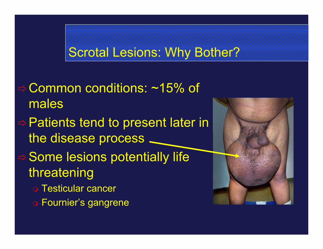
Scrotal Lesions: Why Bother?
Common conditions: ~15% of males
Patients tend to present later in the disease process
Some lesions potentially life threatening Testicular cancer Fournier’s gangrene

Scrotal Lesions: Classification
1. Cutaneous (external) Lesions
2. Intra-scrotal Lesionsa. Extra-testicular
a. Epididymisb. Spermatic Cordc. Appendages
b. Intra-testicular Malignancy

CutaneousLesions

Cutaneous Scrotal Lesions
1 BenignAngiokeratomaPsoriasisEpidermal cystsVitiligo
2. MalignantSquamous cell
carcinomaKaposi’s sarcoma
3. Infectious Lesions:
Condyloma STD’s Tinea cruris Fournier’s gangrene

Benign Cutaneous Lesions: Angiokeratoma
Ectatic dermal blood vessels 1-2mm papules Benign May bleed recurrently &
profusely requiring cauterization

Benign Cutaneous Lesions:Psoriasis
May involve groin & scrotum
Red plaques with white scale patches
Occurs elsewhere Treatment:
Topical steroids Emollients Systemic PUVA
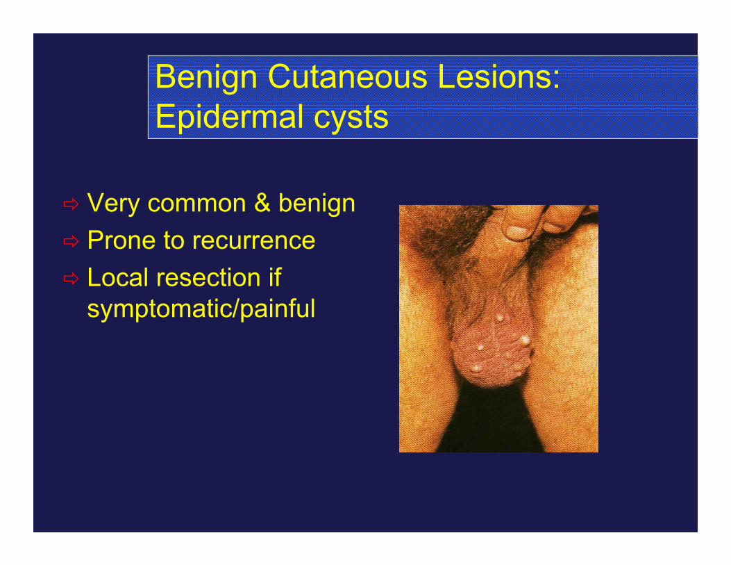
Benign Cutaneous Lesions:Epidermal cysts
Very common & benign Prone to recurrence Local resection if
symptomatic/painful

Benign Cutaneous Lesions:Vitiligo
~1% of population Skin depigmentation Genitals commonly
involved May regress
spontaneously Treatment:
ReassuranceUV lightCorticosteriods prn

Malignant Cutaneous Lesions:Squamous Cell Carcinoma
Etiology: Industrial exposure Viral (HPV) Hygeine
**Chronic ulcerative, enlarging lesion**
Delay in presentation Inguinal metastases
common Wide local excision Outcome:
Dependent on nodal involvement Inanition from local disease

Malignant Cutaneous Lesions: Kaposi’s sarcoma
Increasing in incidence Lesions are violaceous or
light brown in colour Associated with HIV-1 3% of men with AIDS will
initially present with genital Kaposi’s
Treatment if irritating or cosmetically bothersome Intralesional chemotherapeutics Local excision

Infective Cutaneous Lesions: : Condyloma
Caused by human papilloma virus (HPV6 & 11)
Papillary, cauliflower-like proliferations
Treat symptomatic lesions: Podophyllin Imiquimod (Aldara™) Cautery/liquid N2 Laser ablation
Cannot be cured of the underlying viral infection
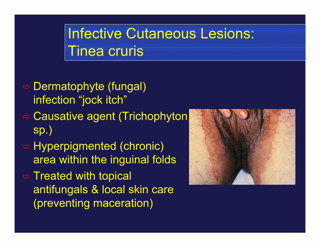
Infective Cutaneous Lesions: Tinea cruris
Dermatophyte (fungal) infection “jock itch”
Causative agent (Trichophyton sp.)
Hyperpigmented (chronic) area within the inguinal folds
Treated with topical antifungals & local skin care (preventing maceration)

Infective Cutaneous Lesions: Fournier’s gangrene
Necrotizing fasciitis of the deep cutaneous structures and fascia
**A LIFE THREATENING CONDITION !**
Requires prompt diagnosis Painful, necrotic, foul
smelling lesions Treatment:
Extensive debridement of affected tissues
Broad spectrum antibiotics

Extra-Testicular Lesions

Scrotal Lesions:Extra-testicular
Benign (non-neoplastic) Hydrocele Spermatocele Epididymitis Torsion of Testicular Appendage Testicular Torsion (spermatic cord is torsed – but pain is
in testicle) Varicocele Hernia
Neoplastic (rare): Sarcoma Adenomatoid tumour, etc.

Scrotal Lesions: Clinical Questions
Clinical Questions:1) What is the patients age?2) Is the onset acute or chronic?3) Is it painful?4) Is it intra- or extra-testicular?5) Does it transilluminate?6) Are there urinalysis findings?

Scrotal Lesions: Hydrocele
Very common benign scrotal mass
1% of all males A collection of serous fluid
between the two layers of the tunica vaginalis
Etiology: Increased production or decreased absorption of fluid by scrotal lymphatics
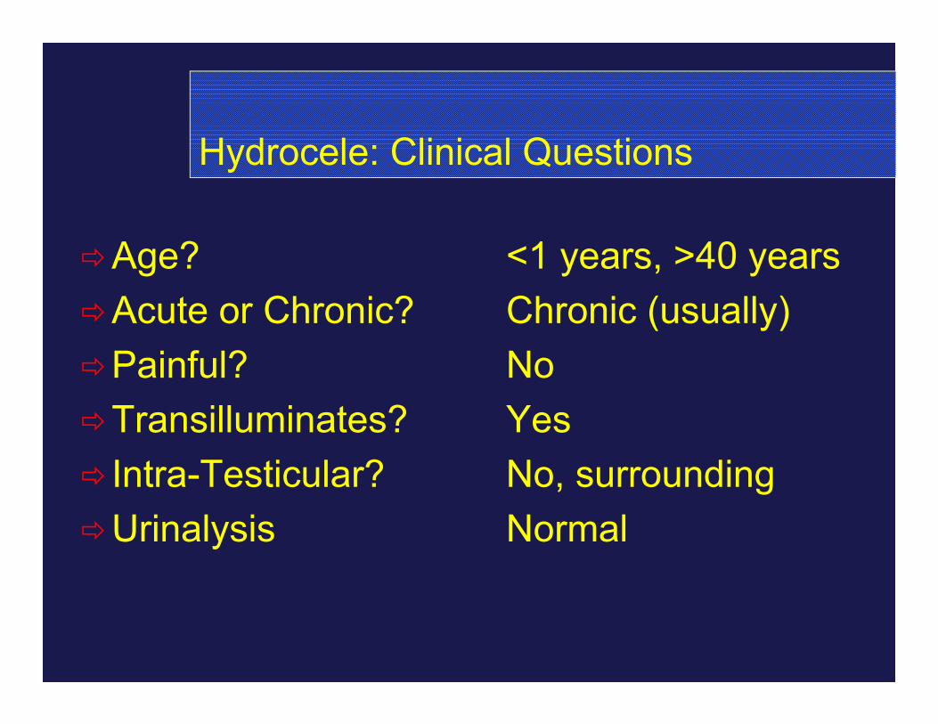
Hydrocele: Clinical Questions
Age? <1 years, >40 yearsAcute or Chronic? Chronic (usually)Painful? NoTransilluminates? Yes Intra-Testicular? No, surroundingUrinalysis Normal

Hydrocele: Transillumination
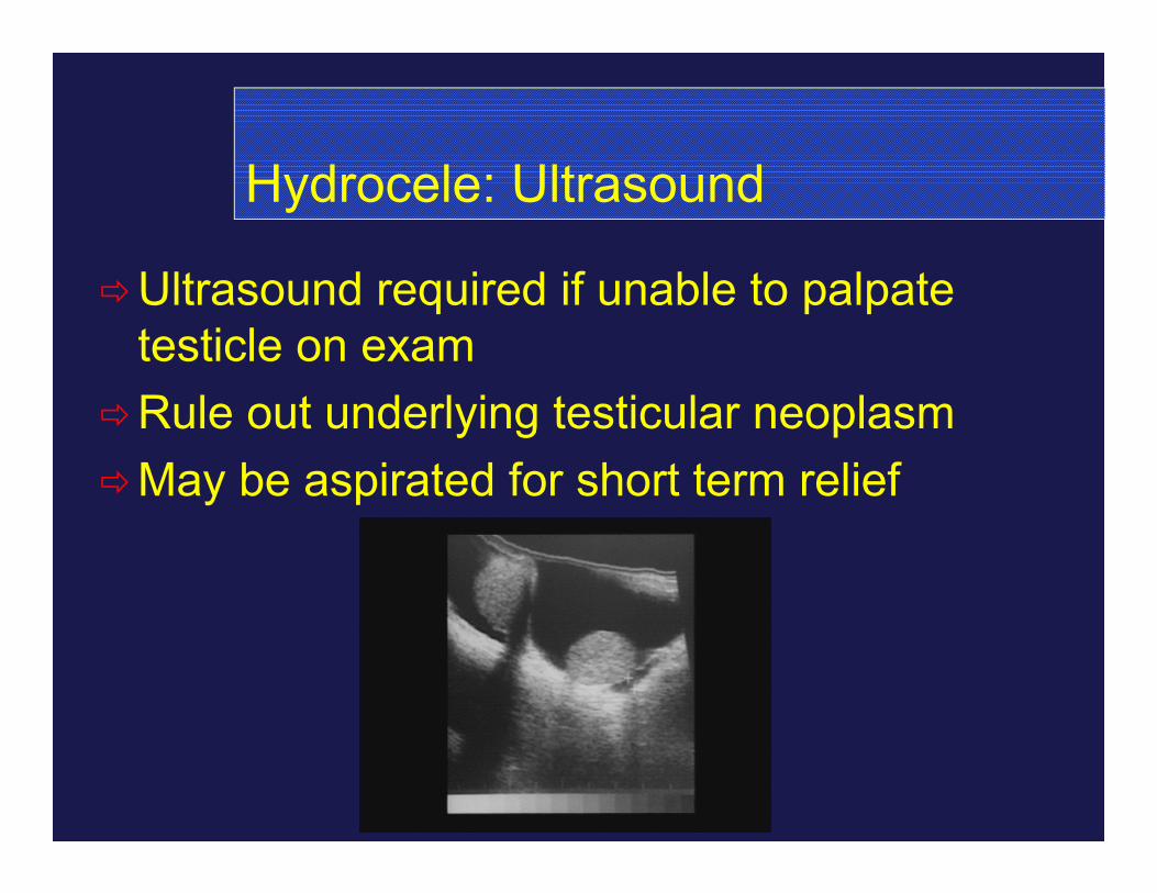
Hydrocele: Ultrasound
Ultrasound required if unable to palpate testicle on exam
Rule out underlying testicular neoplasmMay be aspirated for short term relief
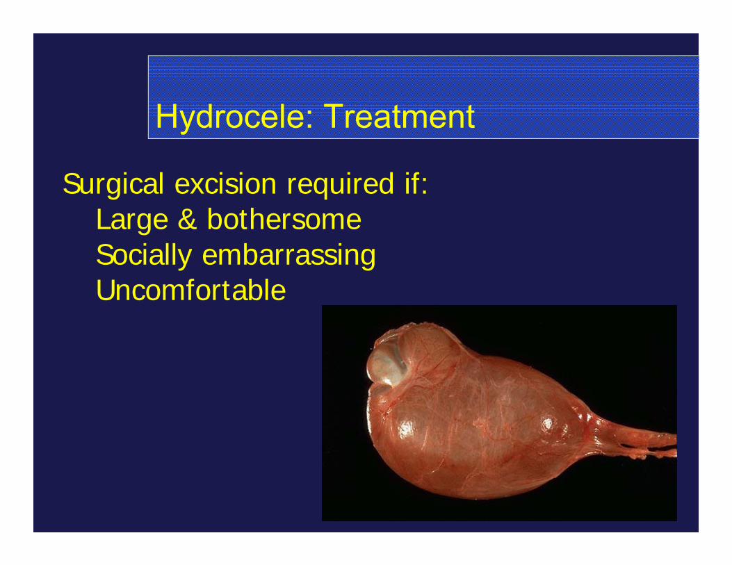
Hydrocele: Treatment
Surgical excision required if:Large & bothersomeSocially embarrassingUncomfortable

Scrotal Lesions: Spermatocele
A sperm containing cyst arising from the head of the epididymis
Caused by ductal obstruction Traumatic Inflammatory Idiopathic
Lesion is usually discrete from the testicle (superior)
Excise if large & bothersome
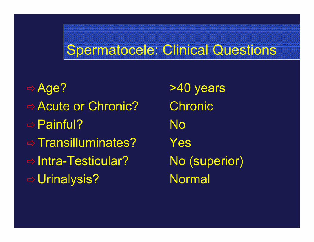
Spermatocele: Clinical Questions
Age? >40 yearsAcute or Chronic? ChronicPainful? NoTransilluminates? Yes Intra-Testicular? No (superior)Urinalysis? Normal

Scrotal Lesions: Epididymitis
Inflammation of the epididymis (<6weeks duration)
Generally due to ascending bacterial infection
Etiology <35 years: C. trachomatis or N. gonorrhea >35 years: Gram negative (E. Coli)

Epididymitis: Clinical Questions
Age? >20 yearsAcute or Chronic? Sub-AcutePainful? YesTransilluminates? No (only if reactive
hydrocele present) Intra-Testicular? NoUrinalysis? Positive (50%)
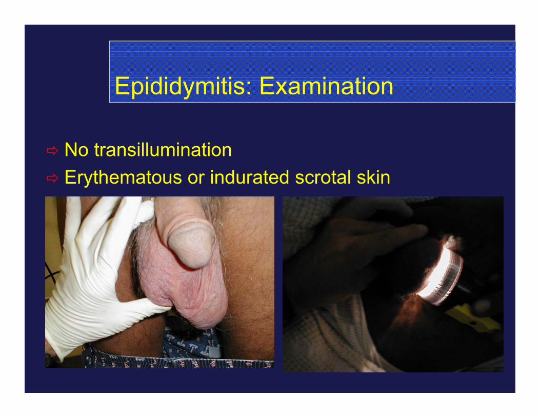
Epididymitis: Examination
No transillumination Erythematous or indurated scrotal skin

Epididymitis: Treatment
NEED TO RULE OUT TESTICULAR TORSION ! Doppler ultrasound or scrotal exploration
Bedrest, scrotal supportNSAID’sAge <35 years: Ceftriaxone 1g IV then
Doxycycline 100mg po bid x 14 daysAge >35 years: TMP-SMX or
fluoroquinolone x14 days

Epididymitis: Complications
AbscessInfertilityTesticular infarctionChronic pain
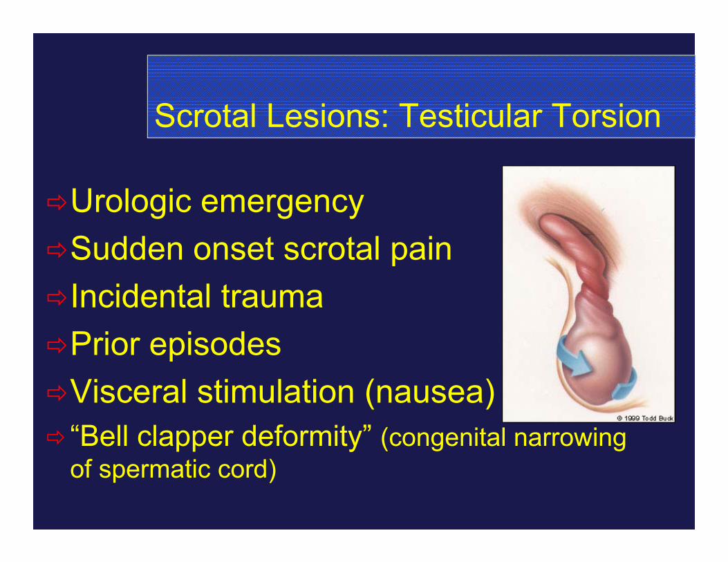
Scrotal Lesions: Testicular Torsion
Urologic emergencySudden onset scrotal pain Incidental traumaPrior episodesVisceral stimulation (nausea) “Bell clapper deformity” (congenital narrowing
of spermatic cord)

Testicular Torsion: Clinical Questions
Age? 12-25 (75%)Acute or Chronic? AcutePainful? Yes, markedlyTransilluminates? No Intra-Testicular? No (Yes -pain)Urinalysis? Negative
** This requires urgent attention**
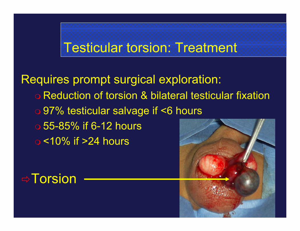
Testicular torsion: Treatment
Requires prompt surgical exploration:Reduction of torsion & bilateral testicular fixation 97% testicular salvage if <6 hours 55-85% if 6-12 hours <10% if >24 hours
Torsion

Testicular Torsion: Doppler Ultrasound
Imaging if diagnosis uncertainDuplex ultrasound:
~82-100% sensitivity Operator dependent Heterogenous testicle with absent flow on Doppler
Normal waveform Absent waveform: Torsion

Approach to Suspected Torsion
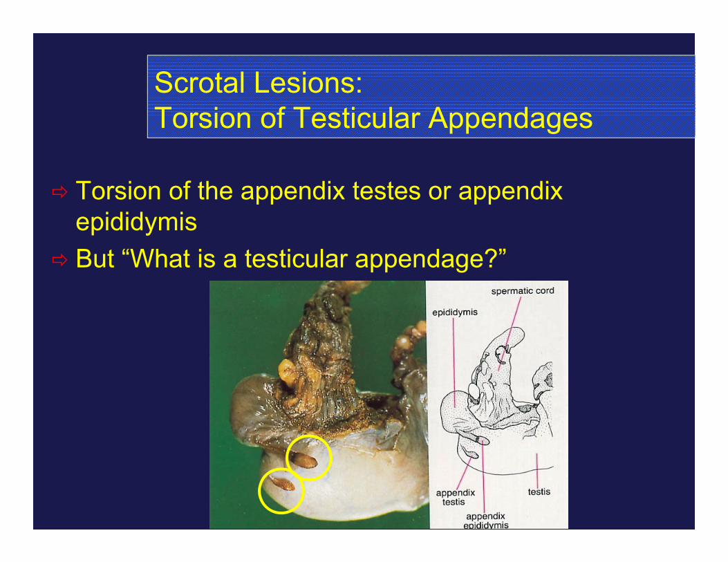
Scrotal Lesions: Torsion of Testicular Appendages
Torsion of the appendix testes or appendix epididymis
But “What is a testicular appendage?”
Torsion of Appendage Testes
“Blue dot” sign (seen on scrotum)More focal pain (upper hemiscrotum)Often difficult to distinguish from other
causesTreatment:
ConservativePain relief (NSAID’s)

Scrotal Lesions: Varicocele
Dilation of the veins of the pampiniform plexus in the spermatic cord
~15% of post-pubertal men Predominantly left sided
(>90%) Associated with infertility
Increased testicular temperature Decreased metabolite clearance Testicular volume loss
Varicocele: Clinical Questions
Age? >12 yearsAcute or Chronic? ChronicPainful? NoTransilluminates? No Intra-Testicular? No (left sided)Urinalysis? Negative

Varicocele: Treatment
Surgical varicocelectomy is required for: Impaired sperm quality (associated with infertility) Loss of testicular volume (in an adolescent) Pain (not a typical indication)
Varicocelectomy involves ligating the offending incompetent vessels of the spermatic cord either: Inguinal Subinguinal Laparoscopically Embolization

Scrotal Lesions: Extratesticular & Neoplastic
A rare group of tumours Adenomatoid tumour of the
epididymis is the most common of these

Intra-Testicular Lesions (**URGENT**)

Scrotal Lesions:Intra-Testicular
All solid intratesticular lesions must be considered malignant until proven otherwise ! (90% are cancer)
Proven otherwise = radical orchiectomy
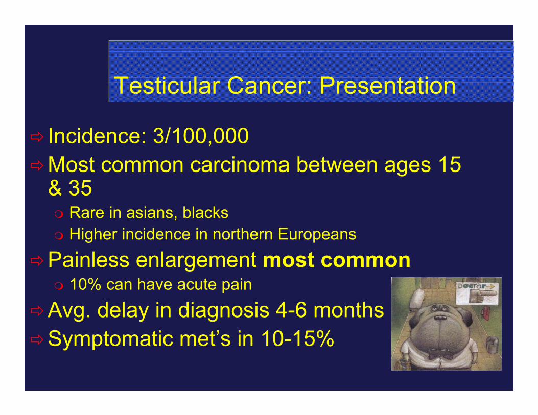
Testicular Cancer: Presentation
Incidence: 3/100,000Most common carcinoma between ages 15
& 35 Rare in asians, blacks Higher incidence in northern Europeans
Painless enlargement most common 10% can have acute pain
Avg. delay in diagnosis 4-6 monthsSymptomatic met’s in 10-15%

Testicular Cancer: Clinical Questions
Age? 15-35 yearsAcute or Chronic? ChronicPainful? NoTransilluminates? No Intra-Testicular? Yes!Urinalysis? Negative
This requires urgent attention

History & Physical
Assess risk factorsCheck for lymphadenopathy, abdominal
massesExamine both testes (2-5% bilateral)
Try to transilluminate If hydrocele prevents exam, get ultrasound
Avoid biopsy Do not breach the scrotum

Testicular Cancer: Risk Factors
Risk Factors:Cryptorchidism (3-14x risk)AtrophyCarcinoma in situ (CIS)HIV “Bad luck”

Metastatic Evaluation
Initial Management Tumor markers CXR Radical (Inguinal) orchiectomyCT scan of chest, abdomen and pelvis Further staging after orchiectomy
Repeat markers serially

Tumour Markers
-fetoprotein Normal <20ng/ml Elevated in:
80% of embryonal carcinoma yolk sac and teratocarcinoma NOT in seminoma or choriocarcinoma
2. HCG Elevated in almost all choriocarcinomas Elevated in 5% of pure seminomas
3. Others LDH, PLAP etc.

Clinical Staging: TNM
T1: Limited to testes & epididymis. No vascular invasion
T2: Invades beyond tunica or vascular invasionT3: Invades spermatic cordT4: Invades scrotum
N0: No nodesN1: Lymph node met’s <2cm and <5 nodesN2: >5 nodes, or nodal mass >2cm or <5cmN3: Nodal mass >5cm

Clinical Staging: Walter Reed
Stage I:Confined to testes
Stage IIa:Retroperitoneal nodes <2cm (small)
Stage IIb:Retroperitoneal nodes >2cm (large)
Stage III:Visceral metastases or supra-diaphragmatic
nodes
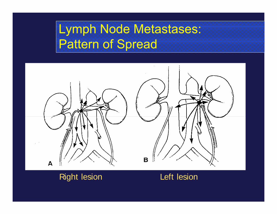
Lymph Node Metastases:Pattern of Spread
Right lesion Left lesion
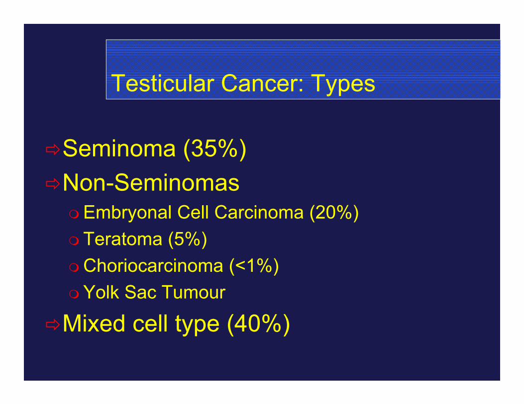
Testicular Cancer: Types
Seminoma (35%)Non-Seminomas
Embryonal Cell Carcinoma (20%) Teratoma (5%)Choriocarcinoma (<1%)Yolk Sac Tumour
Mixed cell type (40%)
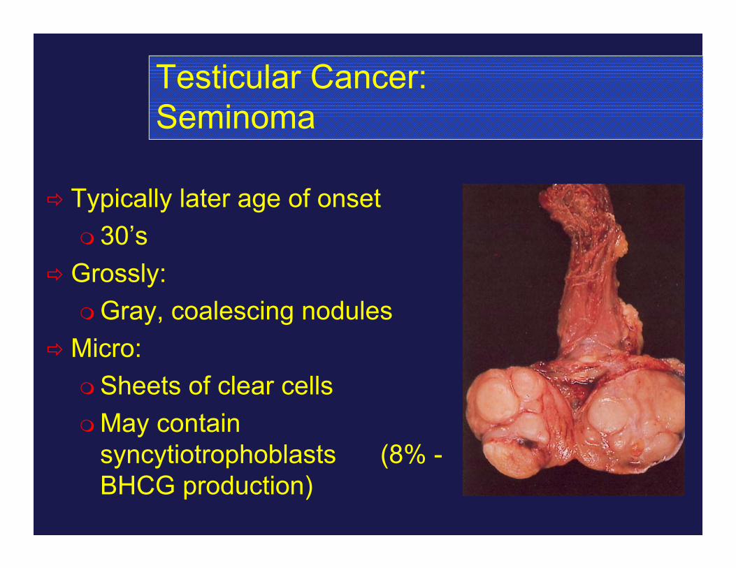
Testicular Cancer: Seminoma
Typically later age of onset 30’s
Grossly:Gray, coalescing nodules
Micro:Sheets of clear cellsMay contain
syncytiotrophoblasts (8% -BHCG production)

Testicular Cancer:Embryonal Carcinoma
Often associated with teratoma
Younger onset Increased AFP levels (>50%) 1/3 have clinically evident
met’s at diagnosis Grossly:
Solid, grey-white with necrosis Micro:
Poorly differentiated

Testicular Cancer:Teratoma
Well differentiated tumours Lesions can become quite large Bimodal age distribution May contain differentiated cell types (i.e. cartilage,
neural)

Testicular Cancer: Choriocarcinoma
Rare neoplasm(<1% germ cell tumours)
Produces BHCGHemorrhagic tumours Hematogenous metastasesPrimary tumour may be
quite smallPresentation commonly due
to metastatic disease
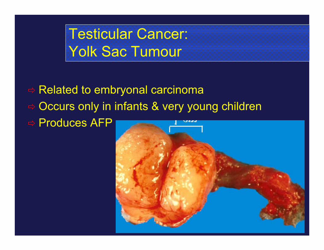
Testicular Cancer: Yolk Sac Tumour
Related to embryonal carcinoma Occurs only in infants & very young children Produces AFP

Testicular Cancer:Treatment Summary
Most curable of all solid neoplasmsAlmost 100% cure rate for low-stage diseaseChemotherapy regimens changing to reduce
morbidityStage I disease = Orchiectomy and
surveillance is an optionMore treatment complex at higher
stages/non-seminomas

Treatment: Seminoma
Stage I & IIaRadical orchiectomy & low dose retroperitoneal
radiation (2500 cGy)
Stage IIb & IIIOrchiectomyANDChemotherapy (Cisplatin, Etoposide, Bleomycin)
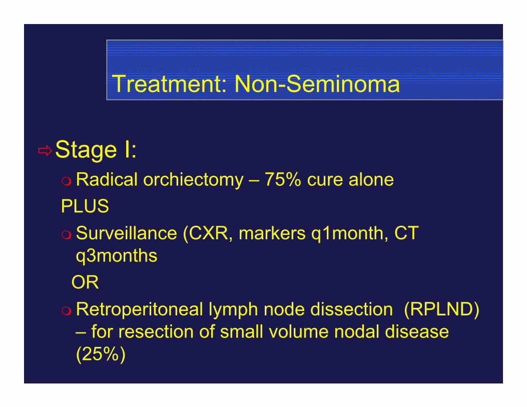
Treatment: Non-Seminoma
Stage I:Radical orchiectomy – 75% cure alonePLUSSurveillance (CXR, markers q1month, CT
q3monthsORRetroperitoneal lymph node dissection (RPLND)
– for resection of small volume nodal disease (25%)

Treatment: Non-Seminomas
Stage II and IIIRadical Orchiectomy ANDChemotherapy (Cisplatin based)Perform RPLND for patients with residual
retroperitoneal nodes after chemo (if tumour markers normalize)

Other Testicular Tumours
Leydig Cell TumoursSertoli Cell Tumours
Rare, <1% of all testicular tumours
LymphomaGonadoblastomaMetastatic Tumours

Summary
Classify and organize lesions logically Cutaneous Extra-testicular Intra-testicular
Benign lesions more common than malignant
Malignant lesions are most likely intra-testicular than paratesticular
Testicular cancer is the most curable solid tumor (esp. if caught early)
Summary (cont’d)
If in doubt, GET AN ULTRASOUND!DO NOT biopsy testicular lesions or
remove them trans-scrotally (**need radical inguinal orchiectomy**)