
Calcium Oxalate Crystals In the Kidney and Thyroidila.ilsl.br/pdfs/v38n3a07.pdfCalcium Oxalate...
8
I NTERNA'fJONAL J OURNAL OF I. EPROSY Volume 38, N umber 3 Printed in fh e U.S .. ·f Calcium Oxalate Crystals In the Kidney and Thyroid of Leprosy Patients l M. Furuta and M. Ozaki 2 Th e pr esence of calcium oxalate cr ys t a. ls in the human kidney ,vas re port ed 111 1959 (1 5) . Thi s was later follo'Ned by many studies in which the same kind ?f crys tals were found in the hum an thY l:Old (17), retina oculi (5, 18 ), and myocardIUm ( 2). Th ese ealciu m oxal at e crystals ha been thought to have a differe nt pathologIC meaning from crystal formation in primary oxalosis. Th ere have been discussions on the relationship between this kind of crys- tal a nd maligna nt tumor (:\) and ur cmia ( I, 7). . In the course of a stud y of 149 autopsi es of leprosy patient s performed in two Japa- nese national le pr osaria (Keifu-en Lepro - sarium a nd Komyo-en Lc prosarium ) dur- ing 1943-1967, similar calcium oxalate crys- tals wer e found in 46 cases of leprosy patients in the kidney and I or thyroid. Although a group of autopsy cases from the 1960's in this study hav e s hown crystals at a higher rat e in the thyroid with distinct correlation with ma li g nant tumors, most of the cases with crystals in the kidney and l or thyroid were not related to ur emia. similar crystals w('ru found fr equ ently 111 autopsy cases at general hospitals , :h e presence of th ese crystals is not a specIfic finding for leprosy (10). In summarizing th e histopathologic ings of 46 cases of leprosy, a hypoth es Is as to the patho genesis of the crystals is prese nt ed together with a discussion of pathologic meaning of such crystal depOSI- tion in leprosy. 1 Received fo r publi ca tion 3 Nove mb er 1969. 2 M. Furuta, M.D., Chief Pa th ologist, K yoto tional Hospital. Depa rtme nt of Pathology, Fushlml' ku. Kyoto. and Th e Ja pan Bapti st Hospital , Sakyo· ku . Kyo to. Japan ; M. Ozaki, M.D ., Staff of National Leprosarium Nagashima Aisei·en. De parlme nt of Chemotherap y. Mu shiak e, Oka ya ma. and Staff of Le prosy R esearch Labora tor y. Kyoto UnI - versity School of Medicine. Kyoto. Japan. 286 MATERIALS AND METHODS Th e autopsy cases were divided into the followin g thr ee groups: Group A: 49 leprosy cases autopsied in 1943 and 1944 in Komyo-en Lepro- sarium in Oka yama, Japan. Group B. 40 leprosy cases autopsied during 1955-1957 in Keifu-en Lepro- sarium in Kumamoto, Japan. Group C: 60 leprosy cases autopsied during 1962-1967 in Kom o-en L cpr::>- s.arium in Okayama, Japan. In each case, paraffin sections of the kidney and th yroid were stained with hematoxylin a nd eosin. Th e sta in ed specimens were examined with a polarizing microscope. X-ray diffraction is the bcst method for the identification of these crystal s. How ev- er, th e crystals in the ti ss u es were often toe scarce for this me thod ; therefore, only one case .showing relatively numerous crystalf in the kidney could be thus examined X-ray Geiger count er diffraction (Norelco ) identified the crystals of this case as calci- um oxalate monohydrat e. In addition, hi s- tochemical methods (1 3) were app lied to four cases, and the results obtained also confirmed the prese nce of calcium oxalate. It is usually very difficult to find calcium oxalate crystals in ti ss ues with an ordinary light microscop e. RESULTS Group A. (49 leprosy cases) Crystals found in sections from 10 cases. Th ese cases ranged in age, at the time of death, from 29 to 54, with an average of 41 years. One 42 year old female showed crystals in the kidney. Th e cause of death in this patie nt was severe pulmonary tuberculosis. In the other nine cases a small quantity of minut e crystals were seen in the thyroid. Most of them died of tuberculosis. Group B. (40 leprosy cases) Twelve showed the pr esence of the crystals; in
Transcript of Calcium Oxalate Crystals In the Kidney and Thyroidila.ilsl.br/pdfs/v38n3a07.pdfCalcium Oxalate...

I NTERNA 'fJONAL J OURNAL OF I. EPROSY Volume 38, Number 3 Printed in fh e U.S .. ·f
Calcium Oxalate Crystals In the Kidney and Thyroid
of Leprosy Patients l
M. Furuta and M. Ozaki 2
The presence of calcium oxalate crysta.ls in the human kidney ,vas reported 111
1959 ( 15) . This was later follo'Ned by many studies in which the same kind ?f crystals were found in the human thYl:Old (17), retina oculi (5, 18), and myocardIUm ( 2). These ealciu m oxal ate crystals ha ~e been thought to have a different pathologIC meaning from crystal formation in primary oxalosis. There have been discussions on the relationship between this kind of crystal and malignant tumor (:\) and urcmia ( I, 7). .
In the course of a study of 149 autopsi es of leprosy patients performed in two Japanese national leprosaria ( Keifu-en Leprosarium and Komyo-en Lcprosarium ) during 1943-1967, similar calcium oxalate crystals were found in 46 cases of leprosy patients in the kidn ey and I or thyroid.
Although a group of autopsy cases from the 1960's in this study have shown crystals at a higher rate in the thyroid with distinct correlation with malignant tumors, most of the cases with crystals in the kidney and l or thyroid were not related to uremia. ~s similar crys tals w('ru found frequently 111
autopsy cases at general hospitals , :he presence of these crystals is not a specIfic finding for leprosy (10).
In summarizing the histopathologic fin~ings of 46 cases of leprosy, a hypothesIs as to the pathogenesis of the crystals is presented together with a discussion of th~ pathologic meaning of such crystal depOSItion in leprosy.
1 Received fo r publica tion 3 November 1969. 2 M. Furuta, M.D., Chief Pathologist , K yoto .N~
tional Hospital . Department of Pathology, Fushlml ' ku. Kyoto. and The J apan Baptist Hosp ital , Sakyo· ku . Kyoto. Japan ; M. Ozaki , M.D ., Staff of National Leprosarium Nagashima Aisei·en. Deparlment of Chemotherapy. Mushiake, Okayama. and Researc~ Staff of Leprosy R esearch Laboratory. Kyoto UnI versity School of Medicine. Kyoto. Japan.
286
MATERIALS AND METHODS
The autopsy cases were divided into the followin g three groups:
Group A: 49 leprosy cases autopsied in 1943 and 1944 in Komyo-en Leprosarium in Okayama, Japan.
Group B. 40 leprosy cases autopsied during 1955-1957 in Keifu-en Leprosarium in Kumamoto, Japan.
Group C: 60 leprosy cases autopsied during 1962-1967 in Kom o-en Lcpr::>s.arium in Okayama, Japan.
In each case, paraffin sections of the kidney and thyroid were stained with hematoxylin and eosin. The stained specimens were examined with a polarizing microscope.
X-ray diffraction is the bcst method for the identification of these crystals. However, the crystals in the tissues were often toe scarce for this method; therefore, only one case .showing relatively numerous crystalf in the kidney could be thus examined X-ray Geiger counter diffraction ( Norelco ) identified the crystals of this case as calcium oxalate monohydrate. In addition, histochemical methods (13) were applied to four cases, and the results obtained also confirmed the presence of calcium oxalate. It is usually very difficult to find calcium oxalate crystals in tissues with an ordinary light microscope.
RESULTS
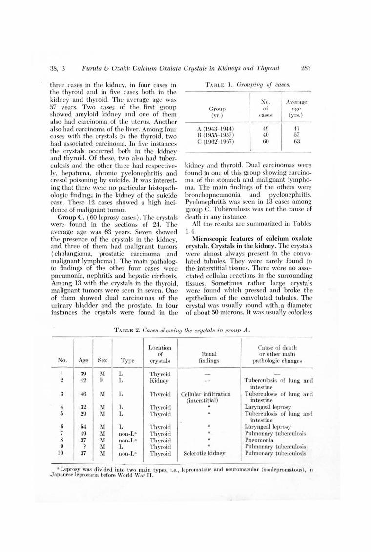
Group A. (49 leprosy cases) Crystals found in sections from 10 cases. These cases ranged in age, at the time of death, from 29 to 54, with an average of 41 years. One 42 year old female showed crystals in the kidney. The cause of death in this patient was severe pulmonary tuberculosis . In the other nine cases a small quantity of minute crystals were seen in the thyroid. Most of them died of tuberculosis.
Group B. (40 leprosy cases) Twelve showed the presence of the crystals; in
38, 3 Furuta & Ozaki: Calcium Oxalate Crystal itl Kidneys and Thyroid 287
three cases in the kidney, in four cases in the thyroid and in five cases both in the kidney and thyroid. The average age was 57 years. Two cases of the first group showed amyloid kidney and one of them also had carcinoma of the uterus. Another .also had carcinoma of the liver. Among four ~ases with the crystals in the thyroid, two had associated carcinoma. In five instances the crystals occurred both in the kidney and thyroid. Of these, two also had tuberculosis and the other three had respectively, hepatoma, chronic pyelonephritis and cresol poisoning by suicide. It was interesting that there were no particular histopathologic findings in the kidney of the suicide case. These 12 cases showed a high incid ence of malignant tumor.
Group C. (60 leprosy cases). The crystals were found in the sectioQ<; of 24. The average age was 63 years. Seven showed the presence of the crystals in the kidney, and three of them had malignant tumors ( cholangioma, prostatic carcinoma and malignant lymphoma). The main pathologic findings of the other four cases were pneumonia, nephritis and hepatic cirrhosis. Among 13 with the crystals in the thyroid, malignant tumors were seen in seven. One of them showed dual carcinomas of the urinary bladder and the prostate. In four instances the crystals were found in the
T A BLE 1. Grou7)ill(j of cases.
Group (yr.)
A (1943- 1944) 13 (1955- ] 957) C (1962'- 1967)
No. of
case::;
49 40 60
Average age
(yrs.)
41 57 63
kidney and thyroid. Dual carcinomas were found in one of this group showing caJ'cinoma of the stomach and malignant lymphoma. The main findings of the others were bronchopneumonia and pyelonephritis. Pyelonephritis was seen in 13 cases among group C. Tuberculosis was not the cause of death in any instance.
All the results are summarized in Tables 1-4.
Microscopic features of calcium oxalate crystals. Crystals in the kidney. The crystals were almost always present in the convoluted tubules. They were rarely found in the interstitial tissues. There were no associated cellular reactions in the surrounding tissues. Sometimes rather large crystals were found which pressed and broke the epithelium of the convoluted tubules. The crystal was usually round with. a diameter of about 50 microns. It was usually colorless
TA BLE 2. Cases showing the crystals in group A.
Location Cause of death of Renal 01' other main
No. Age Sex Type crystals findings pathologic changes -----
1 39 M L Thyroid - -2 42 F L Kidney - Tuberculo is or lung and
intestine 3 46 M L Thyroid Cellular infil tration Tuberculosis or lung and
(interstitial) intestine 4 32 M L Thyroid " Laryngeal leprosy 5 29 M L Thyroid " Tuberculosis of lung and
intestine 6 54 M L Thyroid " Laryngeal leprosy 7 49 M non-V' Thyroid " Pulmonary tuberculosifl
37 M non-La Thyroid " Pneumonia 9 ? 1\1 L Thyroid " Pulmonary tuberculosis
10 37 M non-V ' Thyroid Sclerotic kidney Pulmonary t.uberculosis
a Leprosy was ?ivided into two main types, i.e., lepromatous and neuromacular (nonlepromatous), in ~apanese leprosaria before World War II .

2BB International Journal of Leprosy
FIG. 1. A crys tal in a distal convoluted tubule (arrow) under light microscope. Patient No. 4, Table 3. (Hematoxylin and eos in stain. Magnification X200)
F IG. 2. Amyloid kidney with crystals in a tubule under polarizing microscope. Patient No. 12, Table 3. (Hematoxylin and eosin stain . Magnification X200 )
FIG. 3. High magnifi cation of the crystal. Note the radial stri ations. Patient No. 13, Table 4. (Hematoxylin and eosin sta in . Magnificatioll X400 )
FIG. 4. A crys tal in thyroid follicle. Patient No. 13, Table 4. (Hematoxylin and eos in stain. Magnifica tion X400 )
1970

38, 3 Furuta & Ozaki: Calcium Oxalate C1'ystals in Kidneys and Thyroid 289
T A BL E :3. Cases ShOll' i l/(J the crystals i ll (JI"OUp B. -
Location Cause of death 0 1'
of R enal other main No. Age Sex T ype" cry~ta],; finding~ pathologic change"
-------1 60 :vI: L Thyroid Chroni c nephri tis -
r\ rterioloneph 1'0 -
sclerosis . 2 32 ;\[ L Kidney - Sui cide (cresol poison i ng)
Th yroid :~ .1.f F non-L Kid nry I'urulen t neph ri t is Purulen t cystit is
Carcinoma of uteru~ .f 39 :\[ non-L l"':id ne)" - Carcinoma of li\'er
Thy roid 5 69 :'II non-L Ki dney Amyloid kidney Pulmona ry tubercu losi"
Thy roid Arterionephro- Liver cirrhosis sclerosis
6 78 ;U non-L Thy roid Amyloid kidney Carcinoma of "tomaeh Nephrosclerosis
7 69 ~I non-L Kidney Chroni c pyelone- Ulcer of stomach Thy roid phri t i,.;
8 53 F non-L Kid ney - Carcinoma of li ver Li ver cirrhosis
9 70 F non-L Thy roid Pyelonephri t i,.; Cholangiocarcinoma 10 61 :'II non-L Th y roid - Pneumonia ]] 57 :'II L Ki d ney Chroni c glomerulo- Pulmona ry tuberculosis
Thyroid nephri tis 12 38 :'II L Kidney Amyloid kidney Pericardit is
". The ty pe of lep rosy i II thi ~ table is based on t he ori ginal descript ion in t he protoco ls of K eifll-en Lepro"1lJ'lllm .
and transparent under the light microscope, but sometimes it was yellow or brown. The large ones often showed radial striation, recognizable by light microscopy.
Crystals in the thyroid. Very small crystals were present in the follicles of the thyroid, but they were rarely present in the interstitial tissues. They were colorless and transparent. Their morphology was variable; needle-shaped, sand-like, square, polygonal or irregular. Where they were present, they looked like vacuoles in the follicular colloidal substances by light mic roscopy.
DISCUSSION
Renal lesion. vVe considered whether or not the crystals are correlated with the renal changes of patients who suffer from leprosy. It has been reported that this kind of crystals is more frequently found in severe renal lesions. It is generally accepted that the kidney is often affected by
bacterial infections but usually not by Jeprosy itself. Our pathologic examination revealed that nonleprous inflammatory changes of the kidney were present in a considerable number of the autopsied cases, especially in those of group C. Pyelonephritis was the most common (6) . It is not rare that leprosy patients die in uremi a. The high incidence of pyelonephritis in group C could have been caused by recurrent bacterial infections. In group A, however, pathologic .findings of the kidney were different from that in group C. Earlier reports on autopsies of leprosy patients have agreed as to a high incidence of
. secondary amyloidosis in the kidney. However, in groups B and C, the in cidence of secondary amyloidosis was low. There was no determinable correlation of the appearance of these crys tals and leprosy infection of the kidney spec.incally. .
Characteristics of the pathologic changes of each group. Only one case of group A

290 International Journal of Leprosy 1970
TA BLE 4. Cases showing the crystals ill oroup C.
Location Cause of death or of Renal other main
~o. Age Sex TypeS crystals findings pathologic changes ------
I 77 1\1 T Thyroid Chronic pyelo- Carcinoma of pancreas nephritis
2 57 M L Thyroid Chronic pyelo- Pneumonia nephritis
Arteriolo-nephro-sclerosis
3 · 55 1\1 L Kidney Acute glomerulo- I ~ronchopneu mon ia nephritis
4 46 1\1 T Kidnc~ · Chronic pyelo- Bronchopneumonia Thyroid nephritis
5 55 1\1 L Kidney Chronic pyelo- -
Thyroid nephri t is 6 64 1\1 L Kidney Solitary cyst 1\1alignant lymphoma
Thyroid Chronic pyelo- Carcinoma of stomach nephri tis
7 74 M T Kidney Chronic pyelo- -nephritis
Amyloid d,<)posit 8 78 M T Kidney Chronic pyelo- Bronchopneu mon ia
nephri t is Chronic hepatitis 9 80 F L Kidney Arterionephro- I3ronchopneumonia
sclerosis Liver cirrhosis 10 40 M L Kidney Nephrosclerosis 1\1alignant lymphoma
Hydronephrosis Liver cirrhosis 11 73 M T Thyroid - Carcinoma of esophagu~ 12 63 1\1 L Kidney Renal tubule del!;en- Cholangiocarcinoma
eration Hepatic cirrhosis Chronic pyelo-
nephritis 13 82 M L Kidney
Thyroid Benign adenoma Bronchopneumon ia
14 41 1\1 L Thyroid - Carcinoma of rectum 15 58 M T Thyroid Chronic glomerulo- Carcinoma of stomach
nephri t is Chronic hepatitis 16 57 M L Thyroid - Carcinoma of pancrea;: 17 62 F T Thyroid Arterionephro- Carcinoma of tongu e
sclerosis 18 42 M L Thyroid Pyelonephritis Death from shock 19 81 F T Thyroid Chronic pyelone- Large intestinal ulcer
phritis Arterionephro-
sclerosis 20 77 lVI L Thyroid Chronic pyelo- Carcinoma of prostate
nephritis Carcinoma of urinary bladder
21 74 M L Thyroid - I3ronchopneurnonia 22 50 1\1 L Thyroid - Cerebral hemorrhage 23 73 1\1 T Kidney Chronic pyelo- Hepatic cirrhosis
nephritis Occult carcinoma of pro><-ta te
24 57 1\1 T Thyroid Round cell infi 1- Bronchopneumonia tration Cerebral hemorrhage
• T = tuberculoid; L = lepromatous.

38,3 FU1'llta & Ozaki: Calcium Oxal.ate C1'ystals in Kidneys and Thyroid 291
showed the crystals in the kidney. The cause of death in this patient was pulmonary tuberculosis and the structure of the kidney was almost normal. All nine cases with the crystals in the thyroid showed a mild cellular infiltration in the interstitial tissue of the kidney, and five of them died of pulmonary tuberculosis . In group A, malignant tumors were very few in number.
Among the 12 cases of group B showing crystals, carcinoma was found in five. Not all cases showing carcinoma had the crystal in the kidney and / or thyroid. Renal findin gs in these 12 were mainly nephritis, pyelonephritis and amyloid deposit.
In group C, half of the cases with crystals had pyelonephritis. It is interesting to note the in crease in malignant tumors in this group and the complication of more or less serious bronchopneumonia.
Age. The average age of cases with crystals in group A was 41 years. The averag'3 age of leprosy patients in Japanese leprosaria has been remarkably lengthened after World War II. The average age of the cases with the crystals in groups B and C was 57 and 63 years respectively. The crystals tend to be found more often in the aged or in cases showing pathologic changes in the kidney, although there were some exceptions.
Causes of crystal fonnation. Crystals in the thyroid were frequently found in the cases having also malignant tumors especially in group C. When the cause of death was pneumonia or pulmonary tuberculosis, it seems necessary to consider the possible influences of anorexia or respiratory acidosis in explaining the origin of the crystals. \ iVe also considered the possible effect of medicine, especially vitamins such as vitamin C (H) or vitamin B-6 ( 12) in crystal formation. The history of medical trea tment was examined in the protocols of the cases of group C which showed no remarkable renal les ions or malignant tu ~ mol's in th e internal organs. Some of them were not given any vitamin compounds. It was impossible to explain the crystallization of calcium oxalate by the metabolism of vitamins.
It seems likely that destruction of renal tissues might be correlated with the
presence of the crystals in the kidney. The most suggestive case in group B was a 32 year old male who committed suicide by taking a large quantity of cresol. Crystals were present both in the kidneys and thyroid of this patient. There were no pathologic changes noticeable in these tissues by light microscopic study, althou gh cresol must have' caused dysfunction of renal tubules. This raised the question of how long it takes to crystallize calcium oxalate in the human body. The death of this patient occurred less than a day after taking the cresol. If it is correct to assume that the crys tals of the suicide case resulted from renal dysfunction caused by poisoning, then calcium oxalate had to crys tallize within a few hours in the kidney and thyroid. The cause of crystallization in this case, however, remains unexplained because we could not confirm the evidence of the renal dysfunction. With respect to the rapidity of crystallization, there has been a suggestive report relating to acute ethylene glycol poisoning (10) . In this report, calcium oxalate crystals were found in the kidneys and central nervous systems of soldiers who almost all died one or two days after drinking lethal quantities of antifreeze solution of the ethylene glycol type. The same type of crystal was also found in newborn infan ts with congel1ital oxalosis ( 8). It might be possible to suppose that calcium oxalate would crystallize in a few hours or days, or at most, within several months.
An 82 year old male patient in group C, who had increased dysacousis (!l), showed the largest quantity of crystals in the kidney and thyroid. The cause of death was bronchopneumonia. It was this case to which the x-ray diffraction method was applied and the crystals were identified as calcium oxalate monohydrate. The metabolism of oxalate is so complicated that it is difficult to decide the exact process of crystal form ation . Special attention was paid to possible disturbances in the metabolism of amino acids such as glycine. The patient had had difficulty in hearing from his youth. This raised the possibility that dysacousis had been correlated with abnormal deposits of oxalate in the neural tissues (11).

292 International J oumal of Leprosy 1970
Crystallization of calcium oxalate is also thought to be closely related to the concentration of oxalate in the serum. H yperoxalemia would be a preceding condition before producing the crystals in the kidney.
Localization of crystals. The crystals never accompanied pathologic changes of the thyroid, but sometimes accompanied changes in the kidney. This led to the assumption that the cause of crystallization leaves no visible morphologic evidence in the tissues. Our present study was limited to the kidney and the thyrOid, because of more frequent localization of the crystals in these organs. The cause of such constant localization of the crystals in certain organs remains unexplained. As for the kidney, it might be supposed that the presence of byperoxaluria due to some renal dysfunction, for example, poor reabsorption of tlmino acids through renal tubules might playa role. In some cases such dysfunction would leave no traces in the tissue. In the kidney the crystals were mostly found in the renal tubules of the cortex and rarely in the interstitial tissue. In the case of crytals occurring without reactional cellular infiltration we think that conditions preceding crystallization might have been more harmful than the presence of the crystal itself.
Relation to oxalosis. Oxalosis has been defined as an extra-renal deposit of calcium oxalate crystals. If so, many cases in the present study having crystals might be thought to have had oxalosis (4). However, the crystalline calcium oxalate deposition in kidneys and thyroids in these cases seems to have been caused by hyperoxalemia, and for this reason we think it is not suitable to call these cases "oxalosis." The same kind of crystal were found in tissues of nonl eprous patients who were autopsied in general hospitals. For this reason, the presence of the crystal seems not to be essentially related to leprosy.
SUMMARY
Polarizing microscopic study of 149 autopsied cases of leprosy patients revealed the presence of crystalline calcium oxalate in the kidneys and thyroid in 46 cases. The
presence of the crystal was rare in the kidneys of patients who died in 194.3 and 1944. Crystals in the thyroid were frequently found in cases of malignant tumor, especially in the group which died in 1962-1967. The possible relationship of the presence of the crystal to hyperoxalemia, metabolic disturbances of amino acids , oxalosis and medical treatment arc discussed. Generally, crystals were found in agcd people, in cases with renal les ion s, malignant tumor or pulmonary les ions, although there were some exceptions. No definite pathogenic relationship could be made to leprosy.
RESUMEN
Un estudio con microscopio de~luzrpolarizada de 149 casos de a utopsia de pacientes con lepra revelo la presencia de oxalato de calcio cristalino en el rinon y tiroides en 46 casos. El cristal se encontro muy pocas veces en los rinones de pacientes que murieron en 1943 y 1944. Los cristales en el tiroides se encontraron frecuentemente en casos de tumores malignos, especialmente en el grupo que murio entre ]962- 1967. Se considera la positile relacion entre la presencia del cristal e hiperoxalemia, alteraciones metabolicas de amino acidos, oxalosis y tratamiento medico. En general, los cristales se encontraron en personas de edad, en casos con lesiones renales, tumores malignos 0 lesiones pulmonares, aunque hubieron algunas excepciones. No se pudo establecer una relacion patogenica definitiva con la lepra.
RESUME On a procede a une etude au microscope
polarisant de ]49 cas autopsies de malades de la lepre. Cette etude a revele la presence de cristaux d'oxalate de calcium au niveau du rein et de la thyroide chez 46 de ces malades. La presence de crista ux etait rare dans les reins des malades decedes en 1943 et 1944. Les cristaux ont ete frequemment observes au niveau de la thyroide dans des cas de tumeurs malignes et partiClIlierement dans les groupes decedes entre 1962 et ] 967. On discute de la relation qui pourrait exister entre la presence de cristaux d' une part, et d'autre part l'hyperoxalemie, les troubles metaboliques des acides amines, l'oxalose et Ie traitement medical. En regie generale les cristaux ont ete trouves chez des personnes agees, de meme que chez des cas soutfrant de lesions renales, de tumeurs malignes ou de lesions pulmonaires, encore qu'il y ait eu certaines exceptions. Aucune relation pathogenique nette n'a pu eIre elablie entre ces observations et la lepre.

38, 3 Furuta & O;;aki: Ca.lcium Oxalate Crystals in Kidneys and T ItYI'o i.d 293
REFERENCES 1. BENDNAR, B., ]IRASEK, A. , STE]SKEL, J.
and CHYTIL, M. Die sekundiire udimische Oxalose. Zbl. all g. Path . & Path. Anat. 102 ( 1961 ) 289-297.
2. BENNET, B. and ROSE NBLUM, C. Identification of calcium oxalate crystals in the myocardium in patients with uremia. Lab. Invest. 10 ( 1961 ) 947-955.
3. BENNINGTON, J. L., HAllEH, S. H ., SM ITH, J. V. and WAHNEn, N. E. Crystals of ca lcium oxalate in the human kidney. American J. Cli n. Path . 41 ( 1964 ) 8-14.
4. CHISHOLM, C. D. and HEAHD, B. E. Oxalos is. British J. Surg. SO (1962) 78-92.
5. COGAN, D. C., KUWA BAHA, T ., SILBEHT, J., McMUHHAY, V. and HURLBUT, C. Calci ll m oxalate and calcium phosphate crystals in de tached re tinitis. Arch. Ophth. 60 ( 1958) 366-371.
6. DESIKAN, K. V. and JOB, C. K. A review of postmortem findings in 37 cases of leprosy. Internat. J. L eprosy 36 (1968) 32-44.
7. FANGEH, H. and ESPAHAZA, A. Crystals of calcium oxalate in kidn eys in uremia. American J. Clin . Path. 41 (1964) 597-603.
8 .. FUHUTA, ~vr. and TORII, S. Congenital oxalosis. First report of two neonatal cases. Ann . Pediat. (Japan) 13 (1967) 196-206.
9. FURUTA, lVI., OZAKI, M. and HAHADA, N. An autopsy case of d ysacousis with pres-
ence of calcium oxalate crysta ls. Reported at 37th Setouchi-shudankai Leprosy Meeting, 12 July 1968. (To be published )
10. FURUTA, M. Calcium oxalate crysta ls in the kidn ey and thyroid . Trans. Soc. Pa th . Jap. 58 ( 1969) 64-65.
11. FURUTA, M. and HAYASm, M. D ysacousis and oxalosis. Nippon Rinsho (Med. Clin. Japan ) . 27 (1969) 197-200.
12. CEHSlfOFF, S. N., FARAGALLA, F. F., NEl.SON, D. A. and ANDHUS, S. B. Vitamin 13-6 defici ency and oxalate nephrocalcinosis in ca t. Ameri can J. Med. 27 (1959) 72-80.
J.3. JOl-I NSON, F. B. an d PA NT, K. Histochemical identification of calcium oxalate. Arch. Path. 74 ( 1962) 347-351.
J4. LAJ'.1DEN, M. P. and CI-IHYSTOWSKT, C. A. Urinary oxalate excretion by man fo llowing ascorbic acid inges tion. Proc. Soc. Expel'. BioI. & Med. 85 (1964) 190-192.
IS. MACALUSO, M. P. and BERG, N. O. Calcium oxalate crysta ls in kidn eys in acute tubular nephrosis and other disease with functional fai lure. Acta. Path. Microbiol. Scand. 46 (1959) 197-205.
16. PONS, C. A. and CUSTEH, R. P . Acute e thylene glycol poisoning. American J. Med. Sci. 211 (1946) 544-552.
17. RICHTER, M. N. and MCCARTY, K. S. Anisotropic crysta ls in the human thyroid gland. American J. Path . 30 ( 1954 ) 545-553.
18. ZI;-..r;-"TEHM .-\ N, L. E. and JOHNSON, F. B. Calcium oxa late crystals within ocular tissues. Arch. Ophth. 60 ( 1958,) 372-383.


