Caffeic Acid Phenethyl Ester Reduces Ischemia-Induced ...
12
Research Article Caffeic Acid Phenethyl Ester Reduces Ischemia-Induced Kidney Mitochondrial Injury in Rats Sonata Trumbeckaite, 1,2 Neringa Pauziene, 3 Darius Trumbeckas, 4 Mindaugas Jievaltas, 4 and Rasa Baniene 1,5 1 Neuroscience Institute, Lithuanian University of Health Sciences, Eiveniu Str. 4, LT-50161 Kaunas, Lithuania 2 Department of Pharmacognosy, Medical Academy, Lithuanian University of Health Sciences, Eiveniu Str. 13, LT-50166 Kaunas, Lithuania 3 Institute of Anatomy, Lithuanian University of Health Sciences, Mickeviciaus Str. 9, LT-44307 Kaunas, Lithuania 4 Department of Urology, Medical Academy, Lithuanian University of Health Sciences, Eivenių g. 2, LT-50161 Kaunas, Lithuania 5 Department of Biochemistry, Medical Academy, Lithuanian University of Health Sciences, Eiveniu Str. 4, LT-50161 Kaunas, Lithuania Correspondence should be addressed to Sonata Trumbeckaite; [email protected] Received 31 March 2017; Accepted 19 June 2017; Published 13 August 2017 Academic Editor: Moh H. Malek Copyright © 2017 Sonata Trumbeckaite et al. This is an open access article distributed under the Creative Commons Attribution License, which permits unrestricted use, distribution, and reproduction in any medium, provided the original work is properly cited. During partial nephrectomy, the avoidance of ischemic renal damage is extremely important as duration of renal artery clamping (i.e., ischemia) influences postoperative kidney function. Mitochondria (main producer of ATP in the cell) are very sensitive to ischemia and undergo damage during oxidative stress. Finding of a compound which diminishes ischemic injury to kidney is of great importance. Caffeic acid phenethyl ester (CAPE), biologically active compound of propolis, might be one of the promising therapeutic agents against ischemia-caused damage. Despite wide range of biological activities of CAPE, detailed biochemical mechanisms of its action at the level of mitochondria during ischemia are poorly described and need to be investigated. We investigated if CAPE (22 mg/kg and 34 mg/kg, injected intraperitoneally) has protective effects against short (20 min) and longer time (40 min) rat kidney ischemia in an in vitro ischemia model. CAPE ameliorates in part ischemia-induced renal mitochondrial injury, improves oxidative phosphorylation with complex I-dependent substrate glutamate/malate, increases Ca 2+ uptake by mitochondria, blocks ischemia-induced caspase-3 activation, and protects kidney cells from ischemia-induced necrosis. The protective effects on mitochondrial respiration rates were seen after shorter (20 min) time of ischemia whereas reduction of apotosis and necrosis and increase in Ca 2+ uptake were revealed after both, shorter and longer time of ischemia. 1. Introduction Kidney ischemia-reperfusion (I/R) injury is characterized by restriction of blood supply to an organ followed by restora- tion of blood flow and reoxygenation. Kidney injury may occur after infarction and sepsis, during partial nephrectomy and a surgical procedure, and when kidney tumor is removed after clamping renal artery [1]. Clamping time (i.e., duration of ischemia) is thought to be a major factor in determining postoperative kidney dysfunction. During partial nephrec- tomy, the avoidance of ischemic renal damage is extremely important as duration of renal artery clamping influences postoperative kidney function. It is well described that mitochondria are very sensitive to ischemia-induced injury and undergo damage during oxidative stress [2]. Impairment in Ca 2+ homeostasis, formation of reactive oxygen species (ROS), release of proapoptotic proteins, and loss of ATP syn- thesis occur during ischemia [3], and all these processes might lead to cell death in the form of apoptosis or necrosis. Recently, when more and more partial kidney resections are performed for bigger kidney tumors, the time of ischemia is extremely important for postoperative kidney function. Completion of open partial nephrectomy with 30 minutes renal artery clamping is generally easily achieved in standard Hindawi Oxidative Medicine and Cellular Longevity Volume 2017, Article ID 1697018, 11 pages https://doi.org/10.1155/2017/1697018
Transcript of Caffeic Acid Phenethyl Ester Reduces Ischemia-Induced ...

Research ArticleCaffeic Acid Phenethyl Ester Reduces Ischemia-Induced KidneyMitochondrial Injury in Rats
Sonata Trumbeckaite,1,2Neringa Pauziene,3Darius Trumbeckas,4Mindaugas Jievaltas,4 andRasa Baniene1,5
1Neuroscience Institute, Lithuanian University of Health Sciences, Eiveniu Str. 4, LT-50161 Kaunas, Lithuania2Department of Pharmacognosy, Medical Academy, Lithuanian University of Health Sciences, Eiveniu Str. 13,LT-50166 Kaunas, Lithuania3Institute of Anatomy, Lithuanian University of Health Sciences, Mickeviciaus Str. 9, LT-44307 Kaunas, Lithuania4Department of Urology, Medical Academy, Lithuanian University of Health Sciences, Eivenių g. 2, LT-50161 Kaunas, Lithuania5Department of Biochemistry, Medical Academy, Lithuanian University of Health Sciences, Eiveniu Str. 4,LT-50161 Kaunas, Lithuania
Correspondence should be addressed to Sonata Trumbeckaite; [email protected]
Received 31 March 2017; Accepted 19 June 2017; Published 13 August 2017
Academic Editor: Moh H. Malek
Copyright © 2017 Sonata Trumbeckaite et al. This is an open access article distributed under the Creative CommonsAttribution License, which permits unrestricted use, distribution, and reproduction in any medium, provided the originalwork is properly cited.
During partial nephrectomy, the avoidance of ischemic renal damage is extremely important as duration of renal artery clamping(i.e., ischemia) influences postoperative kidney function. Mitochondria (main producer of ATP in the cell) are very sensitive toischemia and undergo damage during oxidative stress. Finding of a compound which diminishes ischemic injury to kidney is ofgreat importance. Caffeic acid phenethyl ester (CAPE), biologically active compound of propolis, might be one of the promisingtherapeutic agents against ischemia-caused damage. Despite wide range of biological activities of CAPE, detailed biochemicalmechanisms of its action at the level of mitochondria during ischemia are poorly described and need to be investigated. Weinvestigated if CAPE (22mg/kg and 34mg/kg, injected intraperitoneally) has protective effects against short (20min) and longertime (40min) rat kidney ischemia in an in vitro ischemia model. CAPE ameliorates in part ischemia-induced renalmitochondrial injury, improves oxidative phosphorylation with complex I-dependent substrate glutamate/malate, increases Ca2+
uptake by mitochondria, blocks ischemia-induced caspase-3 activation, and protects kidney cells from ischemia-inducednecrosis. The protective effects on mitochondrial respiration rates were seen after shorter (20min) time of ischemia whereasreduction of apotosis and necrosis and increase in Ca2+ uptake were revealed after both, shorter and longer time of ischemia.
1. Introduction
Kidney ischemia-reperfusion (I/R) injury is characterized byrestriction of blood supply to an organ followed by restora-tion of blood flow and reoxygenation. Kidney injury mayoccur after infarction and sepsis, during partial nephrectomyand a surgical procedure, and when kidney tumor is removedafter clamping renal artery [1]. Clamping time (i.e., durationof ischemia) is thought to be a major factor in determiningpostoperative kidney dysfunction. During partial nephrec-tomy, the avoidance of ischemic renal damage is extremelyimportant as duration of renal artery clamping influences
postoperative kidney function. It is well described thatmitochondria are very sensitive to ischemia-induced injuryand undergo damage during oxidative stress [2]. Impairmentin Ca2+ homeostasis, formation of reactive oxygen species(ROS), release of proapoptotic proteins, and loss of ATP syn-thesis occur during ischemia [3], and all these processesmight lead to cell death in the form of apoptosis or necrosis.Recently, when more and more partial kidney resections areperformed for bigger kidney tumors, the time of ischemia isextremely important for postoperative kidney function.Completion of open partial nephrectomy with 30 minutesrenal artery clamping is generally easily achieved in standard
HindawiOxidative Medicine and Cellular LongevityVolume 2017, Article ID 1697018, 11 pageshttps://doi.org/10.1155/2017/1697018
T1 stage renal tumor, but longer ischemia time is necessary inbigger tumors or in tumors of unfavorable localization [4].This can be achieved using cold ischemia when kidney cantolerate ischemia up to two hours [5]. However, cooling ofkidney during laparoscopic procedure is technically compli-cated and so rarely used. Therefore, there is a need of anti-ischemic agents in situations when longer time of kidneyclamping is necessary. For improving ischemia tolerance,much attention has focused on new antioxidants or freeradical scavengers with high potency, easy permeability tocellular compartments, and low toxicity. Caffeic acid phe-nethyl ester (CAPE) due to high lipophilicity might beone of the promising therapeutic agents against I/R-causeddamage. Finding of a biologically active compound whichdiminishes negative ischemia impact to kidney functionwould be a solution in situations when longer time ofkidney clamping is necessary.
CAPE is one of the most active compounds of propolis,exhibiting wide range of biological properties. CAPEpossesses antioxidant, anti-inflammatory, and anticanceractivity and regulates apoptosis [6, 7]. It has been demon-strated that CAPE (10 μmol/kg/day for 11 days) preventscyclosporine A and lipid peroxidation-mediated nephrotoxi-city via inhibition of oxidative process [8]. Another studyshowed that pretreatment with intraperitoneal CAPE (10μmol/kg/day) protects kidney from ischemia/reperfusioninjury [9] by partial inhibition of neutrophil sequestrationinto the kidney. In contrast, Roso et al. state [10] that CAPE(10 μmol/kg/day) demonstrated greater functional and ana-tomic renal injury during ischemia and reperfusion in ratsanesthetized with isoflurane [10] and no beneficial CAPEeffect in the glycerol-induced acute renal failure model [11].Wei et al. showed that intraperitoneal injections of CAPE(40mg/kg/day) protected hypoxic ischemia-induced neona-tal rat brain damage by inhibiting caspase-3 activation,expression of inducible nitric oxide synthase, and Ca2+-induced cytochrome c release [12]. Khan et al. observed thatCAPE (1–10mg/kg) protected the brain from ischemia-reperfusion-induced injury, increased nitric oxide andglutathione levels, and decreased lipid peroxidation [13].Parlakpinar et al. indicated that CAPE (50 μmol/kg) hadprotective effect against cardiac ischemia-reperfusion-induced apoptosis and acts in the heart as scavenger offree radicals [14]. Despite all these controversial data,detailed biochemical mechanisms at the level of mitochon-dria during ischemia/reperfusion are poorly described andneed to be investigated.
Thus, the aim of this study was to test our hypothesis ifcaffeic acid phenethyl ester (CAPE) may protect kidneymitochondria from ischemic injury.
2. Materials and Methods
2.1. Animals and Experimental Model. The experimental pro-cedures used in the present study were performed accordingto the permission of the Lithuanian Committee of Good Lab-oratory Animal Use Practice (number 0228/2012). Adultmale Wistar rats weighing 200–250 g were housed understandard laboratory conditions and maintained on natural
light and dark cycle and had free access to food and water.Animals were acclimatized to laboratory conditions beforethe experiment. Animals were pretreated with two doses(22mg/kg and 34mg/kg) of intraperitoneal injections ofCAPE 1.5 h prior induction of ischemia. Then, animals weresacrificed and the kidneys were removed, washed free ofblood in warm (37°C) 0.9% KCl solution, placed in a humid-ified chamber maintained at 37°C, and were exposed for20min, 40min of total (in vitro) ischemia. After that time,kidney tissue was used for isolation of mitochondria.
2.2. Chemicals. Succinic acid, glutamic acid, cytochrome cfrom bovine heart, adenosine-5'-diphosphate sodium salt(ADP), CAPE, malic acid, KH2PO4, ethylene glycol-bis-(b-aminoethylether)-N,N,N',N'-tetraacetic acid (EGTA),ethylenediamine tetraacetic acid (EDTA), Tris, amytal, andatractyloside were obtained from “Sigma.”Mannitol, sucrose,KCl, HEPES, and MgCl2 were obtained from “Roth.”
2.3. Preparation of Renal Mitochondria. Kidney tissue wascut into small pieces and homogenized in the medium con-taining 250mM sucrose, 10mM Tris-HCl, and 1mM EDTA(pH7.3). Cytosolic and mitochondrial fractions were sepa-rated by differential centrifugation (5min at 750×g and10min at 10,000×g, two times), and pellet was suspendedin an isolation medium.
2.4. Measurement of Mitochondrial Respiration. Mitochon-drial respiration (oxygen consumption) rate was measuredat 37°C using Clark-type electrode in 1.5ml incubationmedium containing 150mM KCl, 10mM Tris-HCl, 5mMKH2PO4, and 1mM MgCl2× 6H2O, pH7.2. The mitochon-drial leak respiration (V0) was recorded in the medium sup-plemented with mitochondria and respiratory substrates:complex I dependent (5mM glutamate + 5mM malate) orcomplex II dependent (15mM succinate + 2mM amytal)but without ADP. Glutamate dehydrogenase oxidizes gluta-mate to α-ketoglutarate, and in this reaction, NAD+ isreduced to NADH (NADH is a substrate for complex I ofmitochondrial respiratory chain). Oxidation of succinate iscoupled with reduction of FAD to FADH2 (FADH2 is a sub-strate for complex II of mitochondrial respiratory chain).Then, excess of ADP (1mM) was added in order to measurethe state 3 respiration rate (V3). After addition of cytochromec, respiration rate V3 + cyt c was registered. The increase inV3 + cyt c represents the damage of mitochondrial outermembrane and release of cytochrome c. Nonphosphorylat-ing respiration rate (VATR) was measured in the presenceof excess of atractyloside (0.12mM) in order to inhibitATP/ADP translocator and to block ATP synthesis.
2.5. Measurement of Complex I Activity. Mitochondriaimmediately after isolation were freeze-thawed four times.Complex I activity was determined spectrophotometricallyby following the kinetics of NADH oxidation at 340nm, inthe medium containing 10mM KH2PO4 (pH8.0), 1mg/mlAntimycin A, 0.1mM NADH, 100mM coenzyme Q1,and 0.05mg/ml fractured mitochondria. Rotenone-sensitiveNADH oxidation rate was recorded in the presence of10μmol of rotenone. Complex I activity was calculated
2 Oxidative Medicine and Cellular Longevity

as the difference between NADH oxidation rate without/with rotenone using the NADH extinction coefficient6.22M−1 cm−1.
2.6. Measurement of Mitochondrial Calcium UptakeCapacity. Mitochondrial calcium uptake capacity was mea-sured fluorimetrically (at 37°C) with Calcium Green-5N(excitation at 506nm, emission at 535 nm) in medium con-taining 200mM sucrose, 1mM KH2PO4, 10mM Tris-HCl,10 μM EGTA, 0.3mM pyruvate plus 0.3mM malate,pH7.4, and 0.05mg/ml of mitochondrial protein asdescribed previously [15]. For calibration of the signal,known amounts of CaCl2 (100 μM) were added. Then, CaCl2(100 μM) was added in two-minute intervals until opening ofpermeability transition pore occurred.
2.7. Measurement of Caspase Activity. Postmitochondrialsupernatant was additionally centrifuged for 30min at10000×g, and the resulting supernatant was used fordetermination of caspase activity. For measurement ofcaspase-3-like activity, 1mg/ml of total cytosolic proteinwas incubated for 60min in buffer containing 250mMsucrose, 5mM HEPES, 2mM EGTA (pH7.3 at 37°C),and 0.1mM acetyl-Asp-Glu-Val-Asp-7-amido-4-methyl-coumarin (DEVD). The hydrolysis of caspase substratewas followed fluorimetrically, excitation was set at 380nm,and emission at 460 nm. Substrate cleaving activity wascompletely suppressed by 0.02mM N-acetyl-Asp-Glu-Val-Asp-aldehyde, a reversible inhibitor of caspase-3.
2.8. Electron Microscopy. The control and ischemic samplesof 1-2× 2-3mm from the kidneys were transferred to thefixative buffer containing 2.5% glutaraldehyde in 0.1M phos-phate buffer (pH7.4). The taken samples were stored in
fixative for at least 4 h at room temperature or overnight at4°C and analyzed as described in [15].
2.9. Statistical Analysis.Data are presented as mean± SEM of4 separate experiments. The mean for individual experimentwas obtained from at least three repetitive measurements.Statistical analysis was performed using the software packageSPSS version 16.0 for Windows.
3. Results
3.1. Effect of CAPE on Ischemia-Induced MitochondrialInjury. To investigate if CAPE protects mitochondria fromischemia-induced mitochondrial damage, short time(20min) and longer time periods (40min) of ischemia werechosen. As shown in Figure 1(a), ADP-dependent (state 3)respiration (V3) after 20min of ischemia was decreased by52% with glutamate/malate and by 44% with succinate,p < 0 05 (Figure 2(a)). Longer duration (40min) of ischemiacaused even greater (by 62%, Figure 1(b)) decrease of thestate 3 respiration rate with glutamate/malate and succinate(decreased by 56%, Figure 2(b)). Respiratory control index(RCI) decreased by 58% and 70% with glutamate/malate assubstrate and by 41% and 54% (p < 0 05) with succinate assubstrate (Figures 3(a) and 3(b)) after 20min and 40min ofischemia, respectively, in accordance with the decrease instate 3 respiration rate. Leak respiration rate (i.e., withoutaddition of ADP) remained unchanged (not shown). Afteraddition of exogenous cytochrome c during state 3 respira-tion, respiratory rate (V3 + cyt c) with glutamate +malateincreased by 9% and 12% and with succinate by 32% and93% (p < 0 05) after 20min and 40min of ischemia, respec-tively, as compared to the control group (Figures 2(a) and2(b)), indicating that ischemia induced damage of mitochon-drial outer membrane, which increased with the duration of
50
100
150
200
250
300
ControlIschemia 20 min
Ischemia 20 min + CAPE 22 mg/kgIschemia 20 min + CAPE 34 mg/kg
V3 (Glu) V3 + cyt c (Glu)
⁎#⁎ ⁎#
⁎
⁎
#⁎#
Resp
iratio
n ra
te (n
mol
/min
/mg)
(a)
ControlIschemia 40 min
Ischemia 40 min + CAPE 22 mg/kgIschemia 40 min + CAPE 34 mg/kg
⁎⁎ ⁎
⁎⁎ ⁎
50
100
150
200
250
300
Resp
iratio
n ra
te (n
mol
/min
/mg)
V3 (Glu) V3 + cyt c (Glu)
(b)
Figure 1: Effects of ischemia on renal mitochondrial state 3 respiration rate with glutamate/malate as substrates. Mitochondrial respirationrate was measured as described in Materials andMethods using 6mM glutamate plus 6mMmalate as substrates; V3: state 3 respiration rate inthe presence of 1mM ADP; V3 + cyt c: state 3 respiration rate in the presence of 32 μM cytochrome c. Each column represents the mean± SEM of 4 independent experiments; ∗p < 0 05 versus control; #p < 0 05 versus ischemia alone.
3Oxidative Medicine and Cellular Longevity
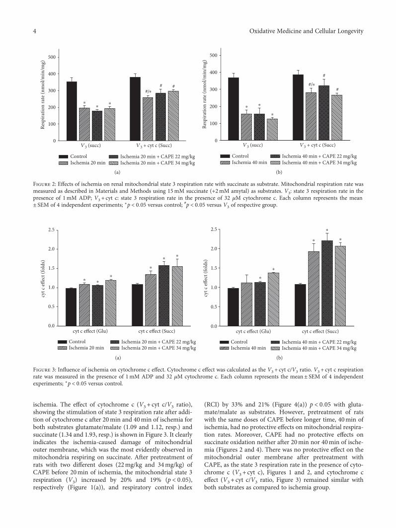
ischemia. The effect of cytochrome c (V3 + cyt c/V3 ratio),showing the stimulation of state 3 respiration rate after addi-tion of cytochrome c after 20min and 40min of ischemia forboth substrates glutamate/malate (1.09 and 1.12, resp.) andsuccinate (1.34 and 1.93, resp.) is shown in Figure 3. It clearlyindicates the ischemia-caused damage of mitochondrialouter membrane, which was the most evidently observed inmitochondria respiring on succinate. After pretreatment ofrats with two different doses (22mg/kg and 34mg/kg) ofCAPE before 20min of ischemia, the mitochondrial state 3respiration (V3) increased by 20% and 19% (p < 0 05),respectively (Figure 1(a)), and respiratory control index
(RCI) by 33% and 21% (Figure 4(a)) p < 0 05 with gluta-mate/malate as substrates. However, pretreatment of ratswith the same doses of CAPE before longer time, 40min ofischemia, had no protective effects on mitochondrial respira-tion rates. Moreover, CAPE had no protective effects onsuccinate oxidation neither after 20min nor 40min of ische-mia (Figures 2 and 4). There was no protective effect on themitochondrial outer membrane after pretreatment withCAPE, as the state 3 respiration rate in the presence of cyto-chrome c (V3 + cyt c), Figures 1 and 2, and cytochrome ceffect (V3 + cyt c/V3 ratio, Figure 3) remained similar withboth substrates as compared to ischemia group.
cyt c
effec
t (fo
lds)
0.0
0.5
1.0
1.5
2.0
2.5
cyt c effect (Glu)
⁎ ⁎⁎
⁎
⁎⁎
cyt c effect (Succ)
ControlIschemia 20 min
Ischemia 20 min + CAPE 22 mg/kgIschemia 20 min + CAPE 34 mg/kg
(a)
cyt c
effec
t (fo
lds)
⁎
⁎
⁎
⁎
⁎
0.0
0.5
1.0
1.5
2.0
2.5
cyt c effect (Glu) cyt c effect (Succ)
ControlIschemia 40 min
Ischemia 40 min + CAPE 22 mg/kgIschemia 40 min + CAPE 34 mg/kg
(b)
Figure 3: Influence of ischemia on cytochrome c effect. Cytochrome c effect was calculated as the V3 + cyt c/V3 ratio. V3 + cyt c respirationrate was measured in the presence of 1mM ADP and 32 μM cytochrome c. Each column represents the mean± SEM of 4 independentexperiments; ∗p < 0 05 versus control.
0
100
200
300
400
500
V3 (succ) V3 + cyt c (Succ)
⁎⁎
⁎
⁎#/# #
ControlIschemia 20 min
Ischemia 20 min + CAPE 22 mg/kgIschemia 20 min + CAPE 34 mg/kg
Resp
iratio
n ra
te (n
mol
/min
/mg)
(a)
V3 (succ) V3 + cyt c (Succ)
⁎ ⁎
⁎
⁎
0
100
200
300
400
500
#/#
#
ControlIschemia 40 min
Ischemia 40 min + CAPE 22 mg/kgIschemia 40 min + CAPE 34 mg/kg
Resp
iratio
n ra
te (n
mol
/min
/mg)
(b)
Figure 2: Effects of ischemia on renal mitochondrial state 3 respiration rate with succinate as substrate. Mitochondrial respiration rate wasmeasured as described in Materials and Methods using 15mM succinate (+2mM amytal) as substrates. V3: state 3 respiration rate in thepresence of 1mM ADP; V3 + cyt c: state 3 respiration rate in the presence of 32 μM cytochrome c. Each column represents the mean± SEM of 4 independent experiments; ∗p < 0 05 versus control; #p < 0 05 versus V3 of respective group.
4 Oxidative Medicine and Cellular Longevity

3.2. Effect of CAPE on Complex I Activity. As our resultsshowed that mitochondrial respiration rate with glutamate/malate (complex I-linked substrates) was clearly reducedafter ischemia, in addition, we measured the effects of ische-mia on mitochondrial complex I activity. Our data revealedthat the reduction of state 3 respiration rate after 20min ofischemia was associated with the decrease in complex I activ-ity by 23% (Figure 5(a)). After 40min of ischemia, mitochon-drial complex I activity was diminished by 54% (p < 0 05,Figure 5(b)). Pretreatment of animals with two different
doses of CAPE (22mg/kg and 34mg/kg) had protective effecton mitochondrial respiratory chain. After pretreatment withCAPE, complex I activity after 20min of ischemia increasedby 18% (22mg/kg CAPE) and by 98%, p < 0 05 (34mg/kgCAPE, Figure 5(a)). After 40min of ischemia, complex Iactivity increased by 77%, p < 0 05 (22mg/kg CAPE), andby 36%, p < 0 05 (34mg/kg CAPE, Figure 5(b)).
3.3. CAPE Increases Mitochondrial Ca2+ Uptake. It is wellknown that mitochondria play a crucial role in intracellular
1
2
3
4
5
RCI (Glu) RCI (Succ)
#
⁎
⁎⁎
⁎⁎
⁎
Rerp
irato
ry co
ntro
l ind
ex (R
CI)
ControlIschemia 20 min
Ischemia 20 min + CAPE 22 mg/kgIschemia 20 min + CAPE 34 mg/kg
(a)
RCI (Glu) RCI (Succ)
⁎
⁎ ⁎
⁎ ⁎ ⁎
1
2
3
4
5
#
Rerp
irato
ry co
ntro
l ind
ex (R
CI)
ControlIschemia 40 min
Ischemia 40 min + CAPE 22 mg/kgIschemia 40 min + CAPE 34 mg/kg
(b)
Figure 4: Effect of ischemia on mitochondrial respiratory control index (RCI). Measurements were performed in the presence of 5mMglutamate + 5mM malate or 15mM succinate (+2mM amytal) as substrates. Mitochondrial respiratory control index (RCI), that is, theratio between oxygen uptake rates in state 3 and routine respiration rate (RCI =V3/V0). Each column represents the mean± SEM of 4independent experiments; ∗p < 0 05 versus control; #p < 0 05 versus ischemia alone.
1
2
3
4
5
6
7
#
NA
DH
oxi
datio
n (�휇
mol
/min
/mg
prot
ein)
Ischemia
ControlIschemia 20 min
Ischemia 20 min + CAPE 22 mg/kgIschemia 20 min + CAPE 34 mg/kg
(a)
NA
DH
oxi
datio
n (�휇
mol
/min
/mg
prot
ein)
1
2
3
4
5
6
7
⁎
#
#
Ischemia
ControlIschemia 40 min
Ischemia 40 min + CAPE 22 mg/kgIschemia 40 min + CAPE 34 mg/kg
(b)
Figure 5: Effect of ischemia on complex I activity in kidney mitochondria. The complex I activity was measured spectrophotometrically at340 nm as described in Materials and Methods. Each column represents the mean± SEM of 4 independent experiments; ∗p < 0 05 versuscontrol; #p < 0 05 versus ischemia alone.
5Oxidative Medicine and Cellular Longevity

Ca2+ signaling, taking up and releasing calcium upon differ-ent cellular conditions such as ischemia, oxidative stress,etc. Elevation of intramitochondrial calcium concentrationafter ischemia can trigger opening of mitochondrial perme-ability transition pore and cell death.
Fluorimetric Ca2+ measurements were performed inorder to measure Ca2+ uptake by mitochondria after 20 and40min of ischemia alone or after pretreatment with CAPE.Our results indicated that in control mitochondria, Ca2+
uptake was 15.99 μmol/minmg protein. Ischemia 20 and40min reduced accumulation of calcium in mitochondriaby 30% (Figures 6(a) and 6(b)). Pretreatment of rats withCAPE (22mg/kg) significantly increased the mitochondrialCa2+ uptake by 50% after 20min of ischemia and by 41%after 40min of ischemia (Figures 6(a) and 6(b)) as comparedto ischemia alone. Pretreatment of animals with higherconcentration of CAPE (34mg/kg) had no protective effecton calcium accumulation in kidney mitochondria afterischemia.
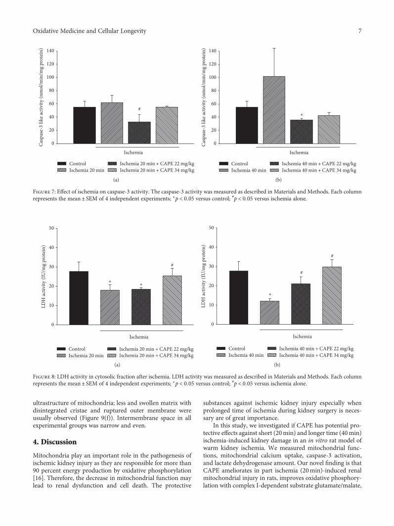
3.4. CAPE Reduces Caspase Activation. As an indicator forapoptosis, we measured DEVD-cleaving caspase-3-like pro-tease activity. After 20min of ischemia, caspase-3-like activ-ity in cytosolic fraction was increased by 1.15-fold ascompared to control, whereas after pretreatment with CAPE(22mg/kg and 34mg/kg), caspase-3-like activity was dimin-ished by 1.52 fold, p < 0 05, and returned to control level(Figure 7(a)). After 40min of ischemia, caspase-3-likeactivity in cytosolic fraction was increased by 1.86-fold ascompared to control. CAPE (22mg/kg and 34mg/kg) dimin-ished caspase-3-like activity to control level (Figure 7(b)).
3.5. CAPE Reduced Lactate Dehydrogenase (LDH) Activity inCytosolic Fraction. As an indicator for necrosis, lactate dehy-drogenase (LDH) activity was measured in cytosolic fractions
in the control group and after ischemia (with and withoutpretreatment with CAPE). LDH activity in cytosolic fractionof control mitochondria was 27.8± 5.1 IU/mg protein anddecreased by 35% and 56% after 20min and 40min of ische-mia, respectively (Figures 8(a) and 8(b)). Pretreatment withCAPE (22mg/kg) had no protective effect after 20min ofischemia, but improved it after 40min of ischemia(Figures 8(a) and 8(b)), that is, LDH activity in cytosolicfraction increased by 74% (to 21.1 IU/mg protein). Afterpretreatment with higher dose (34mg/kg) of CAPE, activityof LDH was restored nearly to control level after both timesof ischemia (Figures 8(a) and 8(b)).
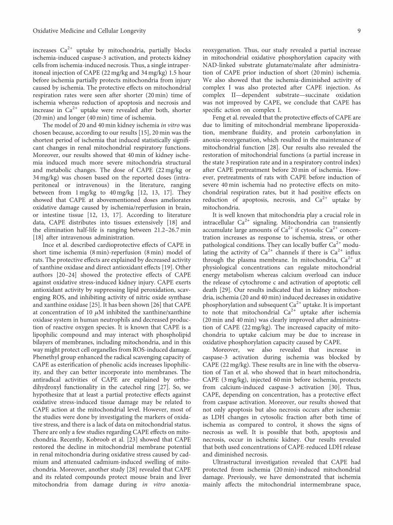
3.6. Kidney ElectronMicroscopy. Electron microscopical find-ings revealed that CAPE (22mg/kg and 34mg/kg) did notaffect the ultrastructure of control mitochondria—theyshowed normal mitochondrial ultrastructure—parallel cris-tae, uniform matrix, and uninterrupted outer membrane.Both parts of intermembrane space—intracristal and periph-eral—are narrow and even (Figures 9(a) and 9(b)). CAPEpretreatment before 20min of ischemia affected mainlymitochondrial matrix making it slightly swollen and perfo-rated by empty patches in some cells. Mitochondria after pre-treatment with CAPE (22mg/kg) showed less swollen matrix(Figure 9(c)) comparing with the higher CAPE concentration(34mg/kg) (Figure 9(d)). Following enlarged matrix cristaelose their parallel arrangement and rearrange to radial loca-tion filling almost all volume of mitochondria (Figure 9(d)).After 40min of ischemia, mitochondria increased in sizedue to enlarged amount of matrix; their cristae lost parallelarrangement. Seldom, mitochondria were seen broken(Figures 9(e) and 9(f)). However, CAPE (22mg/kg)preserved continuous matrix with sporadically seen smallpatches and only partly lost cristae parallelism(Figure 9(e)). Higher dose of CAPE (34mg/kg) preserved
0
Ca2+
upt
ake (�휇
M/m
in/m
g pr
otei
n)
5
10
15
20
25
30
ControlIschemia 20 min
Ischemia 20 min + CAPE 22 mg/kgIschemia 20 min + CAPE 34 mg/kg
Ischemia
⁎
⁎
#
(a)
ControlIschemia 40 min
Ischemia 40 min + CAPE 22 mg/kgIschemia 40 min + CAPE 34 mg/kg
0
5
10
15
20
25
30
#
Ischemia
⁎
⁎#
Ca2+
upt
ake (�휇
M/m
in/m
g pr
otei
n)
(b)
Figure 6: Mitochondrial Ca2+ uptake: effect of ischemia. Ca2+ uptake was measured fluorimetrically (excitation at 506 nm, emission at535 nm) as described in Materials and Methods. Each column represents the mean± SEM of 4 independent experiments; ∗p < 0 05 versuscontrol; #p < 0 05 versus ischemia alone.
6 Oxidative Medicine and Cellular Longevity
ultrastructure of mitochondria; less and swollen matrix withdisintegrated cristae and ruptured outer membrane wereusually observed (Figure 9(f)). Intermembrane space in allexperimental groups was narrow and even.
4. Discussion
Mitochondria play an important role in the pathogenesis ofischemic kidney injury as they are responsible for more than90 percent energy production by oxidative phosphorylation[16]. Therefore, the decrease in mitochondrial function maylead to renal dysfunction and cell death. The protective
substances against ischemic kidney injury especially whenprolonged time of ischemia during kidney surgery is neces-sary are of great importance.
In this study, we investigated if CAPE has potential pro-tective effects against short (20min) and longer time (40min)ischemia-induced kidney damage in an in vitro rat model ofwarm kidney ischemia. We measured mitochondrial func-tions, mitochondrial calcium uptake, caspase-3 activation,and lactate dehydrogenase amount. Our novel finding is thatCAPE ameliorates in part ischemia (20min)-induced renalmitochondrial injury in rats, improves oxidative phosphory-lation with complex I-dependent substrate glutamate/malate,
0
10
20
30
40
50
LDH
activ
ity (I
U/m
g pr
otei
n)
ControlIschemia 20 min
Ischemia 20 min + CAPE 22 mg/kgIschemia 20 min + CAPE 34 mg/kg
#
Ischemia
⁎⁎
(a)
0
10
20
30
40
50
#
#
ControlIschemia 40 min
Ischemia 40 min + CAPE 22 mg/kgIschemia 40 min + CAPE 34 mg/kg
Ischemia
⁎
LDH
activ
ity (I
U/m
g pr
otei
n)
(b)
Figure 8: LDH activity in cytosolic fraction after ischemia. LDH activity was measured as described in Materials and Methods. Each columnrepresents the mean± SEM of 4 independent experiments; ∗p < 0 05 versus control; #p < 0 05 versus ischemia alone.
0Casp
ase-
3 lik
e act
ivity
(nm
ol/m
in/m
g pr
otei
n)
20
40
60
80
100
120
140
ControlIschemia 20 min
Ischemia 20 min + CAPE 22 mg/kgIschemia 20 min + CAPE 34 mg/kg
#
Ischemia
(a)
ControlIschemia 40 min
Ischemia 40 min + CAPE 22 mg/kgIschemia 40 min + CAPE 34 mg/kg
0
20
40
60
80
100
120
140
Ischemia
⁎
Casp
ase-
3 lik
e act
ivity
(nm
ol/m
in/m
g pr
otei
n)
(b)
Figure 7: Effect of ischemia on caspase-3 activity. The caspase-3 activity was measured as described in Materials and Methods. Each columnrepresents the mean± SEM of 4 independent experiments; ∗p < 0 05 versus control; #p < 0 05 versus ischemia alone.
7Oxidative Medicine and Cellular Longevity

Control + CAPE, 22 mg/kg
(a)
Control + CAPE, 34 mg/kg
(b)
Ischemia (20 min) + CAPE, 22 mg/kg
(c)
Ischemia (20 min) + CAPE, 34 mg/kg
(d)
Ischemia (40 min) + CAPE, 22 mg/kg
(e)
Ischemia (40 min) + CAPE, 34 mg/kg
(f)
Figure 9: Ultrastructural changes to mitochondria. (a, b) Control. Mitochondria display normal morphology—shape is from round toelongate depending on the section, cristae are arranged in parallel rows at regular intervals, and the matrix is evenly dense. Note thatnormal morphology is characteristic to all mitochondria despite their irregular distribution in renal cells. (c, d) 20min ischemia affectedmitochondria: matrix is enlarged, uneven, and partly interrupted by empty patches; the outer and inner mitochondrial membranes areclose and parallel. In 22mg/kg CAPE group (c), cristae are parallel ranged; however, in CAPE 34mg/kg group (d), irregular orientation ofcristae can be seen. (e, f) 40min ischemia affected mitochondria. (e) Cell with moderately preserved mitochondria in CAPE 22mg/kggroup: matrix is slightly enlarged with seldom empty patches, cristae are irregularly oriented, and the outer membrane is discontinuous.Note a mitochondrion with fully lost inner structure (∗ ). (f) Mitochondria in CAPE 34mg/kg group possess noticeably swollen matrixand exclusively peripheral cristae, while the outer membrane is ruptured in many places. N: nucleus; RER: rough endoplasmicreticulum; Ex: extracellular space. Scale bar: 1 μm.
8 Oxidative Medicine and Cellular Longevity
increases Ca2+ uptake by mitochondria, partially blocksischemia-induced caspase-3 activation, and protects kidneycells from ischemia-induced necrosis. Thus, a single intraper-itoneal injection of CAPE (22mg/kg and 34mg/kg) 1.5 hourbefore ischemia partially protects mitochondria from injurycaused by ischemia. The protective effects on mitochondrialrespiration rates were seen after shorter (20min) time ofischemia whereas reduction of apoptosis and necrosis andincrease in Ca2+ uptake were revealed after both, shorter(20min) and longer (40min) time of ischemia.
The model of 20 and 40min kidney ischemia in vitro waschosen because, according to our results [15], 20min was theshortest period of ischemia that induced statistically signifi-cant changes in renal mitochondrial respiratory functions.Moreover, our results showed that 40min of kidney ische-mia induced much more severe mitochondria structuraland metabolic changes. The dose of CAPE (22mg/kg or34mg/kg) was chosen based on the reported doses (intra-peritoneal or intravenous) in the literature, rangingbetween from 1mg/kg to 40mg/kg [12, 13, 17]. Theyshowed that CAPE at abovementioned doses amelioratesoxidative damage caused by ischemia/reperfusion in brain,or intestine tissue [12, 13, 17]. According to literaturedata, CAPE distributes into tissues extensively [18] andthe elimination half-life is ranging between 21.2–26.7min[18] after intravenous administration.
Ince et al. described cardioprotective effects of CAPE inshort time ischemia (8min)-reperfusion (8min) model ofrats. The protective effects are explained by decreased activityof xanthine oxidase and direct antioxidant effects [19]. Otherauthors [20–24] showed the protective effects of CAPEagainst oxidative stress-induced kidney injury. CAPE exertsantioxidant activity by suppressing lipid peroxidation, scav-enging ROS, and inhibiting activity of nitric oxide synthaseand xanthine oxidase [25]. It has been shown [26] that CAPEat concentration of 10 μM inhibited the xanthine/xanthineoxidase system in human neutrophils and decreased produc-tion of reactive oxygen species. It is known that CAPE is alipophilic compound and may interact with phospholipidbilayers of membranes, including mitochondria, and in thiswaymight protect cell organelles from ROS-induced damage.Phenethyl group enhanced the radical scavenging capacity ofCAPE as esterification of phenolic acids increases lipophilic-ity, and they can better incorporate into membranes. Theantiradical activities of CAPE are explained by ortho-dihydroxyl functionality in the catechol ring [27]. So, wehypothesize that at least a partial protective effects againstoxidative stress-induced tissue damage may be related toCAPE action at the mitochondrial level. However, most ofthe studies were done by investigating the markers of oxida-tive stress, and there is a lack of data on mitochondrial status.There are only a few studies regarding CAPE effects on mito-chondria. Recently, Kobroob et al. [23] showed that CAPErestored the decline in mitochondrial membrane potentialin renal mitochondria during oxidative stress caused by cad-mium and attenuated cadmium-induced swelling of mito-chondria. Moreover, another study [28] revealed that CAPEand its related compounds protect mouse brain and livermitochondria from damage during in vitro anoxia-
reoxygenation. Thus, our study revealed a partial increasein mitochondrial oxidative phosphorylation capacity withNAD-linked substrate glutamate/malate after administra-tion of CAPE prior induction of short (20min) ischemia.We also showed that the ischemia-diminished activity ofcomplex I was also protected after CAPE injection. Ascomplex II—dependent substrate—succinate oxidationwas not improved by CAPE, we conclude that CAPE hasspecific action on complex I.
Feng et al. revealed that the protective effects of CAPE aredue to limiting of mitochondrial membrane lipoperoxida-tion, membrane fluidity, and protein carbonylation inanoxia-reoxygenation, which resulted in the maintenance ofmitochondrial function [28]. Our results also revealed therestoration of mitochondrial functions (a partial increase inthe state 3 respiration rate and in a respiratory control index)after CAPE pretreatment before 20min of ischemia. How-ever, pretreatments of rats with CAPE before induction ofsevere 40min ischemia had no protective effects on mito-chondrial respiration rates, but it had positive effects onreduction of apoptosis, necrosis, and Ca2+ uptake bymitochondria.
It is well known that mitochondria play a crucial role inintracellular Ca2+ signaling. Mitochondria can transientlyaccumulate large amounts of Ca2+ if cytosolic Ca2+ concen-tration increases as response to ischemia, stress, or otherpathological conditions. They can locally buffer Ca2+ modu-lating the activity of Ca2+ channels if there is Ca2+ influxthrough the plasma membrane. In mitochondria, Ca2+ atphysiological concentrations can regulate mitochondrialenergy metabolism whereas calcium overload can inducethe release of cytochrome c and activation of apoptotic celldeath [29]. Our results indicated that in kidney mitochon-dria, ischemia (20 and 40min) induced decreases in oxidativephosphorylation and subsequent Ca2+ uptake. It is importantto note that mitochondrial Ca2+ uptake after ischemia(20min and 40min) was clearly improved after administra-tion of CAPE (22mg/kg). The increased capacity of mito-chondria to uptake calcium may be due to increase inoxidative phosphorylation capacity caused by CAPE.
Moreover, we also revealed that increase incaspase-3 activation during ischemia was blocked byCAPE (22mg/kg). These results are in line with the observa-tion of Tan et al. who showed that in heart mitochondria,CAPE (3mg/kg), injected 60min before ischemia, protectsfrom calcium-induced caspase-3 activation [30]. Thus,CAPE, depending on concentration, has a protective effectfrom caspase activation. Moreover, our results showed thatnot only apoptosis but also necrosis occurs after ischemia:as LDH changes in cytosolic fraction after both time ofischemia as compared to control, it shows the signs ofnecrosis as well. It is possible that both, apoptosis andnecrosis, occur in ischemic kidney. Our results revealedthat both used concentrations of CAPE-reduced LDH releaseand diminished necrosis.
Ultrastructural investigation revealed that CAPE hadprotected from ischemia (20min)-induced mitochondrialdamage. Previously, we have demonstrated that ischemiamainly affects the mitochondrial intermembrane space,
9Oxidative Medicine and Cellular Longevity

where intracristal space was found enlarged resulting in bal-looned cristae as well as enlarged peripheral space followedby detachment of outer and inner membranes [15]. Applica-tion of CAPE preserved intermembrane space from edemaafter 20min of ischemia, as it was found narrow and uniformin all experimental groups. No ballooned cristae, withenlarged intracristal space or detachment of outer and innermembranes resulting in enlarged peripheral space, wereobserved in this study. Though pretreatment with the higherdose (34mg/kg) of CAPE protected slightly mitochondrialfunctions (oxidative phosphorylation, activity of mitochon-drial complex I, LDH), whereas ultrastructure of mitochon-dria in some kidney tissue slices showed swollen matrixwith disintegrated cristae and ruptured outer mitochondrialmembrane, thus it seems that higher dose of CAPE did notprotect ultrastructure of mitochondria. The obtained effectsmay be associated with the heterogeneity of mitochondria.Our observations suggest that different renal cell types areaffected by ischemia to a different extent. Thus, it would befeasible to investigate the protective properties of CAPE indifferent renal cell types.
In conclusion, our study revealed partial protection ofmitochondrial function after pretreatment of rats with intra-peritoneal injection of CAPE. Since CAPE has beneficial,including antioxidant and anti-inflammatory, effects as wellas partially preserves mitochondrial function, we think thatthis compound may have a potential to protect the kidneyfrom ischemia-induced damage. The mechanisms of protec-tion are not fully understood yet, but at least, partially, it isassociated with the improvement of mitochondrial status inthe cells. Further studies will be required for the better char-acterization of the mechanism of CAPE action.
Disclosure
The manuscript is based on the paper published as anabstract in European Urology Supplements Journal2014 and available at the following link: http://www.eusupplements.europeanurology.com/article/S1569-9056.
Conflicts of Interest
The authors confirm that this article content has no conflictof interests.
References
[1] M. Malek and M. Nematbakhsh, “Renal ischemia/reperfusioninjury; from pathophysiology to treatment,” Journal of RenalInjury Prevention, vol. 4, no. 2, pp. 20–27, 2015.
[2] F. Becker, H. Van Poppel, O. W. Hakenberg et al., “Assessingthe impact of ischaemia time during partial nephrectomy,”European Urology, vol. 56, pp. 625–634, 2009.
[3] G. Kroemer and J. C. Reed, “Mitochondrial control of celldeath,” Nature Medicine, vol. 6, pp. 513–519, 2000.
[4] R. H. Thompson, B. R. Lane, C. M. Lohse et al., “Every minutecounts when the renal hilum is clamped during partialnephrectomy,” European Urology, vol. 58, pp. 340–345, 2010.
[5] D. J. Parekh, J. M. Weinberg, B. Ercole et al., “Tolerance of thehuman kidney to isolated controlled ischemia,” Journal of theAmerican Society of Nephrology, vol. 24, pp. 506–517, 2013.
[6] Y. J. Chen, A. C. Huang, H. H. Chang et al., “Caffeic acid phe-nethyl ester, an antioxidant from propolis, protects peripheralblood mononuclear cells of competitive cyclists againsthyperthermal stress,” Journal of Food Science, vol. 74,pp. H162–H167, 2009.
[7] L. M. LeBlanc, A. F. Pare, J. Jean-Francois, M. J. Hebert, M. E.Surette, and M. Touaibia, “Synthesis and antiradical/antioxi-dant activities of caffeic acid phenethyl ester and its relatedpropionic, acetic, and benzoic acid analogues,” Molecules,vol. 17, pp. 14637–14650, 2012.
[8] A. Gokce, S. Oktar, Z. Yonden et al., “Protective effect of caffeicacid phenethyl ester on cyclosporine A-induced nephrotoxi-city in rats,” Renal Failure, vol. 31, pp. 843–847, 2009.
[9] A. Gurel, F. Armutcu, S. Sahin et al., “Protective role ofalpha-tocopherol and caffeic acid phenethyl ester onischemia-reperfusion injury via nitric oxide and myeloperoxi-dase in rat kidneys,” Clinica Chimica Acta, vol. 339, pp. 33–41,2004.
[10] N. C. Roso, R. R. Correa, Y. M. Castiglia et al., “Caffeic acidphenethyl ester effects in the kidney during ischemia andreperfusion in rats anesthetized with isoflurane,” Transplanta-tion Proceedings, vol. 44, pp. 1211–1213, 2012.
[11] N. Aydogdu, G. Atmaca, O. Yalcin, K. Batcioglu, and K.Kaymak, “Effects of caffeic acid phenethyl ester on glycerol-induced acute renal failure in rats,” Clinical and ExperimentalPharmacology & Physiology, vol. 31, pp. 575–579, 2004.
[12] X. Wei, L. Zhao, Z. Ma et al., “Caffeic acid phenethyl esterprevents neonatal hypoxic-ischaemic brain injury,” Brain,vol. 127, pp. 2629–2635, 2004.
[13] M. Khan, C. Elango, M. A. Ansari, I. Singh, and A. K. Singh,“Caffeic acid phenethyl ester reduces neurovascular inflamma-tion and protects rat brain following transient focal cerebralischemia,” Journal of Neurochemistry, vol. 102, pp. 365–377,2007.
[14] H. Parlakpinar, E. Sahna, A. Acet, B. Mizrak, and A. Polat,“Protective effect of caffeic acid phenethyl ester (CAPE) onmyocardial ischemia-reperfusion-induced apoptotic celldeath,” Toxicology, vol. 209, pp. 1–14, 2005.
[15] R. Baniene, D. Trumbeckas, M. Kincius et al., “Short ischemiainduces rat kidney mitochondria dysfunction,” Journal of Bio-energetics and Biomembranes, vol. 48, no. 1, pp. 77–85, 2016.
[16] R. Che, Y. Yuan, S. Huang, and A. Zhang, “Mitochondrial dys-function in the pathophysiology of renal diseases,” AmericanJournal of Physiology-Renal Physiology, vol. 306, pp. F367–F378, 2014.
[17] Y. Yildiz, M. Serter, R. O. Ek et al., “Protective effects of caffeicacid phenethyl ester on intestinal ischemia-reperfusioninjury,” Digestive Diseases and Sciences, vol. 54, pp. 738–744,2009.
[18] S. Akyol, Z. Ginis, F. Armutcu, G. Ozturk, M. R. Yigitoglu, andO. Akyol, “The potential usage of caffeic acid phenethyl ester(CAPE) against chemotherapy-induced and radiotherapy-induced toxicity,” Cell Biochemistry and Function, vol. 30,pp. 438–443, 2012.
[19] H. Ince, E. Kandemir, C. Bagci, M. Gulec, and O. Akyol,“The effect of caffeic acid phenethyl ester on short-termacute myocardial ischemia,” Medical Science Monitor,vol. 12, pp. BR187–BR193, 2006.
10 Oxidative Medicine and Cellular Longevity

[20] M. Ogeturk, I. Kus, N. Colakoglu, I. Zararsiz, N. Ilhan, and M.Sarsilmaz, “Caffeic acid phenethyl ester protects kidneysagainst carbon tetrachloride toxicity in rats,” Journal of Ethno-pharmacology, vol. 97, pp. 273–280, 2005.
[21] S. Ozen, O. Akyol, M. Iraz et al., “Role of caffeic acid phenethylester, an active component of propolis, against cisplatin-induced nephrotoxicity in rats,” Journal of Applied Toxicology,vol. 24, pp. 27–35, 2004.
[22] O. Wongmekiat, S. Gomonchareonsiri, and K. Thamprasert,“Caffeic acid phenethyl ester protects against oxidative stress-related renal dysfunction in rats treated with cyclosporin A,”Fundamental & Clinical Pharmacology, vol. 25, pp. 619–626,2011.
[23] A. Kobroob, N. Chattipakorn, and O. Wongmekiat, “Caffeicacid phenethyl ester ameliorates cadmium-induced kidneymitochondrial injury,” Chemico-Biological Interactions,vol. 200, pp. 21–27, 2012.
[24] P. Gong, F. Chen, X. Liu, X. Gong, J. Wang, and Y. Ma, “Pro-tective effect of caffeic acid phenethyl ester against cadmium-induced renal damage in mice,” The Journal of ToxicologicalSciences, vol. 37, pp. 415–425, 2012.
[25] M. Hosnuter, A. Gurel, O. Babuccu, F. Armutcu, E. Kargi, andA. Isikdemir, “The effect of CAPE on lipid peroxidation andnitric oxide levels in the plasma of rats following thermalinjury,” Burns, vol. 30, pp. 121–125, 2004.
[26] G. F. Sudina, O. K. Mirzoeva, M. A. Pushkareva, G. A.Korshunova, N. V. Sumbatyan, and S. D. Varfolomeev,“Caffeic acid phenethyl ester as a lipoxygenase inhibitor withantioxidant properties,” FEBS Letters, vol. 329, pp. 21–24,1993.
[27] W. M. Wu, L. Lu, Y. Long et al., “Free radical scavenging andantioxidative activities of caffeic acid phenethyl ester (CAPE)and its related compounds in solution and membranes: astructure–activity insight,” Food Chemistry, vol. 105, no. 1,pp. 107–115, 2007.
[28] Y. Feng, Y. W. Lu, P. H. Xu et al., “Caffeic acid phenethyl esterand its related compounds limit the functional alterations ofthe isolated mouse brain and liver mitochondria submittedto in vitro anoxia-reoxygenation: relationship to their antioxi-dant activities,” Biochimica et Biophysica Acta (BBA) - GeneralSubjects, vol. 1780, pp. 659–672, 2008.
[29] K. S. Kumaran and P. S. Prince, “Caffeic acid protects rat heartmitochondria against isoproterenol-induced oxidative dam-age,” Cell Stress & Chaperones, vol. 15, pp. 791–806, 2010.
[30] K. Nomura, H. Imai, T. Koumura, T. Kobayashi, and Y.Nakagawa, “Mitochondrial phospholipid hydroperoxideglutathione peroxidase inhibits the release of cytochromec from mitochondria by suppressing the peroxidation ofcardiolipin in hypoglycaemia-induced apoptosis,” TheBiochemical Journal, vol. 351, pp. 183–193, 2000.
11Oxidative Medicine and Cellular Longevity

Submit your manuscripts athttps://www.hindawi.com
Stem CellsInternational
Hindawi Publishing Corporationhttp://www.hindawi.com Volume 2014
Hindawi Publishing Corporationhttp://www.hindawi.com Volume 2014
MEDIATORSINFLAMMATION
of
Hindawi Publishing Corporationhttp://www.hindawi.com Volume 2014
Behavioural Neurology
EndocrinologyInternational Journal of
Hindawi Publishing Corporationhttp://www.hindawi.com Volume 2014
Hindawi Publishing Corporationhttp://www.hindawi.com Volume 2014
Disease Markers
Hindawi Publishing Corporationhttp://www.hindawi.com Volume 2014
BioMed Research International
OncologyJournal of
Hindawi Publishing Corporationhttp://www.hindawi.com Volume 2014
Hindawi Publishing Corporationhttp://www.hindawi.com Volume 2014
Oxidative Medicine and Cellular Longevity
Hindawi Publishing Corporationhttp://www.hindawi.com Volume 2014
PPAR Research
The Scientific World JournalHindawi Publishing Corporation http://www.hindawi.com Volume 2014
Immunology ResearchHindawi Publishing Corporationhttp://www.hindawi.com Volume 2014
Journal of
ObesityJournal of
Hindawi Publishing Corporationhttp://www.hindawi.com Volume 2014
Hindawi Publishing Corporationhttp://www.hindawi.com Volume 2014
Computational and Mathematical Methods in Medicine
OphthalmologyJournal of
Hindawi Publishing Corporationhttp://www.hindawi.com Volume 2014
Diabetes ResearchJournal of
Hindawi Publishing Corporationhttp://www.hindawi.com Volume 2014
Hindawi Publishing Corporationhttp://www.hindawi.com Volume 2014
Research and TreatmentAIDS
Hindawi Publishing Corporationhttp://www.hindawi.com Volume 2014
Gastroenterology Research and Practice
Hindawi Publishing Corporationhttp://www.hindawi.com Volume 2014
Parkinson’s Disease
Evidence-Based Complementary and Alternative Medicine
Volume 2014Hindawi Publishing Corporationhttp://www.hindawi.com