c-ETS1FacilitatesG /S-phaseTransitionbyUp-regulating ... · 4 The abbreviations used are: CDK,...
11
c-ETS1 Facilitates G 1 /S-phase Transition by Up-regulating Cyclin E and CDK2 Genes and Cooperates with Hepatitis B Virus X Protein for Their Deregulation * □ S Received for publication, March 8, 2011, and in revised form, April 20, 2011 Published, JBC Papers in Press, April 22, 2011, DOI 10.1074/jbc.M111.238238 Anup Kumar Singh 1 , Manickavinayaham Swarnalatha 2 , and Vijay Kumar 3 From the Virology Group, International Centre for Genetic Engineering and Biotechnology, Aruna Asaf Ali Marg, New Delhi 110067, India Recent studies on the molecular mechanisms responsible for cell cycle deregulation in cancer have puzzled out the role of oncogenes in mediating unscheduled cellular proliferation. This is reminiscence of their activity as proto-oncogenes that drives scheduled cell cycle progression under physiological con- ditions. Working on the cell cycle regulatory activity of proto- oncogene, we observed that c-ETS1 transcriptionally up-regu- lated both cyclin E and CDK2 genes, the master regulators of G 1 /S-phase transition. The process was mediated by kinetic coherence of c-ETS1 expression and its recruitment to both pro- moters during G 1 /S-phase transition. Furthermore, enforced expression of c-ETS1 helped G 0 -arrested cells to progress into G 1 /S-phases apparently due to the activation of cyclin E/CDK2 genes. Physiological induction of c-ETS1 by EGF showed the remodeling of mononucleosomes bound to the c-ETS1 binding site on both promoters during their activation. The exchange of HDAC1 with histone acetyltransferase-p300 was contempora- neous to the chromatin remodeling with consequent increase in histone H3K9 acetylation. Furthermore, the ATP-dependent chromatin remodeler hBRM1 recruitment was also associated with nucleosome remodeling and promoter occupancy of phos- pho-Ser5 RNA polymerase II. Intriguingly, the activity of the HBx viral oncoprotein was dependent on c-ETS1 in a hepato- tropic manner, which led to the activation of cyclin E/CDK2 genes. Thus, cyclin E and CDK2 genes are key physiological effectors of the c-ETS1 proto-oncogene. Furthermore, c-ETS1 is indispensable for the hepatotropic action of HBx in cell cycle deregulation. Mechanisms that coordinate cell cycle progression are driven by sequential activation and inactivation of a family of cyclin-dependent kinases (CDKs). 4 The activation occurs pre- dominately by the periodic expression of its regulatory subunit cyclin and activating phosphorylation of the kinase subunit (1). Redundant nature of cyclins and CDKs implies that the func- tional importance lies in their temporal expression rather than their effector molecules (2). G 1 -specific cyclin E expression periodically oscillates in every cycle of proliferating cells, ensuring its ordered progression (3). Periodicity is mainly controlled at the transcriptional level lead- ing to its peak expression during G 1 /S-phase transition (4, 5). The cyclin E/CDK2 activity assumes special significance as it is the rate-limiting regulator of the G 1 /S-phase transition and act as a switch for various cellular processes including initiation of DNA replication (6, 7). Perturbation of this rate-limiting step by viral oncoproteins is a common theme that causally relates to the plethora of cancers (8). The regulation of the cyclin E promoter by mitogens, various transactivators, and growth factors has been analyzed exten- sively in the recent past (9 –12). In contrast, CDK2 regulation has been studied mainly at post translational level (13), and only a few reports have discussed the oscillation of CDK2 levels before S-phase entry (14, 15). This suggests that the regulation of CDK2 at the transcriptional level could be as important as its post-translational control. Although the cyclin E-CDK2 com- plex is assembled and active during the same window of cell cycle, their transcriptional regulation by common transactiva- tors remains obscure. c-ETS1 (transcription factor E-26 transforming sequence-1) is a classic example of the proto-oncogene and the founding member of ETS family proteins. The unique cis element “GGA(A/T),” known as the ETS binding site (EBS), is among the eight most important DNA motifs in minimal responsive synthetic promoters and identified in the promoter/enhancer regions of 200 genes (16). c-ETS1 is known to be associated with different aspects of cancer, including extracellular matrix remodeling, invasion, angiogenesis (17), and also has an impor- tant role in proliferation and differentiation of hematopoietic cells (18). Moreover, inappropriate expression of c-ETS1 is an early event in a wide variety of cancers, and its overexpression results in a transformed phenotype (19). Furthermore, the serum-inducible nature of the c-ETS1 promoter (20) suggested its plausible role in cell cycle regulation. However, the physio- * This work was supported by a core grant of the International Centre for Genetic Engineering and Biotechnology, New Delhi. □ S The on-line version of this article (available at http://www.jbc.org) contains supplemental Tables 1–3 and Figs. 1–5. 1 Recipient of a Senior Research Fellowship from the Council of Scientific and Industrial Research (New Delhi). 2 Recipient of a Senior Research Fellowship from the Indian Council of Medical Research (New Delhi). 3 To whom correspondence should be addressed: Virology Group, International Centre for Genetic Engineering and Biotechnology, Aruna Asaf Ali Marg, New Delhi 110067, India. Tel.: 91-11-26741680; Fax: 91-11-26742316; E-mail: vijay@ icgeb.res.in. 4 The abbreviations used are: CDK, cyclin-dependent kinase; c-ETS1, E-26 transforming sequence 1; CHART-PCR, chromatin accessibility assay based on real-time PCR; HDAC, histone deacetylase; MNase, micrococcal nuclease; qRT-PCR, quantitative RT-PCR; EBS, Ets binding site; CAT, chlor- amphenicol acetyltransferase; pol II, RNA polymerase II. THE JOURNAL OF BIOLOGICAL CHEMISTRY VOL. 286, NO. 25, pp. 21961–21970, June 24, 2011 © 2011 by The American Society for Biochemistry and Molecular Biology, Inc. Printed in the U.S.A. JUNE 24, 2011 • VOLUME 286 • NUMBER 25 JOURNAL OF BIOLOGICAL CHEMISTRY 21961 by guest on April 21, 2019 http://www.jbc.org/ Downloaded from
Transcript of c-ETS1FacilitatesG /S-phaseTransitionbyUp-regulating ... · 4 The abbreviations used are: CDK,...
c-ETS1 Facilitates G1/S-phase Transition by Up-regulatingCyclin E and CDK2 Genes and Cooperates with Hepatitis BVirus X Protein for Their Deregulation*□S
Received for publication, March 8, 2011, and in revised form, April 20, 2011 Published, JBC Papers in Press, April 22, 2011, DOI 10.1074/jbc.M111.238238
Anup Kumar Singh1, Manickavinayaham Swarnalatha2, and Vijay Kumar3
From the Virology Group, International Centre for Genetic Engineering and Biotechnology, Aruna Asaf Ali Marg,New Delhi 110067, India
Recent studies on the molecular mechanisms responsible forcell cycle deregulation in cancer have puzzled out the role ofoncogenes in mediating unscheduled cellular proliferation.This is reminiscence of their activity as proto-oncogenes thatdrives scheduled cell cycle progression under physiological con-ditions. Working on the cell cycle regulatory activity of proto-oncogene, we observed that c-ETS1 transcriptionally up-regu-lated both cyclin E and CDK2 genes, the master regulators ofG1/S-phase transition. The process was mediated by kineticcoherence of c-ETS1 expression and its recruitment to bothpro-moters during G1/S-phase transition. Furthermore, enforcedexpression of c-ETS1 helped G0-arrested cells to progress intoG1/S-phases apparently due to the activation of cyclin E/CDK2genes. Physiological induction of c-ETS1 by EGF showed theremodeling of mononucleosomes bound to the c-ETS1 bindingsite on both promoters during their activation. The exchange ofHDAC1 with histone acetyltransferase-p300 was contempora-neous to the chromatin remodeling with consequent increase inhistone H3K9 acetylation. Furthermore, the ATP-dependentchromatin remodeler hBRM1 recruitment was also associatedwith nucleosome remodeling and promoter occupancy of phos-pho-Ser5 RNA polymerase II. Intriguingly, the activity of theHBx viral oncoprotein was dependent on c-ETS1 in a hepato-tropic manner, which led to the activation of cyclin E/CDK2genes. Thus, cyclin E and CDK2 genes are key physiologicaleffectors of the c-ETS1proto-oncogene. Furthermore, c-ETS1 isindispensable for the hepatotropic action of HBx in cell cyclederegulation.
Mechanisms that coordinate cell cycle progression aredriven by sequential activation and inactivation of a family ofcyclin-dependent kinases (CDKs).4 The activation occurs pre-
dominately by the periodic expression of its regulatory subunitcyclin and activating phosphorylation of the kinase subunit (1).Redundant nature of cyclins and CDKs implies that the func-tional importance lies in their temporal expression rather thantheir effector molecules (2).G1-specific cyclin E expression periodically oscillates in every
cycle of proliferating cells, ensuring its ordered progression (3).Periodicity ismainly controlled at the transcriptional level lead-ing to its peak expression during G1/S-phase transition (4, 5).The cyclin E/CDK2 activity assumes special significance as it isthe rate-limiting regulator of the G1/S-phase transition and actas a switch for various cellular processes including initiation ofDNA replication (6, 7). Perturbation of this rate-limiting stepby viral oncoproteins is a common theme that causally relatesto the plethora of cancers (8).The regulation of the cyclin E promoter bymitogens, various
transactivators, and growth factors has been analyzed exten-sively in the recent past (9–12). In contrast, CDK2 regulationhas been studiedmainly at post translational level (13), and onlya few reports have discussed the oscillation of CDK2 levelsbefore S-phase entry (14, 15). This suggests that the regulationof CDK2 at the transcriptional level could be as important as itspost-translational control. Although the cyclin E-CDK2 com-plex is assembled and active during the same window of cellcycle, their transcriptional regulation by common transactiva-tors remains obscure.c-ETS1 (transcription factor E-26 transforming sequence-1)
is a classic example of the proto-oncogene and the foundingmember of ETS family proteins. The unique cis element“GGA(A/T),” known as the ETS binding site (EBS), is amongthe eight most important DNA motifs in minimal responsivesynthetic promoters and identified in the promoter/enhancerregions of �200 genes (16). c-ETS1 is known to be associatedwith different aspects of cancer, including extracellular matrixremodeling, invasion, angiogenesis (17), and also has an impor-tant role in proliferation and differentiation of hematopoieticcells (18). Moreover, inappropriate expression of c-ETS1 is anearly event in a wide variety of cancers, and its overexpressionresults in a transformed phenotype (19). Furthermore, theserum-inducible nature of the c-ETS1 promoter (20) suggestedits plausible role in cell cycle regulation. However, the physio-
* This work was supported by a core grant of the International Centre forGenetic Engineering and Biotechnology, New Delhi.
□S The on-line version of this article (available at http://www.jbc.org) containssupplemental Tables 1–3 and Figs. 1–5.
1 Recipient of a Senior Research Fellowship from the Council of Scientific andIndustrial Research (New Delhi).
2 Recipient of a Senior Research Fellowship from the Indian Council of MedicalResearch (New Delhi).
3 To whom correspondence should be addressed: Virology Group, InternationalCentre for Genetic Engineering and Biotechnology, Aruna Asaf Ali Marg, NewDelhi 110067, India. Tel.: 91-11-26741680; Fax: 91-11-26742316; E-mail: [email protected].
4 The abbreviations used are: CDK, cyclin-dependent kinase; c-ETS1, E-26transforming sequence 1; CHART-PCR, chromatin accessibility assay based
on real-time PCR; HDAC, histone deacetylase; MNase, micrococcalnuclease; qRT-PCR, quantitative RT-PCR; EBS, Ets binding site; CAT, chlor-amphenicol acetyltransferase; pol II, RNA polymerase II.
THE JOURNAL OF BIOLOGICAL CHEMISTRY VOL. 286, NO. 25, pp. 21961–21970, June 24, 2011© 2011 by The American Society for Biochemistry and Molecular Biology, Inc. Printed in the U.S.A.
JUNE 24, 2011 • VOLUME 286 • NUMBER 25 JOURNAL OF BIOLOGICAL CHEMISTRY 21961
by guest on April 21, 2019
http://ww
w.jbc.org/
Dow
nloaded from

logical effectors of c-ETS1 involved in the regulation of cellcycle remains enigmatic.In the present study, we have shown that cyclin E and CDK2
genes are the physiological effectors of c-ETS1. The stimulationof c-ETS1 by EGF leads to up-regulation of both genes, whichinvolves chromatin remodeling and cross-talk of associated co-factors. Furthermore, the up-regulation of cyclin E and CDK2genes by viral oncoprotein HBx is dependent on a c-ETS1-re-sponsive element in a tissue-specific manner.
EXPERIMENTAL PROCEDURES
Expression Vectors and Reporter DNAConstructs—The humancyclin E promoter reporter construct pCycECAT (pE-WT)(�1195/�79)was kindly provided by Professor J. R.Nevins (DukeUniversity Medical Center) (9), whereas the CDK2 promoterreporter construct �2400CDK2/LUC (DSC37) (pCDK2-WT)was fromGaryStein (UniversityofMassachusettsMedicalSchool)(21). The c-ETS1 expression construct was from Hiroyuki Sug-imoto (22), andc-ETS1dominantnegative (c-ETS1DN)construct(pAPrEts-Z) was kindly provided by Arthur Gutierrez-Hartmann(23). The �-galactosidase expression plasmid pCH110 (Amer-sham Biosciences) and the EGFP expression plasmid pEGFP-C1(BD bioscience) were used as transfection control.Site-directed Mutagenesis of c-ETS1 Elements—The c-ETS1
response element in the cyclin E promoter (pE-WT) reporterconstruct was mutated by PCR using the QuikChangeTMsite-directedmutagenesis kit (Stratagene, La Jolla, CA) to getpE-mut. Likewise, the CDK2 promoter (pCDK2-WT) wasalso mutated for its proximal (pCDK2-prox.mut), distal(pCDK2-dis.mut), or both c-ETS1 sites (pCDK2-mut). Thefollowing set of primers were used: forward, pE-mut F, 5�-actcagggcccctcgagcggcgtctc-3�; pE-mut R, 5�-gagacgccgctc-gaggggccctgagt-3�; pCDK2-prox.mut F, 5�-agggaaacgctcga-ggcaggggcggg-3�; pCDK2-prox.mut R, 5�-cccgcccctgcctcga-gcgtttccct-3�; pCDK2-dis.mut F, 5�-agattcccggctcgagggtttc-caaa-3�; and pCDK2-dis.mut R, 5�-tttggaaaccctcgagccggga-atct-3�. The mutated bases are boldfaced and underlined.Cell Culture, Reagents, and Antibodies—Maintanance of
human hepatoma Huh7 and HepG2 cells, human embryonickidney HEK293 (ATCC CRL-1573), and human epithelial cer-vical HeLa (ATCC CCL2) cell lines were described elsewhere(24). Transfection was carried out in a 60-mm culture dish(0.6 � 106 cells) with 2.0 �g of indicated plasmids by Lipo-fectamine (Invitrogen) according to the manufacturer’sinstructions. EGFP-C1 (0.5 �g) was used in each experiment asa transfection control (supplemental Fig. 1). For reporterassays, 0.25 �g of CAT or luciferase reporter plasmids werecotransfected with 0.5 �g of indicated expression plasmids.siRNA against c-ETS1 was procured from Santa Cruz Biotech-nology and used for transfection according to manufacturer’sinstructions. Wherever mentioned, 24-h post-starved cellswere treated with EGF (10 ng/ml, Calbiochem) for 18 h. Anti-bodies were obtained from the following sources: Santa CruzBiotechnology for RNA polymerase II (pol II; CTD4H8), phos-pho-Ser5 pol II, HDAC1, p300, cyclin E, CDK2, GAPDH,c-ETS1 and hBRM1; and Upstate Biotechnology for H3K9Ac.mAb (monoclonal antibody) (B-8/2/8) against HBx has beenreported previously (25).
CAT and Luciferase Assay—The chloramphenicol acetyl-transferase (CAT) assay was performed as described previously(24). Luciferase assaywas performed according to themanufac-turer’s instructions (Promega). The relative CAT and luciferaseactivities were measured after normalizing each sample with�-galactosidase activity. Wherever HBx expression vector wasused in a reporter assay, mean EGFP fluorescence was used fornormalization of reporter activity.Flow Cytometry (FACS)—Huh7 cells were starved for 48 h
then stimulated with serum for the indicated time periods.Wherever indicated, 24 h post-transfected cells were subjectedto serum starvation for another 24 h. Flow cytometry of cellswas done as described in Ref. 26.RNA Isolation and Quantitative RT-PCR Assay—Total RNA
was isolated from cells using TRIzol reagent as per the suppli-er’s instructions (Invitrogen). RT-PCR was performed withM-MuLV reverse transcriptase (Fermentas) according to themanufacturer’s guidelines. The real-time quantitative PCR(qPCR) was done using specific primers (supplemental Table 1)as described previously (24).Western Blotting—Protocol for Western blotting of protein
samples can be found elsewhere (26).EMSA—In vitro binding of c-ETS1 to putative EBS elements
was performed by EMSA as described previously (24). The endlabeling of oligonucleotides (supplemental Table 2) was doneusing [�-32P]ATP and T4 Polynucleotide Kinase (Fermentas)per the manufacturer’s instructions.MNase Southern Hybridization Assay—Nuclei isolation
and MNase digestion was done as described earlier (24) withminormodifications. Complete digestion of nuclei withMNase(Fermentas) was performed for 20 min at 37 °C (50 unitsMNase/2� 106 nuclei), whereas incomplete digestion was per-formed for 5 min at 37 °C (5 units MNase/2 � 106 nuclei). Forsouthern hybridization, 10�g ofDNAyielded fromeither com-plete or incomplete digestion of MNase was electrophoresedand transferred to nylonmembrane for hybridizationwith end-labeled DNA probes (supplemental Table 3).MNase CHART-PCR Assay—Mononucleosomes obtained
by complete digestion of MNase as mentioned above wereresolved on agarose gel (1.8%) and eluted using QIAquick Gelextraction kit (Qiagen). The genomic DNA obtained by gel elu-tion was used to perform SYBR green real-time PCR in tripli-cates with cyclin E and CDK2 primers (supplemental Table 3).The Ct values generated by MNase CHART-PCR were con-verted to DNA concentrations as described previously (27)using the standard curve of corresponding primer set and nor-malized for input variations. The standard curve for each primersetwas generated using serial dilutions of genomicDNA (28). Theresultswere expressed as relative nucleosomeoccupancy. BecauseMNase introduces double-stranded breaks in the nucleosomelinker regions, the level of product generated by CHART-PCRassay is inverselyproportional to theamountofnucleosomalDNAdigested. This was expressed as nucleosome occupancy.ChIP-qPCR Assay—ChIP assay was carried out as described
previously (24). Chromatin obtainedwas purified usingQIAquickPCRpurification kit (Qiagen). The eluted genomicDNAwas sub-jected to either semi-quantitative PCR or SYBR green real-timeqPCR with indicated primer sets (supplemental Table 3) as
Regulation of Cyclin E and CDK2 Genes by c-ETS1
21962 JOURNAL OF BIOLOGICAL CHEMISTRY VOLUME 286 • NUMBER 25 • JUNE 24, 2011
by guest on April 21, 2019
http://ww
w.jbc.org/
Dow
nloaded from

described in CHART-PCR assay. The results were expressed asfold enrichment over mock.X15-Myc TransgenicMouseModel—Processing of liver sam-
ple for Western blotting and RNA isolation from X15-Myctransgenic mouse model has been reported earlier (29).Statistical Analysis—Data are expressed as mean � S.D.
Means were compared with one-factor analysis of variance fol-lowed by Fisher protected least significant difference to assessspecific group differences. Differences were considered signif-icant at p � 0.05.
RESULTS
Cyclin E and CDK2 Genes Are Up-regulated at PromoterLevel by c-ETS1—Despite the same window of expression (14),the transcriptional regulation of cyclin E and CDK2 genesinvolving common regulatory cis elements is poorly under-stood. Our bioinformatics analysis revealed the presence of sin-gle c-ETS1 element at the �526 position on the cyclin E pro-moter, whereas two putative sites on the CDK2 promoter atpositions �67 (proximal) and �384 (distal) (supplemental Fig.2, A and B). Because c-ETS1 is a serum-inducible transcriptionfactor (20), we performed reporter assay in the presence ofc-ETS1 using cyclin E-CAT (pE-WT) and CDK2-luciferase(pCDK2-WT) reporter constructs in hepatic (Huh7 andHepG2) and non-hepatic (HEK293 and HeLa) cell lines. SP1expression was used as positive control for both cyclin E (30)and CDK2 (15) reporters. Irrespective of the cell lines used,
both cyclin E andCDK2 promoters were up-regulated nearly 2-and 12-fold, respectively, by c-ETS1 (Fig. 1, A and B). Further-more, a dose-dependent activation of both promoters wasobserved with increasing amount of c-ETS1 (supplemental Fig.3,A andB). The specificity of promoter stimulationwas evidentfrom their competitive inhibition by c-ETS1DN (supplementalFig. 3, C and D).The involvement of the c-ETS1 element in transactivation
was confirmed using the mutated reporter constructs: pE-mut,pCDK2-prox.mut, pCDK2-dis.mut, and pCDK2-mut (supple-mental Fig. 1, A and B). The c-ETS1-mediated transactivationof the pE-mut reporter was significantly reduced in the pres-ence of c-ETS1, whereas the SP1-mediated promoter stimula-tion remained unchanged (Fig. 1C). Likewise, the transactiva-tion of pCDK2-prox.mut and pCDK2-mut was also abolished(Fig. 1D). However, no appreciable reduction was observedwith pCDK2-dis.mut (Fig. 1D), indicating the functionalimportance of the proximal c-ETS1 element. As a reason, allsubsequent studies were performed in relation to the c-ETS1proximal element of theCDK2 promoter. Nevertheless, c-ETS1appeared to be a modulator of both cyclin E and CDK2promoters.c-ETS1-mediated Stimulation of Cyclin E and CDK2 Genes
Accelerate G1/S-phase Progression—Because c-ETS1 stimu-lated both cyclin E andCDK2 promoters, we next directlymon-itored the expression of these genes atmRNAandprotein levels
FIGURE 1. Functional characterization and localization of c-ETS1 element in cyclin E and CDK2 promoters. A and B, Huh7, HepG2, HeLa, and HEK293 cellswere transfected with either pE-WT (A) or pCDK2-WT (B) reporter construct along with the expression vectors of SP1, and c-ETS1 and the relative CAT andluciferase activities were measured. C, pE-WT and pE-mut reporters were transfected in Huh7 cells along with SP1 and c-ETS1 expression vectors, and therelative CAT activity was measured. D, pCDK2-WT, pCDK2-prox.mut, pCDK2-dis.mut, and pCDK2-mut luciferase reporters were transfected in Huh7 cells alongwith the expression constructs of SP1 and c-ETS1, and the relative reporter activity was measured. Data shown in A–D are the mean � S.D. of three independentexperiments. The asterisk and number sign indicate statistically significant difference at p � 0.05 and p � 0.01, respectively.
Regulation of Cyclin E and CDK2 Genes by c-ETS1
JUNE 24, 2011 • VOLUME 286 • NUMBER 25 JOURNAL OF BIOLOGICAL CHEMISTRY 21963
by guest on April 21, 2019
http://ww
w.jbc.org/
Dow
nloaded from

after enforced expression of c-ETS1 or by RNA interference.We observed that c-ETS1 expression led to a significantincrease in the mRNA levels of both cyclin E and CDK2 genesindependent of cell lines used (Fig. 2, A and B). Furthermore, itwas specific because mRNA levels were down-regulated in thepresence of siRNA against c-ETS1 (Fig. 2E). The up-regulationof both cyclin E and CDK2 genes were further confirmed atprotein levels by Western blot analysis. As expected, overex-pression of c-ETS1 showed a significant increase in the level ofboth cyclin E and CDK2 protein (Fig. 2C), which was abrogatedby c-ETS1 siRNA (Fig. 2F and supplemental Fig. 4A). Becausecyclin E-CDK2 complexes play a key role in G1/S-phase transi-tion, we also studied the effect of c-ETS1 overexpression on cellcycle progression. The FACS analysis clearly indicated thateven starved cells had progressed into S-phase in the presenceof c-ETS1 (Fig. 2D). Thus, c-ETS1 has a modulatory role in the
expression of cyclin E and CDK2 genes and cell cycleprogression.c-ETS1 Binds to Its Cognate “cis” Elements in Cyclin E and
CDK2 Promoters—The binding of c-ETS1 to its putative siteson cyclin E andCDK2 promoters was examined by EMSAusingnuclear extracts of Huh7 cells. As shown in Fig. 3, a typicalprotein-DNA complex was observed with the consensusc-ETS1 element as well as those derived from cyclin E (Fig. 3A,lanes 2 and 3) and CDK2 promoters (Fig. 3B, lane 2). Asexpected, the mutant c-ETS1 element for cyclin E (cyc E mut)and CDK2 proximal region (CDK2 mut) did not exhibit suchcomplexes (Fig. 3A, lane 4, and Fig. 3B, lane 3, respectively).Furthermore, the wild type c-ETS1-DNA complexes could becompetitively displaced by excessive unlabeled probes but notby unlabeled mutant probes (Fig. 3A, lanes 5 and 6, and Fig. 3B,lanes 4 and 5, respectively).
FIGURE 2. Effect of c-ETS1 on cyclin E and CDK2 expression and cell cycle progression. A and B, Huh7 and HeLa cells were transfected with SP1 and c-ETS1expression vectors and the mRNA levels of cyclin E (A) and CDK2 (B) were measured by qRT-PCR. The sequences of primers are given in supplemental Table 1.C, Huh7 cells were transfected with either control or c-ETS1 expression vector and 48 h post-transfection, the cell extracts were processed for Western blottingwith cyclin E, CDK2, c-ETS1, and GAPDH antibodies. D, 24 h post-transfected Huh7 cells were subjected to serum starvation for another 24 h and analyzed byflow cytometry. E and F, Huh7 cells transfected with indicated siRNA were analyzed for cyclin E and CDK2 transcripts level by qRT-PCR (E) or Western blotted(WB) for protein levels of cyclin E, CDK2, c-ETS1, and GAPDH (F). Data shown in A, B, and E are the mean � S.D. of three independent experiments. The asteriskand number sign indicate statistically significant difference at p � 0.05 and p � 0.01, respectively.
Regulation of Cyclin E and CDK2 Genes by c-ETS1
21964 JOURNAL OF BIOLOGICAL CHEMISTRY VOLUME 286 • NUMBER 25 • JUNE 24, 2011
by guest on April 21, 2019
http://ww
w.jbc.org/
Dow
nloaded from

FIGURE 3. Kinetics of c-ETS1 expression and recruitment to cyclin E and CDK2 promoters during cell cycle. A and B, EMSA to show the binding of a �-32P-labeledConsensus (A, lane 2), WT c-ETS1 element of cyclin E (Cyc E; A, lane 3), and CDK2 (B, lane 2) to the nuclear extracts of Huh7 cells. Cold competition with 100-fold molarexcess of unlabeled WT and mut c-ETS1 elements of cyclin E (A, lanes 5 and 6) and CDK2 (B, lane 4 and 5) were used to show the binding specificity. Impaired bindingby �-32P-labeled mut c-ETS1 oligonucleotides of cyclin E (A, lane 4) and CDK2 (B, lane 3) further ensured the specific binding of WT c-ETS1 oligonucleotides. The EMSAprimers are given in supplemental Table 2. Arrows indicate the positions of the c-ETS1-DNA complex. C, ChIP analysis showing the recruitment of c-ETS1 on cyclin E andCDK2 promoters. IgG indicates the control antibody. D, the relative c-ETS1 mRNA level (bar) measured by qRT-PCR was plotted against percent G1 and S-phase Huh7cells that were serum-starved for 48 h and stimulated for indicated time points. E, semiquantitative ChIP-PCR analysis showing the recruitment kinetics of c-ETS1 oncyclin E and CDK2 promoters. Huh7 cells were subjected to ChIP-PCR analysis upon serum stimulation for the indicated time periods. IgG indicates the controlantibody. F, ChIP-qPCR showing fold DNA enrichment over mock due to c-ETS1 occupancy on cyclin E and CDK2 promoters (bars) was plotted against the relativec-ETS1 mRNA level (line) measured by qRT-PCR. Data shown in D and F are the mean � S.D. of three independent experiments.
Regulation of Cyclin E and CDK2 Genes by c-ETS1
JUNE 24, 2011 • VOLUME 286 • NUMBER 25 JOURNAL OF BIOLOGICAL CHEMISTRY 21965
by guest on April 21, 2019
http://ww
w.jbc.org/
Dow
nloaded from
Further evidence of c-ETS1 interaction to its responsive ele-ments on cyclin E and CDK2 promoters came from ChIP anal-ysis on asynchronously growing Huh7 cells (Fig. 3C). Thus,c-ETS1 appears to directly interact with cyclin E and CDK2promoters both in vitro as well as in vivo.Recruitment of c-ETS1 on Cyclin E and CDK2 Promoters Par-
allels G1/S-phase Transition—As cyclin E and CDK2 appearedto be the effectors of c-ETS1 and cyclin E/CDK2 activity isessential for G1/S-phase progression (3), we next analyzed theexpression profile of c-ETS1 along the cell cycle. The G0-ar-rested Huh7 cells were released by serum stimulation and har-vested every 4 h window until 24 h. Percent live cells were plot-ted against the endogenous mRNA level of c-ETS1 measuredduring the same time frame. The results (Fig. 3D) clearly indi-cated that the G1/S-phase transition during 8–12 h coincidedwith the peak of c-ETS1 expression. This is consistent with thefact that the c-ETS1 promoter is serum-inducible and exhibitsmaximum activity during 8 h post-serum stimulation (20). Thec-ETS1 expression profile and its recruitment on cyclin E andCDK2 promoters during cell cycle were also analyzed by semi-quantitative and quantitative ChIP-PCR assay (Fig. 3, E and F).As shown in Fig. 3F, the c-ETS1 expression apparently coin-cided with its occupancy on both promoters. Thus, c-ETS1expression and its recruitment on cyclin E and CDK2 promot-ers follow the similar kinetics and parallel the G1/S-phasetransition.EGF-mediated Expression of c-ETS1 Leads to Cyclin E and
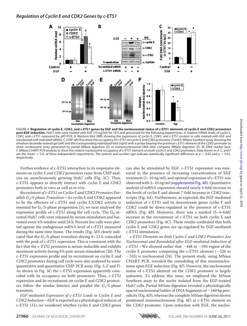
CDK2 Induction—EGF is reported as a physiological inducer ofc-ETS1 (31), we wondered whether cyclin E and CDK2 genes
can also be stimulated by EGF. c-ETS1 expression was mea-sured in the presence of increasing concentrations of EGFtreatment (1–10 ng/ml), and optimal expression of c-ETS1 wasobserved with 5–10 ng/ml (supplemental Fig. 4B). Quantitativeanalysis of mRNA expression showed nearly 5-fold increase inthe levels of cyclin E and almost 7-fold increase in CDK2 tran-scripts (Fig. 4A). Furthermore, as expected, the EGF-mediatedinduction of c-ETS1 and its downstream genes cyclin E andCDK2 could be down-regulated in the presence of c-ETS1siRNA (Fig. 4B). Moreover, there was a marked (5–6-fold)increase in the recruitment of c-ETS1 on both cyclin E andCDK2 promoters (Fig. 4C). These results confirmed that bothcyclin E and CDK2 genes are up-regulated by EGF-mediatedc-ETS1 stimulation.c-ETS1 Elements on Both Cyclin E and CDK2 Promoters Are
Nucleosomal and Remodeled after EGF-mediated Induction ofc-ETS1—We showed earlier that �448 to �595 region of thecyclin E promoter comprising the c-ETS1 element (�526 to�535) is nucleosomal (24). The present study, using MNaseCHART-PCR, revealed the remodeling of this mononucleo-some upon EGF induction (Fig. 4F). However, the nucleosomalstatus of c-ETS1 element on the CDK2 promoter is largelyunknown. To address this issue, we employed the MNaseSouthern assay to the nuclei isolated from the EGF-treatedHuh7 cells. Partial MNase digestion revealed a physiologicallyspaced nucleosomal ladder of DNA fragments of�160 bp peri-odicity (Fig. 4D), whereas the completeMNase digestion shownpositioned mononucleosome (Fig. 4E) at c-ETS1 element onthe CDK2 promoter. Upon induction with EGF, the nucleo-
FIGURE 4. Regulation of cyclin E, CDK2, and c-ETS1 genes by EGF and the nuclesosomal status of c-ETS1 elements of cyclin E and CDK2 promoterspost-EGF induction. Huh7 cells were treated with EGF (10 ng/ml) for 18 h and processed for the following experiments. A, relative mRNA levels of cyclin E,CDK2, and c-ETS1 measured by qRT-PCR. B, Western blot (WB) showing the expression of cyclin E, CDK2, and c-ETS1 protein in cells treated with EGF andtransfected with indicated siRNAs. C, ChIP-qPCR to show the occupancy of c-ETS1 on cyclin E and CDK2 promoters. D and E, MNase Southern assay showing theethidium bromide-stained gel (left) and the corresponding hybridized blot (right) with a probe bearing the proximal c-ETS1 element of the CDK2 promoter toshow nucleosome array generated by partial MNase digestion (D) or mononucleosomal DNA after complete MNase digestion (E). M, DNA marker lane.F, MNase CHART-PCR analysis to show the relative nucleosome occupancy of c-ETS1 element on both cyclin E and CDK2 promoters. Data shown in A, C, and Fare the mean � S.D. of three independent experiments. The asterisk and number sign indicate statistically significant difference at p � 0.05 and p � 0.01,respectively.
Regulation of Cyclin E and CDK2 Genes by c-ETS1
21966 JOURNAL OF BIOLOGICAL CHEMISTRY VOLUME 286 • NUMBER 25 • JUNE 24, 2011
by guest on April 21, 2019
http://ww
w.jbc.org/
Dow
nloaded from

somal status was altered albeit with differences in the inputlevels. This caveat was overcome by the MNase CHART-PCRassay that allows the quantitative measurement of chromatinremodeling (27). Just as cyclin E, the mononucleosome assem-bled on the proximal c-ETS1 element of the CDK2 promoteralso remodeled after EGF treatment (Fig. 4F). Thus, inductionof c-ETS1 leads to remodeling ofmononucleosomes positionedon c-ETS1 responsive elements of both cyclin E and CDK2promoters.Recruitment of c-ETS1 Facilitates Cross-talk of Cofactors over
Cyclin E and CDK2 Promoters—Next, we studied the interplayof co-factors associated with chromatin remodeling and tran-scription initiation cycle upon EGF-mediated c-ETS1 induc-tion. We carried out ChIP-qPCR with HDAC1 and p300 anti-bodies following EGF treatment. HDAC1 occupancy on cyclinE andCDK2 promoters declined upon c-ETS1 induction with aconcomitant increase in the occupancy of histone acetyltrans-ferase-p300 (Fig. 5,A andB). This is consistent with the nucleo-some remodeling of both promoters after EGF treatment (Fig.4E). The increase in the p300 occupancy prompted us to look atthe levels of acetylation of lysine 9 residue of histone H3(H3K9Ac), which is considered as a hallmark of active tran-scription (32). As expected, there was a 4-fold increase in theacetylation of cyclin E promoter and a nearly 2-fold increasewas observed in the acetylation of theCDK2 promoter (Fig. 5,Aand B). Interestingly, the histone H3K9 acetylation correlatedwell with the increased occupancy of phospho-Ser5 pol II tran-
scription initiationmarker on both promoters (Fig. 5,A and B).Furthermore, increased hBRM1 occupancy confirmed theinvolvement of not only the histonemodifiers but also the chro-matin remodelers after EGF treatment (Fig. 5, A and B). Thus,c-ETS1-mediated expression of cyclin E and CDK2 genesinvolves the cross-talk of co-factors associated with chromatinremodeling.HBx Protein Cooperates with c-ETS1 in Up-regulation of
Cyclin E and CDK2—The viral oncoprotein HBx can overcomethe G0 and G1/S checkpoints even in the absence of serum (26).Because cyclin E and CDK2 levels were up-regulated in thepresence of c-ETS1, we wondered whether HBx can aid thisprocess. We performed reporter transactivation assay usingcyclin E and CDK2 reporter constructs in hepatic (Huh7 andHepG2) and non-hepatic (HEK293 and HeLa) cell lines afterco-expressing c-ETS1 and HBx. HBx stimulated both promot-ers and had an additive effect with c-ETS1 in hepatic cell linesbut not in non-hepatic cell lines (Fig. 6,A andB). Stimulation ofboth promoters byHBxwas significantly reducedwithmutatedc-ETS1 reporters in the hepatic cell line (Fig. 6C). Furthermore,the increased level of cyclin E andCDK2 transcripts in presenceof HBx was significantly reduced by siRNA against c-ETS1 andc-ETS1 dominant negative construct (Fig. 6, D–F). Next, toshow c-ETS1 was critical for cell cycle progression throughinduction of cyclin E and CDK2 genes, we performed FACSanalysis after c-ETS1 silencing. Presence of HBx under c-ETS1silenced condition did not lead to S-phase progression, sub-stantiating our earlier observation on c-ETS1 dependence ofHBx activity for G1/S-phase transition. Interestingly, enforcedco-expression of cyclin E and CDK2 bypassed the requirementof c-ETS1 (by knockdown) forG1/S-phase progression (supple-mental Fig. 5).We also investigated the direct regulation of c-ETS1 by HBx.
Amarked increase in c-ETS1mRNA levels was observed in thepresence ofHBx inHuh7 cells but not inHEK293 cells (Fig. 7A).Furthermore, this was also confirmed at protein levels in Huh7cells (Fig. 7B). These results, together with HBx-mediated up-regulation of cyclin E and CDK2 protein levels confirmed themodulation of both genes by HBx via c-ETS1 responsive ele-ments. Interestingly, analysis of c-ETS1 transcript and proteinlevels in the liver of X15-Myc transgenic mice also confirmed asignificant increase in c-ETS1mRNA and protein levels (Fig. 7,C and D). Besides c-ETS1, we also observed increased cyclin EandCDK2mRNAand protein levels in transgenic environment(Fig. 7, C and D). Thus, HBx-mediated direct targeting ofc-ETS1 seems to require a hepatotropic environment for mod-ulating cyclin E and Cdk2 expression.
DISCUSSION
Considering the timing of their expression andbroader rangeof substrates, coregulation of cyclin E and CDK2 is of para-mount importance in the orchestration of S-phase progression.In the current study, we investigated the activation of bothcyclin E and CDK2 promoters by the c-ETS1 proto-oncogene.c-ETS1 is well known to be involved in diverse cellular pro-cesses such as proliferation, differentiation, development,transformation, and apoptosis (33). Intriguingly, c-ETS1 nullanimals do not show any proliferative phenotype (34). Never-
FIGURE 5. Co-factors interplay over c-ETS1 element of cyclin E and CDK2promoter after EGF induction. Occupancy of cofactors pol II, phospho-Ser5pol II, H3K9Ac, p300, HDAC1, and hBRM1 on cyclin E (A) and CDK2 (B) promot-ers measured by ChIP-qPCR. Data shown in A and B are the mean � S.D. ofthree independent experiments. An asterisk and number sign indicate statis-tically significant difference at p � 0.05 and p � 0.01, respectively.
Regulation of Cyclin E and CDK2 Genes by c-ETS1
JUNE 24, 2011 • VOLUME 286 • NUMBER 25 JOURNAL OF BIOLOGICAL CHEMISTRY 21967
by guest on April 21, 2019
http://ww
w.jbc.org/
Dow
nloaded from

theless, the c-ETS1 targeting-mediated cell cycle progressionhas been reported only for the cyclin D1 gene (35). Further-more, c-ETS1 allows rat embryo fibroblasts to grow in serum-free medium (36). Thus, it was noteworthy to investigate therole of c-ETS1 in the regulation of serum-inducible promoterssuch as cyclin E and CDK2.
Our reporter gene studies showed a significant increase inthe cyclin E andCDK2 promoter activity in presence of c-ETS1independent of cell lineage (Fig. 1, A and B) and is of no sur-prise, due to ubiquitous nature of c-ETS1 expression (37).There was a built-in specificity in this mechanism as confirmedby competitive inhibition using c-ETS1 dominant negative ormutation of c-ETS1 elements (supplemental Fig. 3,C andD andFig. 1, C and D). Interestingly for the CDK2 promoter, only theproximal c-ETS1 element appeared to be functionally impor-tant because mutation in the distal element did not impair the
reporter gene activity (Fig. 1D). Furthermore, the occupancy ofc-ETS1 to its responsive elements on both promoters was evi-dent fromour EMSA andChIP studies (Fig. 3,A–C).Moreover,siRNA-mediated silencing of c-ETS1 recapitulated its role intargeting both cyclin E and CDK2 genes (Fig. 2, E and F).The regulation of cell cycle progression by c-ETS1 was
evident from enforced expression of c-ETS1 that helpedovercoming the quiescence state (G0) imposed by depriva-tion of serum. The accelerated G1/S-phase progression wasapparently due to the activation of cyclin E and CDK2 genes(Fig. 2D). Interestingly, a similar effect has been reported forviral oncoprotein HBx in the induction of cyclin A promoter(38). Moreover, c-MYC is also reported to play a critical role insustaining an E2F-independent G1/S-promoting mechanism byregulating cyclin E-CDK2 function (39). Thus, coalescence of ourresults with earlier reports unveiled that the c-ETS1 proto-onco-
FIGURE 6. Induction of cyclin E and CDK2 transcription by HBx. A and B, Huh7, HepG2, HeLa, and HEK293 cells were transfected with either pE-WT (A) orpCDK2-WT (B) reporter constructs along with the expression vectors of c-ETS1 and HBx and the relative reporter activity was measured. C, Huh7 cells weretransfected with either pCDK2-mut (bar) or pE-mut (line) along with the expression construct of HBx, and the relative reporter activity was measured. D–F, therelative mRNA levels of cyclin E (D) and CDK2 (E) genes were measured by qRT-PCR in Huh7 cells transfected with HBx along with either indicated siRNAs orc-ETS1 dominant negative. Data shown in A–F are the mean � S.D. of three independent experiments. The asterisk and number sign indicate statisticallysignificant difference at p � 0.05 and p � 0.01, respectively.
Regulation of Cyclin E and CDK2 Genes by c-ETS1
21968 JOURNAL OF BIOLOGICAL CHEMISTRY VOLUME 286 • NUMBER 25 • JUNE 24, 2011
by guest on April 21, 2019
http://ww
w.jbc.org/
Dow
nloaded from

gene has the same potential as c-MYC and HBx oncoproteins inmediating proliferation function of cells and that its deregulationcould create a microenvironment conducive for cancerousgrowth.Our kinetic studies on c-ETS1 expression clearly showed its
peak during G1/S transition (Fig. 3D), which is in accordance tothe earlier report (20). Furthermore, the expression kinetics ofc-ETS1 was well correlated with its occupancy on both cyclin Eand CDK2 promoters (Fig. 3E) that would be important for theconsequent events in transcription cycle (40). Elucidation of a“transcriptional clock” that directs sequential and combinato-rial assembly of transcriptionally productive complexes wouldbe achieved by induction of promoter elements (41). In thislight, we used EGF as a physiological inducer of c-ETS1 as it isknown to stimulate c-ETS1 transcription and protein expres-sion (31). Corroborating with ectopic expression of c-ETS1, theinduction of endogenous c-ETS1 by EGF also recapitulated asimilar event in the activation of cyclin E and CDK2 genes (Fig.4, A and B).The chromatin structure and remodeling plays a pivotal
role in the control of eukaryotic gene regulation by influenc-ing all stages of transcription (42). Analysis of the nucleo-somal status of EBS indicated that similar to cyclin E (24), theCDK2 promoter EBS also assembled into mononucleoesome(Fig. 4, D and E) and remodeled upon c-ETS1 inductionby EGF (Fig. 4F). The SWItch/Sucrose Non-Fermentable-ATPases, including BRG1 and hBRM complexes (43), areknown to increase the binding of transcription factors tomononucleosomes as well as nucleosomal arrays (44, 45).Consistently, we also observed the increased recruitment of
hBRM1 during activation of cyclin E andCDK2 promoters byc-ETS1 (Fig. 5, A and B). The acetylation of N-terminal tailsof histones is correlated with disruption of higher orderchromatin structure and activation of transcription, whereasits deacetylation relates to its reversal and repression of tran-scription (46). Among the histone acetyltransferases thatcatalyze acetylation of histones, p300/CBP is well estab-lished for its role in the regulation of plethora of genes,including cell cycle regulators (47). Moreover, HDAC1 isreported to be the negative regulator of cyclin E by RB pro-tein (48). In this line, we observed the exchange of HDAC1with histone acetyltransferase p300 on both promoters dur-ing induction of c-ETS1 by EGF treatment (Fig. 5, A and B).Earlier reports suggested that p300 forms co-activator com-plex with PCAF (49), and this complex mediates the histoneH3K9 acetylation (50). Consistently, the increased p300occupancy was observed with the contemporaneous raise inthe acetylation of histone H3K9 levels (Fig. 5, A and B).Moreover, nucleosomes with histone H3-K4me3 and H3K9,K14 acetylation modifications, together with pol II occupythe promoters of most protein-coding genes and serve as thehallmarks of transcription initiation (32). We also observedincreased pol II occupancy on both cyclin E and CDK2 pro-moters with the concomitant rise in histone H3K9 acetyla-tion. Furthermore, the elevation of serine 5-phosphorylatedpol II occupancy confirmed the activation of both genesupon induction of c-ETS1 (Fig. 5, A and B). Collectively, ourresults suggested that chromatin remodeling by both ATP-dependent remodelers and histone modifiers could be theunderlying mechanism of co-factors interplay in the regula-tion of c-ETS1-mediated activation of cyclin E and CDK2genes.Overwhelming evidence suggests that HBx protein of
mammalian hepadnavirus (Hepatitis B virus) with transacti-vator and mitogenic signaling functions has a definitive rolein the development of hepatocellular carcinoma (51). Cellsexpressing HBx show increased rate of entry to S-phase,breakdown of the G1/S-phase checkpoint, and acceleratedG2/M progression due to activation of cyclins and cell divi-sion cycle 2 kinases (52, 26). Because c-ETS1 regulates G1/S-transition by activation of cyclin E and CDK2 genes, weinvestigated whether HBx can deregulate the cell cycle bycooperating with c-ETS1. We indeed observed a synergybetween HBx and c-ETS1 exclusively in a hepatotropic envi-ronment (Fig. 6, A and B), which mimicked the oncogenicnature of HBx in a transgenic environment (53). BecauseHBx is unable to bind directly to any defined DNA-bindingsequences (54, 55), the responsive elements of other tran-scription factors, including EBS (56), could mediate thetransactivation function of HBx. Consistently, mutationstudies and RNA interference against c-ETS1 impaired theup-regulation of both cyclin E and CDK2 promoters in pres-ence of HBx (Fig. 6, C–E). The synergism between HBx andc-ETS1 is likely due to up-regulation of c-ETS1 by HBx asevident from both cell culture studies and in vivo studies inX15-Myc transgenic animals (Fig. 7, A–D).
Collectively, based on these results, it may be concluded thatcyclin E and CDK2 genes are the physiological effector mole-
FIGURE 7. Deregulation of c-ETS1 proto-oncogene by HBx. A, the relativemRNA levels of c-ETS1 were measured by qRT-PCR in both Huh7 and HEK293cells transfected with HBx. C, the relative mRNA levels of cyclin E, CDK2, andc-ETS1 were measured by qRT-PCR in the liver samples of control and X15-Myc transgenic mice. B and D, immunoblots to show the levels of cyclin E,CDK2, c-ETS1, HBx, and GAPDH in the Huh7 cells transfected with HBx (B) or inthe liver samples of 1-month-old X15-Myc transgenic mice (D). Data shown inA and C are the mean � S.D. of three independent experiments. An asteriskand number sign indicate statistically significant difference at p � 0.05 andp � 0.01, respectively.
Regulation of Cyclin E and CDK2 Genes by c-ETS1
JUNE 24, 2011 • VOLUME 286 • NUMBER 25 JOURNAL OF BIOLOGICAL CHEMISTRY 21969
by guest on April 21, 2019
http://ww
w.jbc.org/
Dow
nloaded from

cules of c-ETS1. Thus, direct targeting of c-ETS1 by HBx couldbe associated with the cell cycle regulatory process and posesfresh challenges in understanding of hepatocellular carcinoma.
Acknowledgments—We are grateful to the following scientists for thegenerous gift of the following recombinant constructs: Dr. J. R. Nevins(Duke University Medical Center) for pCycECAT reporter, Dr. GaryStein (University ofMassachusettsMedical School) for�2400CDK2/LUC (DSC37) reporter, Dr. Hiroyuki Sugimoto for the c-ETS1 expres-sion construct, Dr. Arthur Gutierrez-Hartmann for the c-ETS1 DNconstruct, Dr.Michael G. Brattain (Roswell Park Cancer Institute) forSP1 expression vectors, Dr. J. M. Roberts (Fred Hutchinson CancerResearch Center) for cyclin E expression constructs, andDr. E. Harlow(Massachusetts General Hospital Cancer Center) for CDK2 expres-sion constructs. Ravinder Kumar helped in cell culture work.
REFERENCES1. McGowan, C. H. (2003) Prog. Cell. Cycle. Res. 5, 1–42. Santamaria, D., and Ortega, S. (2006) Front. Biosci. 11, 1164–11883. Le Cam, L., Polanowska, J., Fabbrizio, E., Olivier, M., Philips, A., Ng Eaton,
E., Classon, M., Geng, Y., and Sardet, C. (1999) EMBO J. 18, 1878–18904. Dulic, V., Lees, E., and Reed, S. I. (1992) Science 257, 1958–19615. Koff, A., Giordano, A., Desai, D., Yamashita, K., Harper, J. W., Elledge, S.,
Nishimoto, T., Morgan, D. O., Franza, B. R., and Roberts, J. M. (1992)Science 257, 1689–1694
6. Donnellan, R., and Chetty, R. (1999) FASEB J. 13, 773–7807. Krude, T. (2000) J. Biol. Chem. 275, 13699–137078. Schang, L. M. (2003) Prog. Cell. Cycle. Res. 5, 103–1249. Ohtani, K., DeGregori, J., and Nevins, J. R. (1995) Proc. Natl. Acad. Sci.
U.S.A. 92, 12146–1215010. Botz, J., Zerfass-Thome, K., Spitkovsky, D., Delius, H., Vogt, B., Eilers, M.,
Hatzigeorgiou, A., and Jansen-Durr, P. (1996) Mol. Cell. Biol. 16,3401–3409
11. Geng, Y., Eaton, E. N., Picon, M., Roberts, J. M., Lundberg, A. S., Gifford,A., Sardet, C., and Weinberg, R. A. (1996) Oncogene 12, 1173–1180
12. Perez-Roger, I., Solomon, D. L., Sewing, A., and Land, H. (1997)Oncogene14, 2373–2381
13. Morgan, D. O. (1995) Nature 374, 131–13414. Geng, Y., and Weinberg, R. A. (1993) Proc. Natl. Acad. Sci. U.S.A. 90,
10315–1031915. Shiffman, D., Brooks, E. E., Brooks, A. R., Chan, C. S., and Milner, P. G.
(1996) J. Biol. Chem. 271, 12199–1220416. Sementchenko, V. I., and Watson, D. K. (2000) Oncogene 19, 6533–654817. Oda, N., Abe, M., and Sato, Y. (1999) J. Cell. Physiol. 178, 121–13218. Takai, N., Miyazaki, T., Nishida,M., Nasu, K., andMiyakawa, I. (2002) Int.
J. Mol. Med. 9, 287–29219. Seth, A., and Watson, D. K. (2005) Eur. J. Cancer. 41, 2462–247820. Majerus, M. A., Bibollet-Ruche, F., Telliez, J. B., Wasylyk, B., and Bailleul,
B. (1992) Nucleic Acids Res. 20, 2699–270321. Xie, R. L., Gupta, S.,Miele, A., Shiffman, D., Stein, J. L., Stein, G. S., and van
Wijnen, A. J. (2003) J. Biol. Chem. 278, 26589–2659622. Sugimoto, H., Sugimoto, S., Tatei, K., Obinata, H., Bakovic, M., Izumi, T.,
and Vance, D. E. (2003) J. Biol. Chem. 278, 19716–1972223. Bradford, A. P., Conrad, K. E., Wasylyk, C., Wasylyk, B., and Gutierrez-
Hartmann, A. (1995)Mol. Cell. Biol. 15, 2849–285724. Janbandhu, V. C., Singh, A. K., Mukherji, A., and Kumar, V. (2010) J. Biol.
Chem. 285, 17453–1746425. Kumar, V., Jayasuryan, N., Reddi, H., Sahal, D., and Panda, S. K. (1998)
Hybridoma 17, 157–16426. Mukherji, A., Janbandhu, V. C., and Kumar, V. (2007) Biochem. J. 401,
247–25627. Rao, S., Procko, E., and Shannon, M. F. (2001) J. Immunol. 167,
4494–450328. Overbergh, L., Valckx, D., Waer, M., and Mathieu, C. (1999) Cytokine 11,
305–31229. Lakhtakia, R., Kumar, V., Reddi, H., Mathur, M., Dattagupta, S., and
Panda, S. K. (2003) J. Gastroenterol. Hepatol. 18, 80–9130. Kim, S., Kang, J. K., Kim, Y. K., Seo, D.W., Ahn, S. H., Lee, J. C., Lee, C. H.,
You, J. S., Cho, E. J., Lee, H. W., and Han, J. W. (2006) Biochem. Biophys.Res. Commun. 342, 1168–1173
31. Gilles, F., Raes,M. B., Stehelin, D., Vandenbunder, B., and Fafeur, V. (1996)Exp. Cell Res. 222, 370–378
32. Guenther, M. G., Levine, S. S., Boyer, L. A., Jaenisch, R., and Young, R. A.(2007) Cell. 130, 77–88
33. Dittmer, J. (2003)Mol. Cancer. 2, 2934. Muthusamy,N., Barton, K., and Leiden, J.M. (1995)Nature 377, 639–64235. Tanaka, H., Terada, Y., Kobayashi, T., Okado, T., Inoshita, S., Kuwahara,
M., Seth, A., Sato, Y., and Sasaki, S. (2004) J. Am Soc. Nephrol. 15,3083–3092
36. Topol, L. Z., Tatosyan, A. G., Ascione, R., Thompson, D. M., Blair, D. G.,Kola, I., and Seth, A. (1992) Cancer Lett. 67, 71–78
37. Hollenhorst, P. C., Jones, D. A., and Graves, B. J. (2004)Nucleic Acids Res.32, 5693–5702
38. Bouchard, M., Giannakopoulos, S., Wang, E. H., Tanese, N., and Schnei-der, R. J. (2001) J. Virol. 75, 4247–4257
39. Santoni-Rugiu, E., Falck, J., Mailand, N., Bartek, J., and Lukas, J. (2000)Mol. Cell. Biol. 20, 3497–3509
40. Kodadek, T., Sikder, D., and Nalley, K. (2006) Cell 127, 261–26441. Metivier, R., Penot, G., Hubner, M. R., Reid, G., Brand, H., Kos, M., and
Gannon, F. (2003) Cell 115, 751–76342. Li, B., Carey, M., and Workman, J. L. (2007) Cell 128, 707–71943. Becker, P. B., and Horz, W. (2002) Annu. Rev. Biochem. 71, 247–27344. Imbalzano, A. N., Kwon, H., Green, M. R., and Kingston, R. E. (1994)
Nature 370, 481–48545. Kwon, H., Imbalzano, A. N., Khavari, P. A., Kingston, R. E., and Green,
M. R. (1994) Nature. 370, 477–48146. Davie, J. R., and Spencer, V. A. (1999) J. Cell. Biochem. 32, 141–14847. Goodman, R. H., and Smolik, S. (2000) Genes Dev. 14, 1553–157748. Brehm, A., Miska, E. A., McCance, D. J., Reid, J. L., Bannister, A. J., and
Kouzarides, T. (1998) Nature 391, 597–60149. Glass, C. K., and Rosenfeld, M. G. (2000) Genes Dev. 14, 121–14150. Kouzarides, T. (2007) Cell 128, 693–70551. Benhenda, S., Cougot, D., Buendia, M. A., and Neuveut, C. (2009) Adv.
Cancer Res. 103, 75–10952. Benn, J., and Schneider, R. J. (1995) Proc. Natl. Acad. Sci. U.S.A. 92,
11215–1121953. Kumar, V. (2008) inThe Pleiotropic Functions of the Viral ProteinHBx and
the Development of Liver Cancer (Kobarg, J., ed) pp. 221–240, ResearchSignpost, Trivandrum, India
54. Maguire, H. F., Hoeffler, J. P., and Siddiqui, A. (1991) Science 252,842–844
55. Avantaggiati, M. L., Natoli, G., Balsano, C., Chirillo, P., Artini, M., DeMarzio, E., Collepardo, D., and Levrero,M. (1993)Oncogene 8, 1567–1574
56. Yoo, Y. D., Ueda, H., Park, K., Flanders, K. C., Lee, Y. I., Jay, G., and Kim,S. J. (1996) J. Clin. Invest. 97, 388–395
Regulation of Cyclin E and CDK2 Genes by c-ETS1
21970 JOURNAL OF BIOLOGICAL CHEMISTRY VOLUME 286 • NUMBER 25 • JUNE 24, 2011
by guest on April 21, 2019
http://ww
w.jbc.org/
Dow
nloaded from

Anup Kumar Singh, Manickavinayaham Swarnalatha and Vijay KumarGenes and Cooperates with Hepatitis B Virus X Protein for Their Deregulation
CDK2/S-phase Transition by Up-regulating Cyclin E and 1c-ETS1 Facilitates G
doi: 10.1074/jbc.M111.238238 originally published online April 22, 20112011, 286:21961-21970.J. Biol. Chem.
10.1074/jbc.M111.238238Access the most updated version of this article at doi:
Alerts:
When a correction for this article is posted•
When this article is cited•
to choose from all of JBC's e-mail alertsClick here
Supplemental material:
http://www.jbc.org/content/suppl/2011/04/22/M111.238238.DC1
http://www.jbc.org/content/286/25/21961.full.html#ref-list-1
This article cites 55 references, 21 of which can be accessed free at
by guest on April 21, 2019
http://ww
w.jbc.org/
Dow
nloaded from