C e l l S cienc l Journal of Cell Science & Therapy · Tumor milieu contains malignant and non...
12
Biologically Synthesized Gold Nanoparticles using Ocimum sanctum (Tulsi Leaf Extract) Induced Anti-Tumor Response in a T Cell Daltons Lymphoma Pramod Kumar Gautam 1* , Sanjay Kumar 2 , Tomar MS 2 , Rishi Kant Singh 2 , Acharya A 2 , Ritis Shyanti K 1 , Anita 1 , Sonal Swaroop 1 , Sanjay Kumar 3 and B Ram 3 1 Department of Biochemistry, All India Institute of Medical Science, India 2 Department of Zoology, Institute of Science, Banaras Hindu University, India 3 Department of Dravyaguna, Institute of Medical Science, Banaras Hindu University, India * Corresponding author: Pramod Kumar Gautam, Department of Biochemistry, All India Institute of Medical Science, India, 3rd floor, PC block, New Delhi 110029, India, Tel: 011-26546514; E-mail: [email protected] Rec Date: Nov 15, 2017, Acc Date: Dec 15, 2017, Pub Date: Dec 19, 2017 Copyright: © 2017 Gautam PK, et al. This is an open-access article distributed under the terms of the Creative Commons Attribution License, which permits unrestricted use, distribution, and reproduction in any medium, provided the original author and source are credited. Abstract The characterizations of green synthesized gold nanoparticles (AuNPs) prepared using Ocimum sanctum leaf extract were done by UV-Spectrophotometer at 500-540 nm. The XRD data obtained were found similar to gold JCPDS File No- 04-0784. SEM and TEM analysis of AuNPs revealed spherical shape and size of 12-20 nm. Further FT-IR data indicated the various biomolecules present in Ocimum sanctum leaf extract provides stability to gold nanoparticles synthesis. The AuNPs were studied for their anti-cancer activity on Dalton’s lymphoma (DL) cells and the results obtained with IC50 value of <50 ng/ml performed by MTT assay. Further, to confirm anti-tumor potential and the mode of action of the synthesized AuNPs, cell viability assay, nuclear morphology, DNA fragmentation assay, mitochondrial membrane potential (ΔΨm) analysis, and cell cycle analysis were done using DL cells. DL cells treated with the AuNPs showed reduced cell viability, altered nuclear morphology, typical apoptotic DNA ladder formation and apoptosis. From the above finding it can be concluded that the AuNPs have potential to decrease the proliferation of tumor cells and enhanced the production of ROS. Gold nanoparticles used in cancer detection and diagnosis/treatment are mainly in preclinical stages of cancer development. Keywords: AuNPs; Ocimum sanctum; Daltons lymphoma cells; Anti-tumor activity Introduction Nanoscience has focused in the area of drug delivery by utilization of materials at the level of atoms, molecules, supramolecular structures and their distinctive features at nanoscale. In present scenario, nanoparticles (NPs) have drawn marvelous concern due to their valuable contribution on vast fields such as biomedical, sensor, optical, electronic, catalytic application and cancer therapeutics [1]. Several reports have been published for the biosynthetic preparation of nanomaterial. In the traditional system, several plants have been reported for the treatment of diseases. Ocimum sanctum (Toulouse) is one of the potent plant whose all plant parts (leaves, stem, flower, root, seeds and even whole plant) use for the treatment of diseases such as of bronchitis, malaria, diarrhoea, dysentery, arthritis, insect bites, skin disease and so on. Although, plant extract/ phytochemicals are of high- quality options for the cancer treatment, beside there are some restrictions, including poor bioavailability and solubility [2]. In this scenario, nanoscience might be playing a vital and noteworthy role to conquer the restrictions of conventional treatment strategies. Plant extracts have excellent properties for the synthesis of novel nanoparticles, including gold [3-5]. However, which constituents are the major players in plant leaf extracts that lead to the formation of nanoparticles is yet to be established [6]. Biosynthesis of nanoparticles by plant extract has several advantages and cost effective with high potential [7-11]. Gold nanoparticles are the exceptional element which has been used to treat different pathophysiological conditions like anti-malarial and anti-arthritic agent, anti-HIV including cancer diagnostics and therapy. Gold nanoparticles (AuNPs), in particular, it has major role in biomedical applications because they are biocompatible and high surface is easy to modify due to the strong ability of AuNPs to bind to - SH- and -NH 2 - containing molecules such as organic molecules such as drugs, peptides, antibodies, etc. [12]. AuNPs have been used as antiangiogenesis, anti-malarial and anti-arthritic agent, anti-HIV [13]. Moreover, gold nanoparticles are used for delivering molecules into cells to slow down cancer cell growth and/or kill cancerous cells [14]. Tumor milieu contains malignant and non malignant cells such as endothelial cells, fibroblasts, and various cells derived from the bone marrow and suffering from the high scarcity of oxygen, nutrients and metabolic products. Tumor cell produces copious amounts of CC chemokines such as CC chemokine ligand (CCL)1, CCl 2 , CCl 3 , CCl 4 , CCl 5 and VGEF, CSF-1 which attract for immune cells including M1 phenotype of macrophages to the tumor site [15-21]. As a consequence immune system fails to protect form tumor burden by downregulating the function of T cells, B cells, NK cells and dysfunction of visceral organ failure. Keeping the immunomodulatory function of gold nanoparticle in mind, In the present investigation the Ocimum sanctum (Tulsi) induced synthesis of the AuNPs was done. e NPs were characterized by UV-visible spectroscopy, FTIR spectroscopy, XRD, TEM and anti- cancer potential was evaluated on Dalton’s lymphoma. DL cells treated with the AuNPs showed reduced cell viability, altered mitochondrial membrane potential (ΔΨm), change in nuclear morphology and DNA Gautam et al., J Cell Sci Ther 2017, 8:6 DOI: 10.4172/2157-7013. 1000278 Research Article Open Access J Cell Sci er, an open access journal ISSN: 2157-7013 Volume 8 • Issue 6 • 1000278 J o u r n a l o f C e l l S c i e n c e & T h e r a p y ISSN: 2157-7013 J o u r n a l o f C e l l S c i e n c e & T h e r a p y ISSN: 2157-7013 Journal of Cell Science & Therapy Journal of Cell Science & Therapy
Transcript of C e l l S cienc l Journal of Cell Science & Therapy · Tumor milieu contains malignant and non...

Biologically Synthesized Gold Nanoparticles using Ocimum sanctum(Tulsi Leaf Extract) Induced Anti-Tumor Response in a T Cell DaltonsLymphomaPramod Kumar Gautam1*, Sanjay Kumar2, Tomar MS2, Rishi Kant Singh2, Acharya A2, Ritis Shyanti K1, Anita1, Sonal Swaroop1, Sanjay Kumar3 and B Ram3
1Department of Biochemistry, All India Institute of Medical Science, India2Department of Zoology, Institute of Science, Banaras Hindu University, India3Department of Dravyaguna, Institute of Medical Science, Banaras Hindu University, India*Corresponding author: Pramod Kumar Gautam, Department of Biochemistry, All India Institute of Medical Science, India, 3rd floor, PC block, New Delhi 110029, India,Tel: 011-26546514; E-mail: [email protected]
Rec Date: Nov 15, 2017, Acc Date: Dec 15, 2017, Pub Date: Dec 19, 2017
Copyright: © 2017 Gautam PK, et al. This is an open-access article distributed under the terms of the Creative Commons Attribution License, which permits unrestricteduse, distribution, and reproduction in any medium, provided the original author and source are credited.
Abstract
The characterizations of green synthesized gold nanoparticles (AuNPs) prepared using Ocimum sanctum leafextract were done by UV-Spectrophotometer at 500-540 nm. The XRD data obtained were found similar to goldJCPDS File No- 04-0784. SEM and TEM analysis of AuNPs revealed spherical shape and size of 12-20 nm. FurtherFT-IR data indicated the various biomolecules present in Ocimum sanctum leaf extract provides stability to goldnanoparticles synthesis. The AuNPs were studied for their anti-cancer activity on Dalton’s lymphoma (DL) cells andthe results obtained with IC50 value of <50 ng/ml performed by MTT assay. Further, to confirm anti-tumor potentialand the mode of action of the synthesized AuNPs, cell viability assay, nuclear morphology, DNA fragmentationassay, mitochondrial membrane potential (ΔΨm) analysis, and cell cycle analysis were done using DL cells. DL cellstreated with the AuNPs showed reduced cell viability, altered nuclear morphology, typical apoptotic DNA ladderformation and apoptosis. From the above finding it can be concluded that the AuNPs have potential to decrease theproliferation of tumor cells and enhanced the production of ROS. Gold nanoparticles used in cancer detection anddiagnosis/treatment are mainly in preclinical stages of cancer development.
Keywords: AuNPs; Ocimum sanctum; Daltons lymphoma cells;Anti-tumor activity
IntroductionNanoscience has focused in the area of drug delivery by utilization
of materials at the level of atoms, molecules, supramolecular structuresand their distinctive features at nanoscale. In present scenario,nanoparticles (NPs) have drawn marvelous concern due to theirvaluable contribution on vast fields such as biomedical, sensor, optical,electronic, catalytic application and cancer therapeutics [1].
Several reports have been published for the biosynthetic preparationof nanomaterial. In the traditional system, several plants have beenreported for the treatment of diseases. Ocimum sanctum (Toulouse) isone of the potent plant whose all plant parts (leaves, stem, flower, root,seeds and even whole plant) use for the treatment of diseases such as ofbronchitis, malaria, diarrhoea, dysentery, arthritis, insect bites, skindisease and so on. Although, plant extract/ phytochemicals are of high-quality options for the cancer treatment, beside there are somerestrictions, including poor bioavailability and solubility [2]. In thisscenario, nanoscience might be playing a vital and noteworthy role toconquer the restrictions of conventional treatment strategies. Plantextracts have excellent properties for the synthesis of novelnanoparticles, including gold [3-5]. However, which constituents arethe major players in plant leaf extracts that lead to the formation ofnanoparticles is yet to be established [6]. Biosynthesis of nanoparticlesby plant extract has several advantages and cost effective with highpotential [7-11].
Gold nanoparticles are the exceptional element which has been usedto treat different pathophysiological conditions like anti-malarial andanti-arthritic agent, anti-HIV including cancer diagnostics andtherapy. Gold nanoparticles (AuNPs), in particular, it has major role inbiomedical applications because they are biocompatible and highsurface is easy to modify due to the strong ability of AuNPs to bind to -SH- and -NH2- containing molecules such as organic molecules suchas drugs, peptides, antibodies, etc. [12]. AuNPs have been used asantiangiogenesis, anti-malarial and anti-arthritic agent, anti-HIV [13].Moreover, gold nanoparticles are used for delivering molecules intocells to slow down cancer cell growth and/or kill cancerous cells [14].
Tumor milieu contains malignant and non malignant cells such asendothelial cells, fibroblasts, and various cells derived from the bonemarrow and suffering from the high scarcity of oxygen, nutrients andmetabolic products. Tumor cell produces copious amounts of CCchemokines such as CC chemokine ligand (CCL)1, CCl2, CCl3, CCl4,CCl5 and VGEF, CSF-1 which attract for immune cells including M1phenotype of macrophages to the tumor site [15-21]. As a consequenceimmune system fails to protect form tumor burden by downregulatingthe function of T cells, B cells, NK cells and dysfunction of visceralorgan failure.
Keeping the immunomodulatory function of gold nanoparticle inmind, In the present investigation the Ocimum sanctum (Tulsi)induced synthesis of the AuNPs was done. The NPs were characterizedby UV-visible spectroscopy, FTIR spectroscopy, XRD, TEM and anti-cancer potential was evaluated on Dalton’s lymphoma. DL cells treatedwith the AuNPs showed reduced cell viability, altered mitochondrialmembrane potential (ΔΨm), change in nuclear morphology and DNA
Gautam et al., J Cell Sci Ther 2017, 8:6DOI: 10.4172/2157-7013. 1000278
Research Article Open Access
J Cell Sci Ther, an open access journalISSN: 2157-7013
Volume 8 • Issue 6 • 1000278
Jour
nal o
f Cell Science &
Therapy
ISSN: 2157-7013
Jour
nal o
f Cell Science &
Therapy
ISSN: 2157-7013
Journal of Cell Science & TherapyJournal of Cell Science & Therapy

fragmentation ladder pattern as compared to control. These findingsaccounts that the Ocimum sanctum (Tulsi) induced biosynthesis of theAuNPs showed significant anti-tumor potential against invasivegrowth of Dalton’s lymphoma cells.
Materials and Methods
ReagentsGold chloride (HAuCl4) and MTT and concanavalin-A were
purchased from Sigma Aldrich, Bangalore, India. Sodium citratetribasic dehydrate (C6H5Na3O7.2H2O) was purchased from SuperReligare Laboratories (SRL), Mumbai, India. RPMI 1640 culturemedium was obtained from HiMedia, Mumbai, India. Foetal bovineserum (FBS) was obtained from Invitrogen, CA, USA, with PE fromeBiosciences, San Diego, CA, USA. LPS, DCFH-DA, PMA, RH-123,DAPI, Hoechst 33258, PI and Phalloidin were obtained from SigmaChemical Co. (St. Louis, MO, USA). Na2HPO4, KH2PO4,formaldehyde, trypsin and acetone were purchased from Qualigens,Mumbai, India. All other chemicals otherwise stated were obtainedfrom Qualigens.
Animals and tumor modelInbred populations of BALB/c (H2d) strain of mice of either sex
were used at 8-12 weeks of age. BALB/c (H2d) strain of mice was keptin conventional cages (six animals in each cage) and received sterilizedfood and water ad libitum. Experimental animals were inspected dailyfor survival. All animals were kept and maintained with utmost careunder the guidelines of the Animal Ethical Committee, Banaras HinduUniversity, Varanasi, India.
For tumor system, healthy mice of either sex at 8-12 weeks of agewere injected intraperitoneally (i.p.) with 1.0 × 10⁶ non-Hodgkin typeof Dalton’s lymphoma (DL cells) in 0.5 ml sterile PBS. The DL cells fortransplantation were obtained from ascitic fluid of DL-bearing mice,where the yield of the cells is higher and maintained in an ascitic formin vivo by serial transplantation.
Synthesis of gold nanoparticles by Tulsi (Osmium sanctum)Gold nanoparticle was synthesized as per previously described
method [22,23]. In brief, 100 g of freshly harvested leaf of tulsi(Osmium sanctum) was taken and washed gently with distilled water.Then the freshly washed leaf was kept in conical flask in 100 ml ofdeionized water in water bath at 74°C for 1 h. After 1 h leaf extract wascollected and centrifuge at 1500 rpm for 15 min at 4°C. Finallysupernatant was filtered with 0.2 µm Whatman filter paper and collectsin a clean flask and stare at 4°C. Further, 1 mM HAuCl4 solution(Figure 1) was prepared using deionized water of 10 ml of the solutionmixture was added in the 30 ml of leaf extract.
Then the conical flask was kept in a shaker at 37°C for 24 h for thesynthesis of nano-particles and UV spectra were taken at differenttimes at day 1, 2, 3, 4, 5, 10 and 15. After synthesis of Gold nano-particles (Au NPs) the whole supernatant was collected andcentrifuged at 11500 rpm for 15 min at 4°C, the supernatant wasdiscarded and the pellet was washed with dH2O thrice and the finalpellet suspended in dH2O and sonicated. The sample was collected,lyophilized and characterized by following techniques.
Ultraviolet-visible spectroscopic analysis followed bysonication and lyophilization
Synthesized Au NPs by reduction with leaf extract was observedthrough UV-Vis spectroscopy in 300-1200 nm range. Naturallysynthesized AuNPs (540 nm) of diameter gave sharp peak in the visibleregion of the electromagnetic spectrum. [22,24]. In brief, samples werekept in the ice in 20 ml of centrifuge tube and provide ultrasonicvibration for the 2 min with the time gap of 30 second per cycle using.Further sample was Lyophilized using Christ Alpha 1-2 Lyophilizer forpreparation of powdered AuNPs.
Characterization of AuNPsFTIR measurements of the powdered purified AuNPs were assessed
using Perkin Elmer, Spectrum Two FTIR (Waltham, MA, USA) systemin the diffused reflectance mode. To obtain good signal/noise ratio, 512scans/sample were recorded. The spectral range from 4000-400 cm-1with resolution of 4 cm-1 powder samples for the FTIR obtained weresimilar as for powder diffraction measurements [25,26]. To obtainXRD patterns of the AuNPs, the AuNPs were subjected to XRDanalysis at 45 kV and 40 mA with 2θ° in the range from 0to 80angle[27,28] (Figure 2).
The shape and Size of the synthesized AuNPs were obtained incolloidal solution using transmission electron microscope (HR-TEM),Tecnai 20G2 FEI, Oregon, USA [24]. Microphotographs of the NPswere captured at 50-200 KV. The morphological observation andcharacterization of AuNPs samples were done by using scanningelectron microscope (Zieas-EVO LS-10) at 25 kV at LV mode [24].
Anti-cancer effect of the synthesized nanoparticlesTrypan blue dye exclusion and MTTassay: DL cells harvested from
tumor bearing mice were seeded at a cell density of 1 × 10⁶ in theculture medium. DL cells pulsed with or without LPS and Au NPs andincubated for time periods of 24 h in 5% CO2 incubator (Shella,Oregon, CA, USA). After incubation, cell viability was determinedusing exclusion by the trypan blue method. [18]. For MTT assay cellswere treated with 5 mg/ml MTT and incubated for 4 h and then 0.04M HCl/ isopropanol were added. Viable cells were determined by theabsorbance at 570 nm and IC50 value was calculated [29].
Confocal Imaging for Nuclear morphology and mitochondrialmembrane potential (ΔΨm) by Rh-123assay: The DL cells werecultured in 5% CO2 at 37°C with and without LPS and AuNPs for 24 h.Cells were harvested and spread over pre-coated air dried poly-lysinecoated glass slide, air dried and fixed with 4% PFA for 2 h at roomtemperature. Slide was washed and incubated with 0.1% triton X-100for 15 minutes followed by washing with PBS and stained for 15 minwith DAPI (Sigma, USA). The cell was mounted in anti quenching dyeDaboco and observed under confocal microscope [16]. Formitochondrial membrane potential analysis, similar preparations wasdone with the cells fixed over slide and stained for 45 min with RH-123dye in dark conditioned at RT, washed with PBS and further stainedwith Hoechst 33342 for 5 min. Cell mounted in Daboco and observedunder a confocal microscope [30].
Citation: Gautam PK, Kumar S, Tomar MS, Singh RK, Acharya A, et al. (2017) Biologically Synthesized Gold Nanoparticles using Ocimumsanctum (Tulsi Leaf Extract) Induced Anti-Tumor Response in a T Cell Daltons Lymphoma. J Cell Sci Ther 8: 278. doi:10.4172/2157-7013. 1000278
Page 2 of 12
J Cell Sci Ther, an open access journalISSN: 2157-7013
Volume 8 • Issue 6 • 1000278

Figure 1: Visual observations (a) tulsi leaf extract (b) tulsi leaf paste (C) tulsi leaf extract (d) gold chloride solution and (e) formation of goldnanoparticles (e) showing UV–Vis spectra of gold nanoparticles measured at the time of reaction of tulsi leaf extract with in aqueous solution.The inset is a plot of maximum absorbance versus time of reaction for gold solution and also the color change of solution before and afteraddition of leaf extract.
Figure 2: XRD pattern of gold nanoparticles synthesized by tulsi leafextract. The principal Bragg reflections are identified.
Flowcytometry for mitochondrial membrane potential (ΔΨm) byRh-123: For flow cytometry based analysis for mitochondrialmembrane potential (ΔΨm) by Rh-123 cells were treated with andwithout LPS and AuNPs for 24 h. Cell was harvested stained for 45min with RH-123 dye in dark conditioned at RT. The cell was washedtwice with 0.1% sodium azide containing 0.1% BSA in PBS for 10 minat 2500 rpm at 4°C. Further cells were kept in 0.2 ml sheath fluid in icefor Flow cytometry mitochondrial membrane potential observation (BD Biosciences, Mountain View, CA, USA) [31].
DNA fragmentation assay: DL cells with and without LPS andAuNPs treated for 24 h. The whole media of cultures were collectedand centrifuged at 2500 rpm for 12 minutes. TES lysis buffer (20 mMEDTA, 100 mM Tris (pH-8), 0.8% SDS (w/v) in 10 ml distilled water)was added into cultured cell and mixed by wide bore pipette, then 10 µlRNase for 30 min at 37°C, further 10 µl of proteinase K added to thecell and incubate at for 50°C for 90 min. 5 µl of 5x loading dye wereadded to the sample added and observed into 1.5% agarose gel in TAEcontaining 0.5 ug/ml EtBr [29].
Flow cytometry analysis of cell cycle progression assay: Aftertreatment cells were harvested and the pellet was resuspended in 0.5ml saponin-propidium iodide (PI) cocktail. The procedure was carriedout in the dark. All tubes were incubated at 4°C for overnight in darkat 4°C for 20 min and without washing cells were analyzed byFlowcytometry (BD Biosciences, Mountain View, CA, USA) todetermine the percentage of cells in each phase of the cell cycle [32,33].
Citation: Gautam PK, Kumar S, Tomar MS, Singh RK, Acharya A, et al. (2017) Biologically Synthesized Gold Nanoparticles using Ocimumsanctum (Tulsi Leaf Extract) Induced Anti-Tumor Response in a T Cell Daltons Lymphoma. J Cell Sci Ther 8: 278. doi:10.4172/2157-7013. 1000278
Page 3 of 12
J Cell Sci Ther, an open access journalISSN: 2157-7013
Volume 8 • Issue 6 • 1000278

DCFH-DA staining for ROS expression: Macrophage were culturedin medium with or without LPS and SeNPs and incubated for timeperiods of 24 h. After that cell was harvested from the medium bycentrifugation at 1200 rpm for 10 min, cell washed twice with chilledPBS followed by centrifugation and incubated for 20 min with DCFH-DA stain. Cell were spread over pre coated polylysine glass slide andair dried and fixed, mounted with anti-quenching dye DABCO andobserved under florescence microscope equipped with FTIC filter [26].
Fluorescent spectroscopy for estimation of ROS in medium: Aftertreatments, Macrophage was harvested by centrifugation at 2500 rpmfor 10 min, cell washed twice with chilled PBS and incubated for 20min with DCFH-DA stain at the concentration of 20 mM. Cell washarvested from the medium and pellet was discarded and supernatantcollected and observed under florescence spectrometer with FTICfilter range using Hitachi f-2500 spectrophotometer at 519 nm.
RNI estimation assay: Briefly, 50 µl sample (culture supernatant)harvested from cultured medium was seeded into 96-well flat-bottomculture plates and treated with equal volume of Griess reagent (1%sulfanilamide, 0.1% Naphthalene-ethylene-diaminedihidrochlorideand 2.5% H3PO4) and left for 10 min at room temperature.Absorbance was measured at 540 nm with micro plate reader (Bio-Rad, 680, Bangalore, India). Nitrite concentration was determined byusing Sodium nitrite as a standard. The chemicals used for preparationof Griess reagent were obtained from Sigma Chemical Co., St. Louis,USA [26].
ROI estimation assay: ROI production was determined bymeasuring H2O2 production in the culture supernatant of Macrophagecultured in the same conditions of stimulation and pretreatment as inthe case of NO. H2O2 was measured by the TISO4. Briefly,
Cells were harvested by centrifugation at 1200rpm for 10 min,washed twice with chilled PBS. Cells were incubated with TiSO4 (0.1%in 20% H2SO4) for 10 min at RT. Supernatant was collected bycentrifugation and absorbance measured at 410 nm on ELISA platereader.
Statistical analysisEach value represents the Mean SEM of three independent
experiments in each group except for in vitro stimulation experimentswhere three independent experiments were conducted. Data areanalyzed by using two-tailed student’s t-test on statistical softwarepackage Sigma Plot, version 12.0. A value of p<0.05 was consideredsignificant.
Results and Discussion
Synthesis of gold nanoparticlesFormation of gold nanoparticles by reduction of aqueous metal ions
during exposure of heat to the plant extract may be easily followed byUV-visible spectroscopy. It is well known that the gold nanoparticlesexhibit wine red color in solution. The production and stabilization ofthe reduced AuNPs in the colloidal solution were monitored by UV-visible spectrophotometer and is shown in (Figure 1a) UV-visiblespectroscopy is techniques to identify the formation of the AuNPs inaqueous solution. Au NPs are known to exhibit at maximum in therange of 200 to 800 nm. The synthesis of Au NPs was monitored at 24h. The AuNPs synthesized by sodium citrate are positioned at 540 nm.Similarly, Skirtach et al. reported that the gold NPs were absorbed at
540 nm, and it was synthesized using P. aeruginosa. The reduction ofgold ions occurs comparatively slowly, but the AuNPs are found to bevery stable in the colloidal suspension. (Figure 1b). The appearance ofruby wine red color is the characteristics of gold nanoparticles. UVspectra were taken at different time interval, such as 1 day, 2 day, 3 day,4 day 5 day, 10 day and 15 day (Figure 1c). It clearly indicates theformation of gold nanoparticles. This color formation belongs to thesurface Plasmon vibration of the metal nanoparticles. In case of goldnanoparticles, the narrow surface Plasmon resonance band occurred at520 nm as shown in figures.
Structural characterizationsX-ray diffraction (XRD): The X-ray diffraction is used to identify
the phase and crystallinity of the synthesized materials.
The XRD pattern of the AuNPs was recorded in the range of 10 -80°. The XRD patterns match well with the JCPDS File no. 04-0784.The phase of the AU NPs was identified as cubic with space groupFm3m (225) having lattice parameter a = b = c = 4.0786 Å and α = β =γ = 90̊̊. The indexing of the XRD peaks was done using JCPDS File no.04-0784 (Figure 2). The fraction between the intensity of the (200),(220) and (311) diffraction peaks is much lower and the lattice plane(111) is predominant. The remaining peaks may be due to leaf extractand other details of gold nanoparticles were tabulated in Table 1.
Scanning electron microscopy (SEM): In the SEM an electron beamis focused into affine probe and subsequently raster over a smallrectangular zone. As the electron beam interact with the NPs sample, itcreates various signals (secondary electron, internal currents, photonemission, etc.) can be detected. The image scanned by SEM is shown inFigure 3a. The particles are of different shapes and size and they arealso agglomerated.
Transmission electron microscopy (TEM): TEM is a technique toachieve remarkable resolution and is used to see the morphologicalnature of the material. It gives an idea about the particle shape and sizeup to a nanoscale range. In this technique, a thin film of the samplewas prepared over the carbon coated copper grids and extra samplewere removed using blotting paper. Further, the prepared film is placedon the TEM probe and then allowed to dry under a mercury lamp fordesired time.
The TEM micrograph of the AuNPs has been scanned in bright fieldmode to estimate the shape and size of the biosynthesized AuNPsusing leaf extract and is shown in Figure 3b. The figure reveals that theleaves extract derived AuNPs exhibits spherical shape with an averageparticle size 16 nm. The selected-area electron diffraction (SAED)pattern is used to identify the crystalline nature of the material. TheSAED pattern of the AuNPs has been recorded in the dark field modeand is shown in Figure 3c. The figure reveals the formation of circularlattice fringes, which confirms the polycrystalline nature of the AuNPsdue to aggregation [34]. It also reveals that the synthesized AuNPs arestable in solution.
Fourier transform infrared (FTIR) spectroscopy: The FTIR studieshave been carried out to identify the presence of possible vibrationalmodes of different molecules responsible for the reduction andcapping of the Au NPs synthesized by sodium citrate. The FTIR spectraof the AuNPs have been recorded in 400 - 4000 cm-1 and are shown inFigure 4.
The spectra contain various vibrational modes centered at 1625,14.97.6, 1384.25, 1160.5, and 1053.9 cm-1 due to tulsi leaf extract. The
Citation: Gautam PK, Kumar S, Tomar MS, Singh RK, Acharya A, et al. (2017) Biologically Synthesized Gold Nanoparticles using Ocimumsanctum (Tulsi Leaf Extract) Induced Anti-Tumor Response in a T Cell Daltons Lymphoma. J Cell Sci Ther 8: 278. doi:10.4172/2157-7013. 1000278
Page 4 of 12
J Cell Sci Ther, an open access journalISSN: 2157-7013
Volume 8 • Issue 6 • 1000278

strong peak observed at 1630 cm-1 is assigned as C=O stretchingvibrations due to the carbonyl stretch in proteins of the respectiveamide I group of proteins [35,36]. The peaks seen at 1,526 cm−1 and1670 cm−1 are the characteristic asymmetric stretching vibrations ofN-O compounds and the -C=C stretching vibrations corresponding tothe -C=O stretching vibrations due to carboxylic acids and carbonyl
groups, respectively [37,38]. The small peak at 1239 cm−1 is assigned toarise due to C-N stretching vibrations of aliphatic amine groups Table2. The Au NPs is expected to bind to proteins through free carboxylategroups or amine groups in the protein. The presence of the intensepeak at 2248 cm−1 is due to the C=O stretching vibration of carboxylicgroups in the material to bound Au NPs [39].
Figure 3: (a) SEM images of gold nanoparticles produced by tulsi leaf extract, showing unaggregated and probably aggregated nanoparticlesand (b) High resolution transmission electron microscopy (HRTEM) images of biosynthesized gold nanoparticles and selected area electrondiffraction (SAED) pattern of the nanoparticles showing the rings designated 1, 2, 3 and 4 arisen due to the reflections from (111), (200), (220)and (311) and (d) Histogram of percent frequency distribution of gold nanoparticles.
Anti-tumor effect of AuNPs on DL cellIC50 and dose optimization: To optimize the IC50 (the
concentration at which 50% growth inhibition was observed is IC50)of the AuNPs at 24 h. It was observed that treatment with thesynthesized AuNPs showed significantly decreased cell viability ascompared to control. The anti-tumor effect and dose optimization ofAuNPs was done on Daltons lymphoma cells. Prepared AuNPs werelyophilized and dissolved in PBS at the concentration of mg/ml andug/ml for the treatment. Dalton’s lymphoma cells (DL Cells) were
incubated for with 24 h at the dose concentration of 5 to 100 mg forthe1 × 106 cells (Figures 5a and 5b) and cell viability was observed at 6h, 12 h, and 24 h of incubation using trypan blue exclusion method(data not shown).
It was found that at this concentration the cell viability was found0.00 at 6 h of treated cells as compare to control group whose cellviability percent value was 93 ± 11. On this dose DL cells were killedand cell proliferation was inhibited but the dose was not optimized sofurther dose of amps was used in ug/ml for 1 × 10⁶ for 24 h. It was
Citation: Gautam PK, Kumar S, Tomar MS, Singh RK, Acharya A, et al. (2017) Biologically Synthesized Gold Nanoparticles using Ocimumsanctum (Tulsi Leaf Extract) Induced Anti-Tumor Response in a T Cell Daltons Lymphoma. J Cell Sci Ther 8: 278. doi:10.4172/2157-7013. 1000278
Page 5 of 12
J Cell Sci Ther, an open access journalISSN: 2157-7013
Volume 8 • Issue 6 • 1000278

found that more than 40-50% cell viability was found in theconcentration of 5 µg, 10 µg, 20 µg, 40 µg, 60 µg, 80 µg and 100 µg at 24
h of incubation. It was confirmed by MTT assay when cell were treatedin µg/ml for 24 h.
Figure 4: Typical FTIR absorption spectra of the bio-moieties of the macerated extracellular solution C6H5Na3O7.2H2O.
Citation: Gautam PK, Kumar S, Tomar MS, Singh RK, Acharya A, et al. (2017) Biologically Synthesized Gold Nanoparticles using Ocimumsanctum (Tulsi Leaf Extract) Induced Anti-Tumor Response in a T Cell Daltons Lymphoma. J Cell Sci Ther 8: 278. doi:10.4172/2157-7013. 1000278
Page 6 of 12
J Cell Sci Ther, an open access journalISSN: 2157-7013
Volume 8 • Issue 6 • 1000278

Figure 5: Trypen blue exclusion assay and Cell Cytotoxicity assay. results confirming the in vitro cytotoxicity effect of gold nanoparticlesagainst the DL cells for 24 h at the concentration of >70 to 90 ng/ml. Data is expressed as mean ± SD of three experiments. Percentage ofcytotoxicity is expressed relative to untreated controls (*significant p<0.05).
Effect of AuNPs on mitochondrial membrane potential: Further,mitochondrial membrane potential (ΔΨm) was checked to confirm theapoptosis by using Rhodamine 123, is a cationic fluorescent dyequantity mitochondrial membrane potential (ΔΨm). Fluorescence wasmeasured via flow cytometry at an excitation wavelength of 485 nm. Itwas found that the intensity of Red dye (Rh-123) decreases ascompared to the untreated group of DL cells whose intensity wasfound high (Figure 6).
Further flow cytometric analysis was performed. Substantialdecrease in ΔΨm was observed upon exposure to AuNPs whichindicates that AuNPs induces DL cell apoptosis via the mitochondrialinduction pathway (Figures 7a and 7b).
Effect of AuNPs DNA fragmentation assay and analysis of cell cycle:Cell cycle arrest assay, It was found that the proportions of cells in theG0/G1, S and G2/M phases were 50.0%, 24.5%, 13.10% in treating cellwhile in untreated cells the % value of G0/G1, S and G2/M phases wasfound 41.6, 30.5, 14.2% respectively (Figures 7c and 7d). DL cells weretreated with 100µg dose and compared with the control group ofuntreated cells, it was found that the nuclear abrogation of DL cell intreating group more as compared to control whose nuclear
condensation was found higher and more circular which resulted insignificantly higher numbers of cells showing apoptotic feature intumor cells. DNA fragments were observed in treating group 1000 bp,750 bp, 500 bp, 50 bp respectively, as compared to control. AuNPs arecapable of inducing apoptosis in DL cells by inducing apoptoticfactors.
Effect of AuNPs on macrophages enhanced the ROS production:The co-localization study via florescence microscopy showed that inpresence of AuNPs, Macrophage produced enhanced release of ROS ascompared to control (Figure 8a). It was further confirmed by FLspectroscopy of cultured supernatant in which the ROS productionwas found to be more as compared to untreated control (Figure 8b).The ROS includes NO and H2O2 which was estimated by Griessmethod and TiSO4 method. Results showed that Macrophage treatedwith AuNPs produced higher NO production as compared tountreated macrophages at 24 hr of incubation (Figure 8c). It was alsofound that, Macrophage treated with different concentration of AuNPsproduced high amount of H2O2 production as compared to untreatedMacrophage (Figure 8d).
Citation: Gautam PK, Kumar S, Tomar MS, Singh RK, Acharya A, et al. (2017) Biologically Synthesized Gold Nanoparticles using Ocimumsanctum (Tulsi Leaf Extract) Induced Anti-Tumor Response in a T Cell Daltons Lymphoma. J Cell Sci Ther 8: 278. doi:10.4172/2157-7013. 1000278
Page 7 of 12
J Cell Sci Ther, an open access journalISSN: 2157-7013
Volume 8 • Issue 6 • 1000278

Figure 6: Figure (a) Confocal microscopic images of gold nanoparticles induced DL cells nuclear and cytomorphological changes and growthinhibition at different time 24 h. DAPI staining shows apoptotic and necrotic cell death due to the cytotoxicity of biosynthesized goldnanoparticles while (b) showing estimated concentration of isolated DNA was resolved on 2% agarose along with DNA ladder as shown in thefigure and microphotograph were taken under gel-doc system (Bio-Rad) Lane1: DNA ladder; Lane 2: AuNPs and Lane 2: LPS; Lane 3 :Control,respectively.
Figure 7: Figure (7a) showing Mitochondrial membrane potential analysis (ΔΨm). Confocal microscopic images (a) and flocytometry (b) ofgold nanoparticles induced DL cells and mitochondrial membrane potential (ΔΨm) depolarization was detected at 24 h. Fluorescence wasmeasured via flow cytometry at an excitation wavelength of 485 nm while Figure (7b) cell cycle asaay of f gold nanoparticles induced DL cellsprogression inhibition.
Citation: Gautam PK, Kumar S, Tomar MS, Singh RK, Acharya A, et al. (2017) Biologically Synthesized Gold Nanoparticles using Ocimumsanctum (Tulsi Leaf Extract) Induced Anti-Tumor Response in a T Cell Daltons Lymphoma. J Cell Sci Ther 8: 278. doi:10.4172/2157-7013. 1000278
Page 8 of 12
J Cell Sci Ther, an open access journalISSN: 2157-7013
Volume 8 • Issue 6 • 1000278

Figure 8: Oxidative stress response generated by AuNPs on DL tumor cells and effect of AuNPS on macrophages (A&B) Graph representsnitric oxide and H2O2 formation in presence of control, LPS and AuNPs. (C) Effect of control, LPS and AuNPs on macrophages. (D) Dynamiclight scattering and fluorescence measurements were plotted in the form of graph in response to control, LPS and AuNPs treatments after 24 h.
DiscussionThe leaf extract induced synthesized amps were primarily confirmed
by a change in color from a pale yellow to a deep wine red. The changein absorption spectra was firstly carried out by UV spectroscopy in the
range of 500-550 nm. Further FT-IR analysis was carried out to findout the functional group on AuNPs induced by citrate shows variousabsorption peaks which represent the functional group Table 1.
Peak indexing from d – spacing
2θ d 1000/d2 (1000/d2)/60.62 hkl Intensity
38.439 2.34-5.4756 1.82 3 111 28
44.626 2.03-4.1209 242.67 4 200 16
64.947 1.43-2.0449 489.02 8 220 11
Experimental and standard diffraction angles of silver specimen
Experimental diffraction angle [2θ in degrees] Standard diffraction angle [2θin degrees]
JCPDS Gold: File no. 04-0784
38.4386 38.184
Ratio between the intensities of the diffraction peaks
Diffraction Peaks Sample Value Conventional Value
200 and 111 0.57 0.52
220 and 111 0.39 0.32
The grain size of gold nanopowder
2θ degree of intense peak(deg)
hkl FWHM of Intense peak ( β)radians
Size of the partcle (D) nm d-spacing nm Lattice parameter (α) Å
38.4386 111 0.7812 11 0.234196 4.0786 A
44.626 200 0.6298 14 0.203056
64.9466 220 1.152 8 0.14347
The crystallinity index of gold nanoparticle Nanoparticles
Sample Dp (nm) Dcry (nm) Icry (unitless) Particle Type
Citation: Gautam PK, Kumar S, Tomar MS, Singh RK, Acharya A, et al. (2017) Biologically Synthesized Gold Nanoparticles using Ocimumsanctum (Tulsi Leaf Extract) Induced Anti-Tumor Response in a T Cell Daltons Lymphoma. J Cell Sci Ther 8: 278. doi:10.4172/2157-7013. 1000278
Page 9 of 12
J Cell Sci Ther, an open access journalISSN: 2157-7013
Volume 8 • Issue 6 • 1000278
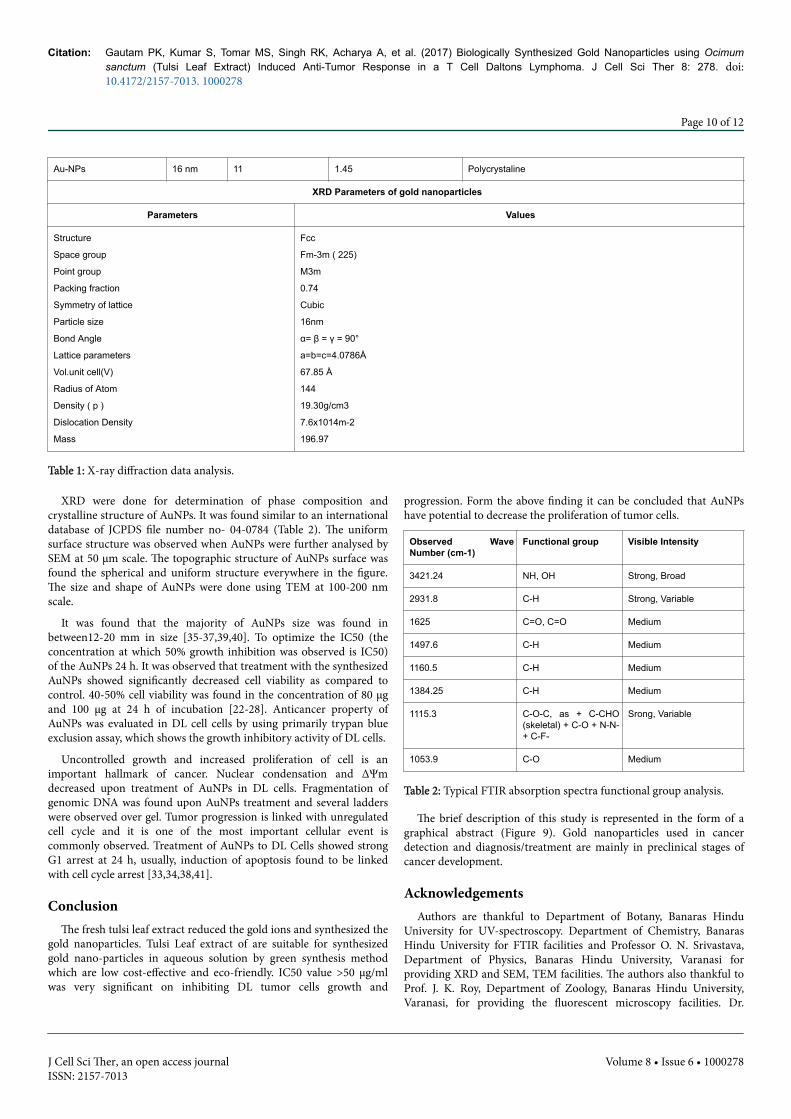
Au-NPs 16 nm 11 1.45 Polycrystaline
XRD Parameters of gold nanoparticles
Parameters Values
Structure
Space group
Point group
Packing fraction
Symmetry of lattice
Particle size
Bond Angle
Lattice parameters
Vol.unit cell(V)
Radius of Atom
Density ( p )
Dislocation Density
Mass
Fcc
Fm-3m ( 225)
M3m
0.74
Cubic
16nm
α= β = γ = 90°
a=b=c=4.0786Å
67.85 Å
144
19.30g/cm3
7.6x10¹⁴m-2
196.97
Table 1: X-ray diffraction data analysis.
XRD were done for determination of phase composition andcrystalline structure of AuNPs. It was found similar to an internationaldatabase of JCPDS file number no- 04-0784 (Table 2). The uniformsurface structure was observed when AuNPs were further analysed bySEM at 50 µm scale. The topographic structure of AuNPs surface wasfound the spherical and uniform structure everywhere in the figure.The size and shape of AuNPs were done using TEM at 100-200 nmscale.
It was found that the majority of AuNPs size was found inbetween12-20 mm in size [35-37,39,40]. To optimize the IC50 (theconcentration at which 50% growth inhibition was observed is IC50)of the AuNPs 24 h. It was observed that treatment with the synthesizedAuNPs showed significantly decreased cell viability as compared tocontrol. 40-50% cell viability was found in the concentration of 80 µgand 100 µg at 24 h of incubation [22-28]. Anticancer property ofAuNPs was evaluated in DL cell cells by using primarily trypan blueexclusion assay, which shows the growth inhibitory activity of DL cells.
Uncontrolled growth and increased proliferation of cell is animportant hallmark of cancer. Nuclear condensation and ΔΨmdecreased upon treatment of AuNPs in DL cells. Fragmentation ofgenomic DNA was found upon AuNPs treatment and several ladderswere observed over gel. Tumor progression is linked with unregulatedcell cycle and it is one of the most important cellular event iscommonly observed. Treatment of AuNPs to DL Cells showed strongG1 arrest at 24 h, usually, induction of apoptosis found to be linkedwith cell cycle arrest [33,34,38,41].
ConclusionThe fresh tulsi leaf extract reduced the gold ions and synthesized the
gold nanoparticles. Tulsi Leaf extract of are suitable for synthesizedgold nano-particles in aqueous solution by green synthesis methodwhich are low cost-effective and eco-friendly. IC50 value >50 μg/mlwas very significant on inhibiting DL tumor cells growth and
progression. Form the above finding it can be concluded that AuNPshave potential to decrease the proliferation of tumor cells.
Observed WaveNumber (cm-1)
Functional group Visible Intensity
3421.24 NH, OH Strong, Broad
2931.8 C-H Strong, Variable
1625 C=O, C=O Medium
1497.6 C-H Medium
1160.5 C-H Medium
1384.25 C-H Medium
1115.3 C-O-C, as + C-CHO(skeletal) + C-O + N-N-+ C-F-
Srong, Variable
1053.9 C-O Medium
Table 2: Typical FTIR absorption spectra functional group analysis.
The brief description of this study is represented in the form of agraphical abstract (Figure 9). Gold nanoparticles used in cancerdetection and diagnosis/treatment are mainly in preclinical stages ofcancer development.
AcknowledgementsAuthors are thankful to Department of Botany, Banaras Hindu
University for UV-spectroscopy. Department of Chemistry, BanarasHindu University for FTIR facilities and Professor O. N. Srivastava,Department of Physics, Banaras Hindu University, Varanasi forproviding XRD and SEM, TEM facilities. The authors also thankful toProf. J. K. Roy, Department of Zoology, Banaras Hindu University,Varanasi, for providing the fluorescent microscopy facilities. Dr.
Citation: Gautam PK, Kumar S, Tomar MS, Singh RK, Acharya A, et al. (2017) Biologically Synthesized Gold Nanoparticles using Ocimumsanctum (Tulsi Leaf Extract) Induced Anti-Tumor Response in a T Cell Daltons Lymphoma. J Cell Sci Ther 8: 278. doi:10.4172/2157-7013. 1000278
Page 10 of 12
J Cell Sci Ther, an open access journalISSN: 2157-7013
Volume 8 • Issue 6 • 1000278

Gautam expresses his appreciation to University Grants Commission,New Delhi for student supports.
Figure 9: Cartoon showing the possible function of AuNPs and their role in possible regressive function upon treatment with cells.
References1. Akbar Y, Kumaraswamy R, Shanmugam K (2014) Optimization and
stabilization of gold nanoparticles by using herbal plant extract withmicrowave heating. Nano Convergence 1:12.
2. Aqil F, Munagala R, Jeyabalan J, Vadhanam MV (2013) Bioavailability ofphytochemicals and its enhancement by drug delivery systems. Cancerletters 334: 133-141.
3. Thakkar KN, Mhatre SS, Parikh RY (2010) Biological synthesis of metallicnanoparticles. Nanomedicine 6: 257-262.
4. Kumar V, Yadav SK (2009) Plant-mediated synthesis of silver and goldnanoparticles and their applications. J chem Techno Biotech 84: 151-157.
5. Shankar SS, Rai A, Ankamwar B, Singh A, Ahmad A, et al. (2004)Biological synthesis of triangular gold nanoprisms. Str Nat Mater 3:482-488.
6. Chauhan A, Zubair S, Sherwani A, Owais M (2012) Aloe vera inducedbiomimetic assemblage of nucleobase into nanosized particles. PLoSONE 7: e32049.
7. Kelm MA, Nair MG, Strasburg GM, DeWitt DL (2000) Antioxidant andcyclooxygenase inhibitory phenolic compounds from OcimumsanctumLinn. Phytomedicine 7: 7-13
8. Singhal G, Bhavesh R, Kasariya K, Sharma AR, Singh RP (2011)Biosynthesis of silver nanoparticles using Ocimum sanctum, Tulsi leafextract and screeningits antimicrobial activity. J Nanopart Res 13:2981-2988.
9. Jaggi RK, Madaan R, Singh B (2003) Anticonvulsant potential of holybasil, Ocimum sanctum Linn, and its cultures. Ind J Exp Biol 41: 1329–33.
10. Pattanayak P, Behera P, Das D, Sangram K (2010) Ocimumsanctum Linn: A reservoir plant for therapeutic applications. Anoverview Pharmacogn Rev 4: 95–105.
11. Ali ME, Hashim U, Mustafa S, Man CYB, Islam KNJ (2012) GoldNanoparticle Sensor for the visual detection of pork adulteration inmeatball formulation. Nanomaterials.
12. Perrault SD, Chan WCW (2010) In vivo assembly of nanoparticlecomponents to improve targeted cancer imaging. Proc Nat Acad SciUSA 107: 11194-11199.
13. Peng GT, Adams UO, Hakim M, Shehada MN, Broza Y, et al.(2009) Detection of lung, breast, colorectal, and prostate cancers fromexhaled breath using a single array of nanosensors. Nature Nanotech 4:669-673.
14. Thompson DT (2007) Using gold nanoparticles for catalysis. NanoToday 2: 40-43.
Citation: Gautam PK, Kumar S, Tomar MS, Singh RK, Acharya A, et al. (2017) Biologically Synthesized Gold Nanoparticles using Ocimumsanctum (Tulsi Leaf Extract) Induced Anti-Tumor Response in a T Cell Daltons Lymphoma. J Cell Sci Ther 8: 278. doi:10.4172/2157-7013. 1000278
Page 11 of 12
J Cell Sci Ther, an open access journalISSN: 2157-7013
Volume 8 • Issue 6 • 1000278

15. Gautam PK, Deepak P, Kumar S, Acharya A (2013) Role of macrophagein tumor microenvironment: prospect in cancer immunotherapy. Euro JInfll 10: 1-14.
16. Gautam PK, Maurya BN, Kumar S, Deepak P, Kumar S Jr, et al. (2013)Progressive growth of a murine T cell lymphoma alters populationkinetics and cell viability of macrophages in a tumor-bearing host. TumBiol 34: 827- 836.
17. Gautam PK, Acharya A (2014) Suppressed expression ofhomotypicmultinucleation, extracellular domains of CD172α (SIRP-α)and CD47 (IAP) receptor in TAMs up-regulated by Hsp70-peptidecomplex in Dalton’s lymphoma. Scan J Immunol 80: 22-35.
18. Gautam PK, Acharya A (2015) Antigenic Hsp70–peptide upregulatealtered cell surface MHC class I expression in TAMs and increases anti-tumor function in Dalton’s lymphoma bearing mice. Tumor Biol 36:2023-2032.
19. Gautam PK, Acharya A (2015) Antigenic Hsp70-peptide upregulatealtered suppressed expression of docking receptor ICAM-1 in TAMsincreases in Dalton's lymphoma bearing mice. Int J Res Sci Tech 5:86-105.
20. Gautam PK, Acharya A (2015) Suppressed expression of cd80 (b7.1) andcd86 (b7.2) receptors in tams up-regulated by autologous hsp70– peptidecomplex in dalton’s lymphoma bearing balb/c mice. IJRST 5: 106-127.
21. Philip D, Unni C, AswathyAromal S, Vidhu VK (2011) Murrayakoenigiileaf-assisted rapid green synthesis of silver and gold nanoparticles.SpectrochimicaActa Part A: Molecular and Biomolecular Spectroscopy78: 899-904.
22. Mallikarjuna K, Narasimaha G, Dillip GR, Praveen B (2011) Greensynthesis of silver nanoparticles using ocimum leafextract and their characterization. Digest J Nanomaterials Biostruc 6:181-186.
23. Lokina S, Narayanan V (2013) A facile phyto-mediated synthesis of goldnanoparticles using aqueous extract ofmomordicacochinchinensisrhizome and their biological activities. ChemSci Trans 2: 105-110.
24. Nakamoto K (1965) Infrared spectra of inorganic and coordinationcompounds. Wiley New York.
25. Goswami PS (2004) Solid state communications. State Comm 132:731-804.
26. Norhayati AB, Shapter JG, Salleh MM, Umar AA (2015) Self-assembly ofhigh density of triangular silver nanoplate films promoted by 3aminopropyltrimethoxysilane. Appl Sci 5: 209-221.
27. Xubin P, Medina-Ramirez I, Mernaugh R, Jingbo L (2010)Nanocharacterization and bactericidal performance of silvermodifiedtitaniaphotocatalyst. Coll Surf B 77: 82-89.
28. Kumar S, Tomar MS, Acharya A (2015) Carboxylic group-inducedsynthesis and characterization of selenium nanoparticles and its anti-tumor potential on Dalton's lymphoma cells. Colloids and Surfaces B:Biointerfaces 126: 546-552.
29. Funakoshi T, Aki T, Nakayama H, Watanuki Y, Imori S, et al. (2011)Reactive oxygen species-independent rapid initiation of mitochondrialapoptotic pathway by chelerythrine. Toxicol in Vitro 25: 1581-1587.
30. Yi M, Parthiban P, Hwang J, Zhang X, Jeong H, et al. (2013) Effect of abispidinone analog on mitochondria mediated apoptosis in HeLacells. Intj oncol 44: 327-335.
31. Prajapati V, Kale RK, Singh RP (2015) Usnic acid inhibits growth andinduces cell cycle arrest and apoptosis in human lung carcinoma A549cells. Nutrition and cancer 67: 647-658.
32. Barath S, Kanth M, Kalishwarala K, Sriram M (2010) Anti-oxidant effectof gold nanoparticles restrains hyperglycemic conditions in diabetic mice.J Nanobiotech 8: 16.
33. Shyanti, Ritis K, Sehrawat A, Singh S, Mishra JPN, et al. (2017)"Zerumbone modulates CD1d expression and lipid antigen presentationpathway in breast cancer cells." Toxicology in Vitro 44: 74-84.
34. Rochelle R, Arvizoa SS, Enfeng W, Robertsonb JD, Bhattacharyaa R, et al.(2013) Inhibition of tumor growth and metastasis by a self-therapeuticnanoparticle. PNAS 110: 6700–6705.
35. Chen PC, Mwakwari SC, Oyelere AK (2008) Gold nanoparticles: Fromnanomedicine to nanosensing. Nanotechnol Sci Appl 2: 145-66.
36. Mousa SA, Bharali DJ (2011) Nanotechnology-based detection andtargeted therapy in cancer: Nano-bio paradigms and applications.Cancers (Basel) 3: 2888-2903.
37. Zaman RT, Diagaradjane P, Krishnan S, Tunnell JW (2011)Magnetomotive molecular nanoprobes. Curr Med Chem 18: 2103–2114.
38. Varahalarao V, Kaladhar D (2014) Green synthesis of silver and goldnanoparticles. J Sci Res 19: 834-842.
39. Akbar Y, Kumaraswamy R, Shanmu RK (2014) Optimization andstabilization of gold nanoparticles by using herbal plant extract withmicrowave heating gam. Nano Conver 1:12.
40. Pellequer Y, Lamprecht A, Lamprecht A (2013) Nanooncology: The futureof cancer diagnosis and treatment. Cancer J Clin 63: 395–441.
41. Brown SD, Nativo P, Smith JA, Stirling D, Edwards PR, et al. (2010) Goldnanoparticles for the improved anticancer drug delivery of the activecomponent of oxaliplatin. Chem J Am Soc 132: 4678–4684.
Citation: Gautam PK, Kumar S, Tomar MS, Singh RK, Acharya A, et al. (2017) Biologically Synthesized Gold Nanoparticles using Ocimumsanctum (Tulsi Leaf Extract) Induced Anti-Tumor Response in a T Cell Daltons Lymphoma. J Cell Sci Ther 8: 278. doi:10.4172/2157-7013. 1000278
Page 12 of 12
J Cell Sci Ther, an open access journalISSN: 2157-7013
Volume 8 • Issue 6 • 1000278