Protein tyrosine phosphatase PRL-3 in malignant cells...
12
Protein tyrosine phosphatase PRL-3 in malignant cells and endothelial cells: expression and function Cecile Rouleau, Andre Roy, Thia St. Martin, Michael R. Dufault, Paula Boutin, Dapei Liu, Mindy Zhang, Kristin Puorro-Radzwill, Lori Rulli, Dave Reczek, Rebecca Bagley, Ann Byrne, William Weber, Bruce Roberts, Katherine Klinger, William Brondyk, Mariana Nacht, Steve Madden, Robert Burrier, Srinivas Shankara, and Beverly A. Teicher Genzyme Corp., Framingham, Massachusetts Abstract Protein tyrosine phosphatase PRL-3 mRNA was found highly expressed in colon cancer endothelium and metas- tases. We sought to associate a function with PRL-3 expression in both endothelial cells and malignant cells using in vitro models. PRL-3 mRNA levels were deter- mined in several normal human endothelial cells exposed or unexposed to the phorbol ester phorbol 12-myristate 13-acetate (PMA) and in 27 human tumor cell lines. In endothelial cells, PRL-3 mRNA expression was increased in human umbilical vascular endothelial cells and human microvascular endothelial cells (HMVEC) exposed to PMA. An oligonucleotide microarray analysis revealed that PRL-3 was among the 10 genes with the largest increase in expression on PMA stimulation. Phenotypically, PMA- treated HMVEC showed increased invasion, tube forma- tion, and growth factor – stimulated proliferation. A flow cytometric analysis of cell surface markers showed that PMA-treated HMVEC retained endothelial characteristics. Infection of HMVEC with an adenovirus expressing PRL-3 resulted in increased tube formation. In tumor cells, PRL-3 mRNA levels varied markedly with high expression in SKNAS neuroblastoma, MCF-7 and BT474 breast carci- noma, Hep3B hepatocellular carcinoma, and HCT116 colon carcinoma. Western blotting analysis of a subset of cell line lysates showed a positive correlation between PRL-3 mRNA and protein levels. PRL-3 was stably trans- fected into DLD-1 colon cancer cells. PRL-3-overexpress- ing DLD-1 subclones were assessed for doubling time and invasion. Although doubling time was similar among parental, empty vector, and PRL-3 subclones, invasion was increased in PRL-3-expressing subclones. In models of endogenous expression, we observed that the MCF-7 cell line, which expresses high levels of PRL-3, was more invasive than the SKBR3 cell line, which expresses low levels of PRL-3. However, the MDA-MB-231 cell line was highly invasive with low levels of PRL-3, suggesting that in some models invasion is PRL-3 independent. Transfection of a PRL-3 small interfering RNA into MCF-7 cells inhibited PRL-3 expression and cell invasion. These results indicate that PRL-3 is functional in both endothelial cells and malignant cells and further validate PRL-3 as a potentially important molecular target for anticancer therapy. [Mol Cancer Ther 2006;5(2):219 – 29] Introduction The family of protein phosphatases of regenerating liver (PRL) comprises three members known as PRL-1, PRL-2, and PRL-3. Rat PRL-1 was originally identified as an immediate-early gene in regenerating liver (1). Murine PRL-2 and PRL-3 were subsequently discovered by amino acid sequence homology and display 87% and 76% sequence identity to murine PRL-1, respectively (2). All three PRL proteins contain a COOH-terminal prenylation motif (2). The human PRL-1, PRL-2, and PRL-3 have been elucidated more recently beginning with the description of human PRL-3 as a human muscle-specific tyrosine phos- phatase (3 – 5). Evidence is accumulating, suggesting that these proteins may be associated with oncogenic states. The first link between PRL expression and cancer comes from studies of tissue distribution showing widespread expression of PRL-1 in embryonic tissues. In the rat embryo, PRL-1 is expressed in the brain, intestine, liver, and esophageal epithelia. In the murine embryo, PRL-1 is expressed in the nervous and skeletal systems (6 – 9). Similarities between cancer cells and embryonic cells have been described in the literature, and the proposition has been made that neoplastic cells are dedifferentiated cells that have reverted to a state of embryonic plasticity (10 – 16). PRL-1 is elevated frequently in breast, ovarian, colon, prostate, and pancre- atic cancers (17, 18). An additional link between PRL proteins and cancer is the expression of PRL-1 in proliferative states. PRL-1 was isolated from regenerating liver, a physiologic process involving cell proliferation (1), which when deregulated may contribute to primary liver cancers (19). In addition, NIH3T3 cells transfected with PRL-1 display abnormal morphology and enhanced growth rate (1), whereas fibroblasts up-regulate PRL-1 mRNA expression on stim- ulation with serum (20). Consistent with these findings are Received 7/29/05; revised 10/19/05; accepted 12/7/05. The costs of publication of this article were defrayed in part by the payment of page charges. This article must therefore be hereby marked advertisement in accordance with 18 U.S.C. Section 1734 solely to indicate this fact. Requests for reprints: Beverly A. Teicher, Genzyme Corp., 1 Mountain Road, Framingham, MA 01701. Phone: 508-271-2843; Fax: 508-620-1203. E-mail: [email protected] Copyright C 2006 American Association for Cancer Research. doi:10.1158/1535-7163.MCT-05-0289 219 Mol Cancer Ther 2006;5(2). February 2006 Research. on September 25, 2018. © 2006 American Association for Cancer mct.aacrjournals.org Downloaded from
Transcript of Protein tyrosine phosphatase PRL-3 in malignant cells...

Protein tyrosine phosphatase PRL-3 in malignant cellsand endothelial cells: expression and function
Cecile Rouleau, Andre Roy, Thia St. Martin,Michael R. Dufault, Paula Boutin, Dapei Liu,Mindy Zhang, Kristin Puorro-Radzwill, Lori Rulli,Dave Reczek, Rebecca Bagley, Ann Byrne,William Weber, Bruce Roberts, Katherine Klinger,William Brondyk, Mariana Nacht, Steve Madden,Robert Burrier, Srinivas Shankara,and Beverly A. Teicher
Genzyme Corp., Framingham, Massachusetts
AbstractProtein tyrosine phosphatase PRL-3 mRNA was foundhighly expressed in colon cancer endothelium and metas-tases. We sought to associate a function with PRL-3expression in both endothelial cells and malignant cellsusing in vitro models. PRL-3 mRNA levels were deter-mined in several normal human endothelial cells exposedor unexposed to the phorbol ester phorbol 12-myristate13-acetate (PMA) and in 27 human tumor cell lines. Inendothelial cells, PRL-3 mRNA expression was increasedin human umbilical vascular endothelial cells and humanmicrovascular endothelial cells (HMVEC) exposed to PMA.An oligonucleotide microarray analysis revealed thatPRL-3 was among the 10 genes with the largest increasein expression on PMA stimulation. Phenotypically, PMA-treated HMVEC showed increased invasion, tube forma-tion, and growth factor–stimulated proliferation. A flowcytometric analysis of cell surface markers showed thatPMA-treated HMVEC retained endothelial characteristics.Infection of HMVEC with an adenovirus expressing PRL-3resulted in increased tube formation. In tumor cells, PRL-3mRNA levels varied markedly with high expression inSKNAS neuroblastoma, MCF-7 and BT474 breast carci-noma, Hep3B hepatocellular carcinoma, and HCT116colon carcinoma. Western blotting analysis of a subsetof cell line lysates showed a positive correlation betweenPRL-3 mRNA and protein levels. PRL-3 was stably trans-fected into DLD-1 colon cancer cells. PRL-3-overexpress-ing DLD-1 subclones were assessed for doubling time and
invasion. Although doubling time was similar amongparental, empty vector, and PRL-3 subclones, invasionwas increased in PRL-3-expressing subclones. In modelsof endogenous expression, we observed that the MCF-7cell line, which expresses high levels of PRL-3, was moreinvasive than the SKBR3 cell line, which expresses lowlevels of PRL-3. However, the MDA-MB-231 cell line washighly invasive with low levels of PRL-3, suggesting that insome models invasion is PRL-3 independent. Transfectionof a PRL-3 small interfering RNA into MCF-7 cells inhibitedPRL-3 expression and cell invasion. These results indicatethat PRL-3 is functional in both endothelial cells andmalignant cells and further validate PRL-3 as a potentiallyimportant molecular target for anticancer therapy. [MolCancer Ther 2006;5(2):219–29]
IntroductionThe family of protein phosphatases of regenerating liver(PRL) comprises three members known as PRL-1, PRL-2,and PRL-3. Rat PRL-1 was originally identified as animmediate-early gene in regenerating liver (1). MurinePRL-2 and PRL-3 were subsequently discovered by aminoacid sequence homology and display 87% and 76%sequence identity to murine PRL-1, respectively (2). Allthree PRL proteins contain a COOH-terminal prenylationmotif (2). The human PRL-1, PRL-2, and PRL-3 have beenelucidated more recently beginning with the description ofhuman PRL-3 as a human muscle-specific tyrosine phos-phatase (3–5).
Evidence is accumulating, suggesting that these proteinsmay be associated with oncogenic states. The first linkbetween PRL expression and cancer comes from studies oftissue distribution showing widespread expression ofPRL-1 in embryonic tissues. In the rat embryo, PRL-1 isexpressed in the brain, intestine, liver, and esophagealepithelia. In the murine embryo, PRL-1 is expressed in thenervous and skeletal systems (6–9). Similarities betweencancer cells and embryonic cells have been described in theliterature, and the proposition has been made thatneoplastic cells are dedifferentiated cells that have revertedto a state of embryonic plasticity (10–16). PRL-1 is elevatedfrequently in breast, ovarian, colon, prostate, and pancre-atic cancers (17, 18).
An additional link between PRL proteins and cancer isthe expression of PRL-1 in proliferative states. PRL-1 wasisolated from regenerating liver, a physiologic processinvolving cell proliferation (1), which when deregulatedmay contribute to primary liver cancers (19). In addition,NIH3T3 cells transfected with PRL-1 display abnormalmorphology and enhanced growth rate (1), whereasfibroblasts up-regulate PRL-1 mRNA expression on stim-ulation with serum (20). Consistent with these findings are
Received 7/29/05; revised 10/19/05; accepted 12/7/05.
The costs of publication of this article were defrayed in part by thepayment of page charges. This article must therefore be hereby markedadvertisement in accordance with 18 U.S.C. Section 1734 solely toindicate this fact.
Requests for reprints: Beverly A. Teicher, Genzyme Corp.,1 Mountain Road, Framingham, MA 01701. Phone: 508-271-2843;Fax: 508-620-1203. E-mail: [email protected]
Copyright C 2006 American Association for Cancer Research.
doi:10.1158/1535-7163.MCT-05-0289
219
Mol Cancer Ther 2006;5(2). February 2006
Research. on September 25, 2018. © 2006 American Association for Cancermct.aacrjournals.org Downloaded from

those suggesting a role for PRL-1 in cell cycle regulation.Investigators have shown that the subcellular localizationof endogenous PRL-1 was cell cycle dependent, with PRL-1located in the endoplasmic reticulum of nonmitotic cellsand to the centrosomes and spindle apparatus of mitoticcells (21, 22).
All three PRL proteins have been implicated directly incancer progression. Overexpression of flag-tagged humanPRL-1 protein in D27 hamster pancreatic ductal epithelialcells led to a loss of contact inhibition in culture andgenerated tumors in athymic nude mice (23). Similarly,human PRL-1 mRNA was shown to be up-regulated inbenign prostatic fibroblast cells after stimulation withconditioned medium from the human prostate tumor cellline LNCaP and to be up-regulated in prostatic tumorfibroblast cells (22). PRL-1 mRNA was expressed inseveral tumor epithelial cell lines, including the HeLacervical adenocarcinoma cell line, the HepG2 hepatoblas-toma cell line, and a Burkitt’s lymphoma cell line (1).PRL-2 mRNA overexpression was detected in prostatecancer cell lines and prostate tumor tissues (22). PRL-1protein and PRL-3 protein were shown to increase cellmotility and cell invasion in vitro (24–30). In the samestudy, PRL-3 protein was shown to be associated withdynamic membrane structures in an in vitro woundhealing assay, and both PRL-1 and PRL-3 proteinsinduced metastasis in mice.
PRL-3 has been strongly implicated in human coloncancer progression. PRL-3 mRNA is a tumor endothelialmarker expressed at higher levels in endothelial cellsisolated from fresh specimens of human colon cancerthan in endothelial cells isolated from normal colonmucosa (31, 32). PRL-3 mRNA was also shown to beexpressed at higher levels in metastases of colorectalcancers compared with nonmetastatic tumors and normalcolorectal epithelium (33). PRL-3 mRNA could not bedetected in normal human colon tissue samples, innonmetastatic colorectal carcinoma samples, or in lungand liver metastases of noncolorectal cancer origin.However, PRL-3 mRNA was detected in 4 of 4 colorectalcancer metastases to lymph nodes, 10 of 11 colorectalmetastases to the liver, 6 of 7 colorectal metastases to thelung, 4 of 4 colorectal metastases to the brain, and 3 of 3colorectal metastases to the ovary (34). These resultsshow the selectivity of PRL-3 expression for colorectalcancer metastasis and suggest that PRL-3 may be animportant mediator of the metastatic process in coloncancer.
We sought to associate a function with PRL-3 expressionin both endothelial cells and malignant cells using in vitromodels. In endothelial cell models, we show that PRL-3expression is associated with increased tube formation,whereas in tumor cell models we show that PRL-3expression is associated with increased invasiveness. Ourdata indicate that PRL-3 may be functional in bothendothelial and malignant compartments of tumors andthat it may play an important role in angiogenic, metastaticcancers.
Materials andMethodsMaterialsPBS, fetal bovine serum (FBS), RPMI, penicillin/strepto-
mycin, Versene, and G418 were purchased from Invitrogen,Inc. (Carlsbad, CA) Phorbol 12-myristate 13-acetate (PMA),DMSO, and protease inhibitor cocktail were purchasedfrom Sigma Chemical Co. (St. Louis, MO). Basic fibroblastgrowth factor (bFGF) and vascular endothelial growthfactor (VEGF) were purchased from R&D Systems, Inc.(Minneapolis, MN).
Cell CultureAll cells were grown at 37jC in 5% CO2 humidified
atmosphere. Human tumor cell lines, DLD-1 colon carci-noma (35, 36), SKNAS and IMR-32 neuroblastoma, andMCF-7, SKBR3, and MDA-MB-231 breast carcinoma(American Type Culture Collection, Manassas, VA) weregrown in RPMI with 10% heat-inactivated FBS and 1%penicillin/streptomycin. Human umbilical vascular endo-thelial cells (HUVEC) and human microvascular endothe-lial cells (HMVEC; Cambrex, East Rutherford, NJ) weregrown in EGM2-MV medium (Cambrex). Endothelialprecursor cells (EPC) were derived from AC133+/CD34+
bone marrow cells (ref. 37; Cambrex) by exposure to 50 ng/mL VEGF, 10 to 50 ng/mL bFGF, and 5 units/mL heparinon fibronectin-coated flasks (Biocoat, Fort Washington, PA)in Iscove’s modified Dulbecco’s medium (Invitrogen) with15% FBS. HMVEC, HUVEC, and EPC were exposed to 100nmol/L PMA in DMSO in serum-free EBM2 (HUVEC andHMVEC) or serum-free Iscove’s modified Dulbecco’smedium (EPC) for 24 hours.
mRNA Extraction and Real-time Reverse Transcrip-tion-PCR
Total RNA was isolated by chloroform extraction usingTrizol reagent (Invitrogen) and purified using QiagenRNeasy method (Valencia, CA). Purified total RNA wassubjected to formaldehyde gel electrophoresis and spectro-photometry (A260 nm and A280 nm) to assess integrity andpurity. Reverse transcription was done using cDNAArchive (Applied Biosystems, Foster City, CA). Forquantification of PRL-3 mRNA, reverse transcription-PCR(RT-PCR) was done in duplicate on a 7700 real-timeTaqman thermal cycler (Applied Biosystems) according tothe manufacturer’s instructions using 900 nmol/L primers(Integrated DNA Technologies, Coralville, IA), 250 nmol/Lfluorogenic probe (Applied Biosystems), and TaqmanUniversal PCR Master Mix (Applied Biosystems). Theforward primer sequence was 5V-ATCACCGTTGTG-GACTGGCC-3V, the reverse primer sequence was 5V-CC-AGTCTTCCACTACCTTGCC-3V, and the probe sequencewas 5V-TTTGACGATGGGGCGCCCCCGC-3V. Loading wasmeasured by 18S mRNA detection using rRNA ControlReagents (Applied Biosystems) in multiplex reactions withPRL-3 primers and probe. The 18S probe final concentra-tion was 250 nmol/L and that of each 18S primer was50 nmol/L. Absolute quantification of PRL-3 transcriptcopy number was done using a cDNA standard curvegenerated by serial dilution of a full-length human PRL-3pcDNA plasmid (Invitrogen). Relative quantification
PRL-3 in Malignant Cells and Endothelial Cells220
Mol Cancer Ther 2006;5(2). February 2006
Research. on September 25, 2018. © 2006 American Association for Cancermct.aacrjournals.org Downloaded from
of PRL-3 mRNA was done according to the comparativecycle threshold method described by the Taqman manu-facturer (Applied Biosystems). Control PCRs substitutingwater for cDNA were used as negative controls. Reversetranscription reactions were done both in the presence(RT+) and absence (RT�) of reverse transcriptase.RT� reactions were used as templates in the RT-PCRexperiment to assess genomic DNA contamination in totalRNA preparations and distinguish it from cDNA amplifi-cation. Most control RT-PCRs reached a cycle thresholdof 40, reflecting absence genomic DNA contaminationin the total RNA preparations and ensuring that thePCR products resulted from cDNA amplification. ForPCR validation of the oligonucleotide microarray analysisof PMA-induced genes in HMVEC, RT-PCR was done on a7900 real-time Taqman thermal cycler according to themanufacturer’s instructions using SYBR Green PCR MasterMix (Applied Biosystems). Loading was measured by18S mRNA detection using rRNA Control Reagents insingleplex reactions. Stanniocalcin-1 forward primer wasGCTGGTGATCAGTGCTTCTGCAA and reverse primerwas TTTGGGCCGCCACTCGGGATTTC. Tissue inhib-itor of matrix metalloproteinase-1 forward primer wasACAGACGGCCTTCTGCAATTC and reverse primer wasTCATCTTGATCTCATAACGCTGGTA. Tissue factorpathway inhibitor-2 forward primer was TGTCACCG-GAACCGGATTGA and reverse primer was TTAAAA-TAATAGCGAGTCACATTGG. Tissue-type plasminogenactivator forward primer was CTGCCTCCCGTGGAATTC-CATGA and reverse primer was AGCCCTCCTTTGATG-CGAAA. CXCR-4 forward primer was GTGTCCTC-ATCCTGGCCTTCAT and reverse primer was GGATCCA-GACGCCAACATAG. Diacylglycerol kinase y forwardprimer was GATGTGGTATGGAGTTCTTGGAA andreverse primer was TGGGAATGTTAAGGACAGCAATT.Nidogen 2 forward primer was TTCCCGGCCATCGCCCC-TTTTC and reverse primer was ACTTT-CCCATTCAC-TCGGTACAG. Galanin forward primer was CCCGAGGC-AGCGCCCTCCTT and reverse primer was GGGTCCAG-CCTCGTTTTTCCTT. Endothelial cell–specific molecule-1forward primer was TGAGGTGTCAGCCTTCTAAT andreverse primer was GAAGGTGCCGTAGGGACAGT. Allprimers were synthesized by Integrated DNA Technolo-gies. For the SYBR Green real-time PCR assay, the finalprimer concentration was 50 nmol/L to ensure the absenceof nonspecific amplification. However, nidogen 2 couldonly be detected using 500 nmol/L primers. PCR productswere run on a PAGE gel to verify size and confirm theabsence of nonspecific bands. Expressed sequence tagswere purchased from Invitrogen and used as positivecontrols.
OligonucleotideMicroarrayAnalysisThe gene expression of PMA-treated HMVEC 1F1645
and DMSO-treated HMVEC 1F1645 was compared. TotalRNA was treated with DNase I (Ambion, Inc., Austin,TX), cleaned using Qiagen RNeasy protocol, and ampli-fied using Ambion Amino Ally MessageAmp AmplifiedRNA kit. Amplified RNAs were labeled with Cy3 or Cy5
dye. Labeled amplified RNAs were fragmented andhybridized on Agilent Human Oligo Microarray usingAgilent hybridization kit (Palo Alto, CA). The experimentused reference design with universal human referencetotal RNA (Invitrogen) with dye swap. The images werequantified using Imagene 5.0 (Biodiscovery, Inc., MarinaDel Ray, CA) imported to GeneSight 3.5 (Biodiscovery) fornormalization and significance analysis. A t test wasapplied and genes were selected that had P V 0.02 (98%confidence).
Flow CytometryPMA-treated, DMSO-treated, and untreated HMVEC
1F1645 were suspended with Versene. Cells (105) wereincubated in PBS/5% FBS with primary antibody (50 AL,1 hour) on ice. After washing, the cells were incubated withsecondary antibody (1 hour), if necessary, on ice. Afterwashing, the cells were suspended in cold buffer (300 AL).Flow cytometry was conducted on a FACSCalibur (BectonDickinson Labware, Franklin Lakes, NJ).
Primary antibodies to CD106, CD54, CD36 (FITC-labeled), CD105 (BD Biosciences, Franklin Lakes, NJ),CD31, VE-cadherin, VEGF receptor-2 (Santa Cruz Biotech-nology, Inc., Santa Cruz, CA), and P1H12 (ChemiconInternational, Inc., Temecula, CA) were used at 5 Ag/mL,and the primary antibody to CD34 (FITC-labeled; BDBiosciences) was used at 3 Ag/mL. A FITC-labeled mouseIgG1 (BD Biosciences) was used as control for CD34 at3 Ag/mL and as control for CD36 at 5 Ag/mL. Rabbit serum(Sigma) was used as a control for CD31 and VEGF receptor-2 at 5 Ag/mL. Goat serum (Sigma) was used as a controlfor VE-cadherin diluted 1:100. Purified mouse IgG1 (BDBiosciences) was used as a control for CD106, CD54,CD105, and P1H12 at 5 Ag/mL. Anti-mouse (JacksonImmunoResearch Laboratories, Inc., West Grove, PA) wasused with CD106, CD54, CD105, and P1H12 at 5 Ag/mL.Anti-rabbit (Jackson ImmunoResearch Laboratories) wasused with CD31 and VEGF receptor-2 at 5 Ag/mL. Anti-goat (Santa Cruz Biotechnology) was used with VE-cadherin at 5 Ag/mL.
Western BlottingConfluent cells in six-well plates were washed with cold
PBS, and cold lysis buffer [50 AL; EB buffer: 1% Triton, 20mmol/L Tris-HCl (pH 7.4), 5 mmol/L EDTA, 10% glycerol,150 mmol/L NaCl, 2 mmol/L sodium orthovanadate,protease inhibitor cocktail] was added on ice (5 minutes).Total cell lysates were collected with rubber scrapers,transferred to a 1.5 mL tube on ice, and then clarified bycentrifugation (14,000 rpm, 20 minutes) at 4jC. Totalprotein concentration was determined by BCA ProteinAssay kit (Pierce, Rockford, IL). Total cell lysates (250 Ag)were loaded onto a 4% to 12% Bis-Tris gel (Invitrogen) andrun at 150 V (2 hours) using MES running buffer(Invitrogen). The protein was transferred onto a polyviny-lidene difluoride membrane at 25 V (45 minutes) using asemidry transfer cell (Bio-Rad Laboratories, Hercules, CA)and then blocked for 1 hour to overnight with 5% dry milkin TBS with 0.01% Tween (Sigma). Anti-PRL-3 monoclonalantibody (no. 14; 1:500; a gift from Drs. Kinzler and
Molecular Cancer Therapeutics 221
Mol Cancer Ther 2006;5(2). February 2006
Research. on September 25, 2018. © 2006 American Association for Cancermct.aacrjournals.org Downloaded from

Vogelstein, Johns Hopkins University School of Medicine,Baltimore, MD) or anti–glyceraldehyde-3-phosphate dehy-drogenase (1:6,000; Abcam, Cambridge, United Kingdom)was incubated for 1 hour. After washing, the membranewas incubated with horseradish peroxidase–labeled anti-mouse antibody (1:10,000; Jackson ImmunoResearchLaboratories) for 1 hour. After washing, the Western blotwas developed using an enhanced chemiluminescence kit(Pierce). For the detection of PRL-3 inhibition in MCF-7cells exposed to the PRL-3 small interfering RNA (siRNA),MCF-7 cells were transfected as described below (see PRL-3siRNA Transfection). Total cell lysates (37 Ag) were loadedonto a 4% to 12% Bis-Tris gel with MES buffer. Detectionwas achieved using an anti-PRL-3 rabbit polyclonal(Genesis Biotech, Inc. Taipei, Taiwan) diluted 1:2,000 andan anti-rabbit horseradish peroxidase–labeled secondaryantibody (Jackson ImmunoResearch Laboratories) diluted1:50,000. Detection was done using the Dura West detectionsystem (Pierce).
ImmunostainingCells were fixed with zinc/formaldehyde for 10 minutes,
rinsed twice in PBS, and permeabilized with 0.2% Tritonfor 10 minutes. Image-iT FX signal enhancer (100 AL) wasadded for 30 minutes at room temperature. The cells wererinsed twice with PBS and anti-PRL-3 (Abcam) was addedin blocking buffer (0.2% horse serum, 0.2% bovine serumalbumin in PBS) for 1 hour at room temperature. Afterwashing, a fluorochrome-labeled secondary antibody wasadded for 1 hour. The cells were then rinsed thrice withPBS. 4V,6-Diamidino-2-phenylindole was counterstainedand the slides were mounted with antifade.
PRL-3 Expression in DLD-1CellsFull-length PRL-3 cDNA (obtained from Drs. Kinzler and
Vogelstein) was subcloned into pcDNA3.1/V5-His-TOPOvector (Invitrogen) expressing neomycin resistance gene,leaving the V5 and His tags out of frame. The pcDNA3.1/V5-His vector (Invitrogen) was the empty vector control.The PRL-3 construct and empty vector were transfectedseparately and stably into DLD-1 cells using Lipofectamine2000 reagent (Invitrogen). Stably transfected DLD-1 cellswere subcloned by limiting dilution and propagated inRPMI with 10% heat-inactivated FBS and 500 Ag/mL G418.
PRL-3 Expression in HMVECPRL-3-expressing adenovirus was made using the
AdEasy XL Adenoviral Vector System (Stratagene, La Jolla,CA). The plasmid construct pcDNA3-V5-HIS-PRL3 wasdigested with KpnI and EcoRV (New England Biolabs,Beverly, MA) and the 596-bp insert was gel isolated. Theinsert was ligated into the KpnI and EcoRV sites ofpShuttle-CMV (Stratagene). The construct pShuttle-CMV-PRL3 was then linearized with PmeI (New EnglandBiolabs) and transformed into electrocompetant BJ5183-Ad-1 cells (Stratagene) according to the manufacturer’sdirections. Recombinant pAd-CMV-PRL3 plasmid DNAwas prepared and recombination was verified with a PacIdigest. PacI-digested recombinant Ad plasmid was thentransfected into Ad293 cells (Stratagene) using Lipofect-amine 2000. When the transfected Ad293 cells reached
complete CPE, the cells were resuspended in a smallvolume of PBS and freeze thawed thrice. The lysed cellswere then spun down and the supernatant was taken asAd-CMV-PRL3 primary viral stock. HMVEC 1F1645 wereinfected with 50 multiplicities of infection of the primaryviral stock and functionally assayed 48 hours postinfection.
PRL-3 siRNATransfectionPRL3-DLD1-IIG5 and MCF-7 cells were grown in RPMI
with 10% FBS, 1% penicillin/streptomycin, and 500 Ag/mL G418. The cells (5 � 104 per well) were plated in a 24-well plate in medium without antibiotics 24 hours beforetransfection. All transfections were carried out in tripli-cate. The protocol for siRNA transfection in a 24-wellformat (Invitrogen) was followed for all transfections. Thegeneral procedure is as follows: (a) siRNA (20 pmol;1 AL of a 20 Amol/L solution) was added to 50 AL Opti-MEM I reduced serum medium, (b) Lipofectamine 2000(1 AL) was added to 50 AL Opti-MEM I reduced serummedium and incubated for 5 minutes at room tempera-ture, (c) The siRNA and Lipofectamine were mixed andincubated at room temperature for 20 minutes, and (d)The 100 AL mixture was added to the cells. The siRNAsused were siRNA Negative-1 (Ambion), siRNA againstlamin A/C (Xeragon/Qiagen, Valencia, CA; target se-quence: AACTGGACTTCCAGAAGAACA), and siRNAagainst PRL-3 (Xeragon/Qiagen; target sequence: TGA-GAGCGGGATGAAGTACGA). Following the 48-hourincubation at 37jC, RNA for Taqman analysis wasisolated using Trizol and pooled.
Proliferation AssayEndothelial cells (5 � 103) in triplicate in a 96-well plate
were incubated with various VEGF and bFGF concentra-tions in the presence or absence of 100 nmol/L PMA or0.00125% DMSO at 37jC in a 5% CO2 humidifiedatmosphere for 96 hours. Tumor cells (2 � 103) in duplicatein a 96-well plate were incubated at 37jC in RPMI with 2%heat-inactivated FBS in a 5% CO2 humidified atmospherefor 96 hours. Proliferation was measured using CellTiter-Glo reagent (Promega, Madison, WI) on a microplateluminometer and Winglow software (Berthold Tech, BadWildbad, Germany).
Endothelial CellTube Formation AssayCells (2 � 104) were seeded in triplicate in a 96-well plate
precoated with Matrigel (Becton Dickinson Labware) inserum-free EBM2 with 20 ng/mL each of bFGF and VEGFand in the presence or absence of 100 nmol/L PMA or0.00125% DMSO at 37jC in a 5% CO2 humidifiedatmosphere for 4 hours. After 24 hours, the cells werestained in 4 Ag/mL calcein (Molecular Probes, Eugene,OR) in PBS at 37jC, imaged, and quantified usingthe image analysis software Scion Image (Scion Corp.,Frederick, MD).
Invasion AssaysA modified Boyden chamber 24-well Transwell with
fluoroblock inserts precoated with Matrigel (ref. 38; BDBiosciences) was used. Cells (2.5 � 104) were seeded perinsert in duplicate in 500 AL serum-free medium. Insertswere placed in wells containing 500 AL medium with 5%
PRL-3 in Malignant Cells and Endothelial Cells222
Mol Cancer Ther 2006;5(2). February 2006
Research. on September 25, 2018. © 2006 American Association for Cancermct.aacrjournals.org Downloaded from

FBS. When appropriate, PMA was added to both chambers.The cells were incubated (48 hours) at 37jC in 5% CO2
humidified atmosphere and the inserts were placed in a24-well plate containing 500 AL calcein (4 Ag/mL, 1 hour)in PBS at 37jC. Fluorescence was read on a CytofluorII fluorescence plate reader (PerSeptive Biosystems,Foster City, CA) with excitation/emission wavelengths of530/590 nm.
Statistical AnalysisAnalysis of statistical significance was done using t
tests for assays consisting of two experimental samplegroups. The ANOVA test and Tukey-Kramer post hoctest were used to determine statistical significance inassays consisting of more than two experimental samplegroups.
ResultsPRL-3 mRNA expression was examined by real-time RT-PCR in human normal endothelial cells and in 27 humantumor cell lines (Fig. 1). HMVEC, HUVEC, and EPC were
examined for PRL-3 mRNA expression without treatmentor after exposure to PMA (100 nmol/L, 24 hours). Understandard conditions, all endothelial cells had <1,000transcripts of PRL-3 mRNA per reaction. Four of fiveendothelial cells tested [lung HMVEC, HMVEC IF1645(neonatal dermal), HMVEC OF1025 (neonatal dermal),and HUVEC] had a marked increase in PRL-3 mRNAafter exposure to PMA. PRL-3 mRNA was increased 27-,23-, 11-, and 23-fold in the lung HMVEC, HMVEC 1F1645,HMVEC OF1025, and HUVEC, respectively, by exposureto PMA compared with DMSO. HMVEC 1F1645 was mostresponsive to PMA with an average of 14,428 transcriptsper reaction after PMA exposure compared with 500 to600 without PMA exposure.
Twenty-seven human tumor cell lines were evaluatedby real-time RT-PCR for expression of PRL-3 mRNA(Fig. 1). The four human melanoma cell lines had lowlevels of PRL-3 mRNA. All three neuroblastoma cell linesexpressed relatively high levels of PRL-3 mRNA rangingfrom an average of 1,252 transcripts per reaction in theIMR-32 cell line to an average of 21,969 transcripts per
Figure 1. RT-PCR analysis of PRL-3 mRNA showing expression in normal human endothelial cells in the presence and absence of PMA exposure in EPC,HMVEC lung, HMVEC 1F1645, HMVEC OF1025, and HUVEC and expression in human tumor cell lines. A cycle threshold of 40 (no signal) was obtained incontrol PCRs where water was substituted for cDNA (data not shown). Control PCRs using mock reverse transcription reactions as templates (reactionswhere water had been substituted for reverse transcriptase) indicated negligible genomic DNA contamination (data not shown).
Molecular Cancer Therapeutics 223
Mol Cancer Ther 2006;5(2). February 2006
Research. on September 25, 2018. © 2006 American Association for Cancermct.aacrjournals.org Downloaded from

reaction in the SKNAS cell line. The five human breastcancer cell lines had varied expression of PRL-3 mRNAranging from an average of 31 transcripts per reaction inthe SKBR3 breast adenocarcinoma cell line to averages of7,571 and 8,082 transcripts per reaction in the MCF-7 breastadenocarcinoma and BT474 breast carcinoma cell lines,respectively. The SKOV3 ovarian adenocarcinoma cell linehad low expression of PRL-3 mRNA with an average of 187transcripts per reaction. All three prostate cancer cell linestested had relatively high levels of PRL-3 mRNA withaverages of 1,125, 4,067, and 1,795 transcripts per reaction,respectively. The 786-O renal cell adenocarcinoma cell linehad negligible levels of PRL-3 mRNA. The Hep3B hepa-tocellular carcinoma cell line had high levels of PRL-3
mRNA with an average of 11,163 transcripts per reaction.The three pancreatic cancer cell lines were positive for PRL-3 mRNA with averages of 866, 2,371, and 6,310 transcriptsper reaction, respectively. The six colorectal cancer celllines had varied PRL-3 mRNA ranging from negligibleamounts of PRL-3 transcripts with an average of a singletranscript per reaction in the DLD-1 colorectal adenocarci-noma cell line to an average of 6,752 and 6,108 transcriptsper reaction in the HCT116 colorectal carcinoma andSW837 rectal adenocarcinoma cell lines, respectively.
Exposure to PMA did not alter expression of cell surfacemarkers on HMVEC 1F1645, including P1H12, VEGFreceptor-2, VE-cadherin, CD105, CD34, CD31, CD36,CD54, and CD106 as determined by flow cytometry (data
Figure 2. A, PMA-induced HMVEC1F1645 gene expression by RT-PCR. mRNAlevels relative to untreated cells. B, relativeinvasion by HMVEC 1F1645 in the presenceand absence of PMA exposure. Representa-tive experiment. C, tube formation byHMVEC IF1645 in the presence and absenceof PMA exposure and after infection witheither PRL-3-expressing adenovirus or emptyadenovirus. Data are compared with undis-turbed cells (untreated and uninfected).Representative experiment. Western blotshowing PRL-3 protein levels in infectedHMVEC 1F1645. ***, P < 0.001; **, P< 0.01; *, P < 0.05 versus control cells.
PRL-3 in Malignant Cells and Endothelial Cells224
Mol Cancer Ther 2006;5(2). February 2006
Research. on September 25, 2018. © 2006 American Association for Cancermct.aacrjournals.org Downloaded from

not shown). However, exposure to PMA (100 nmol/L)enhanced proliferation of HMVEC 1F1645 in the absenceof and over a concentration range of VEGF and bFGF (up to20 ng/mL each; data not shown).
The invasion activity of HMVEC 1F1645 was assessed inthe presence or absence of 100 nmol/L PMA. Exposure toPMA increased the invasiveness of the cells by f4-foldcompared with untreated controls and f6-fold comparedwith DMSO-treated cells (Fig. 2). For tube formationassays, HMVEC 1F1645 were treated with either 0.00125%DMSO or 100 nmol/L PMA in the presence of bFGF andVEGF (20 ng/mL each). PMA exposure increased tubearea compared with both controls (Fig. 2).
The effect of PMA (100 nmol/L, 24 hours) on HMVECgene expression was assessed by oligonucleotide micro-array analysis and subsequent RT-PCR analysis. Themost highly up-regulated mRNAs were stanniocalcin-1,CXCR-4, tissue factor pathway inhibitor-2, tissue inhibitor
of matrix metalloproteinase-1, PRL-3, tissue-type plas-minogen activator, diacylglycerol kinase y, nidogen-2,galanin, and endothelial cell-specific molecule-1 (Fig. 2).Infection of HMVEC 1F1645 with a PRL-3-containingadenovirus increased tube formation compared withinfection with an empty adenovirus or lack of infection,showing that PRL-3 up-regulation can promote tubeformation (Fig. 2).
Total RNA was isolated from parental DLD-1 coloncancer cells and DLD-1 subclones transfected with emptyvector or PRL-3-containing plasmid vector, and cDNA wasprepared and analyzed for PRL-3 mRNA by RT-PCR. Fiveempty vector–transfected DLD-1 subclones and five PRL-3-transfected subclones were selected for characterization(Fig. 3). Protein from the subclones and parental cells wasanalyzed by Western blotting with a mouse monoclonalanti-PRL-3 and by immunostaining with a goat polyclonalanti-PRL-3 to determine PRL-3 protein expression. Western
Figure 3. A, RT-PCR assay showing PRL-3 mRNA in five DLD-1 subclones transfected with a vector containing PRL-3 cDNA, five DLD-1 subclonestransfected with empty vector, and parental DLD-1 cells. B, Western blot showing PRL-3 protein in five DLD-1 subclones transfected with a vectorcontaining PRL-3 cDNA, five DLD-1 subclones transfected with empty vector, and parental DLD-1 cells. C, immunostaining showing the PRL-3 protein infive DLD-1 subclones transfected with a vector containing PRL-3 cDNA and parental DLD-1 cells. Representative experiment. ***, P < 0.001; **, P <0.01 versus control cells.
Molecular Cancer Therapeutics 225
Mol Cancer Ther 2006;5(2). February 2006
Research. on September 25, 2018. © 2006 American Association for Cancermct.aacrjournals.org Downloaded from

blot analysis showed the highest PRL-3 protein insubclones PRL3-IIG11 and PRL3-IID5, moderate PRL-3 insubclone PRL3-IIG5, and lower PRL-3 in subclones PRL3-IID3 and PRL3-IB1. Parental DLD-1 and empty vector–transfected subclones have very low to undetectable PRL-3protein (Fig. 3). Immunostaining showed low PRL-3protein in parental cells, moderate PRL-3 protein insubclones PRL3-IIG11 and PRL3-IID5, and high PRL-3protein in subclones PRL3-IIG5, PRL3-IID3, and PRL3-IB1(Fig. 3). The differences in PRL-3 obtained by Westernblotting and immunostaining may reflect differences in theantibodies used because both antibodies cross-react withthe PRL-2 protein.
Generation times were determined for the five PRL-3-expressing DLD-1 subclones, five empty vector–trans-fected DLD-1 subclones, and parental DLD-1 cells (Fig. 4).The PRL-3 protein level was not associated with genera-tion time, because the population doubling time of thefive PRL-3-expressing subclones (18.5 hours in 10% FBSand 38 hours in 2% FBS), the five empty vector–transfected subclones (14.4 hours in 10% FBS and 33.3hours in 2% FBS), and the parental DLD-1 cells (11.5hours in 10% FBS and 25.3 hours in 2% FBS) were similar.The five PRL-3-expressing subclones, five empty vector–transfected subclones, and parental DLD-1 cells wereassayed for invasion through Matrigel. The PRL-3-expressing DLD-1 subclones invasiveness was 2-foldgreater than that of the parental cells, whereas the emptyvector–transfected subclones invasiveness was 2-fold lessthan that of the parental cells (Fig. 4).
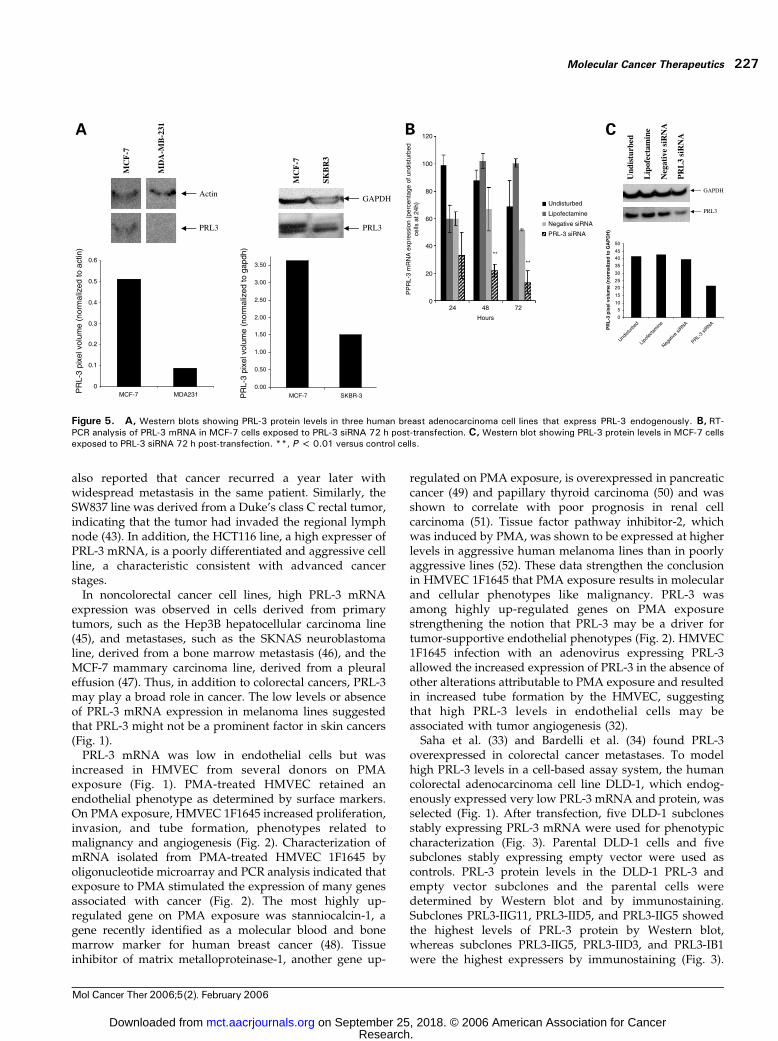
When human breast cancer cell lines endogenouslyexpressing high or low levels of PRL-3 protein (Fig. 5)were assayed for invasion through Matrigel, the MCF-7breast adenocarcinoma cell line was more invasive thanthe SKBR3 breast adenocarcinoma cell line. However, theMDA-MB-231 breast adenocarcinoma cell line (low PRL-3mRNA and protein) was as invasive as the MCF-7 cell line(high PRL-3 mRNA and protein). Therefore, there was atrend between PRL-3 and invasion through Matrigel withtwo of three human tumor cell lines (Fig. 6). MCF-7 cellstransfected with a PRL-3 siRNA had decreased levels ofPRL-3 mRNA and protein (Fig. 5) and a reduced abilityto invade through Matrigel at 72 hours post-transfection(Fig. 6).
DiscussionPhosphatases involved in critical aspects of malignantdisease could be interesting drug targets. PRL-3 mRNAwas detectable in a broad range of human tumor cell linesand endothelial cells. These data reinforce and expand onpreviously published results, indicating that PRL-3 isexpressed in the epithelial and endothelial compartmentsof tumors (31–34, 39, 40).
The expression of PRL-3 mRNA was highly variableamong the primary tumor-derived colorectal cancer celllines, SW837, HT29, DLD-1, SW480, LS174T, and HCT116(Fig. 1; refs. 35, 36, 41–44). Using clinical tissues, Saha et al.
(33) and Bardelli et al. (34) found higher levels of PRL-3mRNA in colorectal tumor metastases compared with theprimary tumor from the same patient. Our findings suggestthat advanced primary tumors may express higher PRL-3mRNA levels than lower grade tumors. Cell lines withmoderate PRL-3 mRNA, such as HT29, SW480, andLST174T colorectal adenocarcinomas, or with high PRL-3mRNA, such as SW837 rectal adenocarcinoma and HCT116colorectal carcinoma, might have been derived fromprimary tumors that had developed a metastatic pheno-type. Indeed, Leibovitz et al. (43) stated that the SW480line was derived from a Duke’s class B primary tumor,indicating that the primary tumor had invaded themuscularis propia of the bowel wall. Leibovitz et al. (43)
Figure 4. A, mean generation times obtained for five PRL-3-expressing subclones, five empty vector– transfected subclones, andparental DLD-1 cells determined in 2% or 10% FBS. Columns, mean oftwo independent experiments; bars, SD. B, relative mean invasionobtained for five PRL-3-expressing subclones and five empty vector–transfected subclones compared with parental DLD-1 cells. Columns,mean of three independent experiments; bars, SD. *, P < 0.05 versuscontrol cells.
PRL-3 in Malignant Cells and Endothelial Cells226
Mol Cancer Ther 2006;5(2). February 2006
Research. on September 25, 2018. © 2006 American Association for Cancermct.aacrjournals.org Downloaded from
also reported that cancer recurred a year later withwidespread metastasis in the same patient. Similarly, theSW837 line was derived from a Duke’s class C rectal tumor,indicating that the tumor had invaded the regional lymphnode (43). In addition, the HCT116 line, a high expresser ofPRL-3 mRNA, is a poorly differentiated and aggressive cellline, a characteristic consistent with advanced cancerstages.
In noncolorectal cancer cell lines, high PRL-3 mRNAexpression was observed in cells derived from primarytumors, such as the Hep3B hepatocellular carcinoma line(45), and metastases, such as the SKNAS neuroblastomaline, derived from a bone marrow metastasis (46), and theMCF-7 mammary carcinoma line, derived from a pleuraleffusion (47). Thus, in addition to colorectal cancers, PRL-3may play a broad role in cancer. The low levels or absenceof PRL-3 mRNA expression in melanoma lines suggestedthat PRL-3 might not be a prominent factor in skin cancers(Fig. 1).
PRL-3 mRNA was low in endothelial cells but wasincreased in HMVEC from several donors on PMAexposure (Fig. 1). PMA-treated HMVEC retained anendothelial phenotype as determined by surface markers.On PMA exposure, HMVEC 1F1645 increased proliferation,invasion, and tube formation, phenotypes related tomalignancy and angiogenesis (Fig. 2). Characterization ofmRNA isolated from PMA-treated HMVEC 1F1645 byoligonucleotide microarray and PCR analysis indicated thatexposure to PMA stimulated the expression of many genesassociated with cancer (Fig. 2). The most highly up-regulated gene on PMA exposure was stanniocalcin-1, agene recently identified as a molecular blood and bonemarrow marker for human breast cancer (48). Tissueinhibitor of matrix metalloproteinase-1, another gene up-
regulated on PMA exposure, is overexpressed in pancreaticcancer (49) and papillary thyroid carcinoma (50) and wasshown to correlate with poor prognosis in renal cellcarcinoma (51). Tissue factor pathway inhibitor-2, whichwas induced by PMA, was shown to be expressed at higherlevels in aggressive human melanoma lines than in poorlyaggressive lines (52). These data strengthen the conclusionin HMVEC 1F1645 that PMA exposure results in molecularand cellular phenotypes like malignancy. PRL-3 wasamong highly up-regulated genes on PMA exposurestrengthening the notion that PRL-3 may be a driver fortumor-supportive endothelial phenotypes (Fig. 2). HMVEC1F1645 infection with an adenovirus expressing PRL-3allowed the increased expression of PRL-3 in the absence ofother alterations attributable to PMA exposure and resultedin increased tube formation by the HMVEC, suggestingthat high PRL-3 levels in endothelial cells may beassociated with tumor angiogenesis (32).
Saha et al. (33) and Bardelli et al. (34) found PRL-3overexpressed in colorectal cancer metastases. To modelhigh PRL-3 levels in a cell-based assay system, the humancolorectal adenocarcinoma cell line DLD-1, which endog-enously expressed very low PRL-3 mRNA and protein, wasselected (Fig. 1). After transfection, five DLD-1 subclonesstably expressing PRL-3 mRNA were used for phenotypiccharacterization (Fig. 3). Parental DLD-1 cells and fivesubclones stably expressing empty vector were used ascontrols. PRL-3 protein levels in the DLD-1 PRL-3 andempty vector subclones and the parental cells weredetermined by Western blot and by immunostaining.Subclones PRL3-IIG11, PRL3-IID5, and PRL3-IIG5 showedthe highest levels of PRL-3 protein by Western blot,whereas subclones PRL3-IIG5, PRL3-IID3, and PRL3-IB1were the highest expressers by immunostaining (Fig. 3).
Figure 5. A, Western blots showing PRL-3 protein levels in three human breast adenocarcinoma cell lines that express PRL-3 endogenously. B, RT-PCR analysis of PRL-3 mRNA in MCF-7 cells exposed to PRL-3 siRNA 72 h post-transfection. C, Western blot showing PRL-3 protein levels in MCF-7 cellsexposed to PRL-3 siRNA 72 h post-transfection. **, P < 0.01 versus control cells.
Molecular Cancer Therapeutics 227
Mol Cancer Ther 2006;5(2). February 2006
Research. on September 25, 2018. © 2006 American Association for Cancermct.aacrjournals.org Downloaded from

The differences may be due to cross-reactivity of theantibodies with PRL-2 protein. The ranking of thesubclones based on ability to invade through Matrigelwas PRL3-IIG5 > PRL3-IID3 and PRL3-IB1 > PRL3-IIG11 >PRL3-IID5, which is in close agreement with the immu-nostaining results for PRL-3 protein (Fig. 3). The generationtime for each of the subclones and parental DLD-1 cellsindicated that PRL-3 expression was not associated withproliferation rate (Fig. 4). However, invasion assaysshowed that higher PRL-3 expression was associated withincreased invasion.
Invasion assays were subsequently done with humantumor cell lines with high and low endogenous PRL-3,including three human breast adenocarcinoma cell lines.The MCF-7 breast adenocarcinoma line, a high PRL-3expresser, was more invasive than the SKBR3 breastadenocarcinoma line, a low PRL-3 expresser. However,
the MDA-MB-231 breast adenocarcinoma line, a low PRL-3mRNA and protein expresser, was as invasive as the MCF-7 cell line (Figs. 1, 5, and 6). Overall, for two of three humantumor cell lines, there was a trend toward high PRL-3expression and greater invasiveness, supporting a role forPRL-3 in invasion (2). However, the MDA-MB-231 breastadenocarcinoma line, which has low PRL-3 mRNA andprotein levels, had robust invasion through Matrigel,indicating that PRL-3 expression is not required forinvasion by all cells. Conclusive demonstration that PRL-3 actively promotes invasiveness in breast cancer cell lineswas achieved by transfecting MCF-7 cells with a PRL-3siRNA. MCF-7 cells exposed to the PRL-3 siRNA showeddecreased PRL-3 mRNA and protein levels 72 hours post-transfection and reduced invasion through Matrigel (Figs. 5and 6).
PRL-3, originally identified as a phosphatase in regener-ating liver, continues to be validated as a potentiallyinteresting drug target in malignant disease. PRL-3 hasbeen found to be up-regulated in tumor endothelium andin metastatic colon cancer cells preclinically and clinically(31–34, 39). PRL-3 is up-regulated by exposure of endo-thelial cells to PMA and can be directly implicated inincreased tube formation by PRL-3 adenovirus-infectedHMVEC and in invasion by stably transfected DLD-1subclones and endogenously expressing tumor cell lines.PRL-3 represents both an antiangiogenic and an antitumortarget in malignant disease (53).
References
1. Diamond RH, Cressman DE, Laz TM, Abrams CA, Taub R. PRL-1, aunique nuclear protein tyrosine phosphatase, affects cell growth. Mol CellBiol 1994;14:3752–62.
2. Zeng Q, Hong W, Tan YH. Mouse PRL-2 and PRL-3, two potentiallyprenylated protein tyrosine phosphatases homologous to PRL-1. BiochemBiophys Res Commun 1998;244:421–7.
3. Matter WF, Estridge T, Zhang C, et al. Role of PRL-3, a human muscle-specific tyrosine phosphatase, in angiotensin-II signaling. BiochemBiophys Res Commun 2001;283:1061–8.
4. Zhou H, Gallina M, Mao H, et al. 1H, 13C and 15N resonanceassignments and secondary structure of the human protein tyrosinephosphatase, PRL-2. J Biomol NMR 2003;27:397–8.
5. Pathak MK, Dhawan D, Lindner DJ, Borden EC, Farver C, Yi T.Pentamidine is an inhibitor or PRL phosphatases with anticancer activity.Mol Cancer Ther 2002;1:1255–64.
6. Haber B, Naji L, Cressman D, Taub R. Coexpression of liver-specific andgrowth-induced genes in perinatal and regenerating liver: attainment andmaintenance of the differentiated state during rapid proliferation. Hepatol-ogy 1995;22:906–14.
7. Takano S, Fukuyama M, Kimura J, Xue J, Ohashi H, Fujita J. PRL-1,a protein tyrosine phosphatase, is expressed in neurons and oligo-dendrocytes in the brain and induced in the cerebral cortexfollowing transient forebrain ischemia. Brain Res Mol Brain Res 1996;40:105–15.
8. Rundle CH, Kappen C. Developmental expression of the murine PRL-1protein tyrosine phosphatase gene. J Exp Zool 1999;283:612–7.
9. Kong W, Swain GP, Li S, Diamond RH. PRL-1 PTPase expres-sion is developmentally regulated with tissue-specific patterns inepithelial tissues. Am J Physiol Gastrointest Liver Physiol 2000;279:G613–21.
10. Chodosh LA. Expression of BRCA1 and BRCA2 in normal andneoplastic cells. J Mammary Gland Biol Neoplasia 1998;3:389–402.
11. Ito T, Noguchi Y, Udaka N, Kitamura H, Satoh S. Glucose transporter
Figure 6. A, relative invasion by human tumor cell lines, including MCF-7, SKBR3, and MDA-MB-231 breast adenocarcinomas that express PRL-3endogenously. Columns, mean of two independent experiments; bars,SD. B, relative invasion by MCF-7 cells exposed to PRL-3 siRNA 72 h post-transfection. **, P < 0.01 for MCF-7 and MDA-MB-231 compared withSKBR3.
PRL-3 in Malignant Cells and Endothelial Cells228
Mol Cancer Ther 2006;5(2). February 2006
Research. on September 25, 2018. © 2006 American Association for Cancermct.aacrjournals.org Downloaded from

expression in developing fetal lungs and lung neoplasms. Histol Histo-pathol 1999;14:895–904.
12. Waltzer L, Bienz M. The control of h-catenin and TCF during embryonicdevelopment and cancer. Cancer Metastasis Rev 1999;18:231–46.
13. Krupp G, Bonatz G, Parwaresch R. Telomerase, immortality andcancer. Biotechnol Annu Rev 2000;6:103–40.
14. Meszoely IM, Means AL, Scoggins CR, Leach SD. Developmentalaspects of early pancreatic cancer. Cancer J 2001;7:242–50.
15. Teicher BA. Malignant cells, directors of the malignant process:role of transforming growth factor-h. Cancer Metastasis Rev 2001;20:133–43.
16. Sood AK, Fletcher MS, Hendrix MJ. The embryonic-like properties ofaggressive human tumor cells. J Soc Gynecol Investig 2002;9:2–9.
17. Werner SR, Lee PA, Cummings OW, Randall SK, Crowell DN, CrowellPL. PRL-1 protein tyrosine phosphatase accelerates progression into Sphase and is found at elevated levels in human ovarian, breast, pancreatic,colon and prostate cancers [abstract 818]. Proc Amer Assoc Cancer Res95th Annu Meet 2004.
18. Stephens BJ, Farnsworth AL, Munoz RM, et al. Lipid phosphataseactivity of PRL-1 [abstract 865]. Proc Amer Assoc Cancer Res 95th AnnuMeet 2004.
19. Diehl AM. Liver regeneration. Front Biosci 2002;7:301–14.
20. Diamond RH, Peters C, Jung SP, et al. Expression of PRL-1 nuclearPTPase is associated with proliferation in liver but with differentiation inintestine. Am J Physiol 1996;271:G121–9.
21. Wang J, Kirby CE, Herbst R. The tyrosine phosphatase PRL-1localizes to the endoplasmic reticulum and the mitotic spindle and isrequired for normal mitosis. J Biol Chem 2002;227:46659–68.
22. Wang Q, Holmes DIR, Powell SM, Lu QL, Waxman J. Analysis ofstromal-epithelial interactions in prostate cancer identifies PTPCAAX2 as apotential oncogene. Cancer Lett 2002;175:63–9.
23. Cates CA, Michael RL, Stayrook KR, et al. Prenylation of oncogenichuman PTP(CAAX) protein tyrosine phosphatases. Cancer Lett 1996;110:49–55.
24. Zeng Q, Dong J-M, Guo K, et al. PRL-3 and PRL-1 promote cellmigration, invasion and metastasis. Cancer Res 2003;63:2716–22.
25. Gou K, Li J, Tang JP, Koh V, Gan BQ, Zeng Q. Catalytic domain ofPRL-3 plays an essential role in tumor metastasis: formation of PRL-3tumors inside the blood vessels. Cancer Biol Ther 2004;3:945–51. Epub2004 Oct 27.
26. Miskad UA, Semba S, Kato H, Yokozaki H. Expression of PRL-3phosphatase in human gastric carcinomas: close correlation with invasionand metastasis. Pathobiology 2004;71:176–84.
27. Wu X, Zeng H, Zhang X, et al. Phosphatase regenerating liver-3promotes motility and metastasis of mouse melanoma cells. Am J Pathol2004;164:2039–54.
28. Peng L, Ning J, Meng L, Shou C. The association of the expressionlevel of protein tyrosine phosphatase PRL-3 protein with liver metastasisand prognosis of patents with colorectal cancer. J Cancer Res Clin Oncol2004;130:521–6.
29. Lim KA, Song JS, Jee J, et al. Structure of human PRL-3, thephosphatase associated with cancer metastasis. FEBS Lett 2004;565:181–7.
30. Kozlov G, Cheng J, Ziomek E, Banville D, Gehring K, Ekiel I. Structuralinsights into molecular function of the metastasis-associated phosphatasePRL-3. Biol Chem 2004;279:11882–9.
31. Sager J, Benvenuti S, Bardelli A. PRL-3: a phosphatase formetastasis? Cancer Biol Ther 2004;3:952–3. Epub 2004 Oct 8.
32. St. Croix B, Rago C, Velculescu V, et al. Genes expressed in humantumor endothelium. Science 2000;289:1197–202.
33. Saha S, Bardelli A, Buckhaults P, et al. A phosphatase associated withmetastasis of colorectal cancer. Science 2001;294:1343–6.
34. Bardelli A, Saha S, Sager J, et al. PRL-3 expression in metastaticcancers. Clin Cancer Res 2003;9:5607–13.
35. Dexter DL, Spremulli EN, Fligiel Z, et al. Heterogeneity of cancer cellsfrom a single human colon carcinoma. Am J Med 1981;71:949–56.
36. Dexter DL. N,N -dimethylformamide-induced alteration of cell culturecharacteristics and loss of tumorigenicity in cultured human coloncarcinoma cells. Cancer Res 1979;39:1020.
37. Bagley R, Walter-Yohrling J, Cao X, et al. Endothelial precursor cellsas a model of tumor endothelium: characterization and comparison tomature endothelial cells. Cancer Res 2003;63:5866–73.
38. Grotendorst GR. Spectrophotometric assay for the quantitation of cellmigration in the Boyden chamber chemotaxis assay. Methods Enzymol1987;147:144–52.
39. Peng Y, Du K, Ramirez S, Diamond RH, Taub R. Mitogenicup-regulation of the PRL-1 protein-tyrosine phosphatase gene by Erg-1:EGR-1 activation is an early event in liver regeneration. J Biol Chem 1999;274:4513–20.
40. Peng L, Ning J, Meng L, Shou C. The association of the expressionlevel of protein tyrosine phosphatase PRL-3 protein with liver metastasisand prognosis of patients with colorectal cancer. J Cancer Res Clin Oncol2004;130:521–6. Epub 2004 May 6.
41. Von Kleist S, Chany E, Burtin P, King M, Fogh J. Immunohistology ofthe antigenic pattern of a continuous cell line from a human colon tumor.J Natl Cancer Inst 1975;55:555–60.
42. Tom BH, Rutsky LP, Jakstys MM, Oyasu R, Kaye CI, Kahan BD.Human colonic adenocarcinoma cells. I. Establishment and description of anew line. In Vitro 1976;12:180–90.
43. Leibovitz A, Stinson JC, McCombs WB III, McCoy CE, Mazur KC,Mabry ND. Classification of human colorectal adenocarcinoma cell lines.Cancer Res 1976;36:4562–9.
44. Boyd D, Florent G, Kim P, Brattain M. Determination of the levels ofurokinase and its receptor in human colon carcinoma cell lines. Cancer Res1988;48:3112–6.
45. Aden DP, Fogel A, Plotkin S, Damjanov I, Knowles BB. Controlledsynthesis of HBsAg in a differentiated human liver carcinoma-derived cellline. Nature 1979;282:615–6.
46. Sugimoto T, Tatsumi E, Kemshead JT, Helson L, Green AA,Minowada J. Determination of cell surface membrane antigens commonto both human neuroblastoma and leukemia-lymphoma cell lines by a panelof 38 monoclonal antibodies. J Natl Cancer Inst 1984;73:51–7.
47. Soule HD, Vazguez J, Long A, Albert S, Brennan M. A human cell linefrom a pleural effusion derived from a breast carcinoma. J Natl Cancer Inst1973;51:1409–16.
48. Wascher RA, Huynh KT, Giuliano AE, et al. Stanniocalcin-1: a novelmolecular blood and bone marrow marker for human breast cancer. ClinCancer Res 2003;9:1427–35.
49. Crnogorac-Jurcevic T, Efthimiou E, Nielsen T, et al. Expressionprofiling of microdissected pancreatic adenocarcinomas. Oncogene 2002;21:4587–94.
50. Huang Y, Prasad M, Lemon WJ, et al. Gene expression in papillarythyroid carcinoma reveals highly consistent profiles. Proc Natl Acad SciU S A 2001;98:15044–9.
51. Kallakury BVS, Karikehalli S, Haholu A, Sheehan CE, Azumi N, RossJS. Increased expression of matrix metalloproteinases 2 and 9 andtissue inhibitors of metalloproteinases 1 and 2 correlate with poorprognostic variables in renal cell carcinoma. Clin Cancer Res 2001;7:3113–9.
52. Ruf W, Seftor EA, Petrovan RJ, et al. Differential role of tissue factorpathway inhibitors 1 and 2 in melanoma vasculogenic mimicry. Cancer Res2003;63:5381–9.
53. Kim K-A, Song J-S, Jee JG, et al. Structure of human PRL-3, thephosphatase associated with cancer metastasis. FEBS Lett 2004;565:181–7.
Molecular Cancer Therapeutics 229
Mol Cancer Ther 2006;5(2). February 2006
Research. on September 25, 2018. © 2006 American Association for Cancermct.aacrjournals.org Downloaded from

2006;5:219-229. Mol Cancer Ther Cecile Rouleau, Andre Roy, Thia St. Martin, et al. endothelial cells: expression and functionProtein tyrosine phosphatase PRL-3 in malignant cells and
Updated version
http://mct.aacrjournals.org/content/5/2/219
Access the most recent version of this article at:
Cited articles
http://mct.aacrjournals.org/content/5/2/219.full#ref-list-1
This article cites 51 articles, 15 of which you can access for free at:
Citing articles
http://mct.aacrjournals.org/content/5/2/219.full#related-urls
This article has been cited by 9 HighWire-hosted articles. Access the articles at:
E-mail alerts related to this article or journal.Sign up to receive free email-alerts
Subscriptions
Reprints and
To order reprints of this article or to subscribe to the journal, contact the AACR Publications
Permissions
Rightslink site. (CCC)Click on "Request Permissions" which will take you to the Copyright Clearance Center's
.http://mct.aacrjournals.org/content/5/2/219To request permission to re-use all or part of this article, use this link
Research. on September 25, 2018. © 2006 American Association for Cancermct.aacrjournals.org Downloaded from